Retinal Disease and Metabolism

A topical collection in Life (ISSN 2075-1729). This collection belongs to the section "Medical Research".

Viewed by 96027

Editors


E-Mail Website
Guest Editor
Boston Children’s Hospital, Harvard Medical School, Boston, MA 02115, USA
Interests: retinal diseases and metabolism; angiogenesis; vitreoretinal surgery; pathologic myopia
Special Issues, Collections and Topics in MDPI journals

E-Mail Website
Guest Editor
Boston Children’s Hospital/Harvard Medical School, Boston, MA 02115, USA
Interests: retinal metabolism; angiogenesis; photoreceptors

E-Mail
Guest Editor
The Scripps Research Institute, Search Results 10550 N Torrey Pines Rd, La Jolla, CA 92037, USA
Interests: retinal diseases; microglia; angiogenesis

Topical Collection Information

Dear Colleagues,

Retinal diseases, such as diabetic retinopathy, age-related macular degeneration, and premature retinopathy, are leading causes of vision loss worldwide. Recently, studies have described the metabolic changes that occur during retinal disease development and progression. In this Collection, we focus on the current research progress of retinal disorders. Both clinical and basic studies are welcome. Original research papers and reviews will be considered. We are focusing on research relevant to retinal disorders, related to the topics outlined below:

  • Cell metabolism, to understand the retinal metabolic needs in RPE, photoreceptors, Müller glia, retinal ganglion cells, endothelial cells, and microglia;
  • Metabolic disorder and retinal diseases, approached from a clinical aspect;
  • Diseases of blood vessels and the vascular system, such as retinal vascular occlusion, arteriosclerosis, hypertension, blood disease, and diabetic fundus disease;
  • Inflammation of the retina;
  • Retinal detachment;
  • Retinal degeneration and malnutrition;
  • Retinal tumors;
  • Glaucoma;
  • Infection of the retina;
  • Myopia.

Dr. Yohei Tomita
Dr. Zhongjie Fu
Dr. Ayumi Ouchi
Guest Editors

Manuscript Submission Information

Manuscripts should be submitted online at www.mdpi.com by registering and logging in to this website. Once you are registered, click here to go to the submission form. Manuscripts can be submitted until the deadline. All submissions that pass pre-check are peer-reviewed. Accepted papers will be published continuously in the journal (as soon as accepted) and will be listed together on the collection website. Research articles, review articles as well as short communications are invited. For planned papers, a title and short abstract (about 100 words) can be sent to the Editorial Office for announcement on this website.

Submitted manuscripts should not have been published previously, nor be under consideration for publication elsewhere (except conference proceedings papers). All manuscripts are thoroughly refereed through a single-blind peer-review process. A guide for authors and other relevant information for submission of manuscripts is available on the Instructions for Authors page. Life is an international peer-reviewed open access monthly journal published by MDPI.

Please visit the Instructions for Authors page before submitting a manuscript. The Article Processing Charge (APC) for publication in this open access journal is 2600 CHF (Swiss Francs). Submitted papers should be well formatted and use good English. Authors may use MDPI's English editing service prior to publication or during author revisions.

Keywords

  • metabolomics
  • diabetic retinopathy
  • macular edema
  • macular degeneration
  • premature retinopathy
  • ocular angiogenesis.

Published Papers (28 papers)

2022

Jump to: 2021, 2020

11 pages, 822 KiB  
Article
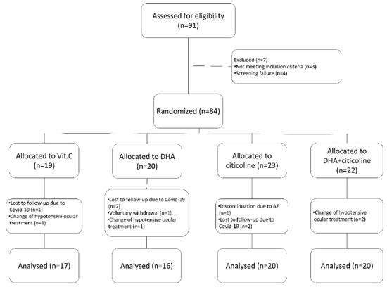
The Effect of Oral Citicoline and Docosahexaenoic Acid on the Visual Field of Patients with Glaucoma: A Randomized Trial
by Alfonso Anton, Virginia Garcia, Marcos Muñoz, Karla Gonzales, Eleonora Ayala, Estela del Mar Sanchez and Antonio Morilla-Grasa
Life 2022, 12(10), 1481; https://doi.org/10.3390/life12101481 - 23 Sep 2022
Cited by 6 | Viewed by 2699
Abstract
The role of nutraceuticals in the treatment of glaucoma remains controversial. The aim of this study was to evaluate the effect of citicoline, vitamin C, and docosahexaenoic acid (DHA) in patients with glaucoma. Methods: This was a prospective, randomized study. Patients with glaucoma [...] Read more.
The role of nutraceuticals in the treatment of glaucoma remains controversial. The aim of this study was to evaluate the effect of citicoline, vitamin C, and docosahexaenoic acid (DHA) in patients with glaucoma. Methods: This was a prospective, randomized study. Patients with glaucoma were randomized to one of four groups and treated for 3 months with vitamin C, DHA, citicoline, or a combination of DHA and citicoline. We conducted a complete ophthalmic examination and visual fields each month and calculated the slopes of field indices. Changes in visual field indices (VFIs) and their slopes were assessed in each group and compared. Results: Seventy-three persons were included in the study. Mean defect (MD) significantly improved (p = 0.001) from −9.52 ± 4.36 to −7.85 ± 4.36 dB during the study period in persons taking DHA + citicoline. Similarly, the mean VFI significantly improved (p = 0.001) in this group. The only treatment group showing a statistically significant improvement (p = 0.006) in the MD (from −0.1041 ± 0.2471 to 0.1383 ± 0.2544 dB/month) and VFI slope was the group treated with DHA+citicoline. Conclusions: The combination of oral treatment with DHA + citicoline significantly improved VF indices and their slopes in patients with glaucoma after 3 months of treatment. Full article
Show Figures

Figure 1

38 pages, 8201 KiB  
Article
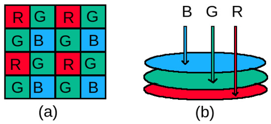
Which Color Channel Is Better for Diagnosing Retinal Diseases Automatically in Color Fundus Photographs?
by Sangeeta Biswas, Md. Iqbal Aziz Khan, Md. Tanvir Hossain, Angkan Biswas, Takayoshi Nakai and Johan Rohdin
Life 2022, 12(7), 973; https://doi.org/10.3390/life12070973 - 28 Jun 2022
Cited by 9 | Viewed by 3379
Abstract
Color fundus photographs are the most common type of image used for automatic diagnosis of retinal diseases and abnormalities. As all color photographs, these images contain information about three primary colors, i.e., red, green, and blue, in three separate color channels. This work [...] Read more.
Color fundus photographs are the most common type of image used for automatic diagnosis of retinal diseases and abnormalities. As all color photographs, these images contain information about three primary colors, i.e., red, green, and blue, in three separate color channels. This work aims to understand the impact of each channel in the automatic diagnosis of retinal diseases and abnormalities. To this end, the existing works are surveyed extensively to explore which color channel is used most commonly for automatically detecting four leading causes of blindness and one retinal abnormality along with segmenting three retinal landmarks. From this survey, it is clear that all channels together are typically used for neural network-based systems, whereas for non-neural network-based systems, the green channel is most commonly used. However, from the previous works, no conclusion can be drawn regarding the importance of the different channels. Therefore, systematic experiments are conducted to analyse this. A well-known U-shaped deep neural network (U-Net) is used to investigate which color channel is best for segmenting one retinal abnormality and three retinal landmarks. Full article
Show Figures

Figure 1

15 pages, 1525 KiB  
Article
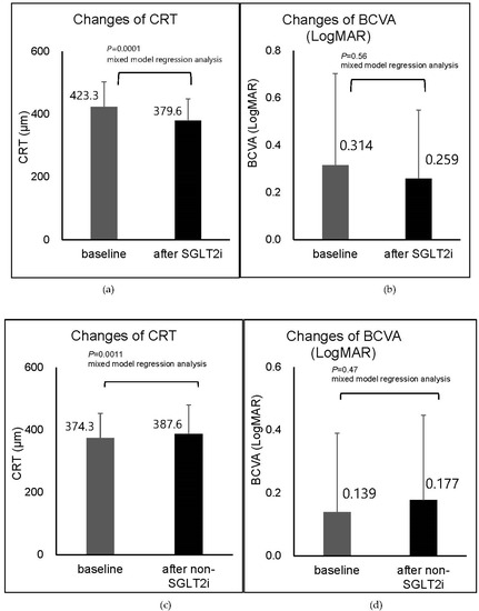
Sodium-Glucose Co-Transporter 2 Inhibitors Reduce Macular Edema in Patients with Diabetes mellitus
by Tomoaki Tatsumi, Toshiyuki Oshitari, Yoko Takatsuna, Ryoichi Ishibashi, Masaya Koshizaka, Yuki Shiko, Takayuki Baba, Koutaro Yokote and Shuichi Yamamoto
Life 2022, 12(5), 692; https://doi.org/10.3390/life12050692 - 6 May 2022
Cited by 14 | Viewed by 2149
Abstract
Purpose: To determine the efficacy of systemic sodium-glucose co-transporter 2 inhibitors (SGLT2i) on diabetic macular edema (DME). Methods: The medical records of patients with DME with a central retinal thickness (CRT) ≥320 µm in men and 305 µm in women, more than 6 [...] Read more.
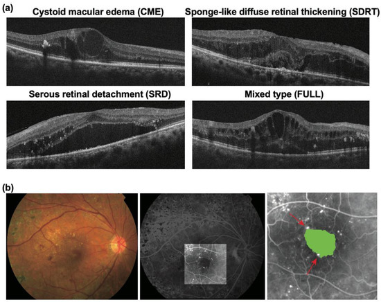
Purpose: To determine the efficacy of systemic sodium-glucose co-transporter 2 inhibitors (SGLT2i) on diabetic macular edema (DME). Methods: The medical records of patients with DME with a central retinal thickness (CRT) ≥320 µm in men and 305 µm in women, more than 6 months after the initiation of diabetes mellitus treatment, were reviewed. The CRT and best-corrected visual acuity (BCVA) were evaluated before and after the initiation of systemic SGLT2i and non-SGLT2i treatments. Results: There were 24 eyes of 19 patients with DME that were treatment naïve or had not received treatments for the DME within four months before the initiation of SGLT2i. In these patients, the BCVA had a 0.31 ± 0.39 logarithm of the minimum angle of resolution (logMAR) units at the baseline, and it did not improve significantly at 0.26 ± 0.29 logMAR units after the initiation of SGLT2i (p = 0.56). However, the SGLT2i treatment significantly reduced the CRT from 423.3 ± 79.8 µm to 379.6 ± 69.5 µm (p = 0.0001). In the same evaluation of 19 eyes of 14 patients with DME that were initiated with non-SGLT2i agents, there was no significant difference between the baseline BCVA and the BCVA after the initiation of non-SGLT2i (p = 0.47). The CRT increased significantly after the initiation of non-SGLT2i (p = 0.0011). In three eyes in which the SGLT2i treatments were administered at the time of anti-vascular endothelial growth factor (VEGF) treatments, the anti-VEGF treatment alone had only a limited effect on the DME, but the reduction in the DME was enhanced after the addition of SGLT2i. Conclusions: These findings indicate that systemic SGLT2i can reduce DMEs, and they suggest that SGLT2i may be an additional treatment option to anti-VEGF treatments for eyes with DMEs. Full article
Show Figures

Figure 1

7 pages, 2916 KiB  
Brief Report
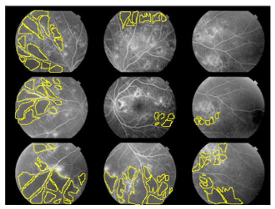
Non-Perfusion Area Index for Prognostic Prediction in Diabetic Retinopathy
by Yoshiko Ofuji, Yusaku Katada, Yohei Tomita, Norihiro Nagai, Hideki Sonobe, Kazuhiro Watanabe, Hajime Shinoda, Yoko Ozawa, Kazuno Negishi, Kazuo Tsubota and Toshihide Kurihara
Life 2022, 12(4), 542; https://doi.org/10.3390/life12040542 - 6 Apr 2022
Cited by 2 | Viewed by 1688
Abstract
Fundus fluorescent angiography is a standard examination in Japan that can directly visualize the circulatory failure in diabetic retinopathy but is not used in Western countries. In this study, we examine the relationship between the non-perfusion area in fundus fluorescent angiography and the [...] Read more.
Fundus fluorescent angiography is a standard examination in Japan that can directly visualize the circulatory failure in diabetic retinopathy but is not used in Western countries. In this study, we examine the relationship between the non-perfusion area in fundus fluorescent angiography and the progression of diabetic retinopathy. We evaluated 22 eyes between 22 patients who had their first fundus fluorescent angiography during a clinical episode at Keio University Hospital from January 2012 to May 2015, were diagnosed as having preproliferative diabetic retinopathy, and could be followed for at least three years. The non-perfusion area index (%) in nine segmented fundi in the initial fundus fluorescent angiography was calculated, and the progression to proliferative diabetic retinopathy over three years was evaluated. Three out of the 22 eyes (13.6%) developed proliferative diabetic retinopathy over three years. The non-perfusion area index for the initial fundus fluorescent angiography was significantly associated with progression to proliferative diabetic retinopathy. The non-perfusion area index in the posterior pole was most strongly correlated with the progression to proliferative diabetic retinopathy. Thus, the non-perfusion area index in the posterior pole among those with preproliferative diabetic retinopathy may predict the progression to proliferative diabetic retinopathy in the subsequent three years. Full article
Show Figures

Figure 1

14 pages, 1415 KiB  
Review
SD-OCT Biomarkers and the Current Status of Artificial Intelligence in Predicting Progression from Intermediate to Advanced AMD
by Ioana Damian and Simona Delia Nicoară
Life 2022, 12(3), 454; https://doi.org/10.3390/life12030454 - 19 Mar 2022
Cited by 7 | Viewed by 4065
Abstract
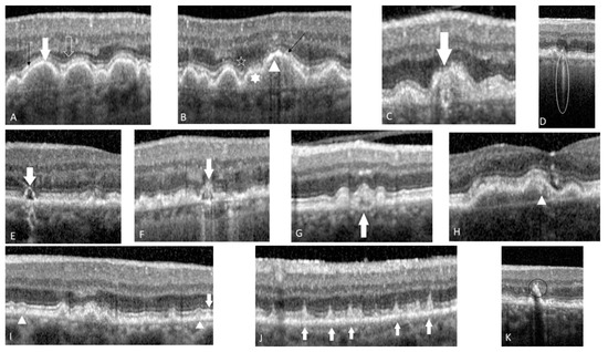
Age-related macular degeneration (AMD) is one of the leading causes of blindness in the Western World. Optical coherence tomography (OCT) has revolutionized the diagnosis and follow-up of AMD patients. This review focuses on SD-OCT imaging biomarkers which were identified as predictors for progression [...] Read more.
Age-related macular degeneration (AMD) is one of the leading causes of blindness in the Western World. Optical coherence tomography (OCT) has revolutionized the diagnosis and follow-up of AMD patients. This review focuses on SD-OCT imaging biomarkers which were identified as predictors for progression in intermediate AMD to late AMD, either geographic atrophy (GA) or choroidal neovascularization (CNV). Structural OCT remains the most compelling modality to study AMD features related to the progression such as drusen characteristics, hyperreflective foci (HRF), reticular pseudo-drusen (RPD), sub-RPE hyper-reflective columns and their impact on retinal layers. Further on, we reviewed articles that attempted to integrate biomarkers that have already proven their involvement in intermediate AMD progression, in their models of artificial intelligence (AI). By combining structural biomarkers with genetic risk and lifestyle the predictive ability becomes more accurate. Full article
Show Figures

Figure 1

31 pages, 2039 KiB  
Review
Pigment Epithelia of the Eye: Cell-Type Conversion in Regeneration and Disease
by Eleonora N. Grigoryan
Life 2022, 12(3), 382; https://doi.org/10.3390/life12030382 - 6 Mar 2022
Cited by 8 | Viewed by 4486
Abstract
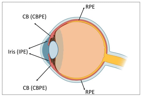
Pigment epithelial cells (PECs) of the retina (RPE), ciliary body, and iris (IPE) are capable of altering their phenotype. The main pathway of phenotypic switching of eye PECs in vertebrates and humans in vivo and/or in vitro is neural/retinal. Besides, cells of amphibian [...] Read more.
Pigment epithelial cells (PECs) of the retina (RPE), ciliary body, and iris (IPE) are capable of altering their phenotype. The main pathway of phenotypic switching of eye PECs in vertebrates and humans in vivo and/or in vitro is neural/retinal. Besides, cells of amphibian IPE give rise to the lens and its derivatives, while mammalian and human RPE can be converted along the mesenchymal pathway. The PECs’ capability of conversion in vivo underlies the lens and retinal regeneration in lower vertebrates and retinal diseases such as proliferative vitreoretinopathy and fibrosis in mammals and humans. The present review considers these processes studied in vitro and in vivo in animal models and in humans. The molecular basis of conversion strategies in PECs is elucidated. Being predetermined onto- and phylogenetically, it includes a species-specific molecular context, differential expression of transcription factors, signaling pathways, and epigenomic changes. The accumulated knowledge regarding the mechanisms of PECs phenotypic switching allows the development of approaches to specified conversion for many purposes: obtaining cells for transplantation, creating conditions to stimulate natural regeneration of the retina and the lens, blocking undesirable conversions associated with eye pathology, and finding molecular markers of pathology to be targets of therapy. Full article
Show Figures

Figure 1

7 pages, 209 KiB  
Editorial
Retinal Disease and Metabolism
by Zhongjie Fu, Ayumi Usui-Ouchi, William Allen and Yohei Tomita
Life 2022, 12(2), 183; https://doi.org/10.3390/life12020183 - 27 Jan 2022
Cited by 2 | Viewed by 2573
Abstract
Retinal diseases, such as diabetic retinopathy (DR), age-related macular degeneration (AMD), and retinopathy of prematurity (ROP), are some of the leading causes of blindness all over the world [...] Full article

2021

Jump to: 2022, 2020

12 pages, 796 KiB  
Article
Impact of Injection Protocol Selection by Retina Specialists on Clinical Outcomes in Patients with Diabetic Macular Edema
by Anika Tanwani, Nida Safdar, Amir Ali, Cina Karimaghaei, Mary Schmitz-Brown, Ahmad Rehmani and Praveena K. Gupta
Life 2022, 12(1), 51; https://doi.org/10.3390/life12010051 - 31 Dec 2021
Cited by 1 | Viewed by 1730
Abstract
Intravitreal anti-VEGF injections are the current gold standard for treating diabetic macular edema (DME). However, injection practice patterns of retina specialists have varied markedly based on physician discretion. This retrospective study analyzes the impact of injection protocol selection on change in best-corrected visual [...] Read more.
Intravitreal anti-VEGF injections are the current gold standard for treating diabetic macular edema (DME). However, injection practice patterns of retina specialists have varied markedly based on physician discretion. This retrospective study analyzes the impact of injection protocol selection on change in best-corrected visual acuity (BCVA) and central macular thickness (CMT) in 170 eyes treated by 4 retina specialists practicing a pro re nata (PRN) strategy between 2010 and 2020. DME patients received an average of 7.25 injections every 6.24 weeks over 56.6 weeks. There were significant differences between retina specialists in mean number of injections (p = 0.0001) and mean length of treatment (p = 0.0007) but not in mean interval between injections. Over the treatment period, average change in BCVA was −0.053 logMAR, and average change in CMT was −51.1 µm, neither of which had significant differences between retina specialists. BCVA and CMT at initial visit were found to be significantly associated with improved BCVA and CMT over the treatment period (p < 0.001). Number of injections administered and interval between injections were not found to be significant factors affecting change in BCVA or CMT. Despite significant differences in injection dosing regimen, retina specialists achieved similar outcomes in change in BCVA and CMT over the treatment period. Full article
Show Figures

Figure 1

11 pages, 2099 KiB  
Brief Report
Caveolin-1 Down-Regulation Reduces VEGF-A Secretion Induced by IGF-1 in ARPE-19 Cells
by Alessandra Puddu, Roberta Sanguineti and Davide Maggi
Life 2022, 12(1), 44; https://doi.org/10.3390/life12010044 - 28 Dec 2021
Cited by 7 | Viewed by 1902
Abstract
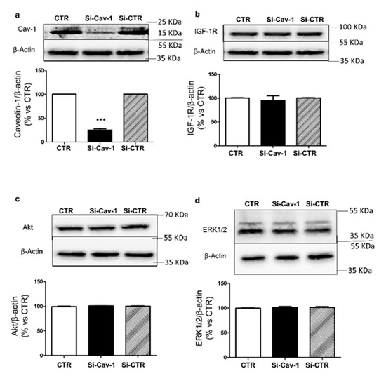
The insulin-like growth factor 1 (IGF-1) stimulates expression and secretion of vascular endothelial growth factor-A (VEGF-A), the main actor in ocular neovascularization, by RPE cells. Activity of IGF-1 is regulated by interaction between its receptor and Caveolin-1 (Cav-1), the main component of caveolae. [...] Read more.
The insulin-like growth factor 1 (IGF-1) stimulates expression and secretion of vascular endothelial growth factor-A (VEGF-A), the main actor in ocular neovascularization, by RPE cells. Activity of IGF-1 is regulated by interaction between its receptor and Caveolin-1 (Cav-1), the main component of caveolae. The aim of this study was to investigate whether modulation of Cav-1 expression affects synthesis and secretion of VEGF-A. ARPE-19 cells were transfected with small interfering RNA for Cav-1 (si-Cav-1) and with control siRNA (si-CTR) and stimulated with IGF-1. We found that down-regulation of Cav-1 did not affect activation of IGF-1R but regulated in an opposite manner the phosphorylation of Akt and Erk1/2. Moreover, we found that IGF-1 increased mRNA levels of VEGF-A in both si-CTR and in si-Cav-1 ARPE-19 cells and that Cav-1 silencing significantly reduced basal and IGF-1-stimulated VEGF-A release. Then we investigated the response of the microvascular endothelial cell line HMEC-1 to secretory products of ARPE-19 cells by evaluating wound healing closure, finding that conditioned media from si-Cav-1-ARPE-19 cells reduced endothelial cell migration rate. These data demonstrate that Cav-1 regulates secretion of VEGF-A, and that the depletion of Cav-1 reduces IGF-1 induced VEGF-A secretion in ARPE-19 cells and the migratory potential of their secretory products. Full article
Show Figures

Figure 1

17 pages, 11707 KiB  
Case Report
Bilateral Optic Disc Swelling as a Plausible Common Ocular Sign of Autoinflammatory Diseases: Report of Three Patients with Blau Syndrome or Cryopyrin-Associated Periodic Syndrome
by Toshihiko Matsuo, Masato Yashiro, Osamu Yamasaki, Takehiro Tanaka and Akira Manki
Life 2021, 11(12), 1433; https://doi.org/10.3390/life11121433 - 19 Dec 2021
Cited by 5 | Viewed by 3013
Abstract
The aim of this study is to describe bilateral optic disc swelling in three consecutive patients with Blau syndrome or cryopyrin-associated periodic syndrome at a single institution. Case 1 was a 30-year-old woman receiving 25 mg etanercept twice weekly who had been diagnosed [...] Read more.
The aim of this study is to describe bilateral optic disc swelling in three consecutive patients with Blau syndrome or cryopyrin-associated periodic syndrome at a single institution. Case 1 was a 30-year-old woman receiving 25 mg etanercept twice weekly who had been diagnosed as early-onset sarcoidosis by biopsy of skin rashes at 5 months old and genetically diagnosed with Blau syndrome with CARD15/NOD2 mutation (N670K) at 13 years old. At 10 years old, she began to have uveitis with optic disc swelling in both eyes, resulting in macular degeneration and optic disc atrophy at 17 years old only when etanercept was introduced. Case 2 was a 21-year-old man receiving adalimumab every 2 weeks who had been diagnosed as early-onset sarcoidosis by biopsy of skin rashes at 1.5 years old and genetically diagnosed as Blau syndrome with CARD15/NOD2 mutation (C495Y) at 5 years old. At 8 years old, around the time of adalimumab introduction, he began to show bilateral optic disc swelling which continued until the age of 16 years when the dose of adalimumab was increased. Case 3 was a 20-year-old woman receiving canakinumab every 8 weeks for systemic symptoms such as fever, headache, vomiting, and abdominal pain and later for sensorineural hearing disturbance on both sides. She had been diagnosed genetically with cryopyrin-associated periodic syndrome with NLRP3 mutation (Y859C) at 7 years old. At 5 years old, she was found to have bilateral optic disc swelling, which continued until the age of 10 years when she began receiving canakinumab (IL-1β inhibitor). Bilateral optic disc swelling might be tentatively designated as a plausible common ocular feature, if it occurred, in autoinflammatory diseases to pay more attention to ophthalmic complications in rare diseases. Full article
Show Figures

Figure 1

12 pages, 1389 KiB  
Article
Decorin Concentrations in Aqueous Humor of Patients with Diabetic Retinopathy
by Shermaine W. Y. Low, Tanuja Vaidya, Santosh G. K. Gadde, Thirumalesh B. Mochi, Devesh Kumar, Iris S. Kassem, Deborah M. Costakos, Baseer Ahmad, Swaminathan Sethu, Arkasubhra Ghosh and Shyam S. Chaurasia
Life 2021, 11(12), 1421; https://doi.org/10.3390/life11121421 - 17 Dec 2021
Cited by 7 | Viewed by 2592
Abstract
Diabetic retinopathy (DR) is a microvascular complication of diabetes in the retina. Chronic hyperglycemia damages retinal microvasculature embedded into the extracellular matrix (ECM), causing fluid leakage and ischemic retinal neovascularization. Current treatment strategies include intravitreal anti-vascular endothelial growth factor (VEGF) or steroidal injections, [...] Read more.
Diabetic retinopathy (DR) is a microvascular complication of diabetes in the retina. Chronic hyperglycemia damages retinal microvasculature embedded into the extracellular matrix (ECM), causing fluid leakage and ischemic retinal neovascularization. Current treatment strategies include intravitreal anti-vascular endothelial growth factor (VEGF) or steroidal injections, laser photocoagulation, or vitrectomy in severe cases. However, treatment may require multiple modalities or repeat treatments due to variable response. Though DR management has achieved great success, improved, long-lasting, and predictable treatments are needed, including new biomarkers and therapeutic approaches. Small-leucine rich proteoglycans, such as decorin, constitute an integral component of retinal endothelial ECM. Therefore, any damage to microvasculature can trigger its antifibrotic and antiangiogenic response against retinal vascular pathologies, including DR. We conducted a cross-sectional study to examine the association between aqueous humor (AH) decorin levels, if any, and severity of DR. A total of 82 subjects (26 control, 56 DR) were recruited. AH was collected and decorin concentrations were measured using an enzyme-linked immunosorbent assay (ELISA). Decorin was significantly increased in the AH of DR subjects compared to controls (p = 0.0034). AH decorin levels were increased in severe DR groups in ETDRS and Gloucestershire classifications. Decorin concentrations also displayed a significant association with visual acuity (LogMAR) measurements. In conclusion, aqueous humor decorin concentrations were found elevated in DR subjects, possibly due to a compensatory response to the retinal microvascular changes during hyperglycemia. Full article
Show Figures

Figure 1

13 pages, 1407 KiB  
Review
PPARα Modulation-Based Therapy in Central Nervous System Diseases
by Deokho Lee, Yohei Tomita, William Allen, Kazuo Tsubota, Kazuno Negishi and Toshihide Kurihara
Life 2021, 11(11), 1168; https://doi.org/10.3390/life11111168 - 2 Nov 2021
Cited by 10 | Viewed by 2973
Abstract
The burden of neurodegenerative diseases in the central nervous system (CNS) is increasing globally. There are various risk factors for the development and progression of CNS diseases, such as inflammatory responses and metabolic derangements. Thus, curing CNS diseases requires the modulation of damaging [...] Read more.
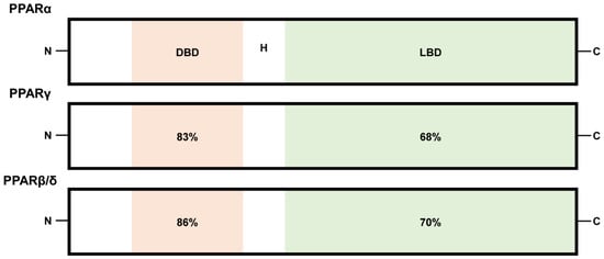
The burden of neurodegenerative diseases in the central nervous system (CNS) is increasing globally. There are various risk factors for the development and progression of CNS diseases, such as inflammatory responses and metabolic derangements. Thus, curing CNS diseases requires the modulation of damaging signaling pathways through a multitude of mechanisms. Peroxisome proliferator-activated receptors (PPARs) are a family of nuclear hormone receptors (PPARα, PPARβ/δ, and PPARγ), and they work as master sensors and modulators of cellular metabolism. In this regard, PPARs have recently been suggested as promising therapeutic targets for suppressing the development of CNS diseases and their progressions. While the therapeutic role of PPARγ modulation in CNS diseases has been well reviewed, the role of PPARα modulation in these diseases has not been comprehensively summarized. The current review focuses on the therapeutic roles of PPARα modulation in CNS diseases, including those affecting the brain, spinal cord, and eye, with recent advances. Our review will enable more comprehensive therapeutic approaches to modulate PPARα for the prevention of and protection from various CNS diseases. Full article
Show Figures

Figure 1

20 pages, 1581 KiB  
Review
Metabolism in Retinopathy of Prematurity
by Yohei Tomita, Ayumi Usui-Ouchi, Anders K. Nilsson, Jay Yang, Minji Ko, Ann Hellström and Zhongjie Fu
Life 2021, 11(11), 1119; https://doi.org/10.3390/life11111119 - 21 Oct 2021
Cited by 20 | Viewed by 10512
Abstract
Retinopathy of prematurity is defined as retinal abnormalities that occur during development as a consequence of disturbed oxygen conditions and nutrient supply after preterm birth. Both neuronal maturation and retinal vascularization are impaired, leading to the compensatory but uncontrolled retinal neovessel growth. Current [...] Read more.
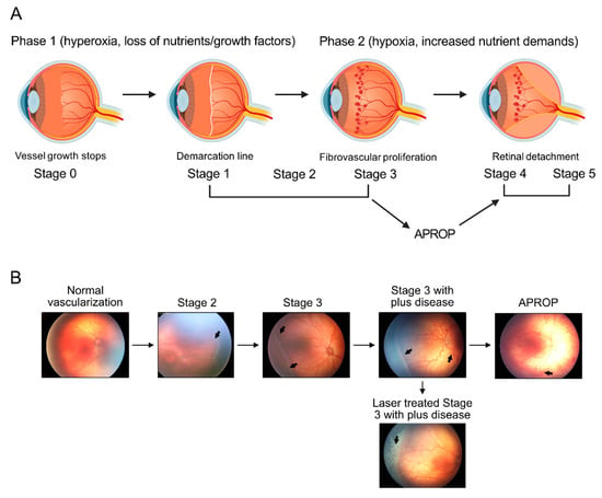
Retinopathy of prematurity is defined as retinal abnormalities that occur during development as a consequence of disturbed oxygen conditions and nutrient supply after preterm birth. Both neuronal maturation and retinal vascularization are impaired, leading to the compensatory but uncontrolled retinal neovessel growth. Current therapeutic interventions target the hypoxia-induced neovessels but negatively impact retinal neurons and normal vessels. Emerging evidence suggests that metabolic disturbance is a significant and underexplored risk factor in the disease pathogenesis. Hyperglycemia and dyslipidemia correlate with the retinal neurovascular dysfunction in infants born prematurely. Nutritional and hormonal supplementation relieve metabolic stress and improve retinal maturation. Here we focus on the mechanisms through which metabolism is involved in preterm-birth-related retinal disorder from clinical and experimental investigations. We will review and discuss potential therapeutic targets through the restoration of metabolic responses to prevent disease development and progression. Full article
Show Figures

Figure 1

15 pages, 853 KiB  
Review
Interactions between Apolipoprotein E Metabolism and Retinal Inflammation in Age-Related Macular Degeneration
by Monica L. Hu, Joel Quinn and Kanmin Xue
Life 2021, 11(7), 635; https://doi.org/10.3390/life11070635 - 29 Jun 2021
Cited by 23 | Viewed by 5271
Abstract
Age-related macular degeneration (AMD) is a multifactorial retinal disorder that is a major global cause of severe visual impairment. The development of an effective therapy to treat geographic atrophy, the predominant form of AMD, remains elusive due to the incomplete understanding of its [...] Read more.
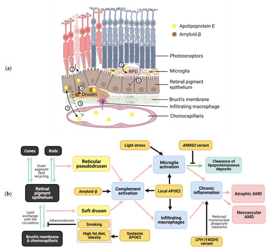
Age-related macular degeneration (AMD) is a multifactorial retinal disorder that is a major global cause of severe visual impairment. The development of an effective therapy to treat geographic atrophy, the predominant form of AMD, remains elusive due to the incomplete understanding of its pathogenesis. Central to AMD diagnosis and pathology are the hallmark lipid and proteinaceous deposits, drusen and reticular pseudodrusen, that accumulate in the subretinal pigment epithelium and subretinal spaces, respectively. Age-related changes and environmental stressors, such as smoking and a high-fat diet, are believed to interact with the many genetic risk variants that have been identified in several major biochemical pathways, including lipoprotein metabolism and the complement system. The APOE gene, encoding apolipoprotein E (APOE), is a major genetic risk factor for AMD, with the APOE2 allele conferring increased risk and APOE4 conferring reduced risk, in comparison to the wildtype APOE3. Paradoxically, APOE4 is the main genetic risk factor in Alzheimer’s disease, a disease with features of neuroinflammation and amyloid-beta deposition in common with AMD. The potential interactions of APOE with the complement system and amyloid-beta are discussed here to shed light on their roles in AMD pathogenesis, including in drusen biogenesis, immune cell activation and recruitment, and retinal inflammation. Full article
Show Figures

Figure 1

11 pages, 2677 KiB  
Brief Report
Hyperoxia Inhibits Proliferation of Retinal Endothelial Cells in a Myc-Dependent Manner
by Charandeep Singh, Andrew Benos, Allison Grenell, Sujata Rao, Bela Anand-Apte and Jonathan E. Sears
Life 2021, 11(7), 614; https://doi.org/10.3390/life11070614 - 25 Jun 2021
Cited by 3 | Viewed by 2744
Abstract
Oxygen supplementation is necessary to prevent mortality in severely premature infants. However, the supraphysiological concentration of oxygen utilized in these infants simultaneously creates retinovascular growth attenuation and vasoobliteration that induces the retinopathy of prematurity. Here, we report that hyperoxia regulates the cell cycle [...] Read more.
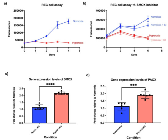
Oxygen supplementation is necessary to prevent mortality in severely premature infants. However, the supraphysiological concentration of oxygen utilized in these infants simultaneously creates retinovascular growth attenuation and vasoobliteration that induces the retinopathy of prematurity. Here, we report that hyperoxia regulates the cell cycle and retinal endothelial cell proliferation in a previously unknown Myc-dependent manner, which contributes to oxygen-induced retinopathy. Full article
Show Figures

Figure 1

18 pages, 4195 KiB  
Article
Photoelectric Dye, NK-5962, as a Potential Drug for Preventing Retinal Neurons from Apoptosis: Pharmacokinetic Studies Based on Review of the Evidence
by Toshihiko Matsuo, Shihui Liu, Tetsuya Uchida, Satomi Onoue, Shinsaku Nakagawa, Mayumi Ishii and Kayoko Kanamitsu
Life 2021, 11(6), 591; https://doi.org/10.3390/life11060591 - 21 Jun 2021
Cited by 4 | Viewed by 3565
Abstract
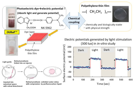
NK-5962 is a key component of photoelectric dye-based retinal prosthesis (OUReP). In testing the safety and efficacy, NK-5962 was safe in all tests for the biological evaluation of medical devices (ISO 10993) and effective in preventing retinal cells from death even under dark [...] Read more.
NK-5962 is a key component of photoelectric dye-based retinal prosthesis (OUReP). In testing the safety and efficacy, NK-5962 was safe in all tests for the biological evaluation of medical devices (ISO 10993) and effective in preventing retinal cells from death even under dark conditions. The long-term implantation of the photoelectric dye-coupled polyethylene film in the subretinal space of hereditary retinal dystrophic (RCS) rats prevented neurons from apoptosis in the adjacent retinal tissue. The intravitreous injection of NK-5962 in the eyes of RCS rats, indeed, reduced the number of apoptotic cells in the retinal outer nuclear layer irrespective of light or dark conditions. In this study, we reviewed the in vitro and in vivo evidence of neuroprotective effect of NK-5962 and designed pharmacokinetic experiments. The in vitro IC50 of 1.7 μM, based on the protective effect on retinal cells in culture, could explain the in vivo EC50 of 3 μM that is calculated from concentrations of intravitreous injection to prevent retinal neurons from apoptosis. Pharmacokinetics of NK-5962 showed that intravenous administration, but not oral administration, led to the effective concentration in the eye of rats. NK-5962 would be a candidate drug for delaying the deterioration of retinal dystrophy, such as retinitis pigmentosa. Full article
Show Figures

Figure 1

9 pages, 925 KiB  
Review
Diabetic Retinopathy in Children with Type 1 Diabetes—Occurrence and Screening Using Optical Coherence Tomography
by Marta Wysocka-Mincewicz, Joanna Gołębiewska, Andrzej Olechowski and Mieczysław Szalecki
Life 2021, 11(6), 590; https://doi.org/10.3390/life11060590 - 21 Jun 2021
Cited by 11 | Viewed by 3272
Abstract
Purpose: To describe the occurrence of diabetic retinopathy, the principles for pediatric care of patients with diabetes, and the utility of optical coherence tomography. Pediatric patients with type 1 diabetes should be screened for diabetic retinopathy upon the lapse of 5 years following [...] Read more.
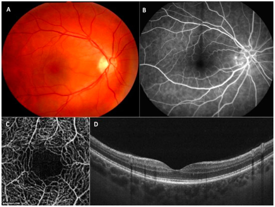
Purpose: To describe the occurrence of diabetic retinopathy, the principles for pediatric care of patients with diabetes, and the utility of optical coherence tomography. Pediatric patients with type 1 diabetes should be screened for diabetic retinopathy upon the lapse of 5 years following the diagnosis. The patients in the time of puberty, who should be screened promptly after the diabetes diagnosis, and patients with type 2 diabetes are the exceptions. Special attention must be paid not only to retinopathy, but also to other possible concomitant conditions, such as cataract, refractive errors, or neuropathy. New techniques, such as optical coherence tomography angiography (OCTA), may contribute greatly to the early detection of retinopathy, facilitating the decision to modify the treatment. The application of modern insulin pumps with continuous glucose monitoring systems has greatly diminished the incidence rate of early symptoms of diabetic retinopathy in the pediatric population. Full article
Show Figures

Figure 1

6 pages, 884 KiB  
Article
Subfoveal Choroidal Thickness and Treatment Outcomes of Intravitreal Aflibercept for Branch Retinal Vein Occlusion
by Yoshihito Sakanishi, Syu Morita, Keitaro Mashimo, Kazunori Tamaki and Nobuyuki Ebihara
Life 2021, 11(6), 572; https://doi.org/10.3390/life11060572 - 17 Jun 2021
Cited by 8 | Viewed by 1971
Abstract
We aimed to investigate the relationship between subfoveal choroidal thickness (SCT) and treatment outcomes of intravitreal aflibercept (IVA) for macular edema (ME) due to branch retinal vein occlusion (BRVO). We retrospectively evaluated 46 patients with treatment-naive BRVO-ME who underwent IVA treatment between March [...] Read more.
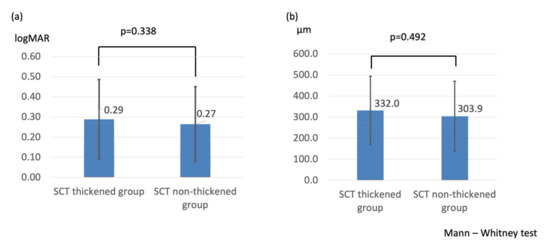
We aimed to investigate the relationship between subfoveal choroidal thickness (SCT) and treatment outcomes of intravitreal aflibercept (IVA) for macular edema (ME) due to branch retinal vein occlusion (BRVO). We retrospectively evaluated 46 patients with treatment-naive BRVO-ME who underwent IVA treatment between March 2016 and February 2017. There was no significant difference in visual acuity within 6 months (0.29 ± 0.20 vs. 0.27 ± 0.19, p = 0.338), the mean central foveal thickness improvement (332.0 ± 162.2 μm vs. 303.9 ± 166.6 μm, p = 0.492), and the mean number of IVA injections (1.7 ± 0.7 vs. 1.6 ± 0.7 times, p = 0.658) between the SCT thickened (n = 26 patients, 26 eyes) and SCT non-thickened groups (n = 20 patients, 20 eyes). The rate of ME recurrence was significantly lower in the SCT decreased group (6/17 eyes (35.2%) vs. 19/30 eyes (63.3%); p = 0.038). In conclusion, pretreatment choroidal thickening does not affect the therapeutic effect of IVA for BRVO, but ME recurrence was lower in cases of treatment-related choroidal thinning. Thus, changes in SCT may be a therapeutic indicator of IVA for acute BRVO. Full article
Show Figures

Figure 1

13 pages, 2823 KiB  
Article
Fluorescence Lifetime Changes Induced by Laser Irradiation: A Preclinical Study towards the Evaluation of Retinal Metabolic States
by Svenja Rebecca Sonntag, Eric Seifert, Maximilian Hamann, Britta Lewke, Dirk Theisen-Kunde, Salvatore Grisanti, Ralf Brinkmann and Yoko Miura
Life 2021, 11(6), 555; https://doi.org/10.3390/life11060555 - 13 Jun 2021
Cited by 5 | Viewed by 2487
Abstract
Fluorescence Lifetime (FLT) of intrinsic fluorophores may alter under the change in metabolic state. In this study, the FLT of rabbit retina was investigated in vivo after laser irradiation using fluorescence lifetime imaging ophthalmoscopy (FLIO). The retina of the Chinchilla bastard rabbits was [...] Read more.
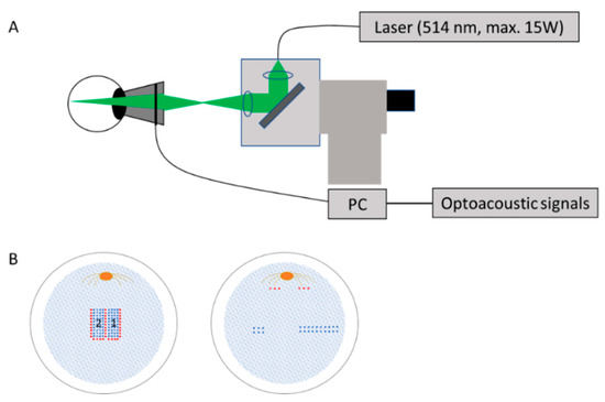
Fluorescence Lifetime (FLT) of intrinsic fluorophores may alter under the change in metabolic state. In this study, the FLT of rabbit retina was investigated in vivo after laser irradiation using fluorescence lifetime imaging ophthalmoscopy (FLIO). The retina of the Chinchilla bastard rabbits was irradiated with a 514 nm diode laser. FLIO, fundus photography, and optical coherence tomography (OCT) were conducted 30 min and 1 to 3 weeks after treatment. After strong coagulation, the FLT at laser spots was significantly elongated immediately after irradiation, conversely shortened after more than a week. Histological examination showed eosinophilic substance and melanin clumping in subretinal space at the coagulation spots older than one week. The FLT was also elongated right around the coagulation spots, which corresponded to the discontinuous ellipsoid zone (EZ) on OCT. This EZ change was recovered after one week, and the FLT became the same level as the surroundings. In addition, there was a region around the laser spot where the FLT was temporarily shorter than the surrounding area. When weak pulse energy was applied to selectively destroy only the RPE, a shortening of the FLT was observed immediately around the laser spot within one week after irradiation. FLIO could serve as a tool to evaluate the structural and metabolic response of the retina to laser treatments. Full article
Show Figures

Figure 1

14 pages, 1901 KiB  
Article
Electroretinography and Gene Expression Measures Implicate Phototransduction and Metabolic Shifts in Chick Myopia and Hyperopia Models
by Nina Riddell, Melanie J. Murphy and Sheila G. Crewther
Life 2021, 11(6), 501; https://doi.org/10.3390/life11060501 - 29 May 2021
Cited by 6 | Viewed by 3068
Abstract
The Retinal Ion-Driven Fluid Efflux (RIDE) model theorizes that phototransduction-driven changes in trans-retinal ion and fluid transport underlie the development of myopia (short-sightedness). In support of this model, previous functional studies have identified the attenuation of outer retinal contributions to the global flash [...] Read more.
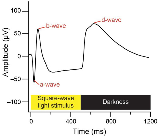
The Retinal Ion-Driven Fluid Efflux (RIDE) model theorizes that phototransduction-driven changes in trans-retinal ion and fluid transport underlie the development of myopia (short-sightedness). In support of this model, previous functional studies have identified the attenuation of outer retinal contributions to the global flash electroretinogram (gfERG) following weeks of myopia induction in chicks, while discovery-driven transcriptome studies have identified changes to the expression of ATP-driven ion transport and mitochondrial metabolism genes in the retina/RPE/choroid at the mid- to late-induction time-points. Less is known about the early time-points despite biometric analyses demonstrating changes in eye growth by 3 h in the chick lens defocus model. Thus, the present study compared gfERG and transcriptome profiles between 3 h and 3 days of negative lens-induced myopia and positive lens-induced hyperopia in chicks. Photoreceptor (a-wave and d-wave) and bipolar (b-wave and late-stage d-wave) cell responses were suppressed following negative lens-wear, particularly at the 3–4 h and 3-day time-points when active shifts in the rate of ocular growth were expected. Transcriptome measures revealed the up-regulation of oxidative phosphorylation genes following 6 h of negative lens-wear, concordant with previous reports at 2 days in this model. Signal transduction pathways, with core genes involved in glutamate and G-protein coupled receptor signalling, were down-regulated at 6 h. These findings contribute to a growing body of evidence for the dysregulation of phototransduction and mitochondrial metabolism in animal models of myopia. Full article
Show Figures

Figure 1

11 pages, 398 KiB  
Article
Antioxidant Status in the Vitreous of Eyes with Rhegmatogenous Retinal Detachment with and without Proliferative Vitreoretinopathy, Macular Hole and Epiretinal Membrane
by Agata Pietras-Baczewska, Katarzyna Nowomiejska, Agnieszka Brzozowska, Mario Damiano Toro, Wojciech Załuska, Małgorzata Sztanke, Krzysztof Sztanke and Robert Rejdak
Life 2021, 11(5), 453; https://doi.org/10.3390/life11050453 - 19 May 2021
Cited by 12 | Viewed by 2296
Abstract
(1) Background: The aim of the study was to test the hypothesis that the antioxidant status in the vitreous body of eyes, which had been vitrectomized due to rhegmatogenous retinal detachment (RRD) with or without proliferative vitreoretinopathy (PVR), is higher than in eyes [...] Read more.
(1) Background: The aim of the study was to test the hypothesis that the antioxidant status in the vitreous body of eyes, which had been vitrectomized due to rhegmatogenous retinal detachment (RRD) with or without proliferative vitreoretinopathy (PVR), is higher than in eyes vitrectomized due to other retinal diseases. (2) Methods: four patient groups were analyzed: 22 eyes of patients with RRD without PVR, 27 eyes with RRD and PVR, 22 eyes with macular hole (MH) and 10 eyes with epiretinal membrane (ERM). Spectrophotometric methods were used to determine the total antioxidant status (TAS) values as well as superoxide dismutase (SOD) and glutathione reductase (GR) activities in the vitreous fluid samples. (3) Results: no significant differences in TAS values and antioxidant enzyme activities were observed among patient with RRD with and without PVR and with MH and ERM. The longer the duration of RRD leading to PVR and better postoperative visual acuity, the higher the TAS level. No significant differences were found between “macula on” and “macula off” subgroups within the RRD group and the RRD combined with PVR group. (4) Conclusions: The preliminary results do not support the thesis that the antioxidant status of vitrectomized eyes is different in patients with RRD with or without PVR in comparison to patients with MH and ERM. In patients with RRD, PVR presence and detached macula do not affect the values of TAS, SOD and GR in the vitreous fluid. The duration of the disease influences TAS in the vitreous in eyes with RRD complicated with PVR. Full article
Show Figures

Figure 1

15 pages, 3685 KiB  
Article
Evaluation of Blood Coagulation Parameters and ADMA, NO, IL-6, and IL-18 Serum Levels in Patients with Neovascular AMD before, during, and after the Initial Loading Phase of Intravitreal Aflibercept
by Michał Wiciński, Małgorzata Seredyka-Burduk, Sławomir Liberski, Daria Marczak, Magdalena Pol, Bartosz Malinowski, Katarzyna Pawlak-Osińska and Bartlomiej J. Kaluzny
Life 2021, 11(5), 441; https://doi.org/10.3390/life11050441 - 14 May 2021
Cited by 5 | Viewed by 2526
Abstract
We evaluated the effect of intravitreal injections of aflibercept (IVA) on blood coagulation parameters including prothrombin time (PT), activated partial thromboplastin time (APTT), and thrombin time (TT), as well as asymmetric dimethylarginine (ADMA), nitric oxide (NO), interleukin 6 (IL-6), and interleukin 18 (IL-18) [...] Read more.
We evaluated the effect of intravitreal injections of aflibercept (IVA) on blood coagulation parameters including prothrombin time (PT), activated partial thromboplastin time (APTT), and thrombin time (TT), as well as asymmetric dimethylarginine (ADMA), nitric oxide (NO), interleukin 6 (IL-6), and interleukin 18 (IL-18) serum levels in patients with neovascular AMD (nAMD). Twenty-two eyes of 22 patients with nAMD were included. Parameters were evaluated before and 2–3 days after the first IVA injection, and then immediately before and 2–3 days after the third IVA injection. We revealed prolongation of the TT after the initial loading phase of IVA (p = 0.041) and a significant increase in IL-18 serum concentration immediately before the third IVA administration compared to baseline (p = 0.037). There were no statistically significant differences of other parameters and PT, APTT, ADMA, NO, and IL-6 values remained within the normal range at each of the time points of the study. Our results suggest that repeated IVA administration may affect the common blood coagulation pathway, which manifests as a prolongation of the TT value. Furthermore, we showed a significant increase in serum concentration of the pro-inflammatory cytokineIL-18during the initial loading phase of IVA. Full article
Show Figures

Figure 1

9 pages, 3015 KiB  
Article
Clinical Characteristics, Differential Diagnosis and Genetic Analysis of Concentric Retinitis Pigmentosa
by Mei Nakahara, Akio Oishi, Manabu Miyata, Hanako Ohashi Ikeda, Tomoko Hasegawa, Shogo Numa, Yuki Otsuka, Maho Oishi, Fumihiko Matsuda and Akitaka Tsujikawa
Life 2021, 11(3), 260; https://doi.org/10.3390/life11030260 - 22 Mar 2021
Cited by 5 | Viewed by 3520
Abstract
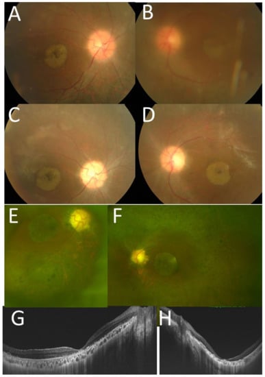
Concentric retinitis pigmentosa (RP), in which retinal degeneration is limited in the periphery, is rare and little information exists to date on the subject. Herein, we describe the clinical and genetic characteristics of this atypical form of RP. We retrospectively reviewed our database [...] Read more.
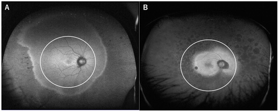
Concentric retinitis pigmentosa (RP), in which retinal degeneration is limited in the periphery, is rare and little information exists to date on the subject. Herein, we describe the clinical and genetic characteristics of this atypical form of RP. We retrospectively reviewed our database and identified 14 patients with concentric RP. Additionally, 14 patients with age-matched typical RP were also included. Patients with concentric RP had better visual acuity (logarithm of minimum angle of resolution −0.04 vs. 0.32, p = 0.047) and preserved ellipsoid zones (7630 µm vs. 2646 µm, p < 0.001) compared to typical RP. The electroretinogram showed subnormal but recordable responses in patients with concentric RP. Genetic testing was done in nine patients with concentric RP and revealed causative mutations in the EYS gene in one patient and the RP9 gene in one patient. Two patients had myotonic dystrophy and the diagnosis was revised as myotonic dystrophy-associated retinopathy. Concentric RP is a rare, atypical form of RP with better visual function. There is some overlap in the causative genes in concentric and typical RP. Myotonic dystrophy-associated retinopathy is an important differential diagnosis. Full article
Show Figures

Figure 1

11 pages, 598 KiB  
Article
Prevalence of Epiretinal Membrane among Subjects in a Health Examination Program in Japan
by Hiroshi Shimizu, Ryo Asaoka, Takashi Omoto, Yuri Fujino, Shingo Mitaki, Keiichi Onoda, Atsushi Nagai, Shuhei Yamaguchi and Masaki Tanito
Life 2021, 11(2), 93; https://doi.org/10.3390/life11020093 - 27 Jan 2021
Cited by 5 | Viewed by 2655
Abstract
The prevalence of an epiretinal membrane (ERM) was elucidated using a dataset from a health examination program database in Japan. From the cohort database, 5042 eyes of 2552 subjects were included. The presence of an ERM, cellophane macular reflex (CMR), or preretinal macular [...] Read more.
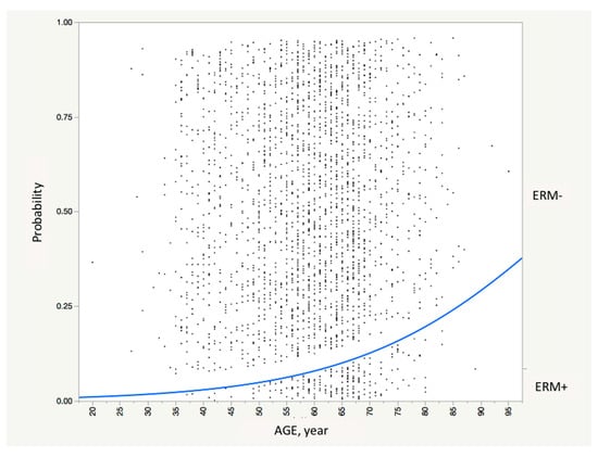
The prevalence of an epiretinal membrane (ERM) was elucidated using a dataset from a health examination program database in Japan. From the cohort database, 5042 eyes of 2552 subjects were included. The presence of an ERM, cellophane macular reflex (CMR), or preretinal macular fibrosis (PMF) was detected using color fundus photographs, and crude and age-standardized prevalence were obtained. To further assess the possible risk factors of ERM, background parameters were compared between ERM+ and − groups, and multiple logistic regression analysis was performed. ERM was detected in fundus photographs of 275 eyes (eye-based prevalence of 5.5%) from 217 subjects (subject-based prevalence of 8.5%). CMR was detected in 169 eyes (3.4%) of 138 subjects (5.4%), and PMF was detected in 106 eyes (2.1%) of 97 subjects (3.8%). By univariate analyses, compared with ERM− eyes or subjects, higher Scheie’s H grade (p < 0.0001), S grade (p < 0.0001), and glaucoma prevalence (p = 0.0440) were found in ERM+ eyes, and older age (p < 0.0001), more frequent histories of hypertension (p = 0.0033) and hyperlipidemia (p = 0.0441), and more frequent uses of medication for hypertension (p = 0.0034) and hyperlipidemia (p = 0.0074), shorter body height (p = 0.0122), and higher systolic blood pressure (p = 0.0078), and thicker intimal medial thickness (p = 0.0318) were found in ERM+ subjects. By multivariate analyses, older age (p < 0.0001, estimate = 0.05/year) was the only significant factor of ERM prevalence. Age-standardized prevalence of ERM was calculated to be 2.4%, 6.7%, and 13.3% for all ages, subjects older than 40 years, and subjects older than 65 years, respectively. We reported the prevalence of ERM and its subclasses in Japanese subjects. Since its prevalence is remarkably high in older subjects, an ERM can be seen as an important cause of visual impairment in Japan and in areas of the world where individuals live to an advanced age. Full article
Show Figures

Figure 1

9 pages, 935 KiB  
Article
Factors Affecting a Short-Term Response to Anti-VEGF Therapy in Diabetic Macular Edema
by Ayumi Usui-Ouchi, Asaka Tamaki, Yoshihito Sakanishi, Kazunori Tamaki, Keitaro Mashimo, Toshiro Sakuma and Nobuyuki Ebihara
Life 2021, 11(2), 83; https://doi.org/10.3390/life11020083 - 25 Jan 2021
Cited by 17 | Viewed by 4075
Abstract
Diabetic macular edema (DME) is a common cause of visual impairment in patients with diabetes. Although intravitreal anti-vascular endothelial growth factor (VEGF) injections were efficacious in clinical trials, several patients exhibited a poor response. This study aimed to compare clinical features between patients [...] Read more.
Diabetic macular edema (DME) is a common cause of visual impairment in patients with diabetes. Although intravitreal anti-vascular endothelial growth factor (VEGF) injections were efficacious in clinical trials, several patients exhibited a poor response. This study aimed to compare clinical features between patients who were susceptible to intravitreal anti-VEGF injections for DME and those who were not. A single-center, retrospective study of 102 such patients was conducted (123 eyes; mean ± standard deviation age, 63.4 ± 10.8 years; 57.8% males). Systemic and ocular data, assessed at baseline and after a month, were compared between good (>20% decrease in central macular thickness (CMT)) and poor (≤20% decrease in CMT) responders using the Mann–Whitney U test/Fisher’s exact test. Eighty-one eyes (65.9%) were good responders. The glycosylated hemoglobin level was higher (p = 0.011) in poor (7.5% ± 0.94%) than in good (7.04% ± 1.19%) responders. The foveal avascular zone was larger (p = 0.0003) in poor (0.67 ± 0.33 μm2) than in good (0.47 ± 0.23 μm2) responders. The number of microaneurysms in the pericapillary network was higher (p = 0.0007) in poor (2.7 ± 2.2) than in good (1.4 ± 2.0) responders. Baseline glycemic control and macular ischemia may be associated with the short-term response to intravitreal anti-VEGF injections. Full article
Show Figures

Figure 1

34 pages, 3035 KiB  
Review
Potential Effects of Nutraceuticals in Retinopathy of Prematurity
by Jessica K. W. Tsang, Susanne A. Wolf, Inga M. Pompoes, Antonia M. Joussen, Wai Ching Lam, Di Yang and Amy C. Y. Lo
Life 2021, 11(2), 79; https://doi.org/10.3390/life11020079 - 22 Jan 2021
Cited by 13 | Viewed by 4043
Abstract
Retinopathy of prematurity (ROP), the most common cause of childhood blindness, is a hypoxia-induced eye disease characterized by retinal neovascularization. In the normal retina, a well-organized vascular network provides oxygen and nutrients as energy sources to maintain a normal visual function; however, it [...] Read more.
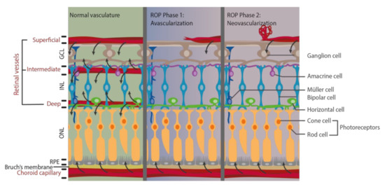
Retinopathy of prematurity (ROP), the most common cause of childhood blindness, is a hypoxia-induced eye disease characterized by retinal neovascularization. In the normal retina, a well-organized vascular network provides oxygen and nutrients as energy sources to maintain a normal visual function; however, it is disrupted when pathological angiogenesis is induced in ROP patients. Under hypoxia, inadequate oxygen and energy supply lead to oxidative stress and stimulate neovasculature formation as well as affecting the function of photoreceptors. In order to meet the metabolic needs in the developing retina, protection against abnormal vascular formation is one way to manage ROP. Although current treatments provide beneficial effects in reducing the severity of ROP, these invasive therapies may also induce life-long consequences such as systemic structural and functional complications as well as neurodevelopment disruption in the developing infants. Nutritional supplements for the newborns are a novel concept for restoring energy supply by protecting the retinal vasculature and may lead to better ROP management. Nutraceuticals are provided in a non-invasive manner without the developmental side effects associated with current treatments. These nutraceuticals have been investigated through various in vitro and in vivo methods and are indicated to protect retinal vasculature. Here, we reviewed and discussed how the use of these nutraceuticals may be beneficial in ROP prevention and management. Full article
Show Figures

Figure 1

2020

Jump to: 2022, 2021

10 pages, 236 KiB  
Article
Association between Systemic Antioxidant Capacity and Retinal Vessel Diameters in Patients with Primary-Open Angle Glaucoma
by Yuji Takayanagi, Yasuyuki Takai, Sachiko Kaidzu and Masaki Tanito
Life 2020, 10(12), 364; https://doi.org/10.3390/life10120364 - 20 Dec 2020
Cited by 5 | Viewed by 2245
Abstract
The retinal vessel narrowing may be implicated in the pathogenesis of glaucoma; however, the association between systemic oxidative stress and retinal vessel diameter remains largely unknown. We examined the relationship between serum oxidative stress markers and retinal vessel diameters in eyes with primary [...] Read more.
The retinal vessel narrowing may be implicated in the pathogenesis of glaucoma; however, the association between systemic oxidative stress and retinal vessel diameter remains largely unknown. We examined the relationship between serum oxidative stress markers and retinal vessel diameters in eyes with primary open-angle glaucoma (POAG) and cataract, using central retinal artery equivalent (CRAE) and central retinal vein equivalent (CRVE). We included 66 eyes of 66 patients with POAG (37 men, 29 women; 65.4 ± 11.7 years) and 20 eyes of 20 patients with cataract (7 men, 13 women; 69.4 ± 9.0 years) as the controls. The CRAE (p < 0.0001), CRVE (p < 0.0001), and serum biological antioxidant potential (BAP) (p = 0.0419) were significantly lower in the POAG group compared to the controls. The BAP showed significant correlation both with CRAE (ρ = 0.2148, p = 0.0471) and systolic blood pressure (ρ = −0.2431, p = 0.0241), while neither Diacron reactive oxygen metabolites nor sulfhydryl test correlated with them. The multivariate analyses indicated that age, best corrected visual acuity, and BAP were independent factors for CRAE or CRVE. The present study suggested that lower systemic antioxidant capacity was significantly associated with the intraocular pressure-independent vascular narrowing in POAG patients. This study provided a novel insight into the pathophysiology of glaucoma and highlighted the clinical impact on systemic antioxidant treatment for patients with glaucoma. Full article
7 pages, 2518 KiB  
Article
Efficacy of the Newly Invented Eyelid Clamper in Ultra-Widefield Fundus Imaging
by Nobuhiro Ozawa, Kiwako Mori, Yusaku Katada, Kazuo Tsubota and Toshihide Kurihara
Life 2020, 10(12), 323; https://doi.org/10.3390/life10120323 - 2 Dec 2020
Cited by 3 | Viewed by 3930
Abstract
Background: Ultra-widefield fundus imaging is widely used for obtaining wide angle images of the retina in one single image. Although it has a potential to obtain a wide area of retinal photographs, images are often obstructed by eyelashes or eye lids. In this [...] Read more.
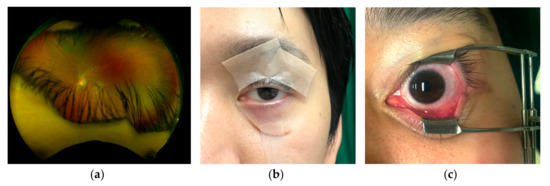
Background: Ultra-widefield fundus imaging is widely used for obtaining wide angle images of the retina in one single image. Although it has a potential to obtain a wide area of retinal photographs, images are often obstructed by eyelashes or eye lids. In this study, we used a newly invented eyelid clamper, which can keep an eye open without touching conjunctiva or lid margin, to assess the efficacy in clinical use by comparing with conventional tape fixation. Methods: Ultra-widefield fundus images were captured with an ultra-widefield imaging system in 19 patients who visited to the outpatient clinic of Department of Ophthalmology, Keio University Hospital with the eyelid clamper or a conventional tape fixation. The area of imaged retinas was outlined and quantified with pixels. After obtaining images, patients answered a questionnaire. Results: The average number of pixels in total areas with the eyelid clamper or with tape fixation were 4.31 ± 0.35 and 4.32 ± 0.34 mega pixels, respectively, showing no significant difference between the groups (p = 0.889). The average face pain scale of the eyelid clamper was 1.13 on a scale of 0 to 5. The number of patients who did not feel any pain was nine (47.4%). Conclusions: The eyelid clamper can be applied in clinical setting and can better support obtaining sufficiently wide fundus images compared to a conventional tape fixation. Full article
Show Figures

Figure 1

Planned Papers

The below list represents only planned manuscripts. Some of these manuscripts have not been received by the Editorial Office yet. Papers submitted to MDPI journals are subject to peer-review.

1. Dr. Yohei Tomita

2. Dr. Margherita Alfonsetti

3. Dr. Zeljka Smit-McBride

Back to TopTop