Mastitis in Dairy Ruminants

A topical collection in Pathogens (ISSN 2076-0817). This collection belongs to the section "Bacterial Pathogens".

Viewed by 86854

Editors


E-Mail Website
Collection Editor
Faculty of Veterinary Medicine, University of Thessaly, 43100 Karditsa, Greece
Interests: small ruminant health management; diseases of small ruminants; welfare of small ruminants; veterinary infectious diseases
Special Issues, Collections and Topics in MDPI journals

E-Mail Website
Collection Editor
Department of Veterinary Medicine, University of Milan, 20133 Milan, Italy
Interests: veterinary infectious diseases; veterinary immunodiagnostics; animal and microbial proteomics; dairy ruminant mastitis; animal microbiome

Topical Collection Information

Dear Colleagues,

Mastitis, which occurs as the result of intramammary infections by various bacteria, is an important disease of dairy ruminants. It is responsible for significant financial problems in cattle, sheep, goat and buffalo farms and it also adversely hampers the welfare of the affected animals; further, the mastitis causal bacteria have are significant public health hazards. The collection will cover all aspects of mastitis: aetiology (including bacterial studies), welfare, epidemiology, pathogenesis, control, zoonotic aspects. The collection will comprise systematic and opinionated reviews, as well as original manuscripts describing field, laboratory or animal experimental work.

Prof. Dr. George Fthenakis
Dr. Maria Filippa Addis
Collection Editors

Manuscript Submission Information

Manuscripts should be submitted online at www.mdpi.com by registering and logging in to this website. Once you are registered, click here to go to the submission form. Manuscripts can be submitted until the deadline. All submissions that pass pre-check are peer-reviewed. Accepted papers will be published continuously in the journal (as soon as accepted) and will be listed together on the collection website. Research articles, review articles as well as short communications are invited. For planned papers, a title and short abstract (about 100 words) can be sent to the Editorial Office for announcement on this website.

Submitted manuscripts should not have been published previously, nor be under consideration for publication elsewhere (except conference proceedings papers). All manuscripts are thoroughly refereed through a single-blind peer-review process. A guide for authors and other relevant information for submission of manuscripts is available on the Instructions for Authors page. Pathogens is an international peer-reviewed open access monthly journal published by MDPI.

Please visit the Instructions for Authors page before submitting a manuscript. The Article Processing Charge (APC) for publication in this open access journal is 2200 CHF (Swiss Francs). Submitted papers should be well formatted and use good English. Authors may use MDPI's English editing service prior to publication or during author revisions.

Keywords

  • mastitis
  • dairy ruminants
  • cattle
  • sheep
  • goat
  • buffalo
  • aetiology
  • pathogenesis
  • control

Published Papers (24 papers)

2024

Jump to: 2023, 2022, 2021, 2020, 2019

21 pages, 318 KiB  
Article
The Influence of Milk Leakage, Udder Pressure and Further Risk Factors on the Development of New Intramammary Infections during the Dry Period of Dairy Cows
by Pauline Katthöfer, Yanchao Zhang, Nicole Wente, Franziska Preine, Julia Nitz and Volker Krömker
Pathogens 2024, 13(5), 430; https://doi.org/10.3390/pathogens13050430 - 20 May 2024
Viewed by 1088
Abstract
Prevention of new intramammary infection (NIMI) during the dry period (DP) is essential to prevent the development of mastitis in dairy cows. To investigate risk factors for NIMI, 212 cows, comprising a total of 848 udder quarters, were examined in this study. Quarter [...] Read more.
Prevention of new intramammary infection (NIMI) during the dry period (DP) is essential to prevent the development of mastitis in dairy cows. To investigate risk factors for NIMI, 212 cows, comprising a total of 848 udder quarters, were examined in this study. Quarter milk samples were taken on the day of drying off and 7 ± 3 days after calving. Cow- and quarter-level associated risk factors were assessed at the beginning of the DP and after calving. In total, 7.1% of the udder quarters developed an NIMI between the samplings. Non-aureus staphylococci (40.4%) and Gram-negative pathogens (22.8%) were most frequently the cause of NIMI. The observed milk leakage prevalence was 16.7%, with a peak 24 h after drying off. Simultaneously, the udder pressure peaked 24 h after drying off. A significant correlation between milk yield on the day before drying off and milk leakage could be proven. Cows with quarters leaking milk produced an average milk yield of 28.32 kg on the day before drying off. Generalised linear mixed models and odds ratios were calculated to determine the significant risk factors for NIMI during the DP and early lactation. Quarters leaking milk had 3.4 higher odds for NIMI between the samplings compared to quarters without milk leakage. Quarters from cows with dirty udders had 3.1 higher odds of developing an NIMI between the samplings compared to quarters from cows with clean udders. The results of this study demonstrate the importance of dry cow management before drying off and during the critical period of active involution of the udder tissue. Full article

2023

Jump to: 2024, 2022, 2021, 2020, 2019

13 pages, 951 KiB  
Article
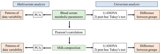
Assessment of Mastitis Patterns in Serbian Dairy Cows: Blood Serum Metabolic Profile and Milk Composition Parameters
by Jovan Stanojević, Mario Kreszinger, Miodrag Radinović, Nebojša Kladar, Dragana Tomanić, Zoran Ružić and Zorana Kovačević
Pathogens 2023, 12(11), 1349; https://doi.org/10.3390/pathogens12111349 - 14 Nov 2023
Cited by 2 | Viewed by 1553
Abstract
Mastitis is one of the most important diseases in dairy cows, leading to substantial economic losses associated with decreased milk production and quality. Early detection of changes in metabolic and milk parameters is crucial for maintaining animal welfare and milk quality. This study [...] Read more.
Mastitis is one of the most important diseases in dairy cows, leading to substantial economic losses associated with decreased milk production and quality. Early detection of changes in metabolic and milk parameters is crucial for maintaining animal welfare and milk quality. This study aimed to detect patterns in metabolic and milk composition parameters in Serbian dairy cows affected by mastitis. It also examined the relationship between these factors in cows with clinical and subclinical mastitis, as well as in healthy cows. This study included 60 Holstein-Friesian cows with the same body score condition that were in the same lactation phase. They were divided into three groups of 20: clinical and subclinical mastitis and a control group of healthy cows. The categorization was based on clinical udder health and the California mastitis test. Blood serum metabolic profiles were measured using a Rayto spectrophotometer (Shenzhen, China), and milk composition was determined using MilcoScanTM (Foss, Hilleroed, Denmark) and FossomaticTM (Foss, Hilleroed, Denmark) instruments. Significant increases in non-esterified fatty acids (NEFAs), beta-hydroxybutyrate (BHB), total protein, globulin, urea, total bilirubin, magnesium, and enzyme activity were noted in mastitis-affected cows compared to healthy ones. Additionally, mastitis-affected cows had higher total protein and globulin levels and increased somatic cell counts (SCCs), while albumin concentrations were decreased. Furthermore, a negative correlation between total protein and lactose suggested inflammation leading to reduced lactose levels due to cell damage, infection, and lactose use by mastitis pathogens. Hence, indicators of the energy and protein status of the metabolic profile, together with the chemical composition of milk, may be significant diagnostic tools for detecting, monitoring, and predicting the outcome of mastitis in cows. Full article
Show Figures

Figure 1

2022

Jump to: 2024, 2023, 2021, 2020, 2019

13 pages, 807 KiB  
Article
Incidence and Treatments of Bovine Mastitis and Other Diseases on 37 Dairy Farms in Wisconsin
by Juliano L. Gonçalves, Juliana L. de Campos, Andrew J. Steinberger, Nasia Safdar, Ashley Kates, Ajay Sethi, John Shutske, Garret Suen, Tony Goldberg, Roger I. Cue and Pamela L. Ruegg
Pathogens 2022, 11(11), 1282; https://doi.org/10.3390/pathogens11111282 - 1 Nov 2022
Cited by 13 | Viewed by 3587
Abstract
The aim of this research was to describe the incidence and treatments of mastitis and other common bovine diseases using one year of retrospective observational data (n = 50,329 cow-lactations) obtained from herd management software of 37 large dairy farms in Wisconsin. Incidence [...] Read more.
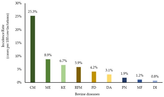
The aim of this research was to describe the incidence and treatments of mastitis and other common bovine diseases using one year of retrospective observational data (n = 50,329 cow-lactations) obtained from herd management software of 37 large dairy farms in Wisconsin. Incidence rate (IR) was defined as the number of first cases of each disease divided by the number of lactations per farm. Clinical mastitis (CM) remains the most diagnosed disease of dairy cows. Across all herds, the mean IR (cases per 100 cow-lactations) was 24.4 for clinical mastitis, 14.5 for foot disorders (FD), 11.2 for metritis (ME), 8.6 for ketosis (KE), 7.4 for retained fetal membranes (RFM), 4.5 for diarrhea (DI), 3.1 for displaced abomasum (DA), 2.9 for pneumonia (PN) and 1.9 for milk fever (MF). More than 30% of cows that had first cases of CM, DA, RFM, DI, and FD did not receive antibiotics. Of those treated, more than 50% of cows diagnosed with PN, ME and CM received ceftiofur as a treatment. The IR of mastitis and most other diseases was greater in older cows (parity ≥ 3) during the first 100 days of lactation and these cows were more likely to receive antibiotic treatments (as compared to younger cows diagnosed in later lactation). Cows of first and second parities in early lactation were more likely to remain in the herd after diagnosis of disease, as compared to older cows and cows in later stages of lactation. Most older cows diagnosed with CM in later lactation were culled before completion of the lactation. These results provide baseline data for disease incidence in dairy cows on modern U.S. dairy farms and reinforce the role of mastitis as an important cause of dairy cow morbidity. Full article
Show Figures

Figure 1

24 pages, 1988 KiB  
Review
Emerging Roles of Noncoding RNAs in Bovine Mastitis Diseases
by Favour Oluwapelumi Oyelami, Tahir Usman, Prashanth Suravajhala, Nawab Ali and Duy N. Do
Pathogens 2022, 11(9), 1009; https://doi.org/10.3390/pathogens11091009 - 3 Sep 2022
Cited by 8 | Viewed by 2818
Abstract
Non-coding RNAs (ncRNAs) are an abundant class of RNA with varying nucleotide lengths. They have been shown to have great potential in eutherians/human disease diagnosis and treatments and are now gaining more importance for the improvement of diseases in livestock. To date, thousands [...] Read more.
Non-coding RNAs (ncRNAs) are an abundant class of RNA with varying nucleotide lengths. They have been shown to have great potential in eutherians/human disease diagnosis and treatments and are now gaining more importance for the improvement of diseases in livestock. To date, thousands of ncRNAs have been discovered in the bovine genome and the continuous advancement in deep sequencing technologies and various bioinformatics tools has enabled the elucidation of their roles in bovine health. Among farm animals’ diseases, mastitis, a common inflammatory disease in cattle, has caused devastating economic losses to dairy farmers over the last few decades. Here, we summarize the biology of bovine mastitis and comprehensively discuss the roles of ncRNAs in different types of mastitis infection. Based on our findings and relevant literature, we highlighted various evidence of ncRNA roles in mastitis. Different approaches (in vivo versus in vitro) for exploring ncRNA roles in mastitis are emphasized. More particularly, the potential applications of emerging genome editing technologies, as well as integrated omics platforms for ncRNA studies and implications for mastitis are presented. Full article
Show Figures

Figure 1

2021

Jump to: 2024, 2023, 2022, 2020, 2019

11 pages, 1784 KiB  
Article
Molecular Typing and Antimicrobial Susceptibility Profiles of Streptococcus uberis Isolated from Sheep Milk
by Nives Maria Rosa, Ilaria Duprè, Elisa Azara, Carla Maria Longheu and Sebastiana Tola
Pathogens 2021, 10(11), 1489; https://doi.org/10.3390/pathogens10111489 - 16 Nov 2021
Cited by 7 | Viewed by 2596
Abstract
Intramammary infections are a major problem for dairy sheep farms, and Streptococcus uberis is one of the main etiological agents of ovine mastitis. Surveys on antimicrobial resistance are still limited in sheep and characterization of isolates is important for acquiring information on resistance [...] Read more.
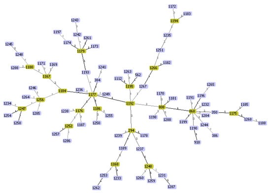
Intramammary infections are a major problem for dairy sheep farms, and Streptococcus uberis is one of the main etiological agents of ovine mastitis. Surveys on antimicrobial resistance are still limited in sheep and characterization of isolates is important for acquiring information on resistance and for optimizing therapy. In this study, a sampling of 124 S. uberis isolates collected in Sardinia (Italy) from sheep milk was analyzed by multilocus-sequence typing (MLST) and pulsed field gel electrophoresis (PFGE) for genetic relatedness. All isolates were also subjected to antimicrobial susceptibility analysis by the disk diffusion test using a panel of 14 antimicrobials. Resistance genes were detected by PCR assays. MLST analysis revealed that the isolates were grouped into 86 sequence types (STs), of which 73 were new genotypes, indicating a highly diverse population of S. uberis. The most frequently detected lineage was the clonal complex (CC)143, although representing only 13.7% of all characterized isolates. A high level of heterogeneity was also observed among the SmaI PFGE profiles, with 121 unique patterns. Almost all (96.8%) isolates were resistant to at least one antimicrobial, while all exhibited phenotypic susceptibility to oxacillin, amoxicillin-clavulanic acid and ceftiofur. Of the antimicrobials tested, the highest resistance rate was found against streptomycin (93.5%), kanamycin (79.8%) and gentamicin (64.5%), followed by novobiocin (25%) and tetracycline-TE (19.3%). Seventy-four (59.7%) isolates were simultaneously resistant to all aminoglycosides tested. Seventeen isolates (13.7%) exhibited multidrug resistance. All aminoglycosides-resistant isolates were PCR negative for aad-6 and aphA-3′ genes. Among the TE-resistant isolates, the tetM gene was predominant, indicating that the resistance mechanism is mainly mediated by the protection of ribosomes and not through the efflux pump. Three isolates were resistant to erythromycin, and two of them harbored the ermB gene. This is the first study reporting a detailed characterization of the S. uberis strains circulating in Sardinian sheep. Further investigations will be needed to understand the relationships between S. uberis genotypes, mastitis severity, and intra-mammary infection dynamics in the flock, as well as to monitor the evolution of antimicrobial resistance. Full article
Show Figures

Figure 1

9 pages, 249 KiB  
Article
Rapid Detection of Mycoplasma bovis, Staphylococcus aureus and Streptococcus agalactiae in Cattle Bulk Tank Milk in Cyprus and Relations with Somatic Cell Counts
by Maria Liapi, George Botsaris, Costas Arsenoglou, Nikolas Markantonis, Christodoulos Michael, Antonis Antoniou and Christodoulos Pipis
Pathogens 2021, 10(7), 841; https://doi.org/10.3390/pathogens10070841 - 4 Jul 2021
Cited by 6 | Viewed by 2961
Abstract
One hundred and seventy-seven (177) bulk tank milk samples were analyzed with a commercially available real-time polymerase chain reaction kit and 11 (6.21%), 41 (23.16%), and 58 (32.77%) tested positive for Mycoplasma bovis, Staphylococcus aureus, and Streptococcus agalactiae, respectively. Statistical analysis [...] Read more.
One hundred and seventy-seven (177) bulk tank milk samples were analyzed with a commercially available real-time polymerase chain reaction kit and 11 (6.21%), 41 (23.16%), and 58 (32.77%) tested positive for Mycoplasma bovis, Staphylococcus aureus, and Streptococcus agalactiae, respectively. Statistical analysis revealed a significant relationship between the presence of S. aureus and S. agalactiae. Enumeration of somatic cells was performed in the same samples by flow cytometry. The somatic cell counts were found higher in S. aureus and S. agalactiae positive samples. No association was found between M. bovis presence and somatic cells counts. Low internal assay control Ct values were found to be related with high somatic cell counts. Noticeably, this is the first report for the presence of M. bovis in Cyprus. Therefore, its presence was confirmed by bulk tank milk culture, conventional PCR, and next generation sequencing. Furthermore, M. bovis was typed with multilocus sequencing typing and was allocated to sequence type 29 (ST 29). Real-time PCR in bulk tank milk samples is a useful tool to detect mammary infections, especially for neglected pathogens such as M. bovis. Full article
12 pages, 940 KiB  
Article
Association of Staphylococcal Populations on Teatcups of Milking Parlours with Vaccination against Staphylococcal Mastitis in Sheep and Goat Farms
by Charalambia K. Michael, Daphne T. Lianou, Natalia G.C. Vasileiou, Katerina Tsilipounidaki, Angeliki I. Katsafadou, Antonis P. Politis, Nikos G. Kordalis, Katerina S. Ioannidi, Dimitris A. Gougoulis, Constantina Trikalinou, Denise C. Orfanou, Ilektra A. Fragkou, Panagiota I. Kontou, Dimitra V. Liagka, Vasia S. Mavrogianni, Efthimia Petinaki and George C. Fthenakis
Pathogens 2021, 10(4), 385; https://doi.org/10.3390/pathogens10040385 - 24 Mar 2021
Cited by 7 | Viewed by 2689
Abstract
There is a paucity of information regarding staphylococcal populations on teatcups of milking parlours in sheep and goat farms. The objectives were to describe the populations of staphylococci on teatcups in milking parlours in sheep or goat farms in two field investigations throughout [...] Read more.
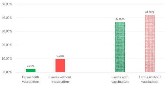
There is a paucity of information regarding staphylococcal populations on teatcups of milking parlours in sheep and goat farms. The objectives were to describe the populations of staphylococci on teatcups in milking parlours in sheep or goat farms in two field investigations throughout Greece and to potentially associate the findings with the use of anti-staphylococcal mastitis vaccinations in the farms visited during the two investigations. In a cross-sectional (255 sheep and 66 goat farms across Greece) and a longitudinal (12 sheep farms, four samplings, throughout lactation) study, swab samples were collected from 1418 teatcups (upper and lower part) for staphylococcal recovery, identification and assessment of biofilm-formation. A total of 328 contaminated teatcups (23.1%) were found in 105 sheep (41.2%) and 35 goat (53.0%) farms. Staphylococci were more frequently recovered from the upper than the lower part of teatcups: 269 versus 139 teatcups, respectively. After identification, 253 staphylococcal isolates were found: Staphylococcus aureus, Staphylococcus equorum, Staphylococcus lentus, and Staphylococcus capitis predominated. Of these isolates, 87.4% were biofilm-forming. The proportion of contaminated teatcups was smaller in farms where vaccination against anti-staphylococcal mastitis in general or vaccination specifically against mastitis caused specifically by biofilm-forming staphylococcal strains was applied, 19.7% or 10.9%, respectively, versus 25.5% in farms without vaccination. In the longitudinal study, contaminated teatcups were identified in 28 (58.3%) sampling occasions, with staphylococci being recovered more frequently from their upper part. The same species as in the cross-sectional study predominated. Of these isolates, 61.9% were biofilm-forming. In farms where vaccination against mastitis caused specifically by biofilm-forming staphylococcal strains was applied, the proportion of contaminated teatcups was smaller: 20.4% versus 48.3% in farms without vaccination. There were no differences in proportions of contaminated teatcups between sampling occasions. In conclusion, the great majority of staphylococci recovered from teatcups of milking parlours in sheep and goat farms included biofilm-forming isolates. Reduced staphylococcal isolation was noted in farms where anti-staphylococcal vaccination was performed; this was possibly the effect of reduced excretion of staphylococci in the milk of vaccinated animals. Full article
Show Figures

Figure 1

19 pages, 319 KiB  
Article
Dry Period or Early Lactation—Time of Onset and Associated Risk Factors for Intramammary Infections in Dairy Cows
by Julia Nitz, Nicole Wente, Yanchao Zhang, Doris Klocke, Martin tho Seeth and Volker Krömker
Pathogens 2021, 10(2), 224; https://doi.org/10.3390/pathogens10020224 - 18 Feb 2021
Cited by 13 | Viewed by 2970
Abstract
The aim of this study was to define the time-related period of intramammary infections and its relation to risk factors for intramammary infections and clinical mastitis at cow and quarter levels. In total, 269 German Holstein Frisian dairy cows on three farms in [...] Read more.
The aim of this study was to define the time-related period of intramammary infections and its relation to risk factors for intramammary infections and clinical mastitis at cow and quarter levels. In total, 269 German Holstein Frisian dairy cows on three farms in Northern and Eastern Germany were included in this study. Quarter milk samples were collected at dry-off, 3 ± 1 days after calving and 17 ± 3 days after calving, for cytomicrobiological examination. Risk factors at quarter- and cow-level associated with intramammary infections and clinical mastitis were recorded during the trial period. Data were analyzed using logistic regression procedures and odds ratios were calculated. Calving for the second time increased the odds of clinical mastitis during the first 100 days of lactation compared to cows calving for the third time or more. A high milk yield after calving was a risk factor for new infections, with environmental pathogens 17 ± 3 days postpartum. A body condition score after calving less than 3.5 was associated with a decreased risk of having an intra-mammary infection (IMI) with non-aureus staphylococci and coryneforms 3 ± 1 days postpartum and consistent body condition between dry-off and early lactation decreased the risk of intramammary infections after calving. The absence of a ring of hyperkeratosis at the teat apex shown at dry-off was associated with a lower risk of intramammary infections with environmental pathogens 17 ± 3 days postpartum. This study shows the important influence of the dry period and early lactation on intramammary infections and clinical mastitis postpartum in dairy cows. Udder quarters may have eliminated pathogens during the dry period in 43.6% of cases in this study. Additionally, new infections occurred during early lactation, so 5.1% more quarters were infected 17 ± 3 days compared to 3 ± 1 days postpartum. New infections can be traced to non-aureus staphylococci and Staphylococcus aureus from dry-off up until 3 ± 1 days postpartum, and to non-aureus staphylococci, Staphylococcus aureus and Streptococcus uberis, after calving. In total, 88.7% of the infected quarters showed new infections with another pathogen species 3 ± 1 days postpartum than at dry-off, and 89.2% of the quarters 17 ± 3 days postpartum than 3 ± 1 days postpartum. In conclusion, the early lactation has just as important an influence on intramammary infections postpartum in dairy cows as the dry period. There is the possibility that udder quarters eliminate pathogens during the early lactation, especially during the dry period. However, there is also the danger that new infections manifest, with a large proportion of new infections occurring after calving. Thus, additional control strategies are of great importance to prevent new infections occurring during early lactation as well as during the dry period to reduce negative effects on milk yield and culling hazards in dairy cows by minimizing the associated risk factors. Full article
15 pages, 297 KiB  
Communication
Characterization of Enterococci- and ESBL-Producing Escherichia coli Isolated from Milk of Bovides with Mastitis in Egypt
by Wedad Ahmed, Heinrich Neubauer, Herbert Tomaso, Fatma Ibrahim El Hofy, Stefan Monecke, Ashraf Awad Abd El-Tawab and Helmut Hotzel
Pathogens 2021, 10(2), 97; https://doi.org/10.3390/pathogens10020097 - 21 Jan 2021
Cited by 21 | Viewed by 3431
Abstract
This study aimed to investigate the prevalence and antimicrobial resistance of enterococci- and ESBL-producing E. coli isolated from milk of bovine mastitis cases in Egypt. Fifty milk samples of dairy animals were collected from localities in the Nile Delta region of Egypt. Isolates [...] Read more.
This study aimed to investigate the prevalence and antimicrobial resistance of enterococci- and ESBL-producing E. coli isolated from milk of bovine mastitis cases in Egypt. Fifty milk samples of dairy animals were collected from localities in the Nile Delta region of Egypt. Isolates were identified using MALDI-TOF MS, and antibiotic susceptibility testing was performed by the broth microdilution method. PCR amplifications were carried out, targeting resistance-associated genes. Seventeen Enterococcus isolates and eight coliform isolates could be cultivated. Vancomycin resistance rate was high in Ent. faecalis. The VITEK 2 system confirmed all E. coli isolates as ESBL-producing. All Ent. faecalis isolates harbored erm(B), tetL and aac-aphD genes. The vanA gene was detected in Ent. faecalis isolate, vanB was found in other Enterococcus, while one isolate of E. casseliflavus exhibited the vanA gene. E. coli isolates exhibited high prevalence of erm(B) and tetL. E. coli isolates were analyzed by DNA microarray analysis. Four isolates were determined by O-serotyping as O8 (n = 1), O86 (n = 2) and O157 (n = 1). H-serotyping resulted in H11, H12, H21 (two isolates each) and one was of H16 type. Different virulence-associated genes were detected in E. coli isolates including lpfA, astA, celB, cmahemL, intI1 and intI2, and the iroN gene was identified by DNA microarray analysis. Full article

2020

Jump to: 2024, 2023, 2022, 2021, 2019

23 pages, 3204 KiB  
Article
A Paradox in Bacterial Pathogenesis: Activation of the Local Macrophage Inflammasome Is Required for Virulence of Streptococcus uberis
by Nathan Archer, Sharon A. Egan, Tracey J. Coffey, Richard D. Emes, M. Filippa Addis, Philip N. Ward, Adam M. Blanchard and James A. Leigh
Pathogens 2020, 9(12), 997; https://doi.org/10.3390/pathogens9120997 - 28 Nov 2020
Cited by 11 | Viewed by 4059
Abstract
Streptococcus uberis is a common cause of intramammary infection and mastitis in dairy cattle. Unlike other mammary pathogens, S. uberis evades detection by mammary epithelial cells, and the host–pathogen interactions during early colonisation are poorly understood. Intramammary challenge of dairy cows with S. [...] Read more.
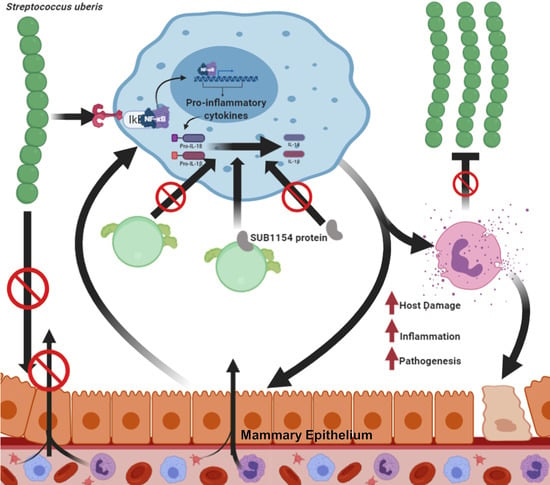
Streptococcus uberis is a common cause of intramammary infection and mastitis in dairy cattle. Unlike other mammary pathogens, S. uberis evades detection by mammary epithelial cells, and the host–pathogen interactions during early colonisation are poorly understood. Intramammary challenge of dairy cows with S. uberis (strain 0140J) or isogenic mutants lacking the surface-anchored serine protease, SUB1154, demonstrated that virulence was dependent on the presence and correct location of this protein. Unlike the wild-type strain, the mutant lacking SUB1154 failed to elicit IL-1β from ex vivo CD14+ cells obtained from milk (bovine mammary macrophages, BMM), but this response was reinstated by complementation with recombinant SUB1154; the protein in isolation elicited no response. Production of IL-1β was ablated in the presence of various inhibitors, indicating dependency on internalisation and activation of NLRP3 and caspase-1, consistent with inflammasome activation. Similar transcriptomic changes were detected in ex vivo BMM in response to the wild-type or the SUB1154 deletion mutant, consistent with S. uberis priming BMM, enabling the SUB1154 protein to activate inflammasome maturation in a transcriptionally independent manner. These data can be reconciled in a novel model of pathogenesis in which, paradoxically, early colonisation is dependent on the innate response to the initial infection. Full article
Show Figures

Graphical abstract

6 pages, 193 KiB  
Communication
Antibacterial Activities of Acetic Acid against Major and Minor Pathogens Isolated from Mastitis in Dairy Cows
by Noppason Pangprasit, Anyaphat Srithanasuwan, Witaya Suriyasathaporn, Surachai Pikulkaew, John K. Bernard and Wasana Chaisri
Pathogens 2020, 9(11), 961; https://doi.org/10.3390/pathogens9110961 - 19 Nov 2020
Cited by 17 | Viewed by 3378
Abstract
The present study evaluated the antimicrobial activities of acetic acid against bovine mastitis pathogens compared to lactic acid and lauric and caprylic saturated fatty acids. Eleven mastitis pathogens were isolated from sub-clinical and clinical bovine mastitis cases for the study. An initial screening [...] Read more.
The present study evaluated the antimicrobial activities of acetic acid against bovine mastitis pathogens compared to lactic acid and lauric and caprylic saturated fatty acids. Eleven mastitis pathogens were isolated from sub-clinical and clinical bovine mastitis cases for the study. An initial screening of their antibacterial activities by agar well diffusion method was performed. The minimum inhibitory concentration (MIC) and minimum bactericidal concentration (MBC) of each acid were obtained using a microdilution method; each acid was diluted from stock solution and then were diluted with culture broth to reach concentrations ranging from 4 to 0.004% w/v. The results showed acetic acid had the highest zone of inhibition against all pathogens except Escherichia coli compared with lauric and caprylic acids. The MIC and MBC were lowest for acetic acid against both Gram-positive (except Staphylococcus chromogenes from the coagulase negative staphylococci (CNS) group) and Gram-negative pathogens, intermediate for lactic and caprylic acids and greatest for lauric acid. In conclusion, acetic acid had antimicrobial activities against most mastitis pathogens compared with other acids. Further studies are needed to optimize the formulation and concentration of acetic acid for teat-dipping agent in the future. Full article
23 pages, 316 KiB  
Review
The Impact of Mastitis on the Biochemical Parameters, Oxidative and Nitrosative Stress Markers in Goat’s Milk: A Review
by Cristiana S. Novac and Sanda Andrei
Pathogens 2020, 9(11), 882; https://doi.org/10.3390/pathogens9110882 - 24 Oct 2020
Cited by 25 | Viewed by 4323
Abstract
Goat mastitis has become one of the most frequently diagnosed conditions in goat farms, with significant economic impact on the dairy industry. Inflammation of the mammary gland poses serious consequences on milk composition, with changes regarding biochemical parameters and oxidative stress markers. The [...] Read more.
Goat mastitis has become one of the most frequently diagnosed conditions in goat farms, with significant economic impact on the dairy industry. Inflammation of the mammary gland poses serious consequences on milk composition, with changes regarding biochemical parameters and oxidative stress markers. The aim of this paper is to present the most recent knowledge on the main biochemical changes that occur in the mastitic milk, as well as the overall effect of the oxidative and nitrosative stress on milk components, focusing on both enzymatic and nonenzymatic antioxidant markers. Mastitis in goats is responsible for a decrease in milk production, change in protein content with pronounced casein hydrolysis, and reduction in lactose concentration and milk fat. Milk enzymatic activity also undergoes changes, regarding indigenous enzymes and those involved in milk synthesis. Furthermore, during mastitis, both the electrical conductivity and the milk somatic cell count are increased. Intramammary infections are associated with a reduced milk antioxidant capacity and changes in catalase, lactoperoxidase, glutathione peroxidase or superoxide dismutase activity, as well as reduced antioxidant vitamin content. Mastitis is also correlated with an increase in the concentration of nitric oxide, nitrite, nitrate and other oxidation compounds, leading to the occurrence of nitrosative stress. Full article
12 pages, 286 KiB  
Article
Concentrations of Acute-Phase Proteins in Milk from Cows with Clinical Mastitis Caused by Different Pathogens
by Felipe M. Dalanezi, Elizabeth M. S. Schmidt, Sâmea F. Joaquim, Felipe F. Guimarães, Simoni T. Guerra, Bruna C. Lopes, Ronaldo L. A. Cerri, Christopher Chadwick and Hélio Langoni
Pathogens 2020, 9(9), 706; https://doi.org/10.3390/pathogens9090706 - 27 Aug 2020
Cited by 19 | Viewed by 3489
Abstract
Among the new diagnostic methods for mastitis detection under development, milk acute-phase proteins (APPs) are receiving special attention. The study aimed to compare the profile of milk APPs from cows with natural clinical mastitis caused by distinct pathogens. The concentrations of haptoglobin (Hp), [...] Read more.
Among the new diagnostic methods for mastitis detection under development, milk acute-phase proteins (APPs) are receiving special attention. The study aimed to compare the profile of milk APPs from cows with natural clinical mastitis caused by distinct pathogens. The concentrations of haptoglobin (Hp), serum amyloid A (SAA), alpha-1-acid glycoprotein (AGP), and C-reactive protein (CRP) were measured by Spatial Proximity Analyte Reagent Capture Luminescence (SPARCL). Each APP was compared across the pathogens causing mastitis. The APPs differed statistically (p < 0.05) among the pathogens causing udder infection. There were significant and positive correlations among the concentration profile, for each pathogen, in three of four APPs studied. It can be concluded that the pathogen causing mastitis could modify the profile of release of the APPs in milk. The profile of Hp, AGP, and CRP demonstrated significant correlation, indicating that the three APPs are suggested as biomarkers, in milk, for bovine mastitis. Full article
13 pages, 2364 KiB  
Communication
Scientometrics Approach to Research in Ovine Mastitis from 1970 to 2019 (with a Complete List of Relevant Literature References)
by Daphne T. Lianou and George C. Fthenakis
Pathogens 2020, 9(7), 585; https://doi.org/10.3390/pathogens9070585 - 17 Jul 2020
Cited by 14 | Viewed by 2768
Abstract
The present study is a scientometrics evaluation of refereed publications on bacterial mastitis in sheep; the objectives were the evaluation of the relevant papers and the presentation of quantitative characteristics regarding their scientific content and bibliometric details. The Web of Science platform was [...] Read more.
The present study is a scientometrics evaluation of refereed publications on bacterial mastitis in sheep; the objectives were the evaluation of the relevant papers and the presentation of quantitative characteristics regarding their scientific content and bibliometric details. The Web of Science platform was used with search terms: [mastitis OR *mammary infection*] AND [sheep OR ewe* OR ovine] for papers from 1970 tο 2019; only ‘articles’, ‘reviews’, ‘proceedings papers’, or ‘data papers’ were evaluated, whilst documents related solely to contagious agalactia, mammary aspects of lentiviral infections, or infections of the teats and the udder skin were excluded. Finally, 580 papers were considered in detail. The number of published papers increased from 8 during the 1970s to 273 during the 2010s. These papers originated from 43 countries (most from Greece or Spain, n = 87 from each) and 240 institutions (145 universities and 95 other establishments), of which 35 produced ≥ 5 papers each. Most papers present original studies (n = 539) with a few reviews (n = 41). The original papers refer to dairy (n = 428), meat (n = 113), or wool (n = 1) production systems and present field (n = 329), laboratory (n = 163), or experimental (n = 67) work; the papers report aetiology (n = 146), risk factors (n = 100), pathogenesis (n = 92), diagnosis (n = 88), effects (n = 66), treatment (n = 50), control (n = 36), or descriptive epidemiology (n = 32) of the disease. Papers related to dairy production present more field and fewer experimental work than papers related to meat production; also, in papers describing work performed in dairy sheep, studies about aetiology, risk factors, and diagnosis of the disease predominate, whilst in papers performed in meat sheep, studies about aetiology, pathogenesis, and effects/diagnosis are reported more often. The papers were published in 175 scientific journals (most in Small Ruminant Research, n = 90, or Journal of Dairy Science, n = 54). On average, the papers received 16.8 total citations and 1.6 yearly citations (h-index = 47). Most papers were published in Scimago classification Q1 (n = 240) or Q2 (n = 230) journals and received 23.4 or 15.4 total citations, respectively. Reviews received more citations than original papers; among the latter, papers with work referring to dairy production received more yearly citations than papers referring to meat production; no differences in citations were seen according to type of work or mastitis aspect covered. Most citations were received by papers from France. Papers published in Journal of Dairy Science or Small Ruminant Research received the most citations. In total, there were 1558 individual authors of the papers, with 24 authors having co-authored > 10 papers each (max: 73 papers); on average, there were 5.2 co-authors per paper (min–max: 1–25). Average number of co-authors progressively increased from 2.1 in the 1970s to 6.3 in the 2010s, with original papers having a higher number of co-authors than reviews: 5.3 and 3.7, respectively. Papers from France had highers number of co-authors (7.9). The findings of this first ever scientometrics study into ovine mastitis indicate that the disease has not been studied as other sheep diseases and that future studies in it should be directed to its control. Full article
Show Figures

Figure 1

2 pages, 181 KiB  
Erratum
Erratum: Fukuyama, K., et al. Evaluation of the Immunomodulatory Ability of Lactic Acid Bacteria Isolated from Feedlot Cattle Against Mastitis Using a Bovine Mammary Epithelial Cells In Vitro Assay. Pathogens 2020, 9, 410
by Kohtaro Fukuyama, Md. Aminul Islam, Michihiro Takagi, Wakako Ikeda-Ohtsubo, Shoichiro Kurata, Hisashi Aso, Maria Elena Fatima Nader-Macías, Graciela Vignolo, Julio Villena and Haruki Kitazawa
Pathogens 2020, 9(7), 574; https://doi.org/10.3390/pathogens9070574 - 16 Jul 2020
Cited by 5 | Viewed by 2244
Abstract
The authors would like to make the following corrections about the published paper [...] Full article
17 pages, 1569 KiB  
Article
Prolactin and Estradiol are Epigenetic Modulators in Bovine Mammary Epithelial Cells during Staphylococcus aureus Infection
by María Guadalupe Salgado-Lora, Ivan Medina-Estrada, Joel Edmundo López-Meza and Alejandra Ochoa-Zarzosa
Pathogens 2020, 9(7), 520; https://doi.org/10.3390/pathogens9070520 - 28 Jun 2020
Cited by 8 | Viewed by 3005
Abstract
Changes in the levels of reproductive hormones compromise the bovine innate immune response (IIR). Changes in 17β-estradiol (E2) and prolactin (bPRL) levels affect the IIR of bovine mammary epithelial cells (bMECs), the target tissue of these hormones. In this work, we explored the [...] Read more.
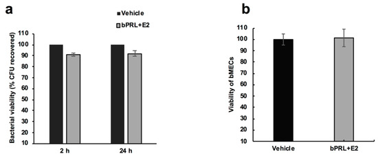
Changes in the levels of reproductive hormones compromise the bovine innate immune response (IIR). Changes in 17β-estradiol (E2) and prolactin (bPRL) levels affect the IIR of bovine mammary epithelial cells (bMECs), the target tissue of these hormones. In this work, we explored the effect of the combined hormones on bMEC IIR during Staphylococcus aureus infection, and if they can modulate epigenetic marks. By gentamicin protection assays, we determined that combined hormones (bPRL (5 ng/mL) and E2 (50 pg/mL)] decrease S. aureus internalization into bMECs (~50%), which was associated with a reduction in integrin α5β1 membrane abundance (MA) (~80%) determined by flow cytometry. Additionally, combined hormones increased Toll-like receptor 2 (TLR2) MA (~25%). By RT-qPCR, we showed that combined hormones induce the expression of pro- and anti-inflammatory cytokine genes, as well as up-regulate antimicrobial peptide gene expression. The combined hormones induced H3K9Ac at 12 h of treatment, which coincides with the reduction in histone deacetylase (HDAC, ~15%) activity. In addition, hormones increased the H3K9me2 mark at 12 h, which correlates with a reduction in the expression of KDM4A. In conclusion, bPRL and E2 modulate the IIR of bMECs, an effect that can be related to the regulation of histone H3 modifications such as H3K9Ac and H3K9me2. Full article
Show Figures

Figure 1

15 pages, 2874 KiB  
Article
Evaluation of the Immunomodulatory Ability of Lactic Acid Bacteria Isolated from Feedlot Cattle Against Mastitis Using a Bovine Mammary Epithelial Cells In Vitro Assay
by Kohtaro Fukuyama, Md. Aminul Islam, Michihiro Takagi, Wakako Ikeda-Ohtsubo, Shoichiro Kurata, Hisashi Aso, Graciela Vignolo, Julio Villena and Haruki Kitazawa
Pathogens 2020, 9(5), 410; https://doi.org/10.3390/pathogens9050410 - 25 May 2020
Cited by 20 | Viewed by 4808
Abstract
Bovine mastitis, the inflammation of the mammary gland, affects the quality and quantity of milk yield. Mastitis control relies on single or multiple combinations of antibiotic therapy. Due to increasing antibiotic resistance in pathogens, the intramammary infusion of lactic acid bacteria (LAB) has [...] Read more.
Bovine mastitis, the inflammation of the mammary gland, affects the quality and quantity of milk yield. Mastitis control relies on single or multiple combinations of antibiotic therapy. Due to increasing antibiotic resistance in pathogens, the intramammary infusion of lactic acid bacteria (LAB) has been considered as a potential alternative to antibiotics for treating and preventing bovine mastitis through the improvement of the host immunity. Probiotic effects are a strain-dependent characteristic; therefore, candidate LAB strains have to be evaluated efficiently to find out the ones with the best potential. Here, we investigated LAB strains originally isolated from feedlot cattle’s environment regarding their ability in inducing the Toll-like receptor (TLR)-triggered inflammatory responses in bovine mammary epithelial (BME) cells in vitro. The BME cells were pre-stimulated with the LAB strains individually for 12, 24, and 48 h and then challenged with Escherichia coli-derived lipopolysaccharide (LPS) for 12 h. The mRNA expression of selected immune genes—interleukin 1 alpha (IL-1α), IL-1β, monocyte chemotactic protein 1 (MCP-1), IL-8, chemokine (C-X-C motif) ligand 2 (CXCL2), and CXCL3 were quantified by real-time quantitative PCR (RT-qPCR). Results indicated that pretreatment with some Lactobacillus strains were able to differentially regulate the LPS inflammatory response in BME cells; however, strain-dependent differences were found. The most remarkable effects were found for Lactobacillus acidophilus CRL2074, which reduced the expression of IL-1α, IL-1β, MCP-1, IL-8, and CXCL3, whereas Lactobacillus rhamnosus CRL2084 diminished IL-1β, MCP-1, and IL-8 expression. The pre-stimulation of BME cells with the CRL2074 strain resulted in the upregulated expression of three negative regulators of the TLRs, including the ubiquitin-editing enzyme A20 (also called tumor necrosis factor alpha-induced protein 3, TNFAIP3), single immunoglobin IL-1 single receptor (SIGIRR), and Toll interacting protein (Tollip) after the LPS challenge. The CRL2084 pre-stimulation upregulated only Tollip expression. Our results demonstrated that the L. acidophilus CRL2074 strain possess remarkable immunomodulatory abilities against LPS-induced inflammation in BME cells. This Lactobacillus strain could be used as candidate for in vivo testing due to its beneficial effects in bovine mastitis through intramammary infusion. Our findings also suggest that the BME cells immunoassay system could be of value for the in vitro evaluation of the immunomodulatory abilities of LAB against the inflammation resulting from the intramammary infection with mastitis-related pathogens. Full article
Show Figures

Figure 1

11 pages, 704 KiB  
Article
Correlation between Milk Bacteriology, Cytology and Mammary Tissue Histology in Cows: Cure from the Pathogen or Recovery from the Inflammation
by Gabriel Leitner, Shlomo E. Blum, Oloeg Krifuks, Nir Edery and Uzi Merin
Pathogens 2020, 9(5), 364; https://doi.org/10.3390/pathogens9050364 - 9 May 2020
Cited by 10 | Viewed by 3679
Abstract
The aim of the current study was to verify the existence of a significant correlation between bacterial isolation (or not) and mammary gland inflammation, using traditional bacterial culturing and PCR, milk leucocytes distributions, and tissue histology. Twenty-two cows were tested at the level [...] Read more.
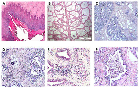
The aim of the current study was to verify the existence of a significant correlation between bacterial isolation (or not) and mammary gland inflammation, using traditional bacterial culturing and PCR, milk leucocytes distributions, and tissue histology. Twenty-two cows were tested at the level of the individual gland for bacteriological culture and real-time PCR (RT-PCR), milk composition, somatic cells count (SCC), and cell differentiation. Post-slaughter samples of teat-ends and mammary tissues were tested for histology and bacteriology by RT-PCR. The 88 glands were assigned to either outcome: 1. Healthy—no inflammation and no bacterial finding (NBF) (n = 33); 2. Inflammation and NBF (n = 26); 3. Inflammation and intra-mammary infection (n = 22) with different bacteria. Bacteriology of milk samples and that of the RT-PCR showed 91.4% agreement. In the lobule’s tissues of healthy glands, ~50% were milk producers and the other glands had dry areas with increased fat globules with a low number of leukocytes. In contrast, ~75% of the infected glands were identified as inflamed, but with no isolation of bacteria. Infiltration of mononuclear cells and neutrophils into the connective tissue was observed but not in the lobule’s lumen. In summary, the study confirms that not every mastitis/inflammation is also an infection. Full article
Show Figures

Figure 1

11 pages, 271 KiB  
Article
Prevalence, Antimicrobial Resistance Profiles, Virulence and Enterotoxins-Determinant Genes of MRSA Isolated from Subclinical Bovine Mastitis in Egypt
by Abdelazeem M. Algammal, Mohamed E. Enany, Reham M. El-Tarabili, Madeha O. I. Ghobashy and Yosra A. Helmy
Pathogens 2020, 9(5), 362; https://doi.org/10.3390/pathogens9050362 - 9 May 2020
Cited by 97 | Viewed by 6544
Abstract
Subclinical mastitis caused by Staphylococcus aureus has worldwide public health significance. Here, we aimed to determine the prevalence of S. aureus, antimicrobial resistance profiles, and the virulence and enterotoxins determinant genes of MRSA strains that caused subclinical bovine mastitis. Milk samples were [...] Read more.
Subclinical mastitis caused by Staphylococcus aureus has worldwide public health significance. Here, we aimed to determine the prevalence of S. aureus, antimicrobial resistance profiles, and the virulence and enterotoxins determinant genes of MRSA strains that caused subclinical bovine mastitis. Milk samples were collected from 120 lactating animals (50 buffaloes and 70 dairy cattle) from different farms located in Ismailia Province (Egypt). The collected samples were investigated for subclinical mastitis using a California mastitis test. The total prevalence of S. aureus was 35.9% (84/234) with 36.3% (53/146) in cattle and 31% (31/88) in buffaloes. Antimicrobial susceptibility testing showed that 35.7% (30/84) of the isolated strains were resistant to cefoxitin, defined as methicillin-resistant S. aureus (MRSA), with 37.7% (20/53) in cattle and 32.2% (10/31) in buffaloes. Using PCR, 100% of the tested strains harbored coa and mecA genes, while 86.6% were positive for spa gene, with remarkable gene size polymorphism. Additionally, 10% of the tested strains contained the pvl gene. Further, using multiplex PCR, 26.6% of the tested samples had sea gene, two strains had sec gene and only one strain had sea and sec genes. The seb and sed genes were absent in the tested strains. In conclusion, mecA, coa and spa virulence genes were widely distributed in MRSA strains isolated from bovine milk, whereas the sea gene was the most predominant enterotoxin gene. Notably, this is the first report that emphasizes the prevalence of pvl gene of MRSA isolated from bovine milk in Egypt. Full article
8 pages, 242 KiB  
Article
Prevention of Intramammary Infections by Prepartum External Application of a Teat Dip Containing Lactic Acid Bacteria with Antimicrobial Properties in Dairy Heifers
by Jan-Hendrik Paduch, Johanna Lücking, Elisabeth Mansion-de Vries, Claudia Zinke, Nicole Wente and Volker Krömker
Pathogens 2020, 9(4), 288; https://doi.org/10.3390/pathogens9040288 - 16 Apr 2020
Cited by 2 | Viewed by 3417
Abstract
The aim of the current study was to investigate the effects of the prepartum external treatment of teats with a combination of four lactic acid bacteria strains viz. Lactobacillus (Lb.) rhamnosus ATCC 7469, Lactococcus lactis subsp. lactis ATCC 11454, Lb. paracasei [...] Read more.
The aim of the current study was to investigate the effects of the prepartum external treatment of teats with a combination of four lactic acid bacteria strains viz. Lactobacillus (Lb.) rhamnosus ATCC 7469, Lactococcus lactis subsp. lactis ATCC 11454, Lb. paracasei 78/37 (DSM 26911), and Lb. plantarum 118/37 (DSM 26912) on the postcalving udder health of dairy heifers. The study used a split-udder design. Two weeks before the expected calving date, one of two contralateral teats of a teat pair was dipped with an aqueous suspension of lactic acid bacteria (final bacterial counts 8.40–8.47 log10-transformed CFU/mL) once in a week until calving; the other teat of the pair was not treated. After calving, quarter foremilk samples were taken and investigated cyto-microbiologically. In total, 629 teat pairs of 319 heifers were included. There was an association between the treatment and intramammary infections caused by the major udder-pathogenic bacteria Staphylococcus aureus, Streptococcus dysgalactiae, and enterococci, as well as clinical mastitis in the first 100 days after calving. The present study indicates that intramammary infections with major pathogens and clinical mastitis may be prevented by regular prepartum external application of lactic acid bacteria in dairy heifers. Full article
18 pages, 1430 KiB  
Article
Transcriptome Analysis of The Inflammatory Responses of Bovine Mammary Epithelial Cells: Exploring Immunomodulatory Target Genes for Bovine Mastitis
by Md. Aminul Islam, Michihiro Takagi, Kohtaro Fukuyama, Ryoya Komatsu, Leonardo Albarracin, Tomonori Nochi, Yoshihito Suda, Wakako Ikeda-Ohtsubo, Victor Rutten, Willem van Eden, Julio Villena, Hisashi Aso and Haruki Kitazawa
Pathogens 2020, 9(3), 200; https://doi.org/10.3390/pathogens9030200 - 9 Mar 2020
Cited by 33 | Viewed by 5396
Abstract
Bovine mastitis is the inflammatory reaction of the mammary gland and is commonly caused by bacterial infections in high-yielding dairy cows. The detailed investigation of the immunotranscriptomic response of bovine mammary epithelial (BME) cells to pattern recognition receptors (PRRs) activation by microbial-associated molecular [...] Read more.
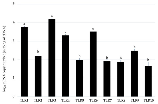
Bovine mastitis is the inflammatory reaction of the mammary gland and is commonly caused by bacterial infections in high-yielding dairy cows. The detailed investigation of the immunotranscriptomic response of bovine mammary epithelial (BME) cells to pattern recognition receptors (PRRs) activation by microbial-associated molecular patterns (MAMPs) can be of great importance for understanding the innate immune defense mechanisms, and for exploring the immunomodulatory candidate genes. In this work, we investigated the transcriptome modifications of BME cells after the in vitro stimulation with Escherichia coli derived lipopolysaccharide (LPS) and heat-killed Staphylococcus aureus JE2 and S. aureus SA003. In addition, the effect of Pam3CSK4 (a synthetic triacylated lipopeptide that activates Toll-like receptor 2 (TLR2)), and the intracellular chemotactic protein cyclophilin A (CyPA), which is secreted by BME cells during mastitis, in the expression changes of selected cytokines and chemokines were evaluated by qPCR. Microarray analysis identified 447, 465 and 520 differentially expressed genes (DEGs) in the BME cells after LPS, S. aureus JE2 and S. aureus SA003 stimulation, respectively. A major differential response in the inflammatory gene expression was noticed between the stimulation of LPS and S. aureus strains. Unlike the S. aureus strains, LPS stimulation resulted in significant upregulation of CCL2, CXCL2, CXCL3, CXCL8, IL1α and IL1β, which were confirmed by qPCR analysis. Pam3CSK4 was not able to induce significant changes in the expression of cytokines and chemokines in challenged BME cells. The exogenous CyPA administration was able to upregulate CXCL2, CXCL3, CXCL8, IL1α and IL1β expression in BME cells indicating its ability to promote inflammation. The identification of transcriptional markers of mastitis specific for individual inflammatory factors such as LPS, Pam3CSK4 or CyPA, which can be evaluated in vitro in BME cells, may enable the development of novel diagnostics and/or immunomodulatory treatments, providing new tools for the effective management of mastitis in dairy cows. The results of this work are an advance in this regard. Full article
Show Figures

Figure 1

15 pages, 2937 KiB  
Article
Witch Hazel Significantly Improves the Efficacy of Commercially Available Teat Dips
by Reuven Rasooly, Adel Molnar, Paula Do, Gianluca Morroni, Lucia Brescini, Oscar Cirioni, Andrea Giacometti and Emmanouil Apostolidis
Pathogens 2020, 9(2), 92; https://doi.org/10.3390/pathogens9020092 - 1 Feb 2020
Cited by 5 | Viewed by 5429
Abstract
Bovine intramammary infections (IMIs) are the main cause of economic loss in milk production. Antibiotics are often ineffective in treating infections due to antimicrobial resistance and the formation of bacterial biofilms that enhance bacterial survival and persistence. Teat dips containing germicides are recommended [...] Read more.
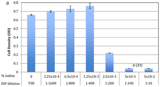
Bovine intramammary infections (IMIs) are the main cause of economic loss in milk production. Antibiotics are often ineffective in treating infections due to antimicrobial resistance and the formation of bacterial biofilms that enhance bacterial survival and persistence. Teat dips containing germicides are recommended to prevent new IMIs and improve udder health and milk quality. IMIs are often caused by staphylococci, which are Gram-positive bacteria that become pathogenic by forming biofilms and producing toxins. As a model for a teat dip (DIP), the BacStop iodine-based teat dip (DIP) was used. Witch hazel extract (whISOBAX (WH)) was tested because it contains a high concentration of the anti-biofilm/anti-toxin phenolic compound hamamelitannin. We found that the minimal inhibitory or bactericidal concentrations of DIP against planktonic S. epidermidis cells increased up to 160-fold in the presence of WH, and that DIP was 10-fold less effective against biofilm cells. While both DIP and WH are effective in inhibiting the growth of S. aureus, only WH inhibits toxin production (tested for enterotoxin-A). Importantly, WH also significantly enhances the antibacterial effect of DIP against Gram-negative bacteria that can cause IMIs, like Escherichia coli and Pseudomonas aeruginosa. Put together, these results suggest that the antibacterial activity of DIP combined with WH is significantly higher, and thus have potential in eradicating bacterial infections, both in acute (planktonic-associated) and in chronic (biofilm-associated) conditions. Full article
Show Figures

Figure 1

10 pages, 1219 KiB  
Article
Relationship of Late Lactation Milk Somatic Cell Count and Cathelicidin with Intramammary Infection in Small Ruminants
by Giulia Maria Grazia Puggioni, Vittorio Tedde, Sergio Uzzau, Simone Dore, Manuele Liciardi, Eugenia Agnese Cannas, Claudia Pollera, Paolo Moroni, Valerio Bronzo and Maria Filippa Addis
Pathogens 2020, 9(1), 37; https://doi.org/10.3390/pathogens9010037 - 1 Jan 2020
Cited by 8 | Viewed by 4424
Abstract
Late lactation is a critical moment for making mastitis management decisions, but in small ruminants the reliability of diagnostic tests is typically lower at this stage. We evaluated somatic cell counts (SCC) and cathelicidins (CATH) in late lactation sheep and goat milk for [...] Read more.
Late lactation is a critical moment for making mastitis management decisions, but in small ruminants the reliability of diagnostic tests is typically lower at this stage. We evaluated somatic cell counts (SCC) and cathelicidins (CATH) in late lactation sheep and goat milk for their relationship with intramammary infections (IMI), as diagnosed by bacteriological culture (BC). A total of 315 sheep and 223 goat half-udder milk samples collected in the last month of lactation were included in the study. IMI prevalence was 10.79% and 15.25%, respectively, and non-aureus staphylococci were the most common finding. Taking BC as a reference, the diagnostic performance of SCC and CATH was quite different in the two species. In sheep, receiver operating characteristic (ROC) analysis produced a higher area under the curve (AUC) value for CATH than SCC (0.9041 versus 0.8829, respectively). Accordingly, CATH demonstrated a higher specificity than SCC (82.92% versus 73.67%, respectively) at comparable sensitivity (91.18%). Therefore, CATH showed a markedly superior diagnostic performance than SCC in late lactation sheep milk. In goats, AUC was <0.67 for both parameters, and CATH was less specific than SCC (61.90% versus 65.08%) at comparable sensitivity (64.71%). Therefore, both CATH and SCC performed poorly in late lactation goats. In conclusion, sheep can be screened for mastitis at the end of lactation, while goats should preferably be tested at peak lactation. In late lactation sheep, CATH should be preferred over SCC for its higher specificity, but careful cost/benefit evaluations will have to be made. Full article
Show Figures

Graphical abstract

2019

Jump to: 2024, 2023, 2022, 2021, 2020

13 pages, 981 KiB  
Article
Detection of Cathelicidin-1 in the Milk as an Early Indicator of Mastitis in Ewes
by Angeliki I. Katsafadou, George Th. Tsangaris, Natalia G.C. Vasileiou, Katerina S. Ioannidi, Athanasios K. Anagnostopoulos, Charalambos Billinis, Ilektra A. Fragkou, Elias Papadopoulos, Vasia S. Mavrogianni, Charalambia K. Michael, M. Filippa Addis and George C. Fthenakis
Pathogens 2019, 8(4), 270; https://doi.org/10.3390/pathogens8040270 - 28 Nov 2019
Cited by 7 | Viewed by 2748
Abstract
The objective of the study was the investigation of the behaviour of cathelicidin-1 in the milk after experimental infection with two prominent bacterial pathogens (experiment 1: Mannheimia haemolytica, experiment 2: M. haemolytica and Staphylococcus chromogenes) as a potential early indicator for [...] Read more.
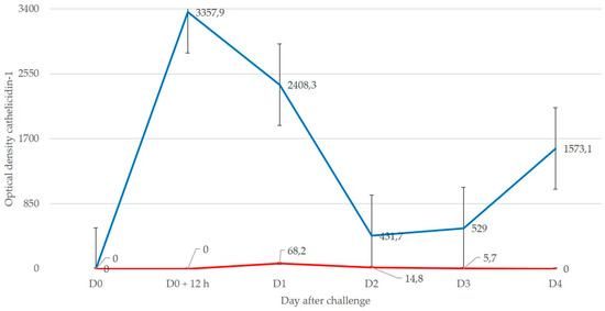
The objective of the study was the investigation of the behaviour of cathelicidin-1 in the milk after experimental infection with two prominent bacterial pathogens (experiment 1: Mannheimia haemolytica, experiment 2: M. haemolytica and Staphylococcus chromogenes) as a potential early indicator for diagnosis of mastitis in sheep. In two experiments, after bacterial inoculation into the udder of ewes, bacteriological and cytological examinations of milk samples as well as proteomics examinations [two-dimensional gel electrophoresis analysis (2-DE) and matrix-assisted laser desorption/ionization time-of-flight mass spectrometer (MALDI-TOF MS) analysis] were performed sequentially. Cathelicidin-1 was detected and spot densities obtained from PDQuest v.8.0 were recorded. Associations were calculated between cell content and spot densities as well as between presence of mastitis in a mammary gland at a given time-point and detection of cathelicidin-1 in the respective milk sample. All inoculated mammary glands developed mastitis, confirmed by the consistent bacterial isolation from mammary secretion and increased leucocyte content therein. Spot density of cathelicidin-1 in samples from inoculated glands increased 3 h post-inoculation; spot density of cathelicidin-1 in samples from inoculated glands was higher than in samples from uninoculated controls. There was clear evidence of correlation between cell content and cathelicidin-1 spot densities in milk samples. There was significant association between presence of mastitis in the mammary gland and detection of cathelicidin-1 in the respective milk sample; overall accuracy was 0.818—this was significantly greater during the first 24 h post-challenge (0.903) than after the first day (0.704). In conclusion, detection of cathelicidin-1 in milk was significantly associated with presence of mastitis in ewes. The associations were stronger during the first 24 h post-infection than after the first day. Cathelicidin-1 has the advantage that it can be a non-specific biomarker, as simply a “positive”/“negative” assessment would be sufficient. Full article
Show Figures

Figure 1

Back to TopTop