sensors-logo

Journal Browser

Journal Browser

Advances in Spectroscopy and Spectral Imaging

A topical collection in Sensors (ISSN 1424-8220). This collection belongs to the section "Sensing and Imaging".

Viewed by 30575

Editors


E-Mail Website
Collection Editor
The Institute for Research in Computer Science, Mathematics, Automation and Signal, IRIMAS UR 7499, University of Haute-Alsace, 68100 Mulhouse, France
Interests: real-time imaging; high dynamic range imaging; polarization imaging; spectral imaging; filter array imaging: from sensor to pre-processing
Special Issues, Collections and Topics in MDPI journals

E-Mail Website
Collection Editor
Okutomi & Tanaka Laboratory, Department of Systems and Control Engineering, School of Engineering, Tokyo Institute of Technology, Tokyo 152-8550, Japan
Interests: spectral imaging; spectral reconstruction; spectral filter arrays; demosaicing

Topical Collection Information

Dear Colleagues,

Spectroscopy aims at recovering the spectral signature of light at a scene point, within a given spectral range and a given spectral resolution. Spectral imaging enhances this functionality by adding spatial dimension, leading to a spatiospectral data representation (i.e., a spectral data cube). On one hand, novel hardware designs dedicated to spectroscopy and spectral imaging (SSI) are demanded to improve the efficiency, flexibility, or compactness of the SSI systems. On the other hand, dedicated data processing is required for the emergence of SSI systems.

Recent advances in the field could potentially lead to the massification of SSI, and a better implication of SSI in applications, such as for computer vision, computer graphics, or remote sensing. To further help SSIs to break through into applications, it is necessary to go beyond our understanding of their limitations.

This Special Issue focuses on these topics, so the different issues, achievements, and progress from different disciplines are available from one single issue.

Potential topics include but are not limited to:
  • Technology: spectral sensors, optical design, camera design, acquisition setup, etc.
  • Computational algorithm: imaging model, data processing, noise reduction, calibration, image enhancement, demosaicing, super-resolution, high dynamic range, etc.
  • Inverse problem: spectral reconstruction, illuminant estimation, reflection mode separation, rendering, matching, etc.
  • Data mining for spectral information: learning, CNN, time series, etc.
  • Applications in computer vision: medical imaging, automotive, cultural heritage (classification, text analysis), etc.
  • Applications in computer graphics: cultural heritage (visual reproduction), etc.
  • Other SSI applications in remote sensing, chemistry, biology, etc.

Dr. Pierre-Jean Lapray
Dr. Jean-Baptiste Thomas
Dr. Yusuke Monno
Collection Editors

Manuscript Submission Information

Manuscripts should be submitted online at www.mdpi.com by registering and logging in to this website. Once you are registered, click here to go to the submission form. Manuscripts can be submitted until the deadline. All submissions that pass pre-check are peer-reviewed. Accepted papers will be published continuously in the journal (as soon as accepted) and will be listed together on the collection website. Research articles, review articles as well as short communications are invited. For planned papers, a title and short abstract (about 100 words) can be sent to the Editorial Office for announcement on this website.

Submitted manuscripts should not have been published previously, nor be under consideration for publication elsewhere (except conference proceedings papers). All manuscripts are thoroughly refereed through a single-blind peer-review process. A guide for authors and other relevant information for submission of manuscripts is available on the Instructions for Authors page. Sensors is an international peer-reviewed open access semimonthly journal published by MDPI.

Please visit the Instructions for Authors page before submitting a manuscript. The Article Processing Charge (APC) for publication in this open access journal is 2600 CHF (Swiss Francs). Submitted papers should be well formatted and use good English. Authors may use MDPI's English editing service prior to publication or during author revisions.

Keywords

  • spectral sensors
  • spectroscopy
  • spectral imaging
  • multispectral imaging
  • hyperspectral imaging
  • spectropolarimetric imaging

Published Papers (11 papers)

2024

Jump to: 2023, 2022, 2021, 2020

7 pages, 8641 KiB  
Communication
Performance Characterization of an Illumination-Based Low-Cost Multispectral Camera
by Hedde van Hoorn, Angel Schraven, Hugo van Dam, Joshua Meijer, Roman Sillé, Arjan Lock and Steven van den Berg
Sensors 2024, 24(16), 5229; https://doi.org/10.3390/s24165229 - 13 Aug 2024
Cited by 1 | Viewed by 1138
Abstract
Spectral imaging has many applications, from methane detection using satellites to disease detection on crops. However, spectral cameras remain a costly solution ranging from 10 thousand to 100 thousand euros for the hardware alone. Here, we present a low-cost multispectral camera (LC-MSC) with [...] Read more.
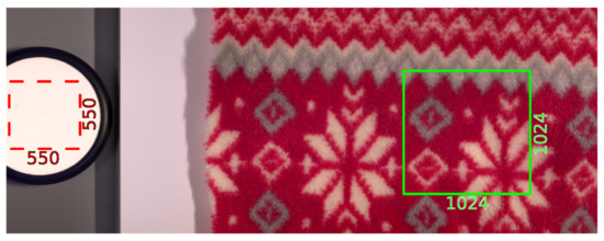
Spectral imaging has many applications, from methane detection using satellites to disease detection on crops. However, spectral cameras remain a costly solution ranging from 10 thousand to 100 thousand euros for the hardware alone. Here, we present a low-cost multispectral camera (LC-MSC) with 64 LEDs in eight different colors and a monochrome camera with a hardware cost of 340 euros. Our prototype reproduces spectra accurately when compared to a reference spectrometer to within the spectral width of the LEDs used and the ±1σ variation over the surface of ceramic reference tiles. The mean absolute difference in reflectance is an overestimate of 0.03 for the LC-MSC as compared to a spectrometer, due to the spectral shape of the tiles. In environmental light levels of 0.5 W m−2 (bright artificial indoor lighting) our approach shows an increase in noise, but still faithfully reproduces discrete reflectance spectra over 400 nm–1000 nm. Our approach is limited in its application by LED bandwidth and availability of specific LED wavelengths. However, unlike with conventional spectral cameras, the pixel pitch of the camera itself is not limited, providing higher image resolution than typical high-end multi- and hyperspectral cameras. For sample conditions where LED illumination bands provide suitable spectral information, our LC-MSC is an interesting low-cost alternative approach to spectral imaging. Full article
Show Figures

Figure 1

19 pages, 7870 KiB  
Article
Lunar Lithium-7 Sensing (δ7Li): Spectral Patterns and Artificial Intelligence Techniques
by Julia Fernandez, Susana Fernandez, Enrique Diez, Noemi Pinilla-Alonso, Saúl Pérez, Santiago Iglesias, Alejandro Buendía, Javier Rodríguez and Javier de Cos
Sensors 2024, 24(12), 3931; https://doi.org/10.3390/s24123931 - 17 Jun 2024
Viewed by 853
Abstract
Lithium, a critical natural resource integral to modern technology, has influenced diverse industries since its discovery in the 1950s. Of particular interest is lithium-7, the most prevalent lithium isotope on Earth, playing a vital role in applications such as batteries, metal alloys, medicine, [...] Read more.
Lithium, a critical natural resource integral to modern technology, has influenced diverse industries since its discovery in the 1950s. Of particular interest is lithium-7, the most prevalent lithium isotope on Earth, playing a vital role in applications such as batteries, metal alloys, medicine, and nuclear research. However, its extraction presents significant environmental and logistical challenges. This article explores the potential for lithium exploration on the Moon, driven by its value as a resource and the prospect of cost reduction due to the Moon’s lower gravity, which holds promise for future space exploration endeavors. Additionally, the presence of lithium in the solar wind and its implications for material transport across celestial bodies are subjects of intrigue. Drawing from a limited dataset collected during the Apollo missions (Apollo 12, 15, 16, and 17) and leveraging artificial intelligence techniques and sample expansion through bootstrapping, this study develops predictive models for lithium-7 concentration based on spectral patterns. The study areas encompass the Aitken crater, Hadley Rima, and the Taurus–Littrow Valley, where higher lithium concentrations are observed in basaltic lunar regions. This research bridges lunar geology and the formation of the solar system, providing valuable insights into celestial resources and enhancing our understanding of space. The data used in this study were obtained from the imaging sensors (infrared, visible, and ultraviolet) of the Clementine satellite, which significantly contributed to the success of our research. Furthermore, the study addresses various aspects related to statistical analysis, sample quality validation, resampling, and bootstrapping. Supervised machine learning model training and validation, as well as data import and export, were explored. The analysis of data generated by the Clementine probe in the near-infrared (NIR) and ultraviolet-visible (UVVIS) spectra revealed evidence of the presence of lithium-7 (Li-7) on the lunar surface. The distribution of Li-7 on the lunar surface is non-uniform, with varying concentrations in different regions of the Moon identified, supporting the initial hypothesis associating surface Li-7 concentration with exposure to solar wind. While a direct numerical relationship between lunar topography and Li-7 concentration has not been established due to morphological diversity and methodological limitations, preliminary results suggest significant economic and technological potential in lunar lithium exploration and extraction. Full article
Show Figures

Figure 1

2023

Jump to: 2024, 2022, 2021, 2020

16 pages, 11117 KiB  
Article
Impact of Exposure and Illumination on Texture Classification Based on Raw Spectral Filter Array Images
by Omar Elezabi, Sebastien Guesney-Bodet and Jean-Baptiste Thomas
Sensors 2023, 23(12), 5443; https://doi.org/10.3390/s23125443 - 8 Jun 2023
Cited by 1 | Viewed by 1503
Abstract
Spectral Filter Array cameras provide a fast and portable solution for spectral imaging. Texture classification from images captured with such a camera usually happens after a demosaicing process, which makes the classification performance rely on the quality of the demosaicing. This work investigates [...] Read more.
Spectral Filter Array cameras provide a fast and portable solution for spectral imaging. Texture classification from images captured with such a camera usually happens after a demosaicing process, which makes the classification performance rely on the quality of the demosaicing. This work investigates texture classification methods applied directly to the raw image. We trained a Convolutional Neural Network and compared its classification performance to the Local Binary Pattern method. The experiment is based on real SFA images of the objects of the HyTexiLa database and not on simulated data as are often used. We also investigate the role of integration time and illumination on the performance of the classification methods. The Convolutional Neural Network outperforms other texture classification methods even with a small amount of training data. Additionally, we demonstrated the model’s ability to adapt and scale for different environmental conditions such as illumination and exposure compared to other methods. In order to explain these results, we analyze the extracted features of our method and show the ability of the model to recognize different shapes, patterns, and marks in different textures. Full article
Show Figures

Figure 1

2022

Jump to: 2024, 2023, 2021, 2020

12 pages, 1645 KiB  
Article
Developing a Method to Estimate the Downstream Metabolite Signals from Hyperpolarized [1-13C]Pyruvate
by Ching-Yi Hsieh, Cheng-Hsuan Sung, Yi-Liang (Eric) Shen, Ying-Chieh Lai, Kuan-Ying Lu and Gigin Lin
Sensors 2022, 22(15), 5480; https://doi.org/10.3390/s22155480 - 22 Jul 2022
Cited by 2 | Viewed by 1825
Abstract
Hyperpolarized carbon-13 MRI has the advantage of allowing the study of glycolytic flow in vivo or in vitro dynamically in real-time. The apparent exchange rate constant of a metabolite dynamic signal reflects the metabolite changes of a disease. Downstream metabolites can have a [...] Read more.
Hyperpolarized carbon-13 MRI has the advantage of allowing the study of glycolytic flow in vivo or in vitro dynamically in real-time. The apparent exchange rate constant of a metabolite dynamic signal reflects the metabolite changes of a disease. Downstream metabolites can have a low signal-to-noise ratio (SNR), causing apparent exchange rate constant inconsistencies. Thus, we developed a method that estimates a more accurate metabolite signal. This method utilizes a kinetic model and background noise to estimate metabolite signals. Simulations and in vitro studies with photon-irradiated and control groups were used to evaluate the procedure. Simulated and in vitro exchange rate constants estimated using our method were compared with the raw signal values. In vitro data were also compared to the Area-Under-Curve (AUC) of the cell medium in 13C Nuclear Magnetic Resonance (NMR). In the simulations and in vitro experiments, our technique minimized metabolite signal fluctuations and maintained reliable apparent exchange rate constants. In addition, the apparent exchange rate constants of the metabolites showed differences between the irradiation and control groups after using our method. Comparing the in vitro results obtained using our method and NMR, both solutions showed consistency when uncertainty was considered, demonstrating that our method can accurately measure metabolite signals and show how glycolytic flow changes. The method enhanced the signals of the metabolites and clarified the metabolic phenotyping of tumor cells, which could benefit personalized health care and patient stratification in the future. Full article
Show Figures

Figure 1

2021

Jump to: 2024, 2023, 2022, 2020

18 pages, 7402 KiB  
Article
Band Selection for Dehazing Algorithms Applied to Hyperspectral Images in the Visible Range
by Sol Fernández-Carvelo, Miguel Ángel Martínez-Domingo, Eva M. Valero, Javier Romero, Juan Luis Nieves and Javier Hernández-Andrés
Sensors 2021, 21(17), 5935; https://doi.org/10.3390/s21175935 - 3 Sep 2021
Cited by 3 | Viewed by 2675
Abstract
Images captured under bad weather conditions (e.g., fog, haze, mist, dust, etc.), suffer from poor contrast and visibility, and color distortions. The severity of this degradation depends on the distance, the density of the atmospheric particles and the wavelength. We analyzed eight single [...] Read more.
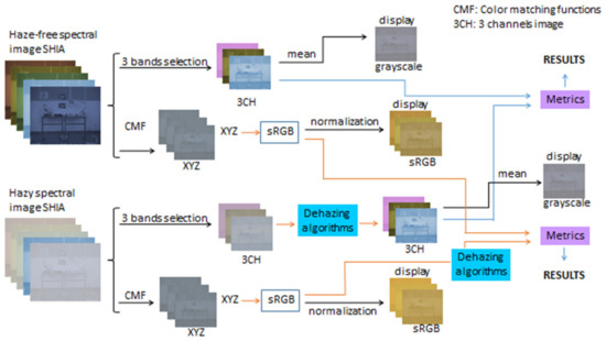
Images captured under bad weather conditions (e.g., fog, haze, mist, dust, etc.), suffer from poor contrast and visibility, and color distortions. The severity of this degradation depends on the distance, the density of the atmospheric particles and the wavelength. We analyzed eight single image dehazing algorithms representative of different strategies and originally developed for RGB images, over a database of hazy spectral images in the visible range. We carried out a brute force search to find the optimum three wavelengths according to a new combined image quality metric. The optimal triplet of monochromatic bands depends on the dehazing algorithm used and, in most cases, the different bands are quite close to each other. According to our proposed combined metric, the best method is the artificial multiple exposure image fusion (AMEF). If all wavelengths within the range 450–720 nm are used to build a sRGB renderization of the imagaes, the two best-performing methods are AMEF and the contrast limited adaptive histogram equalization (CLAHE), with very similar quality of the dehazed images. Our results show that the performance of the algorithms critically depends on the signal balance and the information present in the three channels of the input image. The capture time can be considerably shortened, and the capture device simplified by using a triplet of bands instead of the full wavelength range for dehazing purposes, although the selection of the bands must be performed specifically for a given algorithm. Full article
Show Figures

Figure 1

19 pages, 19217 KiB  
Article
On the Optimization of Regression-Based Spectral Reconstruction
by Yi-Tun Lin and Graham D. Finlayson
Sensors 2021, 21(16), 5586; https://doi.org/10.3390/s21165586 - 19 Aug 2021
Cited by 12 | Viewed by 3459
Abstract
Spectral reconstruction (SR) algorithms attempt to recover hyperspectral information from RGB camera responses. Recently, the most common metric for evaluating the performance of SR algorithms is the Mean Relative Absolute Error (MRAE)—an 1 relative error (also known as percentage error). Unsurprisingly, the [...] Read more.
Spectral reconstruction (SR) algorithms attempt to recover hyperspectral information from RGB camera responses. Recently, the most common metric for evaluating the performance of SR algorithms is the Mean Relative Absolute Error (MRAE)—an 1 relative error (also known as percentage error). Unsurprisingly, the leading algorithms based on Deep Neural Networks (DNN) are trained and tested using the MRAE metric. In contrast, the much simpler regression-based methods (which actually can work tolerably well) are trained to optimize a generic Root Mean Square Error (RMSE) and then tested in MRAE. Another issue with the regression methods is—because in SR the linear systems are large and ill-posed—that they are necessarily solved using regularization. However, hitherto the regularization has been applied at a spectrum level, whereas in MRAE the errors are measured per wavelength (i.e., per spectral channel) and then averaged. The two aims of this paper are, first, to reformulate the simple regressions so that they minimize a relative error metric in training—we formulate both 2 and 1 relative error variants where the latter is MRAE—and, second, we adopt a per-channel regularization strategy. Together, our modifications to how the regressions are formulated and solved leads to up to a 14% increment in mean performance and up to 17% in worst-case performance (measured with MRAE). Importantly, our best result narrows the gap between the regression approaches and the leading DNN model to around 8% in mean accuracy. Full article
Show Figures

Figure 1

19 pages, 20377 KiB  
Article
Band-Selection of a Portal LED-Induced Autofluorescence Multispectral Imager to Improve Oral Cancer Detection
by Yung-Jhe Yan, Nai-Lun Cheng, Chia-Ing Jan, Ming-Hsui Tsai, Jin-Chern Chiou and Mang Ou-Yang
Sensors 2021, 21(9), 3219; https://doi.org/10.3390/s21093219 - 6 May 2021
Cited by 3 | Viewed by 2519
Abstract
This aim of this study was to find effective spectral bands for the early detection of oral cancer. The spectral images in different bands were acquired using a self-made portable light-emitting diode (LED)-induced autofluorescence multispectral imager equipped with 365 and 405 nm excitation [...] Read more.
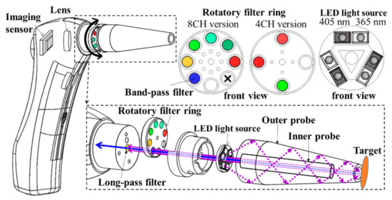
This aim of this study was to find effective spectral bands for the early detection of oral cancer. The spectral images in different bands were acquired using a self-made portable light-emitting diode (LED)-induced autofluorescence multispectral imager equipped with 365 and 405 nm excitation LEDs, emission filters with center wavelengths of 470, 505, 525, 532, 550, 595, 632, 635, and 695 nm, and a color image sensor. The spectral images of 218 healthy points in 62 healthy participants and 218 tumor points in 62 patients were collected in the ex vivo trials at China Medical University Hospital. These ex vivo trials were similar to in vivo because the spectral images of anatomical specimens were immediately acquired after the on-site tumor resection. The spectral images associated with red, blue, and green filters correlated with and without nine emission filters were quantized by four computing method, including summated intensity, the highest number of the intensity level, entropy, and fractional dimension. The combination of four computing methods, two excitation light sources with two intensities, and 30 spectral bands in three experiments formed 264 classifiers. The quantized data in each classifier was divided into two groups: one was the training group optimizing the threshold of the quantized data, and the other was validating group tested under this optimized threshold. The sensitivity, specificity, and accuracy of each classifier were derived from these tests. To identify the influential spectral bands based on the area under the region and the testing results, a single-layer network learning process was used. This was compared to conventional rules-based approaches to show its superior and faster performance. Consequently, four emission filters with the center wavelengths of 470, 505, 532, and 550 nm were selected by an AI-based method and verified using a rule-based approach. The sensitivities of six classifiers using these emission filters were more significant than 90%. The average sensitivity of these was about 96.15%, the average specificity was approximately 69.55%, and the average accuracy was about 82.85%. Full article
Show Figures

Figure 1

10 pages, 1462 KiB  
Communication
Proof of Principle for Direct Reconstruction of Qualitative Depth Information from Turbid Media by a Single Hyper Spectral Image
by Martin Hohmann, Damaris Hecht, Benjamin Lengenfelder, Moritz Späth, Florian Klämpfl and Michael Schmidt
Sensors 2021, 21(8), 2860; https://doi.org/10.3390/s21082860 - 19 Apr 2021
Cited by 3 | Viewed by 2420
Abstract
In medical applications, hyper-spectral imaging is becoming more and more common. It has been shown to be more effective for classification and segmentation than normal RGB imaging because narrower wavelength bands are used, providing a higher contrast. However, until now, the fact that [...] Read more.
In medical applications, hyper-spectral imaging is becoming more and more common. It has been shown to be more effective for classification and segmentation than normal RGB imaging because narrower wavelength bands are used, providing a higher contrast. However, until now, the fact that hyper-spectral images also contain information about the three-dimensional structure of turbid media has been neglected. In this study, it is shown that it is possible to derive information about the depth of inclusions in turbid phantoms from a single hyper-spectral image. Here, the depth information is encoded by a combination of scattering and absorption within the phantom. Although scatter-dominated regions increase the backscattering for deep vessels, absorption has the opposite effect. With this argumentation, it makes sense to assume that, under certain conditions, a wavelength is not influenced by the depth of the inclusion and acts as an iso-point. This iso-point could be used to easily derive information about the depth of an inclusion. In this study, it is shown that the iso-point exists in some cases. Moreover, it is shown that the iso-point can be used to obtain precise depth information. Full article
Show Figures

Figure 1

14 pages, 14069 KiB  
Article
Eight-Channel Multispectral Image Database for Saliency Prediction
by Miguel Ángel Martínez-Domingo, Juan Luis Nieves and Eva M. Valero
Sensors 2021, 21(3), 970; https://doi.org/10.3390/s21030970 - 1 Feb 2021
Cited by 5 | Viewed by 3334
Abstract
Saliency prediction is a very important and challenging task within the computer vision community. Many models exist that try to predict the salient regions on a scene from its RGB image values. Several new models are developed, and spectral imaging techniques may potentially [...] Read more.
Saliency prediction is a very important and challenging task within the computer vision community. Many models exist that try to predict the salient regions on a scene from its RGB image values. Several new models are developed, and spectral imaging techniques may potentially overcome the limitations found when using RGB images. However, the experimental study of such models based on spectral images is difficult because of the lack of available data to work with. This article presents the first eight-channel multispectral image database of outdoor urban scenes together with their gaze data recorded using an eyetracker over several observers performing different visualization tasks. Besides, the information from this database is used to study whether the complexity of the images has an impact on the saliency maps retrieved from the observers. Results show that more complex images do not correlate with higher differences in the saliency maps obtained. Full article
Show Figures

Figure 1

2020

Jump to: 2024, 2023, 2022, 2021

19 pages, 1724 KiB  
Article
Wide Field Spectral Imaging with Shifted Excitation Raman Difference Spectroscopy Using the Nod and Shuffle Technique
by Florian Korinth, Elmar Schmälzlin, Clara Stiebing, Tanya Urrutia, Genoveva Micheva, Christer Sandin, André Müller, Martin Maiwald, Bernd Sumpf, Christoph Krafft, Günther Tränkle, Martin M. Roth and Jürgen Popp
Sensors 2020, 20(23), 6723; https://doi.org/10.3390/s20236723 - 24 Nov 2020
Cited by 9 | Viewed by 3679
Abstract
Wide field Raman imaging using the integral field spectroscopy approach was used as a fast, one shot imaging method for the simultaneous collection of all spectra composing a Raman image. For the suppression of autofluorescence and background signals such as room light, shifted [...] Read more.
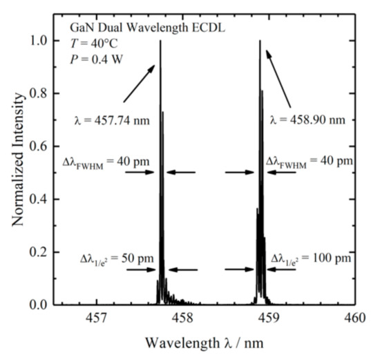
Wide field Raman imaging using the integral field spectroscopy approach was used as a fast, one shot imaging method for the simultaneous collection of all spectra composing a Raman image. For the suppression of autofluorescence and background signals such as room light, shifted excitation Raman difference spectroscopy (SERDS) was applied to remove background artifacts in Raman spectra. To reduce acquisition times in wide field SERDS imaging, we adapted the nod and shuffle technique from astrophysics and implemented it into a wide field SERDS imaging setup. In our adapted version, the nod corresponds to the change in excitation wavelength, whereas the shuffle corresponds to the shifting of charges up and down on a Charge-Coupled Device (CCD) chip synchronous to the change in excitation wavelength. We coupled this improved wide field SERDS imaging setup to diode lasers with 784.4/785.5 and 457.7/458.9 nm excitation and applied it to samples such as paracetamol and aspirin tablets, polystyrene and polymethyl methacrylate beads, as well as pork meat using multiple accumulations with acquisition times in the range of 50 to 200 ms. The results tackle two main challenges of SERDS imaging: gradual photobleaching changes the autofluorescence background, and multiple readouts of CCD detector prolong the acquisition time. Full article
Show Figures

Figure 1

19 pages, 4907 KiB  
Article
Nondestructive Detection for Egg Freshness Based on Hyperspectral Scattering Image Combined with Ensemble Learning
by Dejian Dai, Tao Jiang, Wei Lu, Xuan Shen, Rui Xiu and Jingwei Zhang
Sensors 2020, 20(19), 5484; https://doi.org/10.3390/s20195484 - 25 Sep 2020
Cited by 17 | Viewed by 4939
Abstract
Scattering hyperspectral technology is a nondestructive testing method with many advantages. Here, we propose a method to improve the accuracy of egg freshness, research the influence of incident angles of light source on the accuracy, and explain its mechanism. A variety of weak [...] Read more.
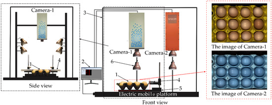
Scattering hyperspectral technology is a nondestructive testing method with many advantages. Here, we propose a method to improve the accuracy of egg freshness, research the influence of incident angles of light source on the accuracy, and explain its mechanism. A variety of weak classifiers classify eggs based on the spectra after preprocessing and feature wavelength extraction to obtain three classifiers with the highest accuracy. The three classifiers are used as metamodels of stacking ensemble learning to improve the highest accuracy from 96.25% to 100%. Moreover, the highest accuracy of scattering, reflection, transmission, and mixed hyperspectral of eggs are 100.00%, 88.75%, 95.00%, and 96.25%, respectively, indicating that the scattering hyperspectral for egg freshness detection is better than that of the others. In addition, the accuracy is inversely proportional to the angle of incidence, i.e., the smaller the incident angle, the camera collects a larger proportion of scattering light, which contains more biochemical parameters of an egg than that of reflection and transmission. These results are very important for improving the accuracy of non-destructive testing and for selecting the incident angle of a light source, and they have potential applications for online non-destructive testing. Full article
Show Figures

Figure 1

Back to TopTop