Article

Antibacterial Activity of a Promising Antibacterial Agent: 22-(4-(2-(4-Nitrophenyl-piperazin-1-yl)-acetyl)-piperazin-1-yl)-22-deoxypleuromutilin

1
Guangdong Provincial Key Laboratory of Veterinary Pharmaceutics Development and Safety Evaluation, College of Veterinary Medicine, South China Agricultural University, Guangzhou 510642, China
2
Guangdong Laboratory for Lingnan Modern Agriculture, Guangzhou 510642, China
*
Author to whom correspondence should be addressed.
These authors contributed equally to this work.
Molecules 2021, 26(12), 3502; https://doi.org/10.3390/molecules26123502
Submission received: 21 May 0202 / Revised: 21 May 2021 / Accepted: 5 June 2021 / Published: 8 June 2021

Abstract

:
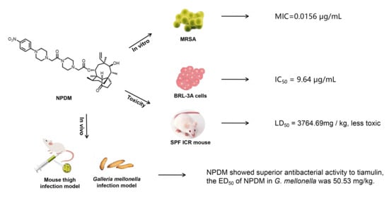
A novel pleuromutilin derivative, 22-(4-(2-(4-nitrophenyl-piperazin-1-yl)-acetyl)-piperazin-1-yl)-22-deoxypleuromutilin (NPDM), was synthesized in our laboratory and proved excellent antibacterial activity against methicillin-resistant Staphylococcus aureus (MRSA). In this study, more methods were used to further study its preliminary pharmacological effect. The antibacterial efficacy and toxicity of NPDM were evaluated using tiamulin as the reference drug. The in vitro antibacterial activity study showed that NPDM is a potent bactericidal agent against MRSA that induced time-dependent growth inhibition and a concentration-dependent post-antibiotic effect (PAE). Toxicity determination showed that the cytotoxicity of NPDM was slightly higher than that of tiamulin, but the acute oral toxicity study proved that NPDM was a low-toxic compound. In an in vivo antibacterial effect study, NPDM exhibited a better therapeutic effect than tiamulin against MRSA in a mouse thigh infection model as well as a mouse systemic infection model with neutropenia. The 50% effective dose (ED50) of NPDM in a Galleria mellonella infection model was 50.53 mg/kg. The pharmacokinetic properties of NPDM were also measured, which showed that NPDM was a rapid elimination drug in mice.

Graphical Abstract

1. Introduction

Staphylococcus aureus is a potentially pathogenic Gram-positive bacteria that can cause infections ranging from mild (such as skin infection) to severe (such as postsurgical wound infections and systemic infections) [1]. Staphylococcus aureus possesses great ability to acquire antibiotic resistance. Only two years after the clinical use of methicillin, Jevons isolated MRSA in 1961 [2]. Nowadays, MRSA is almost universal and has become one of the most common pathogens in clinics [3]. Patients infected with MRSA suffer higher costs, longer hospital stays, and higher morbidity and mortality [4]. In bacteremia, for example, the in-hospital mortality rate for S. aureus bacteremia is nearly 40%, and the mortality rate of bacteremia patients caused by MRSA is 2.15 times higher than that caused by methicillin-sensitive Staphylococcus aureus (MSSA) [5]. Methicillin-resistant S. aureus is particularly resistant to β-lactam antibiotics, and this resistance is known to be caused by the penicillin-binding protein (encoded by genes designated 2’ or 2α) on the cell wall of S. aureus, which reduces the binding affinity between MRSA and β-lactam antibiotics [2]. It is urgent and necessary to develop new antibacterial agents that have different antimicrobial mechanisms against this microorganism.
Pleuromutilin (Figure 1), a natural compound, was isolated from Pleurotus mutiliz and Pleurotus passeckeranius by F. Kavanag in 1951 [6]. It displayed significant antibacterial activity against Gram-positive bacteria and mycoplasma [7]. The pleuromutilin derivatives selectively bind to the bacterial peptidyl transferase center and interfere with the synthesis of bacterial proteins [8,9]. This unique antibacterial mechanism reduces the cross-resistance between pleuromutilin and clinical antibiotics [10]. Nowadays, pleuromutilin has received increasing attention for the development of new antibiotics and the treatment of drug-resistant bacterial infections [11,12]. Lefamulin (Figure 1) is the first intravenous and oral pleuromutilin antibiotic approved by Food and Drug Administration, in 2019, for the treatment of community-acquired bacterial pneumonia [13]. As the first antibiotic with a new structure approved for systemic use in the past 20 years, lefamulin has aroused great interest in pleuromutilin among pharmacological researchers [14].
A series of pleuromutilin compounds were synthesized, and we performed a preliminary antimicrobial analysis. The results revealed that NPDM (Figure 1) possessed distinguished antibacterial effect against MRSA [15]. It motived us to further study its pharmacological characteristics including toxicity determination, in vitro and in vivo antibacterial activity, and pharmacokinetic studies.

2. Results

2.1. In Vitro Efficacy of NPDM

To evaluate the in vitro antibacterial efficacy, the minimum inhibitory concentrations (MICs) and minimum bactericidal concentrations (MBCs) of NPDM were determined against MRSA ATCC 43300, S. aureus ATCC 29213, and two clinical strains of S. aureus (AD3 and 144) using tiamulin as the reference drug. The results are shown in Table 1.
The results of time–kill assays are shown in Figure 2. The NPDM displayed bactericidal activity (i.e., ≥3 log10 CFU/mL decrease) against MRSA at the concentration of ≥2 × MIC after incubation for 24 h, while tiamulin showed bactericidal activity against MRSA at the concentration of ≥4 × MIC. Both NPDM and tiamulin showed a bacteriostatic effect on S. aureus when the drug concentration was no less than 2 × MIC.
To evaluate the in vitro antibacterial pharmacodynamics activity, the PAEs of NPDM and tiamulin against MRSA were then investigated. The results are shown in Figure 3 and Table 2.

2.2. Toxicity Determination

The cytotoxicity of NPDM and tiamulin were determined using the conventional 3-(4,5-dimethyl-2-thiazolyl)-2,5-diphenyl-2H-tetrazolium bromide (MTT) assay on normal rat liver cells (i.e., BRL-3A). The cell viability affected by different concentrations of the tested compounds is indicated in Figure 4. The 50% inhibition concentration (IC50) value of NPDM was 9.64 μg/mL, which was lower than that of tiamulin (13.1 μg/mL).
The results of the acute oral toxicity of NPDM are summarized in Table 3. After receiving a dose of 987.65 mg/kg of NPDM, only one mouse died, and five mice died within a day after receiving a dose of 5000 mg/kg of NPDM. The LD50 was 3764.49 mg/kg calculated by modified Karber’s formula, and the 95% confidence interval was 3006.67–4713.31 mg/kg. Swelling was found in the abdomen and back of some surviving and dead mice. For the dead mice, we also observed the rolling behavior and heard a strong gasping sound while they were alive.

2.3. In Vivo Effect of NPDM

In this study, the mouse thigh infection model and mouse systemic infection model were established to study the in vivo antibacterial activity of NPDM and tiamulin against MRSA or S. aureus ATCC 29213. The results are shown in Figure 5. The amount of inoculum in each thigh of neutropenic mice was 105~6 CFU. After treatment with the test compound for 24 h, the average amount of MRSA inoculum in thighs of the NPDM-treated group, tiamulin-treated group, and untreated control group were (8.16 ± 3.70) × 107 CFU, (2.64 ± 1.95) × 108 CFU, and (3.22 ± 1.09) × 109 CFU, respectively (Figure 5A), and that of the S. aureus were (1.23 ± 0.51) × 108 CFU, (2.00 ± 1.21) × 108 CFU, and (1.00 ± 0.79) × 109 CFU, respectively (Figure 5B). Compared with the untreated group, treatment with NPDM (20 mg/kg) could significantly reduce mean MRSA counts (~1.61 log CFU/mL) in thighs (p < 0.05), indicating that NPDM possessed more potent in vivo antibacterial activity than tiamulin (decrease ~1.15 log CFU/mL). For S. aureus, treatment with NPDM at 20 mg/kg could significantly reduce mean bacterial counts (~0.95 log CFU/mL), which was more effective than tiamulin (~0.68 log CFU/mL).
We evaluated the therapeutic activity of NPDM and tiamulin using the mouse systemic infection model. The survival curves of NPDM and tiamulin at the dose of 30 mg/kg are shown in Figure 6. The untreated mice all died within 72 h following inoculation. Mice injected with sterile saline survived during the experiment. The results showed that the survival rate of NPDM against MRSA was 70% at the endpoint. This result illustrated that NPDM could protect mice from a lethal MRSA attack in vivo.
The Galleria mellonella infection model was utilized to investigate the ED50 of NPDM and tiamulin against MRSA. The survival rate of each group is shown in Figure 7. The dose of NPDM and tiamulin the led to the 50% effectiveness were 50.53 mg/kg and 75.74 mg/kg, respectively.

2.4. Pharmacokinetic Study

In order to assess the pharmacokinetic properties of NPDM, plasma samples were analyzed by LC-MS/MS. Representative LC-MS/MS chromatograms can be found in Figure S1 in the Supplementary Materials including chromatograms of blank plasma samples, blank plasma samples spiked with NPDM, and plasma samples taken 5 min after administration of NPDM. Under this LC-MS/MS condition, the retention time of NPDM was approximately 4.38 min. The results indicated that no interfering peak of endogenous substance was observed to affect the determination of NPDM. The concentration range of the calibration curve was 0~500 ng/mL. The typical equation and R2 value of the calibration curve were as follows: y = 386∙x + 52.1, R2 = 0.9998 (y: peak area of NPDM; x: concentration of NPDM in plasma). The limit of detection of NPDM was 0.1 ng/mL. The limit of quantification of NPDM was 2 ng/mL. The results revealed that the concentration of NPDM in plasma has linearity with its peak area in the range of 2~500 ng/mL. The extraction recovery ranged from 77.2~112%. The intra-day and inter-day precision ranged from 2.75% to 8.74% and 9.16% to 12.77% (see Table S2 in the Supplemental Materials).
The pharmacokinetic parameters of NPDM are shown in Table 4. The plasma concentration–time curve of NPDM after intravenous administration is displayed in Figure 8.

3. Discussion

In a previous study, we determined the MIC and MBC of NPDM and tiamulin against MRSA ATCC 43300 and S. aureus ATCC 29213 [15]. Considering that the high content of organic solvents may affect the value of MIC [16], in this study, we dissolved drugs using 5% DMSO, 5% Tween-80, and 90% normal saline rather than methanol. As shown in Table 1, NPDM showed excellent in vitro antibacterial activity against MRSA ATCC 43300, S. aureus ATCC 29213, AD3, and 144, and it was more potent than the reference drug tiamulin. The MBC/MIC value of NPDM against MRSA was one, which initially indicated that NPDM might possess great bactericidal effect.
The MIC and MBC are static measurements of antibacterial drugs [17]. To explore the profile of antibacterial activity over time, we conducted the time–kill test. As shown in Figure 2, the antibacterial effect of NPDM was positively correlated with time, not with the increase in drug concentration. These results demonstrate that NPDM induced time-dependent growth inhibition instead of dose-dependent growth inhibition. The NPDM displayed bactericidal activity against MRSA at a concentration of 2 × MIC, while tiamulin (2 × MIC) displayed bacteriostatic activity with a rebound growth. It is suggested that NPDM might be more beneficial for clinical use than tiamulin for its more significant bactericidal effect on MRSA.
The PAE was also investigated to evaluate the in vitro antibacterial pharmacodynamic activity of NPDM. The PAE refers to when the growth of bacteria remains under continuous inhibition after brief contact with antibiotics, even if the concentration of antibiotics drops below the minimum inhibitory concentration or disappears. From a clinical perspective, the PAE provides a theoretical basis for adjusting the dosing interval of some antibacterial agents and is of great significance for optimizing the treatment regimen and minimizing the adverse drug reactions [17,18]. At the concentration of 2 × MIC, the PAEs of NPDM after exposure for 1 h and 2 h were 2.58 h and 3.62 h, while at the concentration of 4 × MIC, they were 3.11 h and 4.03 h. The PAE was prolonged with the increase in the concentration of NPDM, which indicated that NPDM displayed a concentration-independent PAE (Figure 3). Additionally, the PAE of NPDM was 1~2 h longer than that of tiamulin (Table 2). It is suggested that NPDM might support longer dosing intervals than tiamulin when formulating a regimen for the treatment of MRSA.
The cytotoxicity study on BRL-3A illustrated that NPDM inhibited cell viability at low concentrations. The IC50 of NPDM was lower than that of tiamulin, which showed that the cytotoxicity of NPDM was slightly higher than that of tiamulin (Figure 4). Because the absorption of various drugs is different, the cytotoxicity may not fully reflect the toxicity of the drug. Therefore, acute toxicity experiments in vivo were conducted to study the toxicity of NPDM. An acute toxicity study identified the toxicity of NPDM and provided information on the maximum dose limit that can be used for a single feeding. The results showed that the LD50 of NPDM in mice was 3764.49 mg/kg, which was in the range of 501~5000 mg/kg. According to the classification of acute toxicity of an exogenous compound [19], NPDM was considered to be a low-toxic compound. The ATTM (Figure 1), the other semi-synthesis pleuromutilin derivative synthesized by Zhang Chao et al. [20], possesses great antibacterial effect on MRSA. The LD50 of ATTM in mice was 2304.4 mg/kg [20]. The oral acute toxicity of NPDM in mice was significantly lower than that of ATTM, which indicated that NPDM exhibits relatively high security and high potential for development as a new drug.
Since NPDM possessed potent antibacterial activity against MRSA in vitro and displayed high safety in the cytotoxicity study, we further utilized the mouse thigh infection model with neutropenia as well as the mouse systemic infection model to evaluate the in vivo antibacterial effect of NPDM against MRSA and S. aureus. Neutropenia mice are difficult to clear the pathogen through autoimmune response after infection. Thus, they can eliminate the interference of self-defense in pharmacodynamic studies [21]. As shown in Figure 5A, the number of bacteria in the thigh muscle of the two compound treatment groups were significantly decreased compared to the bacterial growth control group (p < 0.001), and the difference between the NPDM-treated group and the tiamulin-treated group was statistically significant (p < 0.05). The results indicated that both NPDM and tiamulin have great antibacterial effect on MRSA, and the antibacterial effect of NPDM was stronger than that of tiamulin. It can be seen from Figure 5B that S. aureus was less sensitive to NPDM than MRSA, which was consistent with the results of the in vitro time–kill study. The systemic infection model was utilized to determine the survival rate of mice after the MRSA lethal dose challenge. We found that the survival rate of the NPDM-treated group was 70% at the dose of 30 mg/kg, which was 20% higher than that of the tiamulin-treated group (Figure 6). This result indicated that NPDM possessed superior in vivo therapeutic activity to tiamulin against MRSA infection. We further studied the in vivo therapeutic efficacy of NPDM on the Galleria mellonella infection model. The Galleria mellonella infection model has the advantages of low cost and fast data collection and has been used as an alternative for mammalian models to investigate the in vivo efficacy of antibacterial drugs [22,23]. The ED50 of NPDM and tiamulin were 50.53 mg/kg and 75.74 mg/kg (Figure 7), which showed that NPDM displayed higher activity than tiamulin against MRSA in the Galleria mellonella infection model.
Inspired by the excellent antibacterial activity of NPDM against MRSA both in vivo and in vitro, the pharmacokinetic properties of NPDM were investigated to evaluate its potential for further research. After a single intravenous administration, NPDM displayed an acceptable plasma exposure level (AUC0→ = 3.66 h·μg/mL). The T1/2 was 0.22 h, associated with high clearance (2.76 L/h/kg) and low MRT (0.36 h), which reflected that NPDM belongs to rapid elimination drugs (Table 4, Figure 8). To overcome the disadvantage of the short half-life of NPDM and make it more suitable for clinical application, we will make modifications that can prolong the half-life of NPDM such as using a sustained-release preparation or modifying its chemical structure.

4. Materials and Methods

4.1. Chemicals

The NPDM was synthesized according to our previous report [15]. The purity of NPDM was confirmed by RP-HPLC analysis. Tiamulin (Figure 1, purity: 98%) was purchased from Guangzhou Xiangbo Biotechnology Co., Ltd. (Guangzhou, China). All compounds were dissolved with 5% DMSO, 5% Tween-80, and 90% normal saline and filtered with a disposable 0.22 μm aseptic filter to prepare stock solutions. Stock solutions were stored at −20 °C.

4.2. Cell Culture and Preparation

The BRL-3A cells were purchased from the Shanghai Cell Bank of the Chinese Academy of Sciences (Shanghai, China). The cells were cultured in Dulbecco’s modified Eagle’s medium (DMEM) containing 10% FBS, 100 mg/mL streptomycin, and 100 IU penicillin at 37 °C with 5% CO2 and were sub-cultured every 2 or 3 days.

4.3. Bacterial Strains and Culture Medium

Staphylococcus aureus ATCC 29213 and MRSA ATCC 43300 were obtained from the Guangdong Microbial Culture Collection Center (Guangzhou, China). The S. aureus (AD3 and 144) were isolated from a pig farm in Guangdong Province. Brain heart infusion (BHI), mannitol salt agar (MSA), Mueller–Hinton agar (MHA), and Mueller–Hinton broth (MHB) were purchased from Qingdao Hope Bio-Technology Co. (Qingdao, China), Ltd. All bacterial strains were stored at −20 °C in 40% glycerol. A single colony was selected from the MSA plate, inoculated in BHI broth and pre-incubated at 37 °C to the logarithmic growth phase.

4.4. Animal

Adult specific pathogen-free ICR mice (22~25 g, 5~6 weeks) were purchased from Hunan Slack Jingda Experimental Animal Co., Ltd. (Changsha, China). All mice were adapted for 1 week before the beginning of the experiments. Galleria mellonella (G. mellonella), about 350 mg, were purchased from Tianjin Kaide Ruixin Co., Ltd., (Tianjin, China) and used within ten days. The protocol was approved by the Animal Research Committees of the South China Agriculture University.

4.5. Antimicrobial Activity Evaluation

4.5.1. The MICs Assay

The MICs of NPDM against MRSA were determined by the broth dilution micro-method [24]. Briefly, the stock solution of NPDM and the reference drug tiamulin were diluted by a two-fold serial dilution method with MHB in a 96-well plate to provide concentration ranges of 16~0.032 μg/mL. Then 100 μL of bacterial suspensions in log phase were diluted to 106 CFU/mL with MHB and were added to each well to make the final concentrations of the test compounds ranging from 8 to 0.016 μg/mL. The bacterial suspension (no drugs but solvent) was used as the positive control and MHB (no cultures) was used as the negative control. After incubation at 37 °C for 24 h, the MIC value was observed as the lowest drug concentration which inhibited the visible growth of bacteria in the test group. The MICs assay was carried out in triplicate.

4.5.2. The MBCs Assay

Determination of MBCs refers to the minimum concentrations of antibacterial agents required to kill 99.9% of the test microorganisms after 24 h of incubation [24]. After obtaining the MICs value, the 96-well plates were placed at 37 °C and incubated for 24 h. One hundred microliter aliquots of the bacterial suspensions were taken from the wells without visible bacterial growth and then plated onto MHA plates. All plates were placed at 37 °C and incubated aerobically for 24 h for colony count, and the MBC was determined as the drug concentration corresponding to the number of colonies ≤ 5 on the culture medium. The MBC assay was conducted at least in triplicate.

4.5.3. Time–Kill Assay

The bactericidal activity of NPDM was evaluated by the time–kill curve assay as described previously [25]. The MRSA and S. aureus in the logarithmic phase were diluted to 106 CFU/mL with MHB. Then diverse concentrations of NPDM or tiamulin were added to the bacteria solution reaching the final concentration of 1 × MIC, 2 × MIC, 4 × MIC, and 8 × MIC, respectively. All bacterial inoculums were cultured at 37 °C with shaking. A total of 100 μL samples were taken from the subcultures at 0, 3, 6, 9, and 24 h, respectively, and continuously diluted 10-fold in sterile saline; then, 25 μL diluent was plated on the MHA plate. All plates were placed at 37 °C for 24 h. The total number of colonies on the plate was counted, and the results were expressed as log (CFU/mL).

4.5.4. The PAE Assay

The PAE was carried out as described in the literature [26]. The MRSA was diluted to 106 CFU/mL in MHB. The NPDM or tiamulin was added into the bacteria solution at the final concentrations of 2 × MIC and 4 × MIC. Then all tubes were incubated at 37 °C for 1 h or 2 h. Each of the above inoculums was diluted 1000-fold with pre-warmed MHB to remove the test compounds and placed in a water bath at 37 °C immediately. One hundred microliters of suspension were taken from each culture, diluted 10-fold in sterile saline, and inoculated on MHA plates at 0, 1, 2, 4, and 6 h after inoculation. The total number comprising the colonies was counted after incubation at 37 °C for 20 h. The PAE was calculated referring to published methods [26]. The experiment was performed in triplicate.

4.6. Toxicity Determination

4.6.1. Cytotoxicity Assay

The cytotoxicity of NPDM was evaluated by the MTT assay as previously reported [25]. Briefly, the medium containing 1 × 10⁵ BRL-3A cells per 1 mL was seeded into a 96-well plate at 100 μL per well. After incubation for 48 h, media were replaced with 100 μL of serial dilutions of test compounds and incubated for 16 h. The absorbance (A) was measured at 490 nm by an automatic microplate reader (BIO-TEK Instrument Inc., Winooski, VT, USA). The cell viability was calculated according to Equation (1):
Cell   viability   ( % )   =   ( A simple A negative ) / ( A positive A negative ) × 100 %
A graph was constructed by plotting the inhibition percentage versus various concentrations of NPDM and tiamulin, from which the IC50 of NPDM or tiamulin was calculated. The measurement was repeated in triplicate.

4.6.2. Acute Oral Toxicity Evaluation

The acute oral toxicity of NPDM was evaluated according to the method reported in the literature [27]. Mice were divided into 6 groups (10 animals each group, half male and half female) and were fasted for 12 h before the experiment. The NPDM was dissolved in DMSO and suspended in 0.5% sodium carboxymethyl cellulose solution. The dosage levels of NPDM are shown in Table 3. The volumes of suspension by oral gavages were 0.1 mL/10 g. The vehicle control group received DMSO without drug. All animals were observed twice daily for symptoms and mortality for two weeks. At the end of the study, all surviving animals were sacrificed. The LD50 value and 95% confidence intervals of acute oral toxicity were calculated according to the equation in the literature [28].

4.7. Animal In Vivo Model

4.7.1. Mouse Thigh Infection Model

The mouse thigh infection model was performed as described in the literature with minor modifications [29]. Briefly, female mice had induced neutropenia (neutrophil count ≤ 100/mm3) by administered intraperitoneally cyclophosphamide on day 4 (150 mg/kg) and day 1 (100 mg/kg) before the experiment. A bacterial suspension of 0.1 mL (MRSA or S. aureus) containing approximately 107 CFU/mL microorganism was injected into the posterior thigh of mice to establish infection. Two hours after infection, mice were intravenously injected with NPDM or tiamulin at 20 mg/kg. After 24 h, thighs (six per group) were collected upon euthanasia. Then, thighs were weighed and homogenized with sterile saline. Tissue homogenate was continuously diluted 10-fold and plated on MHA plates for colony counts. SPSS 19 software was used for statistical analysis and t-tests.

4.7.2. Mouse Systemic Infection Model

The survival rate of mice was determined by the systemic infection model according to the literature with some modifications [30]. Neutropenic mice (10 per group, half male and half female) were injected with 0.5 mL MRSA inoculum of 106 CFU/mL (100% minimum lethal dose) intraperitoneally. One hour after infection, the mice were administered with NPDM or tiamulin intravenously at a dose of 30 mg/kg body weight. Tiamulin was used as a reference drug. The survival of the mice was recorded for 7 days.

4.7.3. Galleria Mellonella Infection Model

The survival rate of Galleria mellonella was determined according to the literature with some modifications [22]. Larvae of Galleria mellonella (16 per group) were injected with 10 μL of 108 CFU/mL MRSA inoculum into the last right proleg. Two hours after infection, larvae were injected with 10 μL of various doses (17.14, 25.71, 37.14, 57.14, and 85.71 mg/kg) of NPDM or tiamulin (used as reference drug) into the last left proleg. The larvae were maintained at 37 °C. Survival of the larvae was recorded daily for 5 days, and the ED50 was calculated.

4.8. Pharmacokinetic Studies

Adult female mice fasted for 12 h before the experiment. The NPDM was administered intravenously at the dose of 10 mg/kg. Approximately 0.2 mL blood samples were collected from the ocular venous plexus at 0.083, 0.167, 0.25, 0.5, 0.75, 1, 2, 4, 6, 8, 12, and 24 h after administration. All samples were centrifuged at 3000 rpm for 10 min, then the plasma samples were separated and stored at −20 °C. After thawing at room temperature, samples were mixed with acetonitrile (HPLC grade, sample:acetonitrile = 1:4, v/v) and centrifuged at 12,000 rpm for 10 min. Then, the supernatant was purified by the 0.22 μm cellulose membrane filter and transferred into an autosampler vial for HPLC-MS/MS analysis. Information of the equipment and analysis conditions are available in the Supplemental Materials. The pharmacokinetic parameters of the NPDM were calculated using the non-compartmental model in WinNonlin 5.2 software (Pharsight, Mountain View, CA, USA).

5. Conclusions

NPDM, a new derivative of pleuromutilin, was synthesized in our lab. Compared to tiamulin, NPDM displayed superior bactericidal effect against MRSA both in vitro and in vivo. Furthermore, toxicity studies showed that NPDM was a less-toxic antibacterial agent. The pharmacokinetic study showed that NPDM was a rapid elimination drug in mice. As the harm of multi-drug-resistant bacteria is becoming increasingly serious today, NPDM, given its effectiveness and safety demonstrated in this study, merits further studies as a novel antibacterial agent against MRSA infections.

Supplementary Materials

Table S1. MRM parameters of NPDM. Figure S1. LC-MS/MS chromatogram of a blank plasma sample (A), a blank plasma sample added 500 ng/mL NPDM (B), and a plasma sample 5 min after administration with NPDM (C). Table S2. Intra- and inter-day assay precision and accuracy for NPDM.

Author Contributions

Y.-Z.T. designed the experiments; H.G. and M.-L.G. carried out the experiments; H.G., X.-Y.Z., and Y.-Z.T. analyzed the experimental results; H.G. and X.-Y.Z. wrote the original draft; X.-Y.Z., Z.J., and Y.-Z.T. reviewed and edited the manuscript. All authors have read and agreed to the published version of the manuscript.

Funding

This research received no external funding.

Institutional Review Board Statement

All animal experiments were approved by the Animal Research Committees of the South China Agriculture University (SYXK2019-0136) and carried out in strict accordance with the recommendations in the Guide for the Care and Use of Laboratory Animals of the National Institutes of Health. The approval date was 4 December 2019. The number of the document which verified approval of the study is 2019181.

Informed Consent Statement

Not applicable.

Data Availability Statement

Not available.

Acknowledgments

This work was supported by the National Key Research and Development Program of China (No. 2016YFD0501300), Guangdong Natural Science Funds for Distinguished Young Scholars (No. 2019B151502002), and the National Natural Science Foundation of China (No. 31672602).

Conflicts of Interest

The authors declare no conflict of interest.

Sample Availability

Samples of the compounds are not available from the authors.

References

  1. Diaz, R.; Ramalheira, E.; Afreixo, V.; Gago, B. Methicillin-resistant Staphylococcus aureus carrying the new mecC gene—A meta-analysis. Diagn. Microbiol. Infect. Dis. 2016, 84, 135–140. [Google Scholar] [CrossRef]
  2. Moellering, R.C., Jr. MRSA: The first half century. J. Antimicrob. Chemother. 2012, 67, 4–11. [Google Scholar] [CrossRef] [PubMed] [Green Version]
  3. Frazee, B.W.; Lynn, J.; Charlebois, E.D.; Lambert, L.; Lowery, D.; Perdreau-Remington, F. High Prevalence of Methicillin-Resistant Staphylococcus aureus in Emergency Department Skin and Soft Tissue Infections. Ann. Emerg. Med. 2005, 45, 311–320. [Google Scholar] [CrossRef] [PubMed]
  4. Nyasulu, P.; Chipolombwe, J.; Török, M.E.; Mbelle, N. Methicillin-resistant Staphylococcus aureus multiple sites surveillance: A systemic review of the literature. Infect. Drug Resist. 2016, 9, 35–42. [Google Scholar] [CrossRef] [Green Version]
  5. Kaku, N.; Yanagihara, K.; Morinaga, Y.; Yamada, K.; Harada, Y.; Migiyama, Y.; Nagaoka, K.; Matsuda, J.-I.; Uno, N.; Hasegawa, H.; et al. Influence of antimicrobial regimen on decreased in-hospital mortality of patients with MRSA bacteremia. J. Infect. Chemother. 2014, 20, 350–355. [Google Scholar] [CrossRef] [Green Version]
  6. Kavanagh, F.; Hervey, A.; Robbins, W.J. Antibiotic Substances from Basidiomycetes: VIII. Pleurotus Multilus (Fr.) Sacc. and Pleurotus Passeckerianus Pilat. Proc. Nat. Acad. Sci. USA 1951, 37, 570–574. [Google Scholar] [CrossRef] [PubMed] [Green Version]
  7. Tang, Y.-Z.; Liu, Y.-H.; Chen, J.-X. Pleuromutilin and its derivatives-the lead compounds for novel antibiotics. Mini-Rev. Med. Chem. 2012, 12, 53–61. [Google Scholar] [CrossRef]
  8. Schlünzen, F.; Pyetan, E.; Fucini, P.; Yonath, A.; Harms, J.M. Inhibition of peptide bond formation by pleuromutilins: The structure of the 50S ribosomal subunit from Deinococcus radiodurans in complex with tiamulin. Mol. Microbiol. 2004, 54, 1287–1294. [Google Scholar] [CrossRef] [PubMed]
  9. Dreier, I.; Hansen, L.H.; Nielsen, P.; Vester, B. A click chemistry approach to pleuromutilin derivatives. Part 3: Extended footprinting analysis and excellent MRSA inhibition for a derivative with an adenine phenyl side chain. Bioorg. Med. Chem. Lett. 2014, 24, 1043–1046. [Google Scholar] [CrossRef]
  10. Zhang, Z.-S.; Huang, Y.-Z.; Luo, J.; Wang, B.-F.; Jin, Z.; Liu, Y.-H.; Tang, Y.-Z. Synthesis and Antibacterial Activity against MRSA of Pleuromutilin Derivatives Possessing a Mercaptoethylamine Linker. Med. Chem. 2018, 14, 585–594. [Google Scholar] [CrossRef] [PubMed]
  11. Ling, C.; Fu, L.; Gao, S.; Chu, W.; Wang, H.; Huang, Y.; Chen, X.; Yang, Y. Design, Synthesis, and Structure–Activity Relationship Studies of Novel Thioether Pleuromutilin Derivatives as Potent Antibacterial Agents. J. Med. Chem. 2014, 57, 4772–4795. [Google Scholar] [CrossRef]
  12. Jones, R.N.; Fritsche, T.R.; Sader, H.S.; Ross, J.E. Activity of Retapamulin (SB-275833), a Novel Pleuromutilin, against Selected Resistant Gram-Positive Cocci. Antimicrob. Agents Chemother. 2006, 50, 2583–2586. [Google Scholar] [CrossRef] [Green Version]
  13. Malani, P.N. Lefamulin—A New Antibiotic for Community-Acquired Pneumonia. JAMA 2019, 322, 1671–1672. [Google Scholar] [CrossRef] [PubMed] [Green Version]
  14. Zuo, X.; Fang, X.; Zhang, Z.; Jin, Z.; Xi, G.; Liu, Y.; Tang, Y. Antibacterial Activity and Pharmacokinetic Profile of a Promising Antibacterial Agent: 22-(2-Amino-phenylsulfanyl)-22-Deoxypleuromutilin. Molecules 2020, 25, 878. [Google Scholar] [CrossRef] [Green Version]
  15. Gao, M.-L.; Zeng, J.; Fang, X.; Luo, J.; Jin, Z.; Liu, Y.-H.; Tang, Y.-Z. Design, synthesis and antibacterial evaluation of novel pleuromutilin derivatives possessing piperazine linker. Eur. J. Med. Chem. 2017, 127, 286–295. [Google Scholar] [CrossRef]
  16. Farrell, D.J.; Mendes, R.E.; Rhomberg, P.R.; Jones, R.N. Revised reference broth microdilution method for testing telavancin: Effect on MIC results and correlation with other testing methodologies. Antimicrob. Agents Chemother. 2014, 58, 5547–5551. [Google Scholar] [CrossRef] [PubMed] [Green Version]
  17. Mueller, M.; De La Peña, A.; Derendorf, H. Issues in Pharmacokinetics and Pharmacodynamics of Anti-Infective Agents: Kill Curves versus MIC. Antimicrob. Agents Chemother. 2004, 48, 369–377. [Google Scholar] [CrossRef] [Green Version]
  18. Plachouras, D.; Giamarellos-Bourboulis, E.J.; Kentepozidis, N.; Baziaka, F.; Karagianni, V.; Giamarellou, H. In vitro postantibiotic effect of colistin on multidrug-resistant Acinetobacter baumannii. Diagn. Microbiol. Infect. Dis. 2007, 57, 419–422. [Google Scholar] [CrossRef]
  19. Dadarkar, S.; Deore, M.; Gatne, M. Comparative evaluation of acute toxicity of ivermectin by two methods after single subcutaneous administration in rats. Regul. Toxicol. Pharmacol. 2007, 47, 257–260. [Google Scholar] [CrossRef] [PubMed]
  20. Zhang, C.; Yi, Y.; Chen, J.; Xin, R.; Yang, Z.; Guo, Z.; Liang, J.; Shang, R. In Vivo Efficacy and Toxicity Studies of a Novel Antibacterial Agent: 14-O-[(2-Amino-1,3,4-thiadiazol-5-yl)Thioacetyl] Mutilin. Molecules 2015, 20, 5299–5312. [Google Scholar] [CrossRef] [Green Version]
  21. Zhao, M.; Lepak, A.J.; Andes, D.R. Animal models in the pharmacokinetic/pharmacodynamic evaluation of antimicrobial agents. Bioorg. Med. Chem. 2016, 24, 6390–6400. [Google Scholar] [CrossRef]
  22. Staniszewska, M.; Gizińska, M.; Mikulak, E.; Adamus, K.; Koronkiewicz, M.; Łukowska-Chojnacka, E. New 1,5 and 2,5-disubstituted tetrazoles-dependent activity towards surface barrier of Candida albicans. Eur. J. Med. Chem. 2018, 145, 124–139. [Google Scholar] [CrossRef] [PubMed]
  23. Tsai, C.J.-Y.; Loh, J.M.S.; Proft, T. Galleria mellonella infection models for the study of bacterial diseases and for antimicrobial drug testing. Virulence 2016, 7, 214–229. [Google Scholar] [CrossRef] [Green Version]
  24. Zadrazilova, I.; Pospisilova, S.; Masarikova, M.; Imramovsky, A.; Ferriz, J.M.; Vinsova, J.; Cizek, A.; Jampilek, J. Salicylanilide carbamates: Promising antibacterial agents with high in vitro activity against methicillin-resistant Staphylococcus aureus (MRSA). Eur. J. Pharm. Sci. 2015, 77, 197–207. [Google Scholar] [CrossRef] [PubMed]
  25. Shang, R.; Yi, Y.; Zhang, C.; Fu, Y.; Liang, J.; Pu, W. Antibacterial activity and pharmacokinetic profile of a promising antibacterial agent: 14-O-[(4-Amino-6-hydroxy-pyrimidine-2-yl)thioacetyl] mutilin. Pharmacol. Res. 2018, 129, 424–431. [Google Scholar] [CrossRef]
  26. Jin, Z.; Wang, L.; Gao, H.; Zhou, Y.-H.; Liu, Y.-H.; Tang, Y.-Z. Design, synthesis and biological evaluation of novel pleuromutilin derivatives possessing acetamine phenyl linker. Eur. J. Med. Chem. 2019, 181, 111594. [Google Scholar] [CrossRef] [PubMed]
  27. Fei, C.; She, R.; Li, G.; Zhang, L.; Fan, W.; Xia, S.; Xue, F. Safety and clinical efficacy of tenvermectin, a novel antiparasitic 16-membered macrocyclic lactone antibiotics. Eur. J. Pharm. Sci. 2018, 117, 154–160. [Google Scholar] [CrossRef] [PubMed]
  28. Xiao, W.; Wang, X.; Wang, C.; Wang, M.; Fei, C.; Zhang, L.; Xue, F.; Wang, G.; Zhang, K. Acute and 30-day oral toxicity studies of a novel coccidiostat—ethanamizuril. Toxicol. Res. 2019, 8, 686–695. [Google Scholar] [CrossRef]
  29. Li, Y.; Lin, X.; Yao, X.; Huang, Y.; Liu, W.; Ma, T.; Fang, B. Synergistic Antimicrobial Activity of Colistin in Combination with Rifampin and Azithromycin against Escherichia coli Producing MCR-1. Antimicrob. Agents Chemother. 2018, 62, e01631-18. [Google Scholar] [CrossRef] [Green Version]
  30. Li, C.-R.; Li, Y.; Li, G.-Q.; Yang, X.-Y.; Zhang, W.-X.; Lou, R.-H.; Liu, J.-F.; Yuan, M.; Huang, P.; Cen, S.; et al. In vivo antibacterial activity of nemonoxacin, a novel non-fluorinated quinolone. J. Antimicrob. Chemother. 2010, 65, 2411–2415. [Google Scholar] [CrossRef] [Green Version]
Figure 1. Structure of pleuromutilin, lefamulin, ATTM, tiamulin, and NPDM.
Molecules 26 03502 g001
Figure 2. Time–kill curves for MRSA ATCC 43300 and S. aureus ATCC 29213 with different concentrations of NPDM and tiamulin: (A) NPDM against MRSA; (B) NPDM against S. aureus; (C) tiamulin against MRSA; (D) tiamulin against S. aureus.
Molecules 26 03502 g002
Figure 3. The bacterial growth kinetic curves for MRSA ATCC 43300 exposed to NPDM for 1 h (A) or 2 h (B) and tiamulin for 1 h (C) or 2 h (D).
Molecules 26 03502 g003
Figure 4. Cytotoxicity of NPDM and tiamulin using BRL-3A cells tested using the MTT methodology.
Molecules 26 03502 g004
Figure 5. Efficacy of tiamulin (20 mg/kg) and NPDM (20 mg/kg) against MRSA ATCC 43300 in mouse neutropenic thigh models (A). NPDM vs. growth control, ***; tiamulin vs. growth control, ***; NPDM vs. tiamulin, *. Efficacy of tiamulin (20 mg/kg) and NPDM (20 mg/kg) against S. aureus ATCC 29213 in mouse neutropenic thigh models (B). NPDM vs. growth control, ***; tiamulin vs. growth control, ***; NPDM vs. tiamulin, *. (* 0.01 < p < 0.05; *** p < 0.001).
Molecules 26 03502 g005
Figure 6. Survival of mice challenged with MRSA ATCC 43300 after treatment with 30 mg/kg body weight of NPDM (n = 10). NPDM vs. NPDM vehicle control, ns; NPDM vs. positive control, **; NPDM vs. tiamulin, *. (ns, not significant; * 0.01 < p < 0.05; ** 0.001 < p < 0.01).
Molecules 26 03502 g006
Figure 7. Survival of G. mellonella challenged with MRSA ATCC 43300 after treatment with different doses of NPDM (A) or tiamulin (B).
Molecules 26 03502 g007
Figure 8. The plasma concentration–time curve of NPDM after intravenous administration.
Molecules 26 03502 g008
Table 1. MIC values and MBC values (μg/mL) for NPDM and tiamulin against MRSA ATCC 43300, S. aureus ATCC 29213, and two clinical strains of S. aureus (AD3 and 144).
CompoundsMICs/MBCs (μg/mL)
ATCC 43300ATCC 29213AD3144
NPDM0.125/0.1250.125/0.250.125/0.50.125/0.5
Tiamulin0.5/0.50.5/0.50.5/20.5/2
Table 2. The PAE values of NPDM and tiamulin against MRSA ATCC 43300.
CompoundsConcentrationsPAE (h)
Exposure for 1 hExposure for 2 h
NPDM2 × MIC2.583.62
4 × MIC3.114.03
Tiamulin2 × MIC1.531.65
4 × MIC1.902.04
Table 3. Oral single-dose toxicity of NPDM in mice.
GroupnDose (mg/kg b.w.)Logarithmic DoseMortalityMortality Rate (%)
110987.653.70110
2101481.483.52110
3102222.223.35220
4103333.333.17330
51050002.99550
DMSO100.1 mL/10 g b.w.-00
Table 4. The non-compartmental parameters of NPDM. (Cmax, maximum concentration; Tmax, time to reach Cmax; T1/2, half-life; CL, clearance; MRT, mean resident time; AUC0→t, area under the curve from zero to the last measurable plasma concentration; AUC0→, area under the curve from the last measurable plasma concentration to infinity).
ParametersMean ± SD
Cmax (μg/mL)8.69 ± 0.74
Tmax (h)0.083
T1/2 (h)0.22 ± 0.04
Cl (L/h∙kg)2.76 ± 0.31
MRT (h)0.31 ± 0.05
AUC0-t (h·μg/mL)3.65 ± 0.40
AUC0-∞ (h·μg/mL)3.66 ± 0.37
Publisher’s Note: MDPI stays neutral with regard to jurisdictional claims in published maps and institutional affiliations.