Next Issue
Volume 13, September
Previous Issue
Volume 13, March


J. Funct. Biomater., Volume 13, Issue 2 (June 2022) – 52 articles

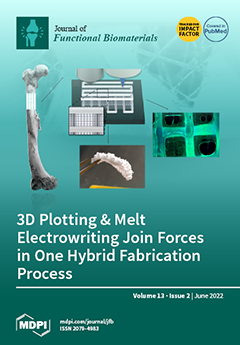
Cover Story (view full-size image): Extrusion-based 3D printing of calcium phosphate cements (CPC) enables the fabrication of patient-specific scaffolds for bone substitutes with high biocompatibility. Due to the brittleness of the material, the stability of such open-pore structures cannot always be guaranteed. Through a multi-technological approach combining CPC printing with melt electrowriting (MEW) of polycaprolactone (PCL) microfibers in one automated process, hybrid and interwoven CPC+PCL scaffolds were successfully fabricated. This not only improved the mechanical properties but also showed partially better pore bridging behavior of human mesenchymal stem cells along the PCL-fibers with increasing CPC strand distances, as well as an elevated proliferation level of murine pre-osteoblasts. View this paper
  • Issues are regarded as officially published after their release is announced to the table of contents alert mailing list.
  • You may sign up for e-mail alerts to receive table of contents of newly released issues.
  • PDF is the official format for papers published in both, html and pdf forms. To view the papers in pdf format, click on the "PDF Full-text" link, and use the free Adobe Reader to open them.
Order results
Result details
Section
Select all
Export citation of selected articles as:
10 pages, 4356 KiB  
Article
Green Synthesis of Antibacterial Nanocomposite of Silver Nanoparticle-Doped Hydroxyapatite Utilizing Curcuma longa Leaf Extract and Land Snail (Achatina fulica) Shell Waste
by Is Fatimah, Habibi Hidayat, Gani Purwiandono, Khoirunisa Khoirunisa, Hasna Azizah Zahra, Rahmania Audita and Suresh Sagadevan
J. Funct. Biomater. 2022, 13(2), 84; https://doi.org/10.3390/jfb13020084 - 20 Jun 2022
Cited by 7 | Viewed by 3145
Abstract
Preparation of green synthesized silver nanoparticle (AgNPs)-doped hydroxyapatite (Ag/HA) utilizing Curcuma longa leaf extract and land snail (Achatina fulica) shell waste was performed. Physicochemical characteristics and antibacterial activity of Ag/HA composite as a function of Ag content was studied. Instrumental analysis [...] Read more.
Preparation of green synthesized silver nanoparticle (AgNPs)-doped hydroxyapatite (Ag/HA) utilizing Curcuma longa leaf extract and land snail (Achatina fulica) shell waste was performed. Physicochemical characteristics and antibacterial activity of Ag/HA composite as a function of Ag content was studied. Instrumental analysis such as XRD, SEM-EDX, TEM, and XPS were employed to characterize the nanocomposites. The physicochemical study revealed the maintained porous structure of HA after Ag immobilization, and from TEM analyses, it was found that the distributed spherical particles are associated with the dispersed Ag and have a particle diameter of around 5–25 nm. Antibacterial activity of the nanocomposite was evaluated against Escherichia coli, Staphylococcus aureus, Kliebsiella pneumonia, and Streptococcus pyogenes. The results showed that the varied Ag content (1.0; 1.6; and 2.4% wt) influenced the nanoparticle distribution in the nanocomposite and enhanced the antibacterial feature. Full article
(This article belongs to the Section Antibacterial Biomaterials)
Show Figures

Figure 1

14 pages, 7533 KiB  
Article
Direct Ink Write Printing of Chitin-Based Gel Fibers with Customizable Fibril Alignment, Porosity, and Mechanical Properties for Biomedical Applications
by Devis Montroni, Takeru Kobayashi, Taige Hao, Derek Lublin, Tomoko Yoshino and David Kisailus
J. Funct. Biomater. 2022, 13(2), 83; https://doi.org/10.3390/jfb13020083 - 16 Jun 2022
Cited by 6 | Viewed by 3067
Abstract
A fine control over different dimensional scales is a challenging target for material science since it could grant control over many properties of the final material. In this study, we developed a multivariable additive manufacturing process, direct ink write printing, to control different [...] Read more.
A fine control over different dimensional scales is a challenging target for material science since it could grant control over many properties of the final material. In this study, we developed a multivariable additive manufacturing process, direct ink write printing, to control different architectural features from the nano- to the millimeter scale during extrusion. Chitin-based gel fibers with a water content of around 1500% were obtained extruding a polymeric solution of chitin into a counter solvent, water, inducing instant solidification of the material. A certain degree of fibrillar alignment was achieved basing on the shear stress induced by the nozzle. In this study we took into account a single variable, the nozzle’s internal diameter (NID). In fact, a positive correlation between NID, fibril alignment, and mechanical resistance was observed. A negative correlation with NID was observed with porosity, exposed surface, and lightly with water content. No correlation was observed with maximum elongation (~50%), and the scaffold’s excellent biocompatibility, which appeared unaltered. Overall, a single variable allowed a customization of different material features, which could be further tuned, adding control over other aspects of the synthetic process. Moreover, this manufacturing could be potentially applied to any polymer. Full article
(This article belongs to the Special Issue Nanoengineered Materials for Biomedical Applications)
Show Figures

Figure 1

12 pages, 4043 KiB  
Case Report
Immediate Tooth Autotransplantation with Root Canal Filling and Partially Demineralized Dentin/Cementum Matrix into Congenital Missing Tooth Region
by Masaru Murata, Yasuhiro Nakanishi, Kaoru Kusano, Yukito Hirose, Junichi Tazaki, Toshiyuki Akazawa and Itaru Mizoguchi
J. Funct. Biomater. 2022, 13(2), 82; https://doi.org/10.3390/jfb13020082 - 16 Jun 2022
Cited by 5 | Viewed by 4526
Abstract
This clinical report describes immediate tooth auto-transplantation with an autograft of partially demineralized dentin/cementum matrix (pDDM), based on an orthodontic treatment plan for a 16-year-old male patient with a congenital missing tooth (#45). First, vital teeth (#14, #24) were extracted, and root canal [...] Read more.
This clinical report describes immediate tooth auto-transplantation with an autograft of partially demineralized dentin/cementum matrix (pDDM), based on an orthodontic treatment plan for a 16-year-old male patient with a congenital missing tooth (#45). First, vital teeth (#14, #24) were extracted, and root canal filling (#14) was immediately performed with the support of a fixation device. Simultaneously, the tooth (#24) was crushed in an electric mill for 1 min, and the crushed granules were partially demineralized in 2% HNO3 solution for 20 min as the graft material. Next, the donor tooth was transplanted into the created socket (#45), and stabilized using an enamel bonding agent. The wet pDDM was loaded into the location of the congenital missing tooth, and the flap was repositioned. The bonding agent for stabilization was removed at 28 days, and also small contact points between the transplanted tooth and the upper premolar (#14) were added using photopolymerizable composite resin. X-ray photos were taken sequentially, and there were no postoperative complications. The radiographic images showed that the periodontal ligament space and alveolar ridge line could be observed at 18 months. The pDDM was harmonized with the mandible, and the remodeled bone-like shadow was observed in the graft region. We concluded that immediate tooth transplantation with root canal fillings and autogenous pDDM may be a valuable alternative to dental implanting or bridge formation for patients with a congenital missing tooth, followed by orthodontic treatment. Full article
(This article belongs to the Special Issue Application of Dentin Matrix in Tissue Regeneration)
Show Figures

Figure 1

18 pages, 3136 KiB  
Review
The Rise of Polymeric Microneedles: Recent Developments, Advances, Challenges, and Applications with Regard to Transdermal Drug Delivery
by Aswani Kumar Gera and Rajesh Kumar Burra
J. Funct. Biomater. 2022, 13(2), 81; https://doi.org/10.3390/jfb13020081 - 15 Jun 2022
Cited by 26 | Viewed by 4910
Abstract
The current scenario of the quest for microneedles (MNs) with biodegradability and biocompatibility properties is a potential research area of interest. Microneedles are considered to be robust, can penetrate the skin’s deep-seated layers, and are easy to manufacture, and their applications from the [...] Read more.
The current scenario of the quest for microneedles (MNs) with biodegradability and biocompatibility properties is a potential research area of interest. Microneedles are considered to be robust, can penetrate the skin’s deep-seated layers, and are easy to manufacture, and their applications from the clinical perspective are still ongoing with standard escalation. This review paper focuses on some of the pivotal variants of polymeric microneedles which are specifically dissolvable and swell-based MNs. It further explores the drug dissolution kinetics and insertion behavior mechanisms with an emphasis on the need for mathematical modeling of MNs. This review further evaluates the multifarious fabrication methods, with an update on the advances in the fabrication of polymeric MNs, the choice of materials used for the fabrication, the challenges in polymeric MN fabrication, and the prospects of polymeric MNs with applications pertinent to healthcare, by exclusively focusing on the procurable literature over the last decade. Full article
Show Figures

Figure 1

23 pages, 5925 KiB  
Article
Osteoconductive Silk Fibroin Binders for Bone Repair in Alveolar Cleft Palate: Fabrication, Structure, Properties, and In Vitro Testing
by Supaporn Sangkert, Kantida Juncheed and Jirut Meesane
J. Funct. Biomater. 2022, 13(2), 80; https://doi.org/10.3390/jfb13020080 - 14 Jun 2022
Cited by 1 | Viewed by 2392
Abstract
Osteoconductive silk fibroin (SF) binders were fabricated for the bone repair of an alveolar cleft defect. Binders were prefigureared by mixing different ratios of a mixture of random coils and SF aggregation with SF fibrils: 100:0 (SFB100), 75:25 (SFB75), 50:50 (SFB50), 25:75 (SFB25), [...] Read more.
Osteoconductive silk fibroin (SF) binders were fabricated for the bone repair of an alveolar cleft defect. Binders were prefigureared by mixing different ratios of a mixture of random coils and SF aggregation with SF fibrils: 100:0 (SFB100), 75:25 (SFB75), 50:50 (SFB50), 25:75 (SFB25), and 0:100 (SFB0). The gelation, molecular organization, structures, topography, and morphology of the binders were characterized and observed. Their physical, mechanical, and biological properties were tested. The SF binders showed gelation via self-assembly of SF aggregation and fibrillation. SFB75, SFB50, and SFB25 had molecular formation via the amide groups and showed more structural stability than SFB100. The morphology of SFB0 demonstrated the largest pore size. SFB0 showed a lowest hydrophilicity. SFB100 showed the highest SF release. SFB25 had the highest maximum load. SFB50 exhibited the lowest elongation at break. Binders with SF fibrils showed more cell viability and higher cell proliferation, ALP activity, calcium deposition, and protein synthesis than without SF fibrils. Finally, the results were deduced: SFB25 demonstrated suitable performance that is promising for the bone repair of an alveolar cleft defect. Full article
Show Figures

Figure 1

13 pages, 2041 KiB  
Article
Effect of a Self-Assembly Peptide on Surface Roughness and Hardness of Bleached Enamel
by Gabriela de A. P. Magalhães, May Anny A Fraga, Isaac J. de Souza Araújo, Rafael R. Pacheco, Américo B. Correr and Regina M. Puppin-Rontani
J. Funct. Biomater. 2022, 13(2), 79; https://doi.org/10.3390/jfb13020079 - 13 Jun 2022
Cited by 4 | Viewed by 3042
Abstract
After bleaching, enamel surfaces are damaged, contributing to erosion and tooth sensitivity. Although fluoride is used after bleaching to try and revert alterations, it is not capable of repairing tooth structure. This study compared the effect of a self-assembly peptide (P11-4), [...] Read more.
After bleaching, enamel surfaces are damaged, contributing to erosion and tooth sensitivity. Although fluoride is used after bleaching to try and revert alterations, it is not capable of repairing tooth structure. This study compared the effect of a self-assembly peptide (P11-4), with and without fluoride, and sodium fluoride (NaF 2%) on the Knoop microhardness (KHN) and surface roughness (Ra (μm)) of bleached enamel with an in-office bleaching regimen. Enamel blocks of bovine teeth (5 × 5 × 2 mm) with standardized surface hardness were bleached with 35% carbamide peroxide, following the manufacturer’s instructions. The teeth were randomly divided into the following groups (n = 7) according to post-bleaching treatment: no treatment (negative control) (C-); 2% NaF (NaF); Curodont™ Repair (Repair); and Curodont™ Protect (Protect). Specimens were stored in artificial saliva at 37 °C. To evaluate the effect of the post-bleaching treatments, KHN and Ra were measured before bleaching (baseline) and 24 h and 7 days after bleaching. Data were submitted to repeated measures ANOVA and Bonferroni tests (α = 0.05). There were significant interactions between the study factors (p = 0.001). After 7 days, Repair (572.50 ± 79.04) and Protect (583.00 ± 74.76) specimens showed increased surface KHN, with values higher than the NaF (465.50 ± 41.50) and C- (475.22 ± 58.95) baseline values. There was no significant difference in KHN at 24 h among groups (p = 0.587). At 24 h after bleaching, Repair was significantly different from all groups (p < 0.05). Repair showed the lowest Ra (μm) values (0.133 ± 0.035). After seven days, there was no significant difference in Ra values among groups when compared to the baseline. The use of P11-4-based materials after bleaching resulted in the fastest recovery to baseline enamel properties. Full article
(This article belongs to the Special Issue Feature Papers in Dental Biomaterials)
Show Figures

Figure 1

14 pages, 9434 KiB  
Article
Cu2+ Release from Polylactic Acid Coating on Titanium Reduces Bone Implant-Related Infection
by Chengdong Zhang, Xingping Li, Dongqin Xiao, Qiao Zhao, Shuo Chen, Fei Yang, Jinhui Liu and Ke Duan
J. Funct. Biomater. 2022, 13(2), 78; https://doi.org/10.3390/jfb13020078 - 10 Jun 2022
Cited by 8 | Viewed by 2640
Abstract
Implant-related infection (IRI) is a major problem in orthopedics. Copper (Cu) is an essential trace element with strong bactericidal activity and, thus, presents potential for reducing IRI. The present study explored a straightforward strategy for releasing Cu2+ from titanium (Ti) implants, and [...] Read more.
Implant-related infection (IRI) is a major problem in orthopedics. Copper (Cu) is an essential trace element with strong bactericidal activity and, thus, presents potential for reducing IRI. The present study explored a straightforward strategy for releasing Cu2+ from titanium (Ti) implants, and we conducted a preliminary study to assess the feasibility of this approach in clinical translation. Polylactic acid (PLA) coatings containing different concentrations of copper ions were prepared on Ti discs. The antibacterial activity and biocompatibility of the copper ion-incorporated Ti implants were evaluated using Staphylococcus aureus (S. aureus), bone marrow mesenchymal stem cells (BMSCs) and animal models. In vitro, the coatings produced burst release of Cu2+ in 12 h, and inhibited S. aureus growth in a dose-dependent manner. The coatings prepared from PLA solutions containing 0.5 or 1.0 mg/mL reduced the viability and osteogenic differentiation of BMSCs, but these effects were negated after the coatings were immersed in culture medium for 6 h. Four weeks after implantation, the Cu-free K-wires challenged with S. aureus had persistent infection and inferior fracture healing to the other three groups, while Cu-coated wires had no evidence of infection. Furthermore, the Cu-coated wires placed in rabbits without S. aureus challenge showed superior fracture healing to the other three groups. These results suggest that PLA coatings containing Cu2+ may be an effective design for reducing IRI without adversely affecting adjacent bone healing. Full article
(This article belongs to the Special Issue Antibacterial Biomaterials)
Show Figures

Figure 1

28 pages, 2047 KiB  
Review
Graphene Oxide (GO) Materials—Applications and Toxicity on Living Organisms and Environment
by Aminah N. Ghulam, Otávio A. L. dos Santos, Layla Hazeem, Bianca Pizzorno Backx, Mohamed Bououdina and Stefano Bellucci
J. Funct. Biomater. 2022, 13(2), 77; https://doi.org/10.3390/jfb13020077 - 10 Jun 2022
Cited by 94 | Viewed by 10073
Abstract
Graphene-based materials have attracted much attention due to their fascinating properties such as hydrophilicity, high dispersion in aqueous media, robust size, high biocompatibility, and surface functionalization ability due to the presence of functional groups and interactions with biomolecules such as proteins and nucleic [...] Read more.
Graphene-based materials have attracted much attention due to their fascinating properties such as hydrophilicity, high dispersion in aqueous media, robust size, high biocompatibility, and surface functionalization ability due to the presence of functional groups and interactions with biomolecules such as proteins and nucleic acid. Modified methods were developed for safe, direct, inexpensive, and eco-friendly synthesis. However, toxicity to the environment and animal health has been reported, raising concerns about their utilization. This review focuses primarily on the synthesis methods of graphene-based materials already developed and the unique properties that make them so interesting for different applications. Different applications are presented and discussed with particular emphasis on biological fields. Furthermore, antimicrobial potential and the factors that affect this activity are reviewed. Finally, questions related to toxicity to the environment and living organisms are revised by highlighting factors that may interfere with it. Full article
(This article belongs to the Special Issue Advanced Bioceramics)
Show Figures

Figure 1

13 pages, 2748 KiB  
Article
Collagenase Administration into Periodontal Ligament Reduces the Forces Required for Tooth Extraction in an Ex situ Porcine Jaw Model
by Ran Tohar, Hen Alali, Tamar Ansbacher, Tamar Brosh, Inbal Sher, Yossi Gafni, Evgeny Weinberg and Maayan Gal
J. Funct. Biomater. 2022, 13(2), 76; https://doi.org/10.3390/jfb13020076 - 8 Jun 2022
Cited by 2 | Viewed by 3328
Abstract
Minimally invasive exodontia is among the long-sought-for development aims of safe dental medicine. In this paper, we aim, for the first time, to examine whether the enzymatic disruption of the periodontal ligament fibers reduces the force required for tooth extraction. To this end, [...] Read more.
Minimally invasive exodontia is among the long-sought-for development aims of safe dental medicine. In this paper, we aim, for the first time, to examine whether the enzymatic disruption of the periodontal ligament fibers reduces the force required for tooth extraction. To this end, recombinantly expressed clostridial collagenase G variant purified from Escherichia coli was injected into the periodontal ligament of mesial and distal roots of the first and second split porcine mandibular premolars. The vehicle solution was injected into the corresponding roots on the contralateral side. Following sixteen hours, the treated mandibles were mounted on a loading machine to measure the extraction force. In addition, the effect of the enzyme on the viability of different cell types was evaluated. An average reduction of 20% in the applied force (albeit with a large variability of 50 to 370 newton) was observed for the enzymatically treated roots, reaching up to 50% reduction in some cases. Importantly, the enzyme showed only a minor and transient effect on cellular viability, without any signs of toxicity. Using an innovative model enabling the analytical measurement of extraction forces, we show, for the first time, that the enzymatic disruption of periodontal ligament fibers substantially reduces the force required for tooth extraction. This novel technique brings us closer to atraumatic exodontia, potentially reducing intra- and post-operative complications and facilitating subsequent implant placement. The development of novel enzymes with enhanced activity may further simplify the tooth extraction process and present additional clinical relevance for the broad range of implications in the oral cavity. Full article
(This article belongs to the Special Issue Biomaterials for Oral Reconstructive Treatment)
Show Figures

Graphical abstract

24 pages, 4731 KiB  
Article
3D Plotting of Calcium Phosphate Cement and Melt Electrowriting of Polycaprolactone Microfibers in One Scaffold: A Hybrid Additive Manufacturing Process
by David Kilian, Max von Witzleben, Matthew Lanaro, Cynthia S. Wong, Corina Vater, Anja Lode, Mark C. Allenby, Maria A. Woodruff and Michael Gelinsky
J. Funct. Biomater. 2022, 13(2), 75; https://doi.org/10.3390/jfb13020075 - 8 Jun 2022
Cited by 12 | Viewed by 3871
Abstract
The fabrication of patient-specific scaffolds for bone substitutes is possible through extrusion-based 3D printing of calcium phosphate cements (CPC) which allows the generation of structures with a high degree of customization and interconnected porosity. Given the brittleness of this clinically approved material, the [...] Read more.
The fabrication of patient-specific scaffolds for bone substitutes is possible through extrusion-based 3D printing of calcium phosphate cements (CPC) which allows the generation of structures with a high degree of customization and interconnected porosity. Given the brittleness of this clinically approved material, the stability of open-porous scaffolds cannot always be secured. Herein, a multi-technological approach allowed the simultaneous combination of CPC printing with melt electrowriting (MEW) of polycaprolactone (PCL) microfibers in an alternating, tunable design in one automated fabrication process. The hybrid CPC+PCL scaffolds with varying CPC strand distance (800–2000 µm) and integrated PCL fibers featured a strong CPC to PCL interface. While no adverse effect on mechanical stiffness was detected by the PCL-supported scaffold design; the microfiber integration led to an improved integrity. The pore distance between CPC strands was gradually increased to identify at which critical CPC porosity the microfibers would have a significant impact on pore bridging behavior and growth of seeded cells. At a CPC strand distance of 1600 µm, after 2 weeks of cultivation, the incorporation of PCL fibers led to pore coverage by a human mesenchymal stem cell line and an elevated proliferation level of murine pre-osteoblasts. The integrated fabrication approach allows versatile design adjustments on different levels. Full article
Show Figures

Graphical abstract

14 pages, 5013 KiB  
Article
In Vivo Evaluation of 3D-Printed Silica-Based Bioactive Glass Scaffolds for Bone Regeneration
by Dilshat U. Tulyaganov, Elisa Fiume, Avzal Akbarov, Nigora Ziyadullaeva, Saidazim Murtazaev, Abbas Rahdar, Jonathan Massera, Enrica Verné and Francesco Baino
J. Funct. Biomater. 2022, 13(2), 74; https://doi.org/10.3390/jfb13020074 - 5 Jun 2022
Cited by 15 | Viewed by 3352
Abstract
Bioactive glasses are often designed as porous implantable templates in which newly-formed bone can grow in three dimensions (3D). This research work aims to investigate the bone regenerative capability of silicate bioactive glass scaffolds produced by robocasting in comparison with powder and granule-like [...] Read more.
Bioactive glasses are often designed as porous implantable templates in which newly-formed bone can grow in three dimensions (3D). This research work aims to investigate the bone regenerative capability of silicate bioactive glass scaffolds produced by robocasting in comparison with powder and granule-like materials (oxide system: 47.5SiO2-10Na2O-10K2O-10MgO-20CaO-2.5P2O5, mol.%). Morphological and compositional analyses performed by scanning electron microscopy (SEM), combined with energy dispersive spectroscopy (EDS) after the bioactivity studies in a simulated body fluid (SBF) confirmed the apatite-forming ability of the scaffolds, which is key to allowing bone-bonding in vivo. The scaffolds exhibited a clear osteogenic effect upon implantation in rabbit femur and underwent gradual resorption followed by ossification. Full resorption in favor of new bone growth was achieved within 6 months. Osseous defect healing was accompanied by the formation of mature bone with abundant osteocytes and bone marrow cells. These in vivo results support the scaffold’s suitability for application in bone tissue engineering and show promise for potential translation to clinical assessment. Full article
Show Figures

Figure 1

15 pages, 2990 KiB  
Article
Rational Design and Characterisation of Novel Mono- and Bimetallic Antibacterial Linde Type A Zeolite Materials
by Emmanuel Oheix, Chloé Reicher, Habiba Nouali, Laure Michelin, Ludovic Josien, T. Jean Daou and Laurent Pieuchot
J. Funct. Biomater. 2022, 13(2), 73; https://doi.org/10.3390/jfb13020073 - 2 Jun 2022
Cited by 6 | Viewed by 3087
Abstract
The development of antimicrobial devices and surfaces requires the setup of suitable materials, able to store and release active principles. In this context, zeolites, which are microporous aluminosilicate minerals, hold great promise, since they are able to serve as a reservoir for metal-ions [...] Read more.
The development of antimicrobial devices and surfaces requires the setup of suitable materials, able to store and release active principles. In this context, zeolites, which are microporous aluminosilicate minerals, hold great promise, since they are able to serve as a reservoir for metal-ions with antimicrobial properties. Here, we report on the preparation of Linde Type A zeolites, partially exchanged with combinations of metal-ions (Ag+, Cu2+, Zn2+) at different loadings (0.1–11.9 wt.%). We combine X-ray fluorescence, scanning electron microscopy, energy-dispersive X-ray spectroscopy, and X-ray diffraction to monitor the metal-ion contents, distribution, and conservation of the zeolite structure after exchange. Then, we evaluate their antimicrobial activity, using agar dilution and optical-density monitoring of Escherichia coli cultures. The results indicate that silver-loaded materials are at least 70-fold more active than the copper-, zinc-, and non-exchanged ones. Moreover, zeolites loaded with lower Ag+ concentrations remain active down to 0.1 wt.%, and their activities are directly proportional to the total Ag content. Sequential exchanges with two metal ions (Ag+ and either Cu2+, Zn2+) display synergetic or antagonist effects, depending on the quantity of the second metal. Altogether, this work shows that, by combining analytical and quantitative methods, it is possible to fine-tune the composition of bi-metal-exchanged zeolites, in order to maximise their antimicrobial potential, opening new ways for the development of next-generation composite zeolite-containing antimicrobial materials, with potential applications for the design of dental or bone implants, as well as biomedical devices and pharmaceutical products. Full article
(This article belongs to the Special Issue Antibacterial Biomaterials)
Show Figures

Figure 1

31 pages, 8743 KiB  
Review
Biodegradable Iron and Porous Iron: Mechanical Properties, Degradation Behaviour, Manufacturing Routes and Biomedical Applications
by Mariana Salama, Maria Fátima Vaz, Rogério Colaço, Catarina Santos and Maria Carmezim
J. Funct. Biomater. 2022, 13(2), 72; https://doi.org/10.3390/jfb13020072 - 1 Jun 2022
Cited by 33 | Viewed by 5359
Abstract
Biodegradable metals have been extensively studied due to their potential use as temporary biomedical devices, on non-load bearing applications. These types of implants are requested to function for the healing period, and should degrade after the tissue heals. A balance between mechanical properties [...] Read more.
Biodegradable metals have been extensively studied due to their potential use as temporary biomedical devices, on non-load bearing applications. These types of implants are requested to function for the healing period, and should degrade after the tissue heals. A balance between mechanical properties requested at the initial stage of implantation and the degradation rate is required. The use of temporary biodegradable implants avoids a second surgery for the removal of the device, which brings high benefits to the patients and avoids high societal costs. Among the biodegradable metals, iron as a biodegradable metal has increased attention over the last few years, especially with the incorporation of additive manufacturing processes to obtain tailored geometries of porous structures, which give rise to higher corrosion rates. Withal by mimic natural bone hierarchical porosity, the mechanical properties of obtained structures tend to equalize that of human bone. This review article presents some of the most important works in the field of iron and porous iron. Fabrication techniques for porous iron are tackled, including conventional and new methods highlighting the unparalleled opportunities given by additive manufacturing. A comparison among the several methods is taken. The effects of the design and the alloying elements on the mechanical properties are also revised. Iron alloys with antibacterial properties are analyzed, as well as the biodegradation behavior and biocompatibility of iron. Although is necessary for further in vivo research, iron is presenting satisfactory results for upcoming biomedical applications, as orthopaedic temporary scaffolds and coronary stents. Full article
(This article belongs to the Special Issue Smart Biomaterials for Soft and Hard Tissue Repair and Regeneration)
Show Figures

Graphical abstract

20 pages, 3723 KiB  
Article
Self-Crosslinkable Oxidized Alginate-Carboxymethyl Chitosan Hydrogels as an Injectable Cell Carrier for In Vitro Dental Enamel Regeneration
by Fatemeh Mohabatpour, Zahra Yazdanpanah, Silvana Papagerakis, Xiongbiao Chen and Petros Papagerakis
J. Funct. Biomater. 2022, 13(2), 71; https://doi.org/10.3390/jfb13020071 - 1 Jun 2022
Cited by 21 | Viewed by 4561
Abstract
Injectable hydrogels, as carriers, offer great potential to incorporate cells or growth factors for dental tissue regeneration. Notably, the development of injectable hydrogels with appropriate structures and properties has been a challenging task, leaving much to be desired in terms of cytocompatibility, antibacterial [...] Read more.
Injectable hydrogels, as carriers, offer great potential to incorporate cells or growth factors for dental tissue regeneration. Notably, the development of injectable hydrogels with appropriate structures and properties has been a challenging task, leaving much to be desired in terms of cytocompatibility, antibacterial and self-healing properties, as well as the ability to support dental stem cell functions. This paper presents our study on the development of a novel self-cross-linkable hydrogel composed of oxidized alginate and carboxymethyl chitosan and its characterization as a cell carrier for dental enamel regeneration in vitro. Oxidized alginate was synthesized with 60% theoretical oxidation degree using periodate oxidation and characterized by Fourier Transform Infrared spectroscopy, proton nuclear magnetic resonance spectroscopy, and Ultraviolet-visible absorption spectroscopy. Then, hydrogels were prepared at three varying weight ratios of oxidized alginate to carboxymethyl chitosan (4:1, 3:1, and 2:1) through Schiff base reactions, which was confirmed by Fourier Transform Infrared spectroscopy. The hydrogels were characterized in terms of gelation time, swelling ratio, structure, injectability, self-healing, antibacterial properties, and in vitro characterization for enamel regeneration. The results demonstrated that, among the three hydrogels examined, the one with the highest ratio of oxidized alginate (i.e., 4:1) had the fastest gelation time and the lowest swelling ability, and that all hydrogels were formed with highly porous structures and were able to be injected through a 20-gauge needle without clogging. The injected hydrogels could be rapidly reformed with the self-healing property. The hydrogels also showed antibacterial properties against two cariogenic bacteria: Streptococcus mutans and Streptococcus sobrinus. For in vitro enamel regeneration, a dental epithelial cell line, HAT-7, was examined, demonstrating a high cell viability in the hydrogels during injection. Furthermore, HAT-7 cells encapsulated in the hydrogels showed alkaline phosphatase production and mineral deposition, as well as maintaining their round morphology, after 14 days of in vitro culture. Taken together, this study has provided evidence that the oxidized alginate-carboxymethyl chitosan hydrogels could be used as an injectable cell carrier for dental enamel tissue engineering applications. Full article
Show Figures

Figure 1

10 pages, 955 KiB  
Article
Surface Modification of Polytetrafluoroethylene and Polycaprolactone Promoting Cell-Selective Adhesion and Growth of Valvular Interstitial Cells
by Matthias Gabriel, Christian Bollensdorff and Christophe Michel Raynaud
J. Funct. Biomater. 2022, 13(2), 70; https://doi.org/10.3390/jfb13020070 - 1 Jun 2022
Cited by 2 | Viewed by 2178
Abstract
Tissue engineering concepts, which are concerned with the attachment and growth of specific cell types, frequently employ immobilized ligands that interact preferentially with cell types of interest. Creating multicellular grafts such as heart valves calls for scaffolds with spatial control over the different [...] Read more.
Tissue engineering concepts, which are concerned with the attachment and growth of specific cell types, frequently employ immobilized ligands that interact preferentially with cell types of interest. Creating multicellular grafts such as heart valves calls for scaffolds with spatial control over the different cells involved. Cardiac heart valves are mainly constituted out of two cell types, endothelial cells and valvular interstitial cells. To have control over where which cell type can be attracted would enable targeted cell settlement and growth contributing to the first step of an engineered construct. For endothelial cells, constituting the outer lining of the valve tissue, several specific peptide ligands have been described. Valvular interstitial cells, representing the bulk of the leaflet, have not been investigated in this regard. Two receptors, the integrin α9β1 and CD44, are known to be highly expressed on valvular interstitial cells. Here, we demonstrate that by covalently grafting the corresponding peptide and polysaccharide ligand onto an erodible, polycaprolactone (PCL), and a non-degradable, polytetrafluoroethylene (PTFE), polymer, surfaces were generated that strongly support valvular interstitial cell colonization with minimal endothelial cell and reduced platelet adhesion. The technology for covalent binding of corresponding ligands is a key element towards tissue engineered cardiac valves for in vitro applications, but also towards future in vivo application, especially in combination with degradable scaffold material. Full article
Show Figures

Graphical abstract

12 pages, 4303 KiB  
Article
A Parametric Study of Flushing Conditions for Improvement of Angioscopy Visibility
by Kohei Mitsuzuka, Yujie Li, Toshio Nakayama, Hitomi Anzai, Daisuke Goanno, Simon Tupin, Mingzi Zhang, Haoran Wang, Kazunori Horie and Makoto Ohta
J. Funct. Biomater. 2022, 13(2), 69; https://doi.org/10.3390/jfb13020069 - 1 Jun 2022
Cited by 3 | Viewed by 2397
Abstract
During an angioscopy operation, a transparent liquid called dextran is sprayed out from a catheter to flush the blood away from the space between the camera and target. Medical doctors usually inject dextran at a constant flow rate. However, they often cannot obtain [...] Read more.
During an angioscopy operation, a transparent liquid called dextran is sprayed out from a catheter to flush the blood away from the space between the camera and target. Medical doctors usually inject dextran at a constant flow rate. However, they often cannot obtain clear angioscopy visibility because the flushing out of the blood is insufficient. Good flushing conditions producing clear angioscopy visibility will increase the rate of success of angioscopy operations. This study aimed to determine a way to improve the clarity for angioscopy under different values for the parameters of the injection waveform, endoscope position, and catheter angle. We also determined the effect of a stepwise waveform for injecting the dextran only during systole while synchronizing the waveform to the cardiac cycle. To evaluate the visibility of the blood-vessel walls, we performed a computational fluid dynamics (CFD) simulation and calculated the visible area ratio (VAR), representing the ratio of the visible wall area to the total area of the wall at each point in time. Additionally, the normalized integration of the VAR called the area ratio (ARVAR) represents the ratio of the visible wall area as a function of the dextran injection period. The results demonstrate that the ARVAR with a stepped waveform, bottom endoscope, and three-degree-angle catheter results in the highest visibility, around 25 times larger than that under the control conditions: a constant waveform, a center endoscope, and 0 degrees. This set of conditions can improve angioscopy visibility. Full article
Show Figures

Figure 1

20 pages, 6923 KiB  
Article
Crossing Phylums: Butterfly Wing as a Natural Perfusable Three-Dimensional (3D) Bioconstruct for Bone Tissue Engineering
by Fatemeh Mostofi, Marzieh Mostofi, Behnaz Niroomand, Saadi Hosseini, Atefeh Alipour, Shahin Homaeigohar, Javad Mohammadi, Mohammad Ali Shokrgozar and Hosein Shahsavarani
J. Funct. Biomater. 2022, 13(2), 68; https://doi.org/10.3390/jfb13020068 - 1 Jun 2022
Cited by 6 | Viewed by 3256
Abstract
Despite the advent of promising technologies in tissue engineering, finding a biomimetic 3D bio-construct capable of enhancing cell attachment, maintenance, and function is still a challenge in producing tailorable scaffolds for bone regeneration. Here, osteostimulatory effects of the butterfly wings as a naturally [...] Read more.
Despite the advent of promising technologies in tissue engineering, finding a biomimetic 3D bio-construct capable of enhancing cell attachment, maintenance, and function is still a challenge in producing tailorable scaffolds for bone regeneration. Here, osteostimulatory effects of the butterfly wings as a naturally porous and non-toxic chitinous scaffold on mesenchymal stromal cells are assessed. The topographical characterization of the butterfly wings implied their ability to mimic bone tissue microenvironment, whereas their regenerative potential was validated after a 14-day cell culture. In vivo analysis showed that the scaffold induced no major inflammatory response in Wistar rats. Topographical features of the bioconstruct upregulated the osteogenic genes, including COL1A1, ALP, BGLAP, SPP1, SP7, and AML3 in differentiated cells compared to the cells cultured in the culture plate. However, butterfly wings were shown to provide a biomimetic microstructure and proper bone regenerative capacity through a unique combination of various structural and material properties. Therefore, this novel platform can be confidently recommended for bone tissue engineering applications. Full article
Show Figures

Graphical abstract

15 pages, 3099 KiB  
Article
Development and Characterization of the Biodegradable Film Derived from Eggshell and Cornstarch
by Joseph Merillyn Vonnie, Kobun Rovina, Rasnarisa Awatif Azhar, Nurul Huda, Kana Husna Erna, Wen Xia Ling Felicia, Md Nasir Nur’Aqilah and Nur Fatihah Abdul Halid
J. Funct. Biomater. 2022, 13(2), 67; https://doi.org/10.3390/jfb13020067 - 27 May 2022
Cited by 13 | Viewed by 5158
Abstract
In the current study, cornstarch (CS) and eggshell powder (ESP) were combined using a casting technique to develop a biodegradable film that was further morphologically and physicochemically characterized using standard methods. Scanning electron microscopy (SEM) and transmission electron microscopy (TEM) were used to [...] Read more.
In the current study, cornstarch (CS) and eggshell powder (ESP) were combined using a casting technique to develop a biodegradable film that was further morphologically and physicochemically characterized using standard methods. Scanning electron microscopy (SEM) and transmission electron microscopy (TEM) were used to characterize the morphology of the ESP/CS film, and the surface of the film was found to have a smooth structure with no cracks, a spherical and porous irregular shape, and visible phase separation, which explains their large surface area. In addition, the energy dispersive X-ray (EDX) analysis indicated that the ESP particles were made of calcium carbonate and the ESP contained carbon in the graphite form. Fourier Transform Infrared Spectroscopy indicated the presence of carbonated minerals in the ESP/CS film which shows that ESP/CS film might serve as a promising adsorbent. Due to the inductive effect of the O–C–O bond on calcium carbonate in the eggshell, it was discovered that the ESP/CS film significantly improves physical properties, moisture content, swelling power, water solubility, and water absorption compared to the control CS film. The enhancement of the physicochemical properties of the ESP/CS film was principally due to the intra and intermolecular interactions between ESP and CS molecules. As a result, this film can potentially be used as a synergistic adsorbent for various target analytes. Full article
(This article belongs to the Special Issue Biodegradable Polymers and Textiles)
Show Figures

Figure 1

8 pages, 1597 KiB  
Case Report
Histological Evidences of Autograft of Dentin/Cementum Granules into Unhealed Socket at 5 Months after Tooth Extraction for Implant Placement
by Masaru Murata, Md Arafat Kabir, Yukito Hirose, Morio Ochi, Naoto Okubo, Toshiyuki Akazawa and Haruhiko Kashiwazaki
J. Funct. Biomater. 2022, 13(2), 66; https://doi.org/10.3390/jfb13020066 - 25 May 2022
Cited by 5 | Viewed by 2440
Abstract
The aim of this clinical case study was to observe biopsy tissues at 5 months after an autograft of a partially demineralized dentin/cementum matrix (pDDM) into a tooth-extracted socket exhibiting healing failure. A 66-year-old female presented with healing failure in the cavity for [...] Read more.
The aim of this clinical case study was to observe biopsy tissues at 5 months after an autograft of a partially demineralized dentin/cementum matrix (pDDM) into a tooth-extracted socket exhibiting healing failure. A 66-year-old female presented with healing failure in the cavity for 2 months after the extraction (#36). Initial X-ray photos showed a clear remainder of lamina dura (#36), a residual root (#37), and a horizontal impaction (#38). The vital tooth (#38) was selected for pDDM. The third molar crushed by electric mill was decalcified in 1.0 L of 2.0% HNO3 for 20 min and rinsed in cold distilled water. The pDDM granules (size: 0.5–2.0 mm) were grafted immediately into the treated socket. X-ray views just after pDDM graft showed radio-opaque granules. At 5 months after pDDM graft, the surface of regenerated bone was harmonized with the mandibular line, and bone-like radio-opacity was found in the graft region. The biopsy tissue (diameter: 3.0 mm) at 5 months after pDDM graft showed that mature bone was interconnected with the remaining pDDM. The novel histological evidence highlighted that newly formed bone was connected directly with both dentin-area and cementum-area matrix of pDDM. We concluded that pDDM contributed to the regeneration of bone in the unhealed socket, and this regeneration prepared the socket for implant placement. Autogenous pDDM could be immediately recycled as an innovative biomaterial for local bone regeneration. Full article
(This article belongs to the Special Issue Application of Dentin Matrix in Tissue Regeneration)
Show Figures

Figure 1

13 pages, 6622 KiB  
Article
Monetite vs. Brushite: Different Influences on Bone Cell Response Modulated by Strontium Functionalization
by Elisa Boanini, Stefania Pagani, Matilde Tschon, Katia Rubini, Milena Fini and Adriana Bigi
J. Funct. Biomater. 2022, 13(2), 65; https://doi.org/10.3390/jfb13020065 - 24 May 2022
Cited by 16 | Viewed by 3189
Abstract
Monetite and brushite are regarded with increasing interest for the preparation of biomaterials for applications in the musculoskeletal system. Herein, we investigated the influence of strontium substitution in the structures of these two phosphates on bone cell response. To achieve this aim, co-cultures [...] Read more.
Monetite and brushite are regarded with increasing interest for the preparation of biomaterials for applications in the musculoskeletal system. Herein, we investigated the influence of strontium substitution in the structures of these two phosphates on bone cell response. To achieve this aim, co-cultures of human primary osteoclasts and human osteoblast-like MG63 cells were tested on strontium-substituted monetite and strontium-substituted brushite, as well as on monetite and brushite, as controls. In both structures, strontium substitution for calcium amounted to about 6 at% and provoked enlargement of the cell parameters and morphologic variations. Cumulative release in physiological solution increased linearly over time and was greater from brushite (up to about 160 and 560 mg/L at 14 days for Sr and Ca, respectively) than from monetite (up to about 90 and 250 mg/L at 14 days for Sr and Ca, respectively). The increasing viability of osteoblast-like cells over time, with the different expression level of some typical bone markers, indicates a more pronounced trigger toward osteoblast differentiation and osteoclast inhibition by brushite materials. In particular, the inhibition of cathepsin K and tartrate-resistant acid phosphatase at the gene and morphological levels suggests strontium-substituted brushite can be applied in diseases characterized by excessive bone resorption. Full article
(This article belongs to the Special Issue Functionalized Biomimetic Calcium Phosphates 2.0)
Show Figures

Graphical abstract

11 pages, 5247 KiB  
Article
Computational Contact Pressure Prediction of CoCrMo, SS 316L and Ti6Al4V Femoral Head against UHMWPE Acetabular Cup under Gait Cycle
by J. Jamari, Muhammad Imam Ammarullah, Gatot Santoso, S. Sugiharto, Toto Supriyono, Akbar Teguh Prakoso, Hasan Basri and Emile van der Heide
J. Funct. Biomater. 2022, 13(2), 64; https://doi.org/10.3390/jfb13020064 - 23 May 2022
Cited by 92 | Viewed by 5151
Abstract
Due to various concerns about the use of metal-on-metal that is detrimental to users, the use of metal as acetabular cup material was later changed to ultra high molecular weight polyethylene (UHMWPE). However, the wear on UHMWPE releases polyethylene wear particles, which can [...] Read more.
Due to various concerns about the use of metal-on-metal that is detrimental to users, the use of metal as acetabular cup material was later changed to ultra high molecular weight polyethylene (UHMWPE). However, the wear on UHMWPE releases polyethylene wear particles, which can trigger a negative body response and contribute to osteolysis. For reducing the wear of polyethylene, one of the efforts is to investigate the selection of metal materials. Cobalt chromium molybdenum (CoCrMo), stainless steel 316L (SS 316L), and titanium alloy (Ti6Al4V) are the frequently employed materials. The computational evaluation of contact pressure was carried out using a two-dimensional axisymmetric model for UHMWPE acetabular cup paired with metal femoral head under gait cycle in this study. The results show Ti6Al4V-on-UHMWPE is able to reduce cumulative contact pressure compared to SS 316L-on-UHMWPE and CoCrMo-on-UHMWPE. Compared to Ti6Al4V-on-UHMWPE at peak loading, the difference in cumulative contact pressure to respective maximum contact pressure is 9.740% for SS 316L-on-UHMWPE and 11.038% for CoCrMo-on-UHMWPE. Full article
(This article belongs to the Special Issue Tribology for Biomedical Applications)
Show Figures

Figure 1

16 pages, 6527 KiB  
Article
Corncob Cellulose Scaffolds: A New Sustainable Temporary Implant for Cartilage Replacement
by Rachel Cordeiro, Marta Henriques, João C. Silva, Filipe Antunes, Nuno Alves and Carla Moura
J. Funct. Biomater. 2022, 13(2), 63; https://doi.org/10.3390/jfb13020063 - 23 May 2022
Cited by 5 | Viewed by 2497
Abstract
Tissue engineering using scaffolds is a promising strategy to repair damaged articular cartilage, whose self-repair is inefficient. Cellulose properties have been recognized for their application in the biomedical field. The aim of this study was to fabricate and characterize novel scaffolds based on [...] Read more.
Tissue engineering using scaffolds is a promising strategy to repair damaged articular cartilage, whose self-repair is inefficient. Cellulose properties have been recognized for their application in the biomedical field. The aim of this study was to fabricate and characterize novel scaffolds based on poly(ɛ-caprolactone) (PCL) and sustainable cellulose. Thus, the performance of corncob-derived cellulose (CC) in scaffolds as an alternative to wood cellulose (WC) was also investigated to reduce the environmental footprint. Two concentrations of CC in scaffolds were tested, 1% and 2% (w/w), and commercial WC using the same concentrations, as a control. Morphologically, all the developed scaffolds presented pore sizes of ~300 µm, 10 layers, a circular shape and well-dispersed cellulose. Thus, all of these characteristics and properties provide the manufactured scaffolds suitable for use in cartilage-replacement strategies. The use of 2% CC results in higher porosity (54.24%), which promotes cell infiltration/migration and nutrient exchange, and has similar mechanical properties to WC. As for the effects of enzymatic degradation of the scaffolds, no significant changes (p > 0.05) were observed in resistance over time. However, the obtained compressive modulus of the scaffold with 2% CC was similar to that of WC. Overall, our results suggest that the integration of 2% corncob cellulose in PCL scaffolds could be a novel way to replace wood-cellulose-containing scaffolds, highlighting its potential for cartilage-replacement strategies. Full article
Show Figures

Graphical abstract

18 pages, 6492 KiB  
Article
Antibacterial and Osteogenic Properties of Ag Nanoparticles and Ag/TiO2 Nanostructures Prepared by Atomic Layer Deposition
by Denis Nazarov, Ilya Ezhov, Natalia Yudintceva, Maxim Shevtsov, Aida Rudakova, Vladimir Kalganov, Vladimir Tolmachev, Yuliya Zharova, Oleksiy Lutakov, Ludmila Kraeva, Elizaveta Rogacheva and Maxim Maximov
J. Funct. Biomater. 2022, 13(2), 62; https://doi.org/10.3390/jfb13020062 - 18 May 2022
Cited by 23 | Viewed by 3308
Abstract
The combination of titania nanofilms and silver nanoparticles (NPs) is a very promising material, with antibacterial and osseointegration-induced properties for titanium implant coatings. In this work, we successfully prepared TiO2 nanolayer/Ag NP structures on titanium disks using atomic layer deposition (ALD). The [...] Read more.
The combination of titania nanofilms and silver nanoparticles (NPs) is a very promising material, with antibacterial and osseointegration-induced properties for titanium implant coatings. In this work, we successfully prepared TiO2 nanolayer/Ag NP structures on titanium disks using atomic layer deposition (ALD). The samples were studied by scanning electron microscopy (SEM), X-ray diffraction, X-ray photoelectron spectroscopy (XPS), contact angle measurements, and SEM-EDS. Antibacterial activity was tested against Staphylococcus aureus. The in vitro cytological response of MG-63 osteosarcoma and human fetal mesenchymal stem cells (FetMSCs) was examined using SEM study of their morphology, MTT test of viability and differentiation using alkaline phosphatase and osteopontin with and without medium-induced differentiation in the osteogenic direction. The samples with TiO2 nanolayers, Ag NPs, and a TiO2/Ag combination showed high antibacterial activity, differentiation in the osteogenic direction, and non-cytotoxicity. The medium for differentiation significantly improved osteogenic differentiation, but the ALD coatings also stimulated differentiation in the absence of the medium. The TiO2/Ag samples showed the best antibacterial ability and differentiation in the osteogenic direction, indicating the success of the combining of TiO2 and Ag to produce a multifunctional biocompatible and bactericidal material. Full article
(This article belongs to the Special Issue Biomaterials in Tissue, Biomedical and Surface Engineering)
Show Figures

Figure 1

4 pages, 214 KiB  
Opinion
From Animal to Human: (Re)using Acellular Extracellular Matrices for Temporomandibular Disc Substitution
by Daniela Trindade, Nuno Alves and Carla Moura
J. Funct. Biomater. 2022, 13(2), 61; https://doi.org/10.3390/jfb13020061 - 18 May 2022
Cited by 2 | Viewed by 1953
Abstract
Current treatments for temporomandibular joint (TMJ) disc dysfunctions are not fully effective and lack regenerative capacity. Therefore, the search for tissue-engineered materials for TMJ disc substitution is critical to fill this gap. Decellularization presents tremendous potential, as it is possible to obtain an [...] Read more.
Current treatments for temporomandibular joint (TMJ) disc dysfunctions are not fully effective and lack regenerative capacity. Therefore, the search for tissue-engineered materials for TMJ disc substitution is critical to fill this gap. Decellularization presents tremendous potential, as it is possible to obtain an extracellular matrix with an adequate biomechanical structure and biochemical components. However, its application to the TMJ disc is still in progress, since there are few studies in the literature, and those that exist have many gaps in terms of characterisation, which is decisive to ensure its success. Ultimately, we intend to emphasize the importance of the decellularization technique for the development of an engineered TMJ disc. Full article
Show Figures

Graphical abstract

12 pages, 2905 KiB  
Article
Restoration in Vertebral Compression Fractures (VCF): Effectiveness Evaluation Based on 3D Technology
by David C. Noriega González, Francisco Ardura Aragón, Jesús Crespo Sanjuan, Silvia Santiago Maniega, Gregorio Labrador Hernández, María Bragado González, Daniel Pérez-Valdecantos, Alberto Caballero-García and Alfredo Córdova
J. Funct. Biomater. 2022, 13(2), 60; https://doi.org/10.3390/jfb13020060 - 17 May 2022
Cited by 1 | Viewed by 4470
Abstract
There are few studies about anatomical reduction of the fractured vertebral body before stabilization for treatment of vertebral compression fracture (VCF). Although restoration on vertebral height has been useful, the reduction of fractured endplates is limited. The vertebra is part of a joint, [...] Read more.
There are few studies about anatomical reduction of the fractured vertebral body before stabilization for treatment of vertebral compression fracture (VCF). Although restoration on vertebral height has been useful, the reduction of fractured endplates is limited. The vertebra is part of a joint, and vertebral endplates must be treated like other weight-bearing joint to avoid complications. The aim of this study was to evaluate the feasibility of anatomic reduction of vertebral compression fracture, in different bone conditions, fracture types, and ages (VCF). Under methodological point of view, we followed different steps: first was the placement of two expandable titanium implants just below the fracture. Later, to push the fractured endplates into a more anatomical position, the implants were expanded. Finally, with the implants perfectly positioned, PMMA cement was injected to avoid any loss of correction. To evaluate the effectiveness of this procedure in anatomical fracture reduction, a method based on 3D CT reconstructions was developed. In this paper, we have developed the procedure in three case studies. In all of them, we were able to demonstrate the efficacy of this procedure to reduce the VCF. The percentage of correction of the kyphotic angle varied range between 49% and 62% with respect to the value after the fracture preoperative value. This was accompanied by a reduction of the pain level on the VAS scale around 50%. In conclusion, this novel approach to the vertebral fracture treatment (VCF) associated with 3D assessment have demonstrated the possibility of reducing the vertebral kyphosis angle and the vertebral endplate fractures. However, given the few cases presented, more studies are necessaries to confirm these results. Full article
Show Figures

Figure 1

12 pages, 2955 KiB  
Article
Autologous Bone Marrow Mononuclear Cells (BMMC)-Associated Anti-Inflammatory Nanoparticles for Cardiac Repair after Myocardial Infarction
by Laercio Uemura, Rossana Baggio Simeoni, Paulo André Bispo Machado Júnior, Gustavo Gavazzoni Blume, Luize Kremer Gamba, Murilo Sgarbossa Tonial, Paulo Ricardo Baggio Simeoni, Victoria Stadler Tasca Ribeiro, Rodrigo Silvestre, Katherine Athayde Teixeira de Carvalho, Marcelo Henrique Napimoga, Júlio Cesar Francisco and Luiz Cesar Guarita-Souza
J. Funct. Biomater. 2022, 13(2), 59; https://doi.org/10.3390/jfb13020059 - 13 May 2022
Viewed by 2630
Abstract
To investigate the effect of transplantation of stem cells from the bone marrow mononuclear cells (BMMC) associated with 15d-PGJ2-loaded nanoparticles in a rat model of chronic MI. Chronic myocardial infarction (MI) was induced by the ligation of the left anterior descending artery in [...] Read more.
To investigate the effect of transplantation of stem cells from the bone marrow mononuclear cells (BMMC) associated with 15d-PGJ2-loaded nanoparticles in a rat model of chronic MI. Chronic myocardial infarction (MI) was induced by the ligation of the left anterior descending artery in 40 male Wistar rats. After surgery, we transplanted bone marrow associated with 15d-PGJ2-loaded nanoparticle by intramyocardial injection (106 cells/per injection) seven days post-MI. Myocardial infarction was confirmed by echocardiography, and histological analyses of infarct morphology, gap junctions, and angiogenesis were obtained. Our results from immunohistochemical analyses demonstrated the presence of angiogenesis identified in the transplanted region and that there was significant expression of connexin-43 gap junctions, showing a more effective electrical and mechanical integration of the host myocardium. This study suggests that the application of nanoparticle technology in the prevention and treatment of MI is an emerging field and can be a strategy for cardiac repair. Full article
Show Figures

Figure 1

13 pages, 5322 KiB  
Article
Electrospun PHB/Chitosan Composite Fibrous Membrane and Its Degradation Behaviours in Different pH Conditions
by Yansheng Zhou, Ying Li, Daqing Li, Yidan Yin and Fenglei Zhou
J. Funct. Biomater. 2022, 13(2), 58; https://doi.org/10.3390/jfb13020058 - 13 May 2022
Cited by 12 | Viewed by 3656
Abstract
Peripheral nerve injury (PNI) is a neurological disorder that causes more than 9 million patients to suffer from dysfunction of moving and sensing. Using biodegradable polymers to fabricate an artificial nerve conduit that replicates the environment of the extracellular matrix and guides neuron [...] Read more.
Peripheral nerve injury (PNI) is a neurological disorder that causes more than 9 million patients to suffer from dysfunction of moving and sensing. Using biodegradable polymers to fabricate an artificial nerve conduit that replicates the environment of the extracellular matrix and guides neuron regeneration through the damaged sites has been researched for decades and has led to promising but primarily pre-clinical outcomes. However, few peripheral nerve conduits (PNCs) have been constructed from controllable biodegradable polymeric materials that can maintain their structural integrity or completely degrade during and after nerve regeneration respectively. In this work, a novel PNC candidate material was developed via the electrospinning of polyhydroxy butyrate/chitosan (PHB/CS) composite polymers. An SEM characterisation revealed the resultant PHB/CS nanofibres with 0, 1 and 2 wt/v% CS had less and smaller beads than the nanofibres at 3 wt/v% CS. The water contact angle (WCA) measurement demonstrated that the wettability of PHB/CS electrospun fibres was significantly improved by additional CS. Furthermore, both the thermogravimetric analysis (TGA) and differentiation scanning calorimetry (DSC) results showed that PHB/CS polymers can be blended in a single phase with a trifluoracetic solvent in all compositions. Besides, the reduction in the degradation temperature (from 286.9 to 229.9 °C) and crystallinity (from 81.0% to 52.1%) with increasing contents of CS were further proven. Moreover, we found that the degradability of the PHB/CS nanofibres subjected to different pH values rated in the order of acidic > alkaline > phosphate buffer solution (PBS). Based on these findings, it can be concluded that PHB/CS electrospun fibres with variable blending ratios may be used for designing PNCs with controlled biodegradability. Full article
(This article belongs to the Topic Advanced Functional Materials for Regenerative Medicine)
Show Figures

Figure 1

11 pages, 2342 KiB  
Article
Physical and Chemical Characterization of Biomineralized Collagen with Different Microstructures
by Tianming Du, Yumiao Niu, Youjun Liu, Haisheng Yang, Aike Qiao and Xufeng Niu
J. Funct. Biomater. 2022, 13(2), 57; https://doi.org/10.3390/jfb13020057 - 13 May 2022
Cited by 7 | Viewed by 3119
Abstract
Mineralized collagen is the basic unit in hierarchically organized natural bone with different structures. Polyacrylic acid (PAA) and periodic fluid shear stress (FSS) are the most common chemical and physical means to induce intrafibrillar mineralization. In the present study, non-mineralized collagen, extrafibrillar mineralized [...] Read more.
Mineralized collagen is the basic unit in hierarchically organized natural bone with different structures. Polyacrylic acid (PAA) and periodic fluid shear stress (FSS) are the most common chemical and physical means to induce intrafibrillar mineralization. In the present study, non-mineralized collagen, extrafibrillar mineralized (EM) collagen, intrafibrillar mineralized (IM) collagen, and hierarchical intrafibrillar mineralized (HIM) collagen induced by PAA and FSS were prepared, respectively. The physical and chemical properties of these mineralized collagens with different microstructures were systematically investigated afterwards. Transmission electron microscopy (TEM) and scanning electron microscopy (SEM) showed that mineralized collagen with different microstructures was prepared successfully. The pore density of the mineralized collagen scaffold is higher under the action of periodic FSS. Fourier transform infrared spectroscopy (FTIR) analysis showed the formation of the hydroxyapatite (HA) crystal. A significant improvement in the pore density, hydrophilicity, enzymatic stability, and thermal stability of the mineralized collagen indicated that the IM collagen under the action of periodic FSS was beneficial for maintaining collagen activity. HIM collagen fibers, which are prepared under the co-action of periodic FSS and sodium tripolyphosphate (TPP), may pave the way for new bone substitute material applications. Full article
Show Figures

Graphical abstract

12 pages, 1295 KiB  
Article
Cytotoxicity of Methacrylate Dental Resins to Human Gingival Fibroblasts
by Jolanta Sulek, Elzbieta Luczaj-Cepowicz, Grazyna Marczuk-Kolada, Maciej Rosłan and Adam Holownia
J. Funct. Biomater. 2022, 13(2), 56; https://doi.org/10.3390/jfb13020056 - 11 May 2022
Cited by 2 | Viewed by 2605
Abstract
This study aimed to assess the acute and delayed cytotoxicity of three, popular light-cured methacrylate-based restorative resins (MRs): Charisma (C), Estelite (E), and Filtek (F), to human gingival fibroblasts in culture. Cells were grown for up to 24 h with light-cured (or pre-cured) [...] Read more.
This study aimed to assess the acute and delayed cytotoxicity of three, popular light-cured methacrylate-based restorative resins (MRs): Charisma (C), Estelite (E), and Filtek (F), to human gingival fibroblasts in culture. Cells were grown for up to 24 h with light-cured (or pre-cured) resins. We evaluated resin cytotoxicity, redox imbalance, necrosis/apoptosis, miR-9, and heat shock protein 70 (HSP70). The role of resin-induced oxidative stress (damage) in HSP70-response (repair) was assessed using binary fluorescence labeling. All MRs decreased viable cell numbers and cell proliferation and damaged cell membranes, and their 24 h-delayed toxicity was lower (C), higher (F), or similar (E) to that induced by freshly-cured resins. Cell membrane damage induced by C and E decreased with time, while F produced a linear increase. All resins generated intracellular oxidative stress with the predominant necrotic outcome, and produced heterogeneous responses in miR-9 and HSP70. The double fluorescence (damage/repair) experiments pointed to common features of E and F but not C. In the subset of cells, the binary response induced by E and F was different from C, similar to each other, and positively interrelated. Experimental data show that selective MR cytotoxicity should be taken into account when considering repetitive use or massive reconstruction. Full article
Show Figures

Graphical abstract

12 pages, 2524 KiB  
Article
Antitumor Efficacy of Doxorubicin-Loaded Electrospun Attapulgite–Poly(lactic-co-glycolic acid) Composite Nanofibers
by Zhe Wang, Yili Zhao, Mingwu Shen, Helena Tomás, Benqing Zhou and Xiangyang Shi
J. Funct. Biomater. 2022, 13(2), 55; https://doi.org/10.3390/jfb13020055 - 10 May 2022
Cited by 10 | Viewed by 4228
Abstract
Currently, cancer chemotherapeutic drugs still have the defects of high toxicity and low bioavailability, so it is critical to design novel drug release systems for cancer chemotherapy. Here, we report a method to fabricate electrospun drug-loaded organic/inorganic hybrid nanofibrous system for antitumor therapy [...] Read more.
Currently, cancer chemotherapeutic drugs still have the defects of high toxicity and low bioavailability, so it is critical to design novel drug release systems for cancer chemotherapy. Here, we report a method to fabricate electrospun drug-loaded organic/inorganic hybrid nanofibrous system for antitumor therapy applications. In this work, rod-like attapulgite (ATT) was utilized to load a model anticancer drug doxorubicin (DOX), and mixed with poly(lactic-co-glycolic acid) (PLGA) to form electrospun hybrid nanofibers. The ATT/DOX/PLGA composite nanofibers were characterized through various techniques. It is feasible to load DOX onto ATT surfaces, and the ATT/DOX/PLGA nanofibers show a smooth and uniform morphology with improved mechanical durability. Under neutral and acidic pH conditions, the loaded DOX was released from ATT/DOX/PLGA nanofibers in a sustained manner. In addition, the released DOX from the nanofibers could significantly inhibit the growth of tumor cells. Owing to the significantly reduced burst release profile and increased mechanical durability of the ATT/DOX/PLGA nanofibers, the designed organic–inorganic hybrid nanofibers may hold great promise as a nanoplatform to encapsulate different drugs for enhanced local tumor therapy applications. Full article
(This article belongs to the Section Biomaterials for Drug Delivery)
Show Figures

Figure 1

Previous Issue
Back to TopTop