Next Issue
Volume 12, February
Previous Issue
Volume 11, December


Dent. J., Volume 12, Issue 1 (January 2024) – 20 articles

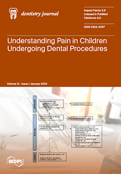
Cover Story (view full-size image): Our systematic review sheds light on how to predict pain and anxiety during routine dental procedures in children. This can help dental practitioners choose and provide appropriate pharmacological and/or non-pharmacological interventions to allow children to cope better with dental care. These interventions should be a priority for future research and implementation as they could help prevent the need for general anaesthesia, which is costly, resource intensive, and has associated risks of morbidity and mortality. View this paper
  • Issues are regarded as officially published after their release is announced to the table of contents alert mailing list.
  • You may sign up for e-mail alerts to receive table of contents of newly released issues.
  • PDF is the official format for papers published in both, html and pdf forms. To view the papers in pdf format, click on the "PDF Full-text" link, and use the free Adobe Reader to open them.
Order results
Result details
Section
Select all
Export citation of selected articles as:
13 pages, 2755 KiB  
Article
The Peri-Implant Microbiome—A Possible Factor Determining the Success of Surgical Peri-Implantitis Treatment?
by Jarno Hakkers, Lei Liu, Diederik F. M. Hentenaar, Gerry M. Raghoebar, Arjan Vissink, Henny J. A. Meijer, Lisa Walters, Hermie J. M. Harmsen and Yvonne C. M. de Waal
Dent. J. 2024, 12(1), 20; https://doi.org/10.3390/dj12010020 - 22 Jan 2024
Cited by 3 | Viewed by 2882
Abstract
The objective was to assess the effect of peri-implantitis surgery on the peri-implant microbiome with a follow-up of one year. A total of 25 peri-implantitis patients in whom non-surgical treatment has failed to solve peri-implantitis underwent resective surgical treatment. Their peri-implant pockets were [...] Read more.
The objective was to assess the effect of peri-implantitis surgery on the peri-implant microbiome with a follow-up of one year. A total of 25 peri-implantitis patients in whom non-surgical treatment has failed to solve peri-implantitis underwent resective surgical treatment. Their peri-implant pockets were sampled prior to surgical treatment (T0) and one year post treatment (T12). The natural dentition was sampled to analyse similarities and differences with the peri-implantitis samples. Treatment success was recorded. The change in microbial relative abundance levels was evaluated. The microbiota was analysed by sequencing the amplified V3-V4 region of the 16S rRNA genes. Sequence data were binned to amplicon sequence variants that were assigned to bacterial genera. Group differences were analysed using principal coordinate analysis, Wilcoxon signed rank tests, and t-tests. Beta diversity analyses reported a significant separation between peri-implantitis and natural dentition samples on T0 and T12, along with significant separations between successfully and non-successfully treated patients. Eubacterium was significantly lower on T12 compared to T0 for the peri-implantitis samples. Treponema and Eubacterium abundance levels were significantly lower in patients with treatment success on T0 and T12 versus no treatment success. Therefore, lower baseline levels of Treponema and Eubacterium seem to be associated with treatment success of peri-implantitis surgery. This study might aid clinicians in determining which peri-implantitis cases might be suitable for treatment and give a prognosis with regard to treatment success. Full article
Show Figures

Figure 1

13 pages, 226 KiB  
Article
Perception of Special-Care Dentistry among Dental Students at Universitas Indonesia: A Cross-Sectional Study
by Atik Ramadhani, Fiki Rizqa Izzati, Diah Ayu Maharani, Normaliza Ab Malik and Febriana Setiawati
Dent. J. 2024, 12(1), 19; https://doi.org/10.3390/dj12010019 - 22 Jan 2024
Cited by 1 | Viewed by 1980
Abstract
Objective: To assess the perceptions of special-care dentistry (SCD) among dental students in the Faculty of Dentistry, Universitas Indonesia, Indonesia. Methods: This cross-sectional study used a self-administered online questionnaire, and all preclinical and clinical students were invited to participate. The survey items comprised [...] Read more.
Objective: To assess the perceptions of special-care dentistry (SCD) among dental students in the Faculty of Dentistry, Universitas Indonesia, Indonesia. Methods: This cross-sectional study used a self-administered online questionnaire, and all preclinical and clinical students were invited to participate. The survey items comprised four domains related to participants’ characteristics, perceptions of SCD, perceptions of managing patients with special needs, and perceptions of SCD education. The chi-square test and Mann–Whitney test were used in the analysis. Results: A total of 572 students participated in this study. The findings showed that the clinical students were more familiar with SCD than the preclinical students were (p = 0.008). A statistically significant relationship was found between the study program and the ability to work independently with special-needs patients after graduation (p < 0.001), the ability to refer special-needs patients to specialists (p = 0.042), the perception of postgraduate training-program needs (p < 0.001), and the opportunity to consider postgraduate training (p = 0.004). Conclusion: Most of the respondents had a favorable perception of SCD. Thus, an improved SCD curriculum and SCD training for undergraduate and postgraduate students should be provided to develop the knowledge and skills needed to provide care to special-needs patients. Full article
(This article belongs to the Section Dental Education)
14 pages, 1751 KiB  
Article
Post-Orthodontic Relapse Prevention through Administration of a Novel Synthetic Carbonated Hydroxyapatite–Chitosan Hydrogel Derived from Blood Cockle Shell (Anadara granosa L.)
by Aanisah Fauziyyah Nurul Hadi, Sabrina Noor Aghniya, Gayuh Abi Haidar, Windy Sepry Marcelina Sihombing, Angelina Sutedjo and Ananto Ali Alhasyimi
Dent. J. 2024, 12(1), 18; https://doi.org/10.3390/dj12010018 - 19 Jan 2024
Cited by 1 | Viewed by 2073
Abstract
Relapse during passive orthodontic treatment is a major issue, with 70–90% frequency. This study examines whether blood cockle shells may be used to extract carbonated hydroxyapatite (CHA)-chitosan (CS). This study also aims to analyze the effect of CHA-CS on orthodontic relapse in rats. [...] Read more.
Relapse during passive orthodontic treatment is a major issue, with 70–90% frequency. This study examines whether blood cockle shells may be used to extract carbonated hydroxyapatite (CHA)-chitosan (CS). This study also aims to analyze the effect of CHA-CS on orthodontic relapse in rats. This study utilized 18 male Wistar rats which were randomly divided into two groups: CHA-CS and the control group (CG). The rats were subjected to a 35 cN orthodontic force for a duration of 7 days, after which the rats were conditioned to be passive. During this phase, the CHA-CS group received daily administration of CHA-CS hydrogel derived from the blood cockle shell. Subsequently, the appliances were detached to facilitate relapse. The distance between the mesial tips was measured using a digital caliper at three consecutive time points: 1, 5, and 7 days after debonding. The number of osteoblasts, osteoclasts, and fibroblasts was examined using hematoxylin–eosin staining. The data were subjected to statistical analysis using a t-test. The relapse distance of the CHA-CS group was lower than that of the control groups on day 7. Histological examinations using hematoxylin–eosin (HE) staining showed a significant increase in osteoblasts, a decrease in osteoclasts, and an increase in fibroblasts during orthodontic relapse movement (p < 0.05). This study found that blood cockle shell-derived CHA-CS may reduce orthodontic relapse by increasing osteoblasts and fibroblasts and by reducing the osteoclast number in rats. Full article
(This article belongs to the Section Dental Materials)
Show Figures

Graphical abstract

17 pages, 5115 KiB  
Review
Late Complications in Long-Term Childhood Cancer Survivors: What the Oral Health Professional Needs to Know
by Sali Al-Ansari, Juliette Stolze, Dorine Bresters, Alan Henry Brook, Alexa M. G. A. Laheij, Henk S. Brand, Göran Dahllöf, Frederik R. Rozema and Judith E. Raber-Durlacher
Dent. J. 2024, 12(1), 17; https://doi.org/10.3390/dj12010017 - 19 Jan 2024
Viewed by 2803
Abstract
With diagnostic and therapeutic advances, over 80% of children diagnosed with cancer become long-term survivors. As the number of childhood cancer survivors (CCS) continues to increase, dental practitioners become more likely to have CCS among their patients. CCS may develop late complications from [...] Read more.
With diagnostic and therapeutic advances, over 80% of children diagnosed with cancer become long-term survivors. As the number of childhood cancer survivors (CCS) continues to increase, dental practitioners become more likely to have CCS among their patients. CCS may develop late complications from damage caused by their cancer treatment to endocrine, cardiovascular, musculoskeletal, and other organ systems. These complications may surface decades after the completion of treatment. Adverse outcomes of childhood cancer treatment frequently involve oral and craniofacial structures including the dentition. Tooth development, salivary gland function, craniofacial growth, and temporomandibular joint function may be disturbed, increasing oral health risks in these individuals. Moreover, CCS are at risk of developing subsequent malignancies, which may manifest in or near the oral cavity. It is important that dental practitioners are aware of the childhood cancer history of their patients and have knowledge of potential late complications. Therefore, this narrative review aims to inform dental practitioners of late oral complications of cancer treatment modalities commonly used in pediatric oncology. Furthermore, selected common non-oral late sequelae of cancer therapy that could have an impact on oral health and on delivering dental care will be discussed. Full article
Show Figures

Figure 1

14 pages, 3394 KiB  
Article
Oral Function and the Oral Microbiome in the Elderly in the Kyotango Area
by Yoshiaki Yamamoto, Toshiro Yamamoto, Nao Miyamoto, Kohei Kinoshita, Satomi Nishikawa, Tetsuya Adachi, Shigeta Takizawa, Ryo Inoue, Satoaki Matoba and Narisato Kanamura
Dent. J. 2024, 12(1), 16; https://doi.org/10.3390/dj12010016 - 18 Jan 2024
Viewed by 2560
Abstract
Introduction: Prevention of tooth loss contributes to an extended life expectancy, namely longevity. Aging-related oral hypofunction, including tooth loss, markedly increases the risks of functional disorder and mortality. Dysbiosis of the oral microbiome has recently been associated with various diseases, such as liver [...] Read more.
Introduction: Prevention of tooth loss contributes to an extended life expectancy, namely longevity. Aging-related oral hypofunction, including tooth loss, markedly increases the risks of functional disorder and mortality. Dysbiosis of the oral microbiome has recently been associated with various diseases, such as liver cirrhosis, pancreatic cancer, colorectal cancer, and inflammatory bowel disease. Therefore, the relationship between the oral microbiome and systemic health has been attracting increasing attention. In the present study, we examined oral function and the oral microbiome in the elderly in a world-leading longevity area. Materials and Methods: An oral examination, chewing ability/tongue-lip motor function/saliva tests, and a metagenomic analysis with a 16S rRNA gene-targeting next-generation sequencer were conducted on 78 subjects aged ≥80 years. Twenty-six healthy individuals aged between 20 and 39 years were also investigated as controls. The data obtained were statistically analyzed. The protocol of the present study was approved by the Ethics Review Board of our university (ERB-C-885). Results: Chewing ability, tongue–lip motor function, and saliva volume were normal in elderly subjects with a current tooth number ≥20, but were significantly lower in those with a current tooth number <20. The oral microbiome in elderly subjects with a current tooth number ≥20 and young controls differed from that in elderly subjects with a current tooth number <20. Conclusion: Tooth number ≥20 in elderly subjects in the longevity area contributed to the maintenance of both oral function and the diversity of the oral microbiome. Full article
Show Figures

Graphical abstract

10 pages, 2023 KiB  
Article
Improving the Communication of Dental Findings in Pediatric Dentistry by Using Intraoral Scans as a Visual Aid: A Randomized Clinical Trial
by Nelly Schulz-Weidner, Marina Gruber, Eva May Schraml, Bernd Wöstmann, Norbert Krämer and Maximiliane Amelie Schlenz
Dent. J. 2024, 12(1), 15; https://doi.org/10.3390/dj12010015 - 17 Jan 2024
Cited by 4 | Viewed by 3351
Abstract
The aim of this first randomized clinical trial (RCT) was to determine whether intraoral scans (IOS) can be used as a visual aid to improve the communication of dental findings in pediatric dentistry. Therefore, 60 children (mean age 10.1 ± 3.3 years) and [...] Read more.
The aim of this first randomized clinical trial (RCT) was to determine whether intraoral scans (IOS) can be used as a visual aid to improve the communication of dental findings in pediatric dentistry. Therefore, 60 children (mean age 10.1 ± 3.3 years) and their accompanying parents/primary caregivers (PGs) were examined between July 2022 and February 2023. Patients were randomly allocated to two groups: half of the participants were informed of the children’s dental findings including treatment plans by verbal explanation alone (control group, n = 30), while the other half were informed using IOS (Trios 4, 3Shape) as a visual aid to support the verbal explanation (study group, n = 30). Both groups then completed a questionnaire regarding their children’s diagnosis, treatment needs, planned therapy, and oral hygiene. Statistical analysis was performed using a t-test (p < 0.05). Overall, there was a significant difference between the two groups (p < 0.001) in terms of understanding the current oral situation of their children. While 85.5 ± 17.3% (mean ± standard deviation) of the answers were correct in the study group, only 57.2 ± 17.8% of the participants in the control group were capable of answering the questions correctly. In particular, the control group had difficulties answering the questions about treatment needs and therapy correctly. Within the limitations of this first pilot study, IOS can be clearly recommended as a visual aid to improve the communication of dental findings with PGs in pediatric dentistry. Full article
(This article belongs to the Special Issue Current Advances in Pediatric Odontology)
Show Figures

Graphical abstract

11 pages, 563 KiB  
Article
Assessment of Dental Student Satisfaction after Internships in Collaborative Dental Practices in Saxony—A Retrospective Questionnaire Analysis
by Annette Wolf, Mihaela Pricop-Jeckstad, Ute Botzenhart and Tomasz Gredes
Dent. J. 2024, 12(1), 14; https://doi.org/10.3390/dj12010014 - 13 Jan 2024
Viewed by 2105
Abstract
The goal for dental students of a university-based program should be to learn about practice procedures in a dental office as part of their studies in order to gain insight into day-to-day activities, such as organizational management, patient communication, and problem-solving strategies. All [...] Read more.
The goal for dental students of a university-based program should be to learn about practice procedures in a dental office as part of their studies in order to gain insight into day-to-day activities, such as organizational management, patient communication, and problem-solving strategies. All dental students from the Faculty of Medicine at the University of Dresden in Germany, who completed a one-week internship in an external dental office in the last year before taking the final exam, were invited to participate in the survey (total n = 182 in years 2017–2019 and 2022). After completing the internship, the students were asked to anonymously rate the distinctive competencies they had acquired during their dental studies in terms of clinical and social communication skills. The results of the survey showed a good practicability of the acquired dental knowledge and a general satisfaction of students during their internships. No significant influence of the COVID-19 outbreak and the resulting special regulations in dental practices during the pandemic on student satisfaction was found. Students were more satisfied with their completed internships in smaller cities. Therefore, a stronger inclusion of practices outside the big cities should be considered in the current implementation of the new Dental Licensure Act in Germany. Full article
(This article belongs to the Special Issue New Trends in Dental Education and Dental Care)
Show Figures

Figure 1

20 pages, 875 KiB  
Systematic Review
Efficacy and Safety of Propolis for Treating Recurrent Aphthous Stomatitis (RAS): A Systematic Review and Meta-Analysis
by Tina Roberts, Idriss Ibrahim Kallon and Anel Schoonees
Dent. J. 2024, 12(1), 13; https://doi.org/10.3390/dj12010013 - 6 Jan 2024
Cited by 1 | Viewed by 2647
Abstract
The systematic review assessed the efficacy and safety of propolis for treating recurrent aphthous stomatitis (RAS). The review adopted the PICO framework to examine the effects of topical and systemic propolis on RAS while also comparing it to established treatments, placebos, or no [...] Read more.
The systematic review assessed the efficacy and safety of propolis for treating recurrent aphthous stomatitis (RAS). The review adopted the PICO framework to examine the effects of topical and systemic propolis on RAS while also comparing it to established treatments, placebos, or no treatment. The main focus was on the healing time, pain levels, adverse effects, the likelihood of ulcer recurrence, and accompanying symptoms such as redness. The team included randomised controlled trials (RCTs) and quasi-randomised trials, excluding case reports and studies on oral ulcers other than RAS. In May 2022, the review team comprehensively searched nine databases and trial registries following the PRISMA guidelines. The protocol was registered in the PROSPERO database under the registration number CRD42022327123. Two review authors conducted a comprehensive and autonomous search for pertinent papers and extracted essential data. Where data permitted, the team utilised Review Manager 5 to conduct a random-effects meta-analysis, assessing the risk of bias and heterogeneity of the included studies. Where possible, the GRADE Pro programme was used to assess the certainty of the evidence for all the outcomes. This review included 10 RCTs, comprising 825 participants aged between 18 and 69 years. Seven studies evaluated the efficacy and safety of propolis when applied topically, all of which used different formulations, concentrations, and carriers. The remaining three studies assessed systemic administration in tablet form. The duration of investigations ranged from 5 days to 3 years. The review team classified two studies as having an overall ‘high risk’ of bias, while the remaining studies were categorised as having an overall ‘uncertain risk’. The overall certainty of the evidence was ‘very low’. The results indicate that topical and systemic propolis may decrease the duration of healing, alleviate pain, and reduce redness in patients with RAS compared to a placebo. However, the certainty of the evidence is very low. These may be due to the high risk of bias, substantial heterogeneity, and limited sample sizes in the included studies. For these reasons, the results of this review should be interpreted with caution. Nevertheless, the limited number of adverse effects observed suggests that propolis may have a favourable safety profile when used for a short period in treating RAS. Full article
Show Figures

Figure 1

12 pages, 746 KiB  
Article
Upper Airway Dimensions among Different Skeletal Malocclusions: A Retrospective Observational Study by Cephalometric Analysis
by Maria Francesca Sfondrini, Simone Gallo, Maurizio Pascadopoli, Paola Gandini, Caterina Roncoroni and Andrea Scribante
Dent. J. 2024, 12(1), 12; https://doi.org/10.3390/dj12010012 - 3 Jan 2024
Cited by 4 | Viewed by 2655
Abstract
The aim of the present work was to investigate the upper airway dimensions in adult non-orthodontic patients, equally divided according to their skeletal class. Methods: In this retrospective cross-sectional study, lateral cephalometric radiographs of adult patients referred for orthodontic consultation were collected. Cephalometric [...] Read more.
The aim of the present work was to investigate the upper airway dimensions in adult non-orthodontic patients, equally divided according to their skeletal class. Methods: In this retrospective cross-sectional study, lateral cephalometric radiographs of adult patients referred for orthodontic consultation were collected. Cephalometric tracing was performed with dedicated software. For each measure, descriptive statistics were calculated. Cephalometric measurements between the different skeletal classes were compared. Linear regressions were performed between upper airway diameters and cephalometric measurements, sex and age. Significance was predetermined for p < 0.05. Results: Lateral cephalometric radiographs of 120 patients were reviewed. Nasopharynx length (NL) and depth (PD) measurements were significantly shorter in skeletal class III patients (p < 0.05). The superior pharyngeal airway space (SPAS) was found to be significantly shorter in class III patients as compared to class II patients (p < 0.05), and the mean airway space (MAS) of class I patients was found to be significantly shorter compared to class II patients (p < 0.05). Palate length (PL) values were found to be significantly longer in class I (p < 0.05). Linear regressions showed that the sella-nasion-A point angle (SNA) and Riedel’s angle between point A, the nasion and point B (ANB) significantly influenced NL and PD (p < 0.05). Conclusions: Class III patients show significantly shorter nasopharynx measurements; clinicians should consider that this sagittal discrepancy could be related to an altered anatomy of the upper respiratory tract. Full article
(This article belongs to the Special Issue Current Research Topics in Orthodontics)
Show Figures

Figure 1

10 pages, 1516 KiB  
Article
The Use of Enamel Matrix Derivative during Surgical Therapy for Peri-Implantitis: A Case Series
by Thomas G. Wilson, Jr., Stephen K. Harrel and Martha E. Nunn
Dent. J. 2024, 12(1), 11; https://doi.org/10.3390/dj12010011 - 30 Dec 2023
Cited by 1 | Viewed by 1847
Abstract
Peri-implantitis is a growing concern and currently, there is no agreement on the best method for treating this condition. This study looked at surgical intervention with the use of enamel matrix derivative (EMD) for treating this condition. A cohort of 25 (34 implants) [...] Read more.
Peri-implantitis is a growing concern and currently, there is no agreement on the best method for treating this condition. This study looked at surgical intervention with the use of enamel matrix derivative (EMD) for treating this condition. A cohort of 25 (34 implants) consecutive patients treated with EMD for peri-implantitis was followed for up to 6.4 years. The survival of the implants as well as changes in clinical parameters are reported. Statistical analysis was performed using paired t tests and general estimating equations. The mean length of time implants were followed post-surgery was 3.05 ± 1.53 years. All but two of the treated implants survived in function (94%). Both failed implants were lost in the same patient, who was a heavy smoker. The changes in mean probing depth (1.94 ± 1.18 mm), change in deepest probing depth (3.12 ± 1.45 mm), and reduction in bleeding on probing (73.6 ± 43.9%) according to patient means were all highly significant (p < 0.001 for all changes). When EMD is used during surgical treatment of peri-implantitis, there is a high survival rate of implants and significant improvements in clinical parameters. Full article
(This article belongs to the Special Issue Advances in Periodontal and Peri-Implant Tissues Health Management)
Show Figures

Figure 1

10 pages, 225 KiB  
Article
Changes in the Perceptions of Oral Symptoms Due to COVID-19 in Korean Adolescents
by Sun-Kyoung Lee and Jeong-Min Seong
Dent. J. 2024, 12(1), 10; https://doi.org/10.3390/dj12010010 - 30 Dec 2023
Viewed by 1912
Abstract
This study investigated the oral symptoms and subjective health perceptions of Korean adolescents due to COVID-19. Data from the 17th Korea Youth Risk Behavior Survey (KYRBS; 2021) were analyzed, and 54,848 Korean middle and high school students were included in the study. Frequency, [...] Read more.
This study investigated the oral symptoms and subjective health perceptions of Korean adolescents due to COVID-19. Data from the 17th Korea Youth Risk Behavior Survey (KYRBS; 2021) were analyzed, and 54,848 Korean middle and high school students were included in the study. Frequency, cross-tabulation, and logistic regression analyses were performed using IBM SPSS (v. 22.0; IBM Corp., Armonk, NY, USA). Statistical significance was set at p < 0.05. The survey results on subjective health perception showed that 64.8% considered themselves “healthy”, 26.1% rated themselves “moderate” in health, and 9.26% rated themselves “unhealthy.” When asked about brushing their teeth after lunch at school, students answered “no” more frequently than “yes”. Participants provided significantly different responses to questions related to receiving “sealant treatment”, “having broken teeth”, “experiencing tooth pain when eating”, “having throbbing tooth pain”, and “having pain and bleeding in gums”. Regarding the relationship between oral symptoms and subjective health perceptions due to COVID-19, students perceived themselves as healthy when they had no throbbing tooth pain, risk of pain, bleeding in the gums, or symptoms of toothache when eating. Results reveal a relationship between oral symptoms and subjective health perceptions due to COVID-19. Thus, appropriate oral health management for adolescents is needed in response to the COVID-related changes. Full article
Show Figures

Graphical abstract

10 pages, 245 KiB  
Article
Effect of the Teeth Whitening Procedure on the Mineral Composition of Oral Fluid
by Elena A. Ryskina, Oksana A. Magsumova, Mikhail A. Postnikov, Tatiana A. Lobaeva, Bahovaddin B. Ahmedzhanov, Anastasia N. Shishparenok and Dmitry D. Zhdanov
Dent. J. 2024, 12(1), 9; https://doi.org/10.3390/dj12010009 - 29 Dec 2023
Viewed by 2605
Abstract
The basis of modern tooth whitening systems is the use of a whitening gel, which usually contains hydrogen peroxide or carbamide peroxide. The study included 81 patients aged 22 to 35 years with a tooth color A2 and a darker color on the [...] Read more.
The basis of modern tooth whitening systems is the use of a whitening gel, which usually contains hydrogen peroxide or carbamide peroxide. The study included 81 patients aged 22 to 35 years with a tooth color A2 and a darker color on the Vita Classic scale. The purpose of our research was to identify a new approach to whitening teeth to improve safety and gentleness. To perform this, we assessed the effect of the tooth whitening procedure on the mineral composition of the oral fluid. A new approach to the teeth whitening procedure was to use a mouth retractor and a tool for aspirating the whitening gel, which we developed. Before the procedure, a protective film-forming aerosol, which included sodium ascorbate, was applied. After the tooth whitening procedure, the enamel was remineralized with a sealing liquid for 14 days. The concentrations of calcium and phosphorus in the oral fluid were determined using a spectrophotometer with a set of reagents (Human). The results obtained indicate that the new approach to the teeth whitening procedure contributed to less pronounced changes in the concentrations of calcium (+29.07, p < 0.001) and phosphorus (−14%, p < 0.001) in the oral fluid immediately after the procedure and in combination with the standard procedure for teeth whitening; immediately after this procedure, the calcium concentration increased by 74.4% (p < 0.001), and the phosphorus concentration decreased by 23.07% (p < 0.001). The use of remineralizing agents led to a faster recovery of the initial levels of calcium and phosphorus in the oral fluid. Full article
(This article belongs to the Special Issue Preventive Dental Care, Chairside and Beyond)
13 pages, 2811 KiB  
Review
Automation in Dentistry with Mechanical Drills and Lasers for Implant Osteotomy: A Narrative-Scoping Review
by Gopala Krishna Ganta, Rodrigo Crespo Mosca, Ridham Varsani, Venkata Ramana Murthy, Kamala Cheruvu, Michael Lu and Praveen R. Arany
Dent. J. 2024, 12(1), 8; https://doi.org/10.3390/dj12010008 - 29 Dec 2023
Cited by 1 | Viewed by 2365
Abstract
The popularity of implants is increasing with the aging population requiring oral–dental rehabilitation. There are several critical steps in the implant workflow, including case selection, implant design, surgical procedure, biological tissue responses, and functional restoration. Among these steps, surgical osteotomy procedures are a [...] Read more.
The popularity of implants is increasing with the aging population requiring oral–dental rehabilitation. There are several critical steps in the implant workflow, including case selection, implant design, surgical procedure, biological tissue responses, and functional restoration. Among these steps, surgical osteotomy procedures are a crucial determinant of clinical success. This brief review was aimed at outlining the current state of the field in automation-assisted implant surgical osteotomy technologies. A broad search of the literature was performed to identify current literature. The results are outlined in three broad categories: semi-automated static (image-guided) or dynamic (navigation-assisted) systems, and fully-automated robotic systems. As well as the current mechanical rotary approaches, the literature supporting the use of lasers in further refinement of these approaches is reviewed. The advantages and limitations of adopting autonomous technologies in practical clinical dental practices are discussed. In summary, advances in clinical technologies enable improved precision and efficacious clinical outcomes with implant dentistry. Hard-tissue lasers offer further advancements in precision, improved biological responses, and favorable clinical outcomes that require further investigation. Full article
Show Figures

Figure 1

10 pages, 1504 KiB  
Article
Clinical Assessment of Plaque Removal Using a Novel Dentifrice Containing Cellulose Microfibrils
by Mohamed E. Labib, Antonio Perazzo, James Manganaro, Yacoob Tabani, Kimberly R. Milleman, Jeffery L. Milleman and Laurence J. Walsh
Dent. J. 2024, 12(1), 7; https://doi.org/10.3390/dj12010007 - 29 Dec 2023
Cited by 1 | Viewed by 3186
Abstract
Dentifrice performance in the removal of dental plaque is influenced by the interactions of dentifrice components with tooth surfaces. This randomized controlled clinical study assessed the effectiveness and safety of a novel fluoride dentifrice formulation that included a micro-fibrillated cellulose network with entangled [...] Read more.
Dentifrice performance in the removal of dental plaque is influenced by the interactions of dentifrice components with tooth surfaces. This randomized controlled clinical study assessed the effectiveness and safety of a novel fluoride dentifrice formulation that included a micro-fibrillated cellulose network with entangled microcrystalline cellulose and silica particles (ProtegeraTM), and compared this to a positive control fluoride dentifrice (Crest Cavity Protection™). Whole mouth dental plaque levels in 82 healthy adults were measured after the first supervised use, and following a week of twice daily use at home. Overall, the test dentifrice was at least three times and up to four times more effective in whole-mouth plaque reductions, with a 38.6% reduction on first use, a 30.9% reduction at day 7, and a 41.6% reduction from day 1 to day 7, compared to reductions of 12.0%, 9.6% and 11.6%, respectively for the positive control, and up to seven times more effective in lingual plaque reductions, than the reference dentifrice (p < 0.001), with a 27.7% reduction on first use, a 22.3% reduction at day 7, and a 31.0% reduction from day 1 to day 7, compared to reductions of 4.4%, 2.2%, and 4.5%, respectively, for the positive control. No safety issues arose from the use of the test dentifrice. These results indicate that including micro-fibrillated cellulose enhances plaque removal effectiveness, without causing adverse changes to oral soft tissues. Full article
Show Figures

Graphical abstract

15 pages, 273 KiB  
Article
Awareness of Forensic Odontology among Dental Students and Faculty in Cyprus: A Survey-Based Study
by Kostis Giannakopoulos, Persefoni Lambrou-Christodoulou and Eleftherios G. Kaklamanos
Dent. J. 2024, 12(1), 6; https://doi.org/10.3390/dj12010006 - 26 Dec 2023
Viewed by 2474
Abstract
This study aimed to evaluate the awareness, comprehension, and practices concerning forensic odontology among dental students and faculty at a Dental School in Cyprus. An online, cross-sectional, descriptive survey, employing an adapted, self-administered questionnaire, was disseminated to all dental students and faculty at [...] Read more.
This study aimed to evaluate the awareness, comprehension, and practices concerning forensic odontology among dental students and faculty at a Dental School in Cyprus. An online, cross-sectional, descriptive survey, employing an adapted, self-administered questionnaire, was disseminated to all dental students and faculty at the School of Dentistry, European University Cyprus, in November 2022. The survey assessed participants’ demographic information and explored their awareness with questions alluding to knowledge, attitudes and practices in forensic dentistry. Of those surveyed, 47 faculty members and 304 students responded, yielding response rates of 66.2% and 80%, respectively. Statistical analysis, including Kendall’s tau test and χ2 test were employed to examine correlations and associations, with Cramer’s V used to measure the strength of significant associations. The predetermined significance level was α = 0.05. Awareness levels were assessed through participants’ responses to specific questions in the survey. It was revealed that 87% of faculty and 65% of students were familiar with forensic odontology. A noteworthy 94% of faculty and 85% of students recognized teeth as DNA repositories. A high percentage, 98% of faculty and 89% of students, acknowledged the role of forensic odontology in the identification of criminals and deceased individuals. Awareness of age estimation through dental eruption patterns was evident in 85% of faculty and 81.6% of students. A substantial proportion (80% of faculty) maintained dental records, while 78% of students recognized the importance of dental record-keeping in ensuring quality care. Interestingly, 57% of students and 64% of faculty were aware of the possibility of dentists testifying as expert witnesses. The majority, 95.7% of faculty and 85% of students, concurred that physical harm, scars, and behavioral alterations predominantly indicate child abuse. The findings, revealing robust awareness among respondents, underscore the importance of enhancing faculty engagement in relevant seminars to further strengthen their knowledge. Additionally, emphasizing improved record-keeping practices for potential forensic applications emerges as a crucial aspect. These insights have implications for refining dental education in Cyprus and enhancing forensic practices by promoting ongoing professional development and emphasizing meticulous record-keeping within the dental community. Full article
(This article belongs to the Special Issue Forensic Dentistry)
13 pages, 609 KiB  
Systematic Review
The Potential Clinical Applications of a Microfluidic Lab-on-a-Chip for the Identification and Antibiotic Susceptibility Testing of Enterococcus faecalis-Associated Endodontic Infections: A Systematic Review
by Carlos M. Ardila, Gustavo A. Jiménez-Arbeláez and Annie Marcela Vivares-Builes
Dent. J. 2024, 12(1), 5; https://doi.org/10.3390/dj12010005 - 26 Dec 2023
Cited by 4 | Viewed by 1998
Abstract
This systematic review evaluated the potential clinical use of microfluidic lab-on-a-chip (LOC) technology in the identification and antibiotic susceptibility testing of E. faecalis in endodontic infections. The search methodology employed in this review adhered to the PRISMA guidelines. Multiple scientific databases, including PubMed/MEDLINE, [...] Read more.
This systematic review evaluated the potential clinical use of microfluidic lab-on-a-chip (LOC) technology in the identification and antibiotic susceptibility testing of E. faecalis in endodontic infections. The search methodology employed in this review adhered to the PRISMA guidelines. Multiple scientific databases, including PubMed/MEDLINE, SCOPUS, and SCIELO, were utilized, along with exploration of grey literature sources. Up to September 2023, these resources were searched using specific keywords and MeSH terms. An initial comprehensive search yielded 202 articles. Ultimately, this systematic review incorporated 12 studies. Out of these, seven aimed to identify E. faecalis, while the remaining five evaluated its susceptibility to different antibiotics. All studies observed that the newly developed microfluidic chip significantly reduces detection time compared to traditional methods. This enhanced speed is accompanied by a high degree of accuracy, efficiency, and sensitivity. Most research findings indicated that the entire process took anywhere from less than an hour to five hours. It is important to note that this approach bypasses the need for minimum inhibitory concentration measurements, as it does not rely on traditional methodologies. Microfluidic devices enable the rapid identification and accurate antimicrobial susceptibility testing of E. faecalis, which are crucial for timely diagnosis and treatment in endodontic infections. Full article
(This article belongs to the Special Issue Feature Review Papers in Dentistry)
Show Figures

Figure 1

13 pages, 956 KiB  
Systematic Review
Predictors of Intra-Operative and Post-Operative Pain Associated with Routine Dental Procedures in Children: A Systematic Review and Meta-Analysis
by Mohammed A. Alzubaidi, Bernadette K. Drummond, Jianhua Wu, Adam Jones, Jinous F. Tahmassebi and Vishal R. Aggarwal
Dent. J. 2024, 12(1), 4; https://doi.org/10.3390/dj12010004 - 25 Dec 2023
Cited by 1 | Viewed by 2100
Abstract
Background: Understanding predictors of pain associated with paediatric dental procedures could play an important role in preventing loss of cooperation, which often leads to the procedure having to be performed under general anaesthesia. Aim: We aimed to identify predictors of intra-operative and post-operative [...] Read more.
Background: Understanding predictors of pain associated with paediatric dental procedures could play an important role in preventing loss of cooperation, which often leads to the procedure having to be performed under general anaesthesia. Aim: We aimed to identify predictors of intra-operative and post-operative pain associated with routine dental procedures in children. Materials and Methods: A systematic review of observational studies was performed using electronic searches on MEDLINE, EMBASE, PsycINFO, Global Health via OVID, PubMed, Scopus, and SciELO. The NIH Quality Assessment Tool for Observational Cohort and Cross-Sectional Studies was used to evaluate the quality of the included studies, which were meta-analysed to estimate the impact of dental procedures and anxiety on children’s pain perception. A meta-regression analysis was also performed to determine the relative effect of predictors on children’s pain perception measured as mean differences on a visual analogue scale (VAS). Results: The search identified 532 articles; 53 were retrieved for full-text screening; 6 studies were included in the review; and 4 were eligible for the meta-analysis. The meta-analysis showed the types of procedures that predicted intra-operative pain, with dental extractions being the most painful (Mean VAS Difference [MD] 46.51 mm, 95% confidence interval [CI] 40.40 to 52.62 mm). The meta-regression showed that pain scores for dental extractions were significantly higher than polishing (the least painful procedure (reference category)) by VAS MD = 23.80 mm (95% CI 5.13–42.46 mm, p-value = 0.012). It also showed that highly anxious children reported significantly higher pain scores during dental procedures by a 12.31 mm MD VAS score (95% CI 5.23–19.40 mm, p-value = 0.001) compared to those with low anxiety levels. Conclusions: This systematic review demonstrates that the strongest predictors of intra-operative pain associated with paediatric dental procedures are dental extractions followed by drilling. Children with high anxiety also reported more pain for similar procedures. Tailoring interventions to reduce pain associated with paediatric dental procedures should be a priority for future research, as reducing pain can impact compliance and could reduce the need for general anaesthesia in dental treatment. Full article
Show Figures

Figure 1

15 pages, 1221 KiB  
Review
Maxillary Anterior Teeth Dimensions and Relative Width Proportions: A Narrative Literature Review
by Francesca Cinelli, Francesco Piva, Fabio Bertini, Daniele Scaminaci Russo and Luca Giachetti
Dent. J. 2024, 12(1), 3; https://doi.org/10.3390/dj12010003 - 25 Dec 2023
Cited by 3 | Viewed by 5518
Abstract
Predictable results in the aesthetic treatment of anterior teeth can be obtained by resorting to the concept of dental aesthetics and, in particular, defining the ideal tooth dimensions and proportions to obtain a harmonious smile. Considering the great variety of articles dealing with [...] Read more.
Predictable results in the aesthetic treatment of anterior teeth can be obtained by resorting to the concept of dental aesthetics and, in particular, defining the ideal tooth dimensions and proportions to obtain a harmonious smile. Considering the great variety of articles dealing with the topic, and the lack of updated reviews, this narrative literature review aims to evaluate current knowledge on anterior teeth dimensions and to verify the existence and the potential applications of the anterior teeth proportioning theories (Golden Proportion, Golden Percentage, RED Proportion, and Golden Rectangle). PubMed, Embase, Cochrane Library, and Google Scholar databases were comprehensively searched using different keywords and term combinations. The research includes articles published up to June 2023, no time limits were set, and only articles in English were included. Inclusion criteria comprehended reviews, clinical studies, and in-vitro studies. A total of 66 articles were selected. Two main topics were identified: “Anterior teeth dimensions”, “Golden Proportions, Golden Percentage, RED Proportions, and Golden Rectangle”. As far as tooth dimensions are concerned, different width ranges are recognized for men and women and for different ethnic groups. Perfectly symmetric contralateral elements are found in low percentages of subjects. The correlation between dental dimensions and facial parameters is not always present, and it strongly depends on the sample’s ethnicity and gender. Ideal tooth proportions were only partially identified. Full article
Show Figures

Figure 1

12 pages, 3243 KiB  
Article
The Influence of Dental Virtualization, Restoration Types, and Placement Angles on the Trueness and Contact Space in 3D-Printed Crowns: A Comprehensive Exploration
by Tsung-Yueh Lu, Wei-Chun Lin, Tzu-Hsuan Yang, Citra Dewi Sahrir, Yung-Kang Shen and Sheng-Wei Feng
Dent. J. 2024, 12(1), 2; https://doi.org/10.3390/dj12010002 - 19 Dec 2023
Cited by 4 | Viewed by 2152
Abstract
The current digital dentistry workflow has streamlined dental restoration production, but the effectiveness of digital virtual design and 3D printing for restorations still needs evaluation. This study explores the impact of model-free digital design and 3D-printing placement angles on restorations, including single crowns [...] Read more.
The current digital dentistry workflow has streamlined dental restoration production, but the effectiveness of digital virtual design and 3D printing for restorations still needs evaluation. This study explores the impact of model-free digital design and 3D-printing placement angles on restorations, including single crowns and long bridges produced with and without casts. The restorations are 3D printed using resin at placement angles of 0°, 60°, and 90°. Each group of samples was replicated ten times, resulting in a total of 120 restorations. The Root Mean Square Error (RMSE) value was used to evaluate the surface integrity of the restoration. In addition, the contact space, edge gap, and occlusal space of restorations produced by different processes were recorded. The results indicate that there was no significant difference in the RMSE value of the crown group (p > 0.05). Changing the bridge restoration angle from 0° to 90° resulted in RMSE values increasing by 2.02 times (without casts) and 2.39 times (with casts). Furthermore, the marginal gaps in the crown group were all less than 60 μm, indicating good adaptation. In contrast, the bridge group showed a significant increase in marginal gaps at higher placement angles (p > 0.05). Based on the findings, virtual fabrication without casts does not compromise the accuracy of dental restorations. When the position of the long bridge exceeds 60 degrees, the error will increase. Therefore, designs without casts and parallel placement result in higher accuracy for dental restorations. Full article
(This article belongs to the Special Issue 3D Printing and Restorative Dentistry)
Show Figures

Graphical abstract

15 pages, 1544 KiB  
Review
Materials and Applications of 3D Printing Technology in Dentistry: An Overview
by Min Jeong, Kyle Radomski, Diana Lopez, Jack T. Liu, Jason D. Lee and Sang J. Lee
Dent. J. 2024, 12(1), 1; https://doi.org/10.3390/dj12010001 - 19 Dec 2023
Cited by 13 | Viewed by 9275
Abstract
Purpose. This narrative review aims to provide an overview of the mechanisms of 3D printing, the dental materials relevant to each mechanism, and the possible applications of these materials within different areas of dentistry. Methods. Subtopics within 3D printing technology in dentistry were [...] Read more.
Purpose. This narrative review aims to provide an overview of the mechanisms of 3D printing, the dental materials relevant to each mechanism, and the possible applications of these materials within different areas of dentistry. Methods. Subtopics within 3D printing technology in dentistry were identified and divided among five reviewers. Electronic searches of the Medline (PubMed) database were performed with the following search keywords: 3D printing, digital light processing, stereolithography, digital dentistry, dental materials, and a combination of the keywords. For this review, only studies or review papers investigating 3D printing technology for dental or medical applications were included. Due to the nature of this review, no formal evidence-based quality assessment was performed, and the search was limited to the English language without further restrictions. Results. A total of 64 articles were included. The significant applications, applied materials, limitations, and future directions of 3D printing technology were reviewed. Subtopics include the chronological evolution of 3D printing technology, the mechanisms of 3D printing technologies along with different printable materials with unique biomechanical properties, and the wide range of applications for 3D printing in dentistry. Conclusions: This review article gives an overview of the history and evolution of 3D printing technology, as well as its associated advantages and disadvantages. Current 3D printing technologies include stereolithography, digital light processing, fused deposition modeling, selective laser sintering/melting, photopolymer jetting, powder binder, and 3D laser bioprinting. The main categories of 3D printing materials are polymers, metals, and ceramics. Despite limitations in printing accuracy and quality, 3D printing technology is now able to offer us a wide variety of potential applications in different fields of dentistry, including prosthodontics, implantology, oral and maxillofacial, orthodontics, endodontics, and periodontics. Understanding the existing spectrum of 3D printing applications in dentistry will serve to further expand its use in the dental field. Three-dimensional printing technology has brought about a paradigm shift in the delivery of clinical care in medicine and dentistry. The clinical use of 3D printing has created versatile applications which streamline our digital workflow. Technological advancements have also paved the way for the integration of new dental materials into dentistry. Full article
(This article belongs to the Special Issue Feature Papers in Digital Dentistry)
Show Figures

Figure 1

Previous Issue
Next Issue
Back to TopTop