Next Issue
Volume 12, March
Previous Issue
Volume 12, January


Dent. J., Volume 12, Issue 2 (February 2024) – 22 articles

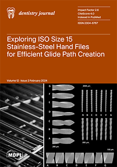
Cover Story (view full-size image): Discovering the optimal path for successful root canal procedures is paramount. This paper explores the unique characteristics of size 15 files, which are commonly the last small files used in creating a glide path during endodontic treatment. Studying the mechanical properties, metal composition, and design of seven ISO size 15 stainless-steel hand files (K-File and C-File+) revealed variations in blade area, tip geometries, surface imperfections, differential microhardness values, and buckling strengths. These observations highlight the importance of proper glide file selection in achieving successful root canal treatment. View this paper
  • Issues are regarded as officially published after their release is announced to the table of contents alert mailing list.
  • You may sign up for e-mail alerts to receive table of contents of newly released issues.
  • PDF is the official format for papers published in both, html and pdf forms. To view the papers in pdf format, click on the "PDF Full-text" link, and use the free Adobe Reader to open them.
Order results
Result details
Section
Select all
Export citation of selected articles as:
19 pages, 5623 KiB  
Article
Clinical and Biological Validation of an Allogeneous Cancellous Bone Block for Alveolar Maxillary Ridge Reconstruction: A Case Series
by Alexandre Perez, Elena Pierantozzi, Roberto Di Felice and Tommaso Lombardi
Dent. J. 2024, 12(2), 42; https://doi.org/10.3390/dj12020042 - 19 Feb 2024
Viewed by 1807
Abstract
This exploratory case series clinically and histologically investigated the performance of allogeneic cancellous freeze-dried bone allograft (FDBA) bone blocks (Maxgraft®) for the lateral augmentation of local alveolar defects in the posterior maxilla as part of two-staged implant therapy. Five patients receiving [...] Read more.
This exploratory case series clinically and histologically investigated the performance of allogeneic cancellous freeze-dried bone allograft (FDBA) bone blocks (Maxgraft®) for the lateral augmentation of local alveolar defects in the posterior maxilla as part of two-staged implant therapy. Five patients receiving eight implants 5 months after block augmentation with a follow-up period of up to 3 years were documented and analyzed. Horizontal alveolar dimensions before and 5 months after block augmentation were quantified using CBCT. Radiographic marginal bone level changes were quantified at implant placement, loading, and 1 year post-placement. Graft integration and resorption were histologically qualitatively evaluated from core biopsies retrieved at implant placement. Block augmentations resulted in a pronounced horizontal median bone gain of 7.0 (5.5 to 7.8) mm. Marginal implant bone levels in block-augmented bone remained constant over the 1 year follow-up period. Block grafts appeared histologically well integrated. Histologic analysis also revealed signs of progressive resorption and new bone formation at the lateral aspects of the grafts. The results of this case series support using Maxgraft® cancellous FDBA blocks as suitable materials for the lateral augmentation of local alveolar defects. Full article
Show Figures

Graphical abstract

12 pages, 457 KiB  
Review
Retreatment Strategies for Cases Containing Calcium Silicate-Based Root Canal Sealers: A Comprehensive Review
by Hussain Al akam, Hyeon-Cheol Kim and Ji Wook Jeong
Dent. J. 2024, 12(2), 41; https://doi.org/10.3390/dj12020041 - 18 Feb 2024
Cited by 1 | Viewed by 2871
Abstract
This review explores the field of retreatment strategies for cases filled with calcium silicate-based root canal sealers. Since the introduction of calcium silicate-based materials in dentistry, calcium silicate-based root canal sealers have become popular among dentists because of their biocompatibility, bioactivity, and sealing [...] Read more.
This review explores the field of retreatment strategies for cases filled with calcium silicate-based root canal sealers. Since the introduction of calcium silicate-based materials in dentistry, calcium silicate-based root canal sealers have become popular among dentists because of their biocompatibility, bioactivity, and sealing ability. Therefore, effective retreatment strategies are indispensable. This article aims to identify the challenges associated with the removal of calcium silicate-based sealers themselves and removal of gutta-percha with the sealers during retreatment, evaluate current techniques and materials, and provide future directions for research in this field. Regarding the strategies of removal of root canal sealers, calcium silicate-based sealers are still relatively new materials for clinicians compared with traditional sealers such as epoxy- or eugenol-based sealers. First, no clinically established solvents have been reported. Second, calcium silicate-based sealers are currently utilized by clinicians in either the cold sealer-based technique or the warm vertical condensation technique. Third, the setting process of calcium silicate-based sealers generates byproducts, primarily calcium hydroxide and secondarily hydroxyapatite, that could interact with dentine. Lastly, there is a lack of clinical studies evaluating the efficacy of retreatment protocols for teeth filled with calcium silicate-based sealers. Therefore, it is important to investigate the chemo-mechanical properties of calcium silicate-based sealers themselves and their reactions to solvents and/or mechanical instruments and identify the interfacial properties of calcium silicate-based sealers with respect to dentine and gutta-percha. In addition, researchers in the clinical field need to actively gather and report data on retreatments of teeth filled with calcium silicate-based sealers. Full article
(This article belongs to the Special Issue Endodontics: From Technique to Regeneration)
Show Figures

Figure 1

11 pages, 1656 KiB  
Article
Pine-Oil-Derived Sodium Resinate Inhibits Growth and Acid Production of Streptococcus mutans In Vitro
by Otto Rajala, Matias Mäntynen and Vuokko Loimaranta
Dent. J. 2024, 12(2), 40; https://doi.org/10.3390/dj12020040 - 17 Feb 2024
Viewed by 1653
Abstract
S. mutans is a key pathogen in dental caries initiation and progression. It promotes oral biofilm dysbiosis and biofilm acidification. Sodium resinate is a salt of pine-oil-derived resin which has antimicrobial properties. Pine-oil-derived resin consists of terpenes, diterpenes, and abietic acids. The aim [...] Read more.
S. mutans is a key pathogen in dental caries initiation and progression. It promotes oral biofilm dysbiosis and biofilm acidification. Sodium resinate is a salt of pine-oil-derived resin which has antimicrobial properties. Pine-oil-derived resin consists of terpenes, diterpenes, and abietic acids. The aim of this study was to determine the effects of pine (Pinus sylvestris) oil resinate (RS) on growth and acid production of cariogenic S. mutans strains in planktonic form and biofilm. The S. mutans type strain NCTC10449 and clinical isolate CI2366 were grown on 96-well plates for testing of RS effects on growth and biofilm formation, and on plates with integrated pH-sensitive optical ensors for real-time measurements of the effects of RS on bacterial acid production. We found that even short-time exposure to RS inhibits the growth and acid production of S. mutans in the planktonic phase and biofilms. In addition, RS was able to penetrate the biofilm matrix and reduce acid production inside S. mutans biofilm. RS thus shows potential as a novel antibacterial agent against cariogenic bacteria in biofilm. Full article
(This article belongs to the Special Issue Oral Microbiology and Related Research)
Show Figures

Figure 1

15 pages, 3453 KiB  
Article
GDF15 Contributes to the Regulation of the Mechanosensitive Responses of PdL Fibroblasts through the Modulation of IL-37
by Julia Steinmetz, Albert Stemmler, Christoph-Ludwig Hennig, Judit Symmank and Collin Jacobs
Dent. J. 2024, 12(2), 39; https://doi.org/10.3390/dj12020039 - 13 Feb 2024
Viewed by 2068
Abstract
During orthodontic tooth movement (OTM), areas of compressive and tensile forces are generated in the periodontal ligament (PdL), a mechanoreactive connective tissue between the teeth and alveolar bone. Mechanically stimulated PdL fibroblasts (PdLFs), the main cell type of PdL, express significantly increased levels [...] Read more.
During orthodontic tooth movement (OTM), areas of compressive and tensile forces are generated in the periodontal ligament (PdL), a mechanoreactive connective tissue between the teeth and alveolar bone. Mechanically stimulated PdL fibroblasts (PdLFs), the main cell type of PdL, express significantly increased levels of growth differentiation factor 15 (GDF15). In compressed PdL areas, GDF15 plays a fundamental role in modulating relevant OTM processes, including inflammation and osteoclast activation. However, the specific function of this factor in tensile areas has not yet been investigated. Thus, the aim of this study was to investigate the role of GDF15 in the mechanoresponse of human PdLFs (hPdLFs) that were exposed to biaxial tensile forces in vitro. Using siRNA-mediated knockdown experiments, we demonstrated that GDF15 had no impact on the anti-inflammatory force response of elongated hPdLFs. Although the anti-inflammatory markers IL1RN and IL10, as well as the activation of immune cells remained unaffected, we demonstrated an inhibitory role of GDF15 for the IL-37 expression. By analyzing osteogenic markers, including ALPL and RUNX2, along with an assessment of alkaline phosphatase activation, we further showed that the regulation of IL-37 by GDF15 modulates the osteogenic differentiation potential of hPdLFs. Despite bone resorption in tensile areas being rather limited, GDF15 was also found to positively modulate osteoclast activation in those areas, potentially by adjusting the IL-37 levels. In light of our new findings, we hypothesize that GDF15 modulates force-induced processes in tissue and bone remodeling through its various intra- and extracellular signaling pathways as well as interaction partners. Potentially acting as a master regulator, the modulation of GDF15 levels may hold relevance for clinical implications. Full article
(This article belongs to the Special Issue Current Research Topics in Orthodontics)
Show Figures

Graphical abstract

19 pages, 904 KiB  
Review
A Scoping Review of the Oral Health Status, Oral Health Behaviours and Interventions to Improve the Oral Health of Children and Young People in Care and Care Leavers
by Joelle Booth, Jo Erwin, Lorna Burns, Nick Axford, Jane Horrell, Hannah Wheat, Robert Witton, Jill Shawe, Janine Doughty, Sarah Kaddour, Skye Boswell, Urshla Devalia, Abigail Nelder and Martha Paisi
Dent. J. 2024, 12(2), 38; https://doi.org/10.3390/dj12020038 - 9 Feb 2024
Cited by 2 | Viewed by 3046
Abstract
Background: Children and young people (CYP) in care experience poorer physical health and overall wellbeing in comparison to their peers. Despite this, relatively little is known about what their oral health needs and behaviours are. The aim of this scoping review was to [...] Read more.
Background: Children and young people (CYP) in care experience poorer physical health and overall wellbeing in comparison to their peers. Despite this, relatively little is known about what their oral health needs and behaviours are. The aim of this scoping review was to provide a global perspective on the oral health status and behaviours of CYP in care and care leavers. It also aimed to synthesise interventions that have been trialled in this population to improve oral health. Methods: Five databases were searched, Ovid Embase, Ovid MEDLINE, CINAHL (EBSCOhost), SocINDEX (EBSCOhost) and Dentistry and Oral Sciences Source (EBSCOhost), alongside grey literature sources up to January 2023. Eligibility criteria were studies that (i) reported on children and adolescents aged 25 years or younger who are currently in formal/informal foster or residential care and care leavers, (ii) pertained to oral health profile, behaviours or oral health promotion interventions (iii) and were published in the English language. Thematic analysis was used to develop the domains for oral health behaviours and interventions. Results: Seventy-one papers were included. Most papers were published from very high or medium Human Development Index countries. CYP in care were found to experience high levels of decay, dental trauma, periodontal disease and poorer oral health-related quality of life. Oral health behaviours included limited oral health self-care behaviours and a lack of oral health-based knowledge. The trialled interventions involved oral health education, supervised brushing and treatment or preventative dental care. Conclusions: This scoping review reveals that CYP in care experience poorer oral health in comparison to their peers. They are also less likely to carry out oral health self-care behaviours. This review highlights a scarcity of interventions to improve the oral health of this population and a paucity of evidence surrounding the oral health needs of care leavers. Full article
(This article belongs to the Special Issue A Commemorative Issue of the Work of Prof. Dr. Ruth Freeman)
Show Figures

Figure 1

18 pages, 471 KiB  
Review
Access to Dental Care for Children and Young People in Care and Care Leavers: A Global Scoping Review
by Jo Erwin, Jane Horrell, Hannah Wheat, Nick Axford, Lorna Burns, Joelle Booth, Robert Witton, Jill Shawe, Janine Doughty, Sarah Kaddour, Skye Boswell, Urshla Devalia, Abigail Nelder and Martha Paisi
Dent. J. 2024, 12(2), 37; https://doi.org/10.3390/dj12020037 - 8 Feb 2024
Cited by 5 | Viewed by 2931
Abstract
Aims: This scoping review aimed to explore three research questions: 1. What is the dental care access for children and young people (CYP) in care and care leavers? 2. What factors influence CYP in care and care leavers’ access to dental care? 3. [...] Read more.
Aims: This scoping review aimed to explore three research questions: 1. What is the dental care access for children and young people (CYP) in care and care leavers? 2. What factors influence CYP in care and care leavers’ access to dental care? 3. What pathways have been developed to improve access to oral health care for CYP in care and care leavers? Methods: Five databases (Ovid MEDLINE, Ovid Embase, CINAHL, SocINDEX and Dentistry and Oral Sciences Source) and grey literature sources were systematically searched. Articles relating to CYP in care or care leavers aged 0–25 years old, published up to January 2023 were included. Abstracts, posters and publications not in the English language were excluded. The data relating to dental care access were analysed using thematic analysis. Results: The search identified 942 articles, of which 247 were excluded as duplicates. A review of the titles and abstracts yielded 149 studies. Thirty-eight were eligible for inclusion in the review: thirty-three peer-reviewed articles, one PhD thesis and four grey literature sources. All papers were published from very high or medium Human Development Index countries. The studies indicate that despite having higher treatment needs, CYP in care and care leavers experience greater difficulty in accessing dental services than those not care-experienced. Organisational, psycho-social and logistical factors influence their access to dental care. Their experience of dental care may be impacted by adverse childhood events. Pathways to dental care have been developed, but little is known of their impact on access. There are very few studies that include care leavers. The voices of care-experienced CYP are missing from dental access research. Conclusions: care-experienced CYP are disadvantaged in their access to dental care, and there are significant barriers to their treatment needs being met. Full article
(This article belongs to the Special Issue A Commemorative Issue of the Work of Prof. Dr. Ruth Freeman)
Show Figures

Figure 1

10 pages, 518 KiB  
Article
Appointments Needed for Complete Denture for Frail Older Adults Residing in Long-Term Care Facilities: A Cross-Sectional Study
by Sahr H. Altuwaijri, Tharee Champirat and Chris Wyatt
Dent. J. 2024, 12(2), 36; https://doi.org/10.3390/dj12020036 - 7 Feb 2024
Cited by 1 | Viewed by 1536
Abstract
Frail older adults who reside in long-term care (LTC) facilities face multiple barriers in receiving dental care. In edentulous LTC patients, the fabrication of complete dentures (CDs) can present challenges, leading to an increase in procedural or post-insertion appointments. The aim of this [...] Read more.
Frail older adults who reside in long-term care (LTC) facilities face multiple barriers in receiving dental care. In edentulous LTC patients, the fabrication of complete dentures (CDs) can present challenges, leading to an increase in procedural or post-insertion appointments. The aim of this cross-sectional study was to document the number of fabrication and post-insertion follow-up appointments for CDs in frail older adults residing in LTC facilities. Data were collected from electronic patient records (AxiUm) and the Index of Clinical Oral Disorder in Elders (CODE) software utilized by the University of British Columbia Geriatric Dentistry Program from 2002 to 2018. A total of 362 CDs were fabricated between 2002 and 2018 in 272 patients. The mean number of visits required was 4.13 and 4.32, with standard deviations (Std) of 1.45 and 1.25 needed to fabricate maxillary CDs and mandibular CDs, respectively. The mean number of follow-up visits was 1.04 for maxillary dentures and 1.09 for mandibular dentures, with an Std of 1.25 for both, similar to the results obtained for adult patients in community dental clinics. Several factors were found to be associated with an increased number of CD fabrication and follow-up visits. Pre-operative assessment of the patient’s cognitive/physical status and intra-oral condition may indicate the estimated time needed to fabricate CDs. Full article
Show Figures

Graphical abstract

8 pages, 1281 KiB  
Article
Classifying Maxillary Sinuses of Polish Patients for Sinus Lift: A Pilot Study
by Radosław Jadach, Farah Asa’ad, Giulio Rasperini and Karolina Osypko
Dent. J. 2024, 12(2), 35; https://doi.org/10.3390/dj12020035 - 5 Feb 2024
Cited by 1 | Viewed by 2495
Abstract
To date, there is no systematic anatomical classification available that could help clinicians in choosing between the lateral and palatal approach in sinus lift procedures. The aim was to provide a simple-to-use and memorable classification of the maxillary sinus concerning the thickness of [...] Read more.
To date, there is no systematic anatomical classification available that could help clinicians in choosing between the lateral and palatal approach in sinus lift procedures. The aim was to provide a simple-to-use and memorable classification of the maxillary sinus concerning the thickness of lateral and palatal walls to facilitate the most adequate choice for the window location during direct sinus floor elevation. Cone beam computed tomography scans were consecutively obtained for 200 maxillary sinuses of patients needing dental implant placement with potential maxillary sinus augmentation. The thickness and height of the alveolar bone of the lateral and palatal walls of the maxillary sinuses were assessed. Four variants were distinguished. Class 0: an adequate sub-sinus residual bone height; without the need for sinus floor augmentation. Classes 1–3 had a reduced sub-sinus residual bone height. Class 1: a thinner lateral than palatal sinus wall. Class 2 (the most frequent; 49%): the comparable thickness of both walls in which either lateral, palatal, or crestal window osteotomies can be applied. Class 3 (the least frequent; 3%): a thinner palatal sinus wall in comparison to the lateral wall. The presented anatomical classification simplifies the decision-making process of choosing the most adequate window location and osteotomy technique. Full article
(This article belongs to the Special Issue Oral Implantology and Bone Regeneration)
Show Figures

Graphical abstract

12 pages, 3827 KiB  
Article
Different Methods of Scan Alignment in Erosive Tooth Wear Measurements: An In Vitro Study
by Nikolaos Loumprinis, Stavroula Michou and Christos Rahiotis
Dent. J. 2024, 12(2), 34; https://doi.org/10.3390/dj12020034 - 5 Feb 2024
Cited by 1 | Viewed by 1943
Abstract
Background: Model alignment in cases of erosive tooth wear can be challenging, and no method has been reported to outweigh the others. Methods: Extracted human teeth were mounted on two models and scanned at different times, from 1 h to 2 weeks, with [...] Read more.
Background: Model alignment in cases of erosive tooth wear can be challenging, and no method has been reported to outweigh the others. Methods: Extracted human teeth were mounted on two models and scanned at different times, from 1 h to 2 weeks, with an intraoral scanner (3Shape TRIOS 4) before and after immersion in Monster® energy drink and tap water. The scans were superimposed (3Shape TRIOS Patient Monitoring, Version 2.2.3.3, 3Shape A/S, Copengagen, Denmark). Best fit, best-fit tooth comparison, reference best fit using fillings, and palatal rugae as reference points were used for alignment. Surface profile differences were calculated in a cross-section view. The nonparametric Bland–Altman and Kruskal–Wallis tests were used. Results: First, statistically significant differences were marked after 4 days of immersion. The measurements obtained after 2 weeks of immersion were statistically significantly different from the measurements obtained at the different time points until 1 week. No statistically significant differences (p < 0.05) were observed among the alignment methods at any time. Conclusion: In comparison to the best-fit model, both palatal rugae and fillings can be used. The best-fit tooth comparison method is a reliable option; however, it should be used with caution in cases of major surface loss. Full article
Show Figures

Graphical abstract

13 pages, 23160 KiB  
Article
Maxillary Sinus Augmentation with Xenogenic Collagen-Retained Heterologous Cortico-Cancellous Bone: A 3-Year Follow-Up Randomized Controlled Trial
by Francisco Correia, Sónia Gouveia, António Campos Felino, Ricardo Faria-Almeida and Daniel H. Pozza
Dent. J. 2024, 12(2), 33; https://doi.org/10.3390/dj12020033 - 3 Feb 2024
Cited by 2 | Viewed by 2326
Abstract
Sinus augmentation procedures have become a valuable solution for patients with posterior maxillary edentulism. The objective of this study was to explore the efficacy and safety of porcine xenograft with collagen supplementation as a potential alternative to autologous bone grafts in lateral sinus [...] Read more.
Sinus augmentation procedures have become a valuable solution for patients with posterior maxillary edentulism. The objective of this study was to explore the efficacy and safety of porcine xenograft with collagen supplementation as a potential alternative to autologous bone grafts in lateral sinus augmentation over a three-year follow-up period. Twelve patients, each with bilateral posterior maxillary edentulism, were enrolled and randomly allocated to receive either a porcine xenograft or an autologous graft. Comprehensive assessments, including clinical and radiographic evaluations, were conducted at specific intervals, including implant stability, marginal bone loss, prosthetic and biological complications, and patient preferences. The results demonstrated no significant differences between the two graft materials in terms of implant survival, marginal bone loss, and patient preferences after three years of follow-up. Only one implant was affected by peri-implantitis, and prosthesis-related complications were present in one patient possibly due to bruxism. In conclusion, these findings suggest that a porcine xenograft with collagen supplementation may be a viable alternative to an autograft in lateral sinus augmentation procedures. The high implant survival rate, minimal complications, and patient satisfaction indicate the potential clinical relevance of this graft material and should be further investigated to confirm these promising results. Full article
Show Figures

Figure 1

12 pages, 3157 KiB  
Article
Effect of Duplication Techniques on the Fitting Accuracy of CAD-CAM Milled, 3D-Printed, and Injection-Molded Mandibular Complete Denture Bases
by Abdel-Naser M. Emam, Ahmed Ayman El-Esawy, Mohammed Hamad Alyami, Yasser Baraka, Mohammed M. Gad and Mohamed Ahmed Helal
Dent. J. 2024, 12(2), 32; https://doi.org/10.3390/dj12020032 - 2 Feb 2024
Cited by 2 | Viewed by 2863
Abstract
Background: Digital technology has been introduced in prosthodontics, and it has been widely used in denture duplication instead of a conventional denture duplication technique. However, research comparing different denture duplication techniques and how they affect the fitting accuracy of the denture base is [...] Read more.
Background: Digital technology has been introduced in prosthodontics, and it has been widely used in denture duplication instead of a conventional denture duplication technique. However, research comparing different denture duplication techniques and how they affect the fitting accuracy of the denture base is scarce. Objectives: The aim was to assess the impact of duplication techniques on the accuracy of the fitting surface of computer-aided design and manufacturing (CAD-CAM) milled, 3D-printed, and injection-molded complete denture bases (CDBs). Methodology: This study involved fabricating a mandibular complete denture base with three marked dimples as reference marks (A, B, and C at the incisive papilla, right molar, and left molar areas) using a conventional compression molded technique. This denture was then scanned to generate a standard tessellation language (STL) file; after that, it was duplicated using three different techniques (milling, 3D printing, and injection molding) and five denture base resin materials—two milled CAD-CAM materials (AvaDent and IvoBase), two 3D-printed materials (NextDent and HARZ Labs), and one injection-molded material (iFlextm). Based on the denture base type, the study divided them into five groups (each with n = 10). An evaluation of duplication accuracy was conducted on the fitting surface of each complete denture base (CDB) using two assessment methods. The first method was a two-dimensional evaluation, which entailed linear measurements of the distances (A–B, A–C, and B–C) between reference points on both the scanned reference mandibular denture and the duplicated dentures. Additionally, a three-dimensional superimposition technique was employed, involving the overlay of the STL files of the dentures onto the reference denture’s STL file. The collected data underwent statistical analysis using a one-way analysis of variance and Tukey’s pairwise post hoc tests. Results: Both evaluation techniques showed significant differences in fitting surface accuracy between the tested CDBs (p ˂ 0.001), as indicated by one-way ANOVA. In addition, the milled CDBs (AvaDent and IvoBase) had significantly higher fitting surface accuracy than the other groups (p ˂ 0.001) and were followed by 3D-printed CDBs (NextDent and HARZ Labs), while the injection-molded (iFlextm) CDBs had the lowest accuracy (p ˂ 0.001). Conclusions: The duplication technique of complete dentures using a CAD-CAM milling system produced superior fitting surface accuracy compared to the 3D-printing and injection-molded techniques. Full article
Show Figures

Figure 1

13 pages, 295 KiB  
Review
Psoriatic Arthritis Involving TMJ: A Review on Pathogenesis and Consideration on Eventual Gender Differences
by Sara Bernardi, Lucia Memè, Chiara Belfioretti, Fabrizio Bambini, Davide Gerardi, Guido Macchiarelli, Serena Bianchi and Stefano Mummolo
Dent. J. 2024, 12(2), 31; https://doi.org/10.3390/dj12020031 - 2 Feb 2024
Cited by 2 | Viewed by 2423
Abstract
Psoriatic arthritis is defined as chronic inflammatory arthritis associated with psoriasis. The current data regarding gender differences in clinical manifestation and therapeutic outcomes of psoriatic arthritis are limited. Generally, men show a peripheral disease manifestation, while women have an axial distribution of the [...] Read more.
Psoriatic arthritis is defined as chronic inflammatory arthritis associated with psoriasis. The current data regarding gender differences in clinical manifestation and therapeutic outcomes of psoriatic arthritis are limited. Generally, men show a peripheral disease manifestation, while women have an axial distribution of the lesions. If we look at temporomandibular joint (TMJ) involvement, epidemiological data on the involvement of the TMJ are hard to find. Few studies on therapeutic management and the related impact on the quality of life are reported in the literature. Given the morpho-functional peculiarities of the TMJ and the different pain burdens between male and female genders, when manifestation of psoriatic arthritis occurs, clinicians should face it using a multidisciplinary approach for a correct diagnosis and successful treatment. This review aims to examine the diagnostic signs of psoriatic arthritis in the TMJ, the eventual variations of this disease in male and female patients, and the therapeutical strategies. The coordination of different specialties is fundamental to the remission of clinical symptoms and lesion regression. Full article
14 pages, 285 KiB  
Review
Oral Health and Older Adults: A Narrative Review
by Martin S. Lipsky, Tejasvi Singh, Golnoush Zakeri and Man Hung
Dent. J. 2024, 12(2), 30; https://doi.org/10.3390/dj12020030 - 1 Feb 2024
Cited by 9 | Viewed by 5261
Abstract
Oral health’s association with general health, morbidity, and mortality in older adults highlights its importance for healthy aging. Poor oral health is not an inevitable consequence of aging, and a proactive, multidisciplinary approach to early recognition and treatment of common pathologies increases the [...] Read more.
Oral health’s association with general health, morbidity, and mortality in older adults highlights its importance for healthy aging. Poor oral health is not an inevitable consequence of aging, and a proactive, multidisciplinary approach to early recognition and treatment of common pathologies increases the likelihood of maintaining good oral health. Some individuals may not have regular access to a dentist, and opportunities to improve oral health may be lost if health professionals fail to appreciate the importance of oral health on overall well-being and quality of life. The authors of this narrative review examined government websites, the American Dental Association Aging and Dental Health website, and the Healthy People 2030 oral objectives and identified xerostomia, edentulism, caries, periodontitis, and oral cancer as five key topics for the non-dental provider. These conditions are associated with nutritional deficiencies, poorer quality of life, increased risk of disease development and poorer outcomes for cardiovascular disease, diabetes, and other systemic conditions prevalent among older adults. It is important to note that there is a bi-directional dimension to oral health and chronic diseases, underscoring the value of a multidisciplinary approach to maintaining oral health in older adults. Full article
(This article belongs to the Special Issue Feature Review Papers in Dentistry)
11 pages, 1387 KiB  
Article
Comparative Analysis of Endodontic 0.15 Stainless-Steel K-Files: Exploring Design, Composition, and Mechanical Performance
by Abayomi Omokeji Baruwa, Filipa Chasqueira, Sofia Arantes-Oliveira, João Caramês, Duarte Marques, Jaime Portugal and Jorge N. R. Martins
Dent. J. 2024, 12(2), 29; https://doi.org/10.3390/dj12020029 - 31 Jan 2024
Cited by 2 | Viewed by 2022
Abstract
To establish a glide path, smaller files (up to size 0.15) with tapers of 2% are commonly used as pathfinding files. They pre-shape the root canal space before transitioning to larger taper endodontic instruments, aiming to prevent procedural errors. This study aimed to [...] Read more.
To establish a glide path, smaller files (up to size 0.15) with tapers of 2% are commonly used as pathfinding files. They pre-shape the root canal space before transitioning to larger taper endodontic instruments, aiming to prevent procedural errors. This study aimed to compare the design, metal wire composition, and mechanical characteristics of seven different ISO size 15 stainless-steel hand files (K-File and C-File+). Ninety-one new stainless-steel ISO 15 K-files were mechanically tested. All files were inspected for deformations before the assessment. Dental operating microscope, scanning electron microscope (SEM), and optical microscope analyses were conducted on four randomly selected instruments from each group, and two instruments per group underwent an energy-dispersive X-ray spectroscopy (EDS) analysis. Buckling mechanical tests were performed using an Instron universal testing machine, and microhardness was assessed using a Vickers hardness tester. The statistical analysis employed the nonparametric Mood’s median test, with a significance level set at 0.05. The instrument design analysis unveiled variations in the active blade area length and the number of spirals, while maintaining consistent cross-sections and symmetrical blades. Distinct tip geometries and surface irregularities were observed. While the energy-dispersive X-ray spectroscopy confirmed similar compositions, the buckling strength and microhardness values exhibited variability across for all tested files. Notably, the Dentsply ReadySteel C-File+ recorded the highest buckling value (2.10 N), and the Dentsply ReadySteel K-File exhibited the lowest (1.00 N) (p < 0.05). Moreover, the Dentsply ReadySteel K-File recorded the highest microhardness value (703 HVN), while the SybronEndo Triple-Flex had the lowest (549 HVN) (p < 0.05). While similarities in cross-section design and metal wire composition were noted among the files, variations in the number of spirals and mechanical performance were also observed. Thus, all of these factors should be considered when selecting suitable files for an efficient root canal treatment. Full article
(This article belongs to the Special Issue Endodontics: From Technique to Regeneration)
Show Figures

Figure 1

20 pages, 2305 KiB  
Systematic Review
Mapping the Landscape of the Digital Workflow of Esthetic Veneers from Design to Cementation: A Systematic Review
by Walaa Magdy Ahmed, Amr Ahmed Azhari, Lamer Sedayo, Alanod Alhaid, Reem Alhandar, Amirah Almalki, Aishah Jahlan, Afnan Almutairi and Waad Kheder
Dent. J. 2024, 12(2), 28; https://doi.org/10.3390/dj12020028 - 31 Jan 2024
Cited by 2 | Viewed by 2555
Abstract
The purpose of this systematic review was to map all the existing literature on digitally designed and fabricated esthetic veneers. We aimed to compare the accuracy of digitally designed preparation and cementation guides for esthetic indirect veneers with the conventional workflow. We evaluated [...] Read more.
The purpose of this systematic review was to map all the existing literature on digitally designed and fabricated esthetic veneers. We aimed to compare the accuracy of digitally designed preparation and cementation guides for esthetic indirect veneers with the conventional workflow. We evaluated studies comparing the accuracy and predictability of workflows between digitally fabricated indirect-esthetic veneers and conventional indirect veneers. Searches were performed in August 2023 across three databases, specifically Google Scholar, Cochrane, and PubMed, and were restricted to English-language publications. The search strategy was based on the PICO criteria. Reference lists of identified articles were manually checked to find further pertinent studies that were not discovered during the electronic search. The titles and abstracts were reviewed in the first stage, and then the full article texts were reviewed and cross-matched against the predetermined inclusion criteria. Following the search, 169 articles were identified: 41 from Google Scholar, 44 from Cochrane, and 71 from PubMed, with 13 added manually. Of these, 20 were chosen for a detailed quality assessment of the digital veneer workflow and the accuracy of digital preparations and cementation guides for laminate veneers. Based on our findings, the digitally fabricated laminate-veneer workflow demonstrated superior predictability and accuracy compared to the conventional workflow. Full article
Show Figures

Figure 1

18 pages, 4054 KiB  
Article
Evaluating the Effectiveness of a Virtual Simulation Platform for Apexification Learning
by Lan Ma, Hongbin Lai and Wei Zhao
Dent. J. 2024, 12(2), 27; https://doi.org/10.3390/dj12020027 - 29 Jan 2024
Viewed by 1843
Abstract
The traditional teaching methods for apexification face difficulties in meeting dental students’ practical training needs. Herein, we aimed to find optimal techniques of learning apexification and to evaluate whether a virtual simulation platform for apexification learning was effective. A virtual simulation learning platform [...] Read more.
The traditional teaching methods for apexification face difficulties in meeting dental students’ practical training needs. Herein, we aimed to find optimal techniques of learning apexification and to evaluate whether a virtual simulation platform for apexification learning was effective. A virtual simulation learning platform for apexification was developed. Ninety-nine 4th-year dentistry students were classified randomly into the control group (Con, receiving conventional teaching) and the experimental group (Exp, receiving virtual simulation teaching). Theoretical tests before (test 1) and after the virtual simulation training (test 2) assessed the effect of learning. In the Exp group, a questionnaire was used to assess student understanding of the virtual simulation platform. In the Exp group, the test-2 scores were significantly better compared those in the Con group (p < 0.001). Furthermore, in the Exp group, the test-2 scores exceeded those of test 1 (p < 0.001). Feedback using the questionnaire covered the evaluation of the Exp group for the virtual reality platform and offered useful suggestions. Applying the virtual simulation learning platform had positive effects on improving learning quality related to apexification. Full article
(This article belongs to the Collection Feature Papers in Dental Education)
Show Figures

Graphical abstract

13 pages, 975 KiB  
Article
Periodontal Health Status in Adults Exposed to Tobacco Heating System Aerosol and Cigarette Smoke vs. Non-Smokers: A Cross-Sectional Study
by Ivana Mišković, Davor Kuiš, Stjepan Špalj, Aleksandar Pupovac and Jelena Prpić
Dent. J. 2024, 12(2), 26; https://doi.org/10.3390/dj12020026 - 29 Jan 2024
Cited by 1 | Viewed by 4465
Abstract
Tobacco heating systems (THS) are new products on the market, advertised as a less harmful alternative for smokers, in which tobacco is heated and not burned like in conventional cigarettes. This research explored the effect on periodontal tissues in contact with heating and [...] Read more.
Tobacco heating systems (THS) are new products on the market, advertised as a less harmful alternative for smokers, in which tobacco is heated and not burned like in conventional cigarettes. This research explored the effect on periodontal tissues in contact with heating and burning tobacco residual products (smoke and tobacco). Methods: The sample included 66 subjects, patients of the Clinic of Dentistry in Rijeka, Croatia, aged 26–56 (median 38), 64% females. Three age- and gender-matched groups were formed (each N = 22): non-smokers, classic cigarettes smokers and THS smokers. Probing depth (PD) and clinical attachment loss (CAL) were primary research parameters. Results: Three groups differed in average PD and CAL (p ≤ 0.002), with cigarette smokers having the highest and non-smokers the lowest values (p ≤ 0.002). THS consumers generally had lower values of periodontal indices than smokers, but only CAL differed significantly (p = 0.011). Periodontal indices CAL and PD were worse in THS consumers than non-smokers, but they did not reach a level of statistical significance. Cigarette smoking was the only predictor of periodontitis (average CAL ≥ 4 mm) in logistic regression models, with an odds ratio of 4.7 (95% confidence interval 1.2–18.3; p = 0.027). Conclusions: Exposure to nicotine-containing aerosol of THS in adults has a less harmful effect on periodontal tissues, measurable through periodontal indices (PD and CAL), compared to burning tobacco of conventional cigarettes. THS, presented as an alternative product to classic cigarettes, also has a detrimental effect on the periodontium. Full article
(This article belongs to the Special Issue Oral Hygiene and Public Health)
Show Figures

Figure 1

25 pages, 891 KiB  
Systematic Review
Exploring the Risk Factors for Oral Cancer in Pakistan: A Systematic Literature Review
by Muhammad Feroz Khan, Richard P. Hayhoe and Russell Kabir
Dent. J. 2024, 12(2), 25; https://doi.org/10.3390/dj12020025 - 29 Jan 2024
Cited by 2 | Viewed by 3578
Abstract
Background: Oral cancer is one of the major public health issues in Pakistan and is the second most common malignancy in the country. This is mainly attributed to the widespread use of smokeless tobacco products, cigarettes, and paan without tobacco. This review aims [...] Read more.
Background: Oral cancer is one of the major public health issues in Pakistan and is the second most common malignancy in the country. This is mainly attributed to the widespread use of smokeless tobacco products, cigarettes, and paan without tobacco. This review aims to go beyond commonly discussed factors and, consequently, to provide a comprehensive picture of all the multi-faceted contributors to the high prevalence of the carcinoma of the oral cavity, including the role of human papillomavirus and genetic predisposition. The aim is to synthesise all available evidence on the predisposing factors of oral carcinoma in Pakistan. Methods: This is a comprehensive systematic review of all observational studies investigating the contributing factors of malignancy of the oral cavity in Pakistan, and it strictly follows the PRISMA guidelines. Multiple databases, such as PubMed, EBSCO CINAHL Plus, SCOPUS, and Ovid Medline, were used to find studies, and the Cochrane Database of Systematic Reviews was searched for existing/ongoing reviews carried out on the same topic. A meta-synthesis of selected studies was carried out to create robust and statistically valid conclusions. Results: ST and cigarette smoking were found to be the major contributors to the burden of carcinoma of the lip and oral cavity. The included studies indicated that genetic predisposition and human papillomavirus could be major risk factors for the disease in the Pakistani population, but not enough research has been carried out to find their true impact. Conclusions: Smokeless tobacco, cigarette smoking, genetic predisposition, and human papillomavirus can be considered significant risk factors for oral cancer in Pakistan. Full article
(This article belongs to the Special Issue Dental Oncology)
Show Figures

Graphical abstract

13 pages, 1994 KiB  
Article
Visual Versus Digital Color Determination of 3D-Printed Teeth as an Exercise in Dental Students’ Education
by Elisabeth Prause, Wolfgang B. Hannak, Robert Nicic, Holger A. Jakstat, Klaus Böning and Thomas Klinke
Dent. J. 2024, 12(2), 24; https://doi.org/10.3390/dj12020024 - 26 Jan 2024
Cited by 1 | Viewed by 2077
Abstract
Visual color determination is part of the daily routine in dental practice. However, it is not a part of dental education so far. The aim of this study was to evaluate whether visual or digital tooth color determination of 3D-printed teeth is a [...] Read more.
Visual color determination is part of the daily routine in dental practice. However, it is not a part of dental education so far. The aim of this study was to evaluate whether visual or digital tooth color determination of 3D-printed teeth is a reliable tool for inexperienced dentistry students. Preclinical dental students evaluated eleven 3D-printed, tooth-shaped samples (VarseoSmile Crown plus, BEGO, Bremen, Germany) of different color shades. Visual shade determination using a reference scale (3D-Master Toothguide (3DM_TG), VITA Zahnfabrik, Bad Säckingen, Germany), followed by a digital color determination using a spectrophotometer (VITA Easyshade V, (ES_V), VITA Zahnfabrik), was performed. Color deviation was calculated in the Lab* color space (ΔE00) and converted into CIELAB 2000. The results were evaluated using the Mann–Whitney U test and the Wilcoxon Rank Sum test (α = 0.05). Significant differences between visual and digital color determination were proven (p < 0.001). Visual color determination (3DM_TG) showed a mean deviation (ΔE00 ± 95%CI) of 6.49 ± 0.47. Digital color determination (ES_V) showed significantly lower mean deviations of ΔE00 of 1.44 ± 0.58. Digital tooth color measurement using a spectrophotometer was a more reliable tool for the color determination of 3D-printed teeth for inexperienced dentistry students. Full article
(This article belongs to the Special Issue New Trends in Dental Education and Dental Care)
Show Figures

Figure 1

10 pages, 597 KiB  
Article
Assessment of Dental Anxiety in Children: Reliability and Validity of the Questionnaire to Assess Dental Anxiety in Children (QADA-C)
by Jutta Margraf-Stiksrud and Klaus Pieper
Dent. J. 2024, 12(2), 23; https://doi.org/10.3390/dj12020023 - 26 Jan 2024
Viewed by 2044
Abstract
This study presents a measure to assess dental anxiety in children. To gain a better understanding of children’s fear and anxiety in the dental setting, instruments to gather data with high quality are necessary and missing, especially in the German language. Based upon [...] Read more.
This study presents a measure to assess dental anxiety in children. To gain a better understanding of children’s fear and anxiety in the dental setting, instruments to gather data with high quality are necessary and missing, especially in the German language. Based upon the facet approach, the Questionnaire to Assess Dental Anxiety in Children (QADA-C) includes items concerning anxiety reactions in thinking, behavior, and feelings in the form of short statements. The item formulation was adapted for children of the target age (9–11 years), and items were presented with pictures of dental situations. Item and reliability analyses in a sample of 1019 children showed the good quality of the instrument (Cronbach’s alpha = 0.89), and the validity indicators revealed its ability to differentiate high-anxiety, low-anxiety, and non-anxious children with an overall sum score. This score was proven to correlate with oral health criteria (decayed/missing/filled teeth, oral health behavior, oral health knowledge). Its easy administration and appropriateness for children make the questionnaire applicable in the dental office for clinical purposes as well as in research contexts. Full article
(This article belongs to the Special Issue Current Advances in Pediatric Odontology)
Show Figures

Figure 1

10 pages, 1042 KiB  
Article
Short- and Long-Term Effects of Maxillary Expander with Tongue Crib in Growing Open-Bite and Skeletal Class II Patients: A Retrospective Study
by Selene Barone, Francesco Bennardo, Federica Diodati, Marianna Salviati, Elena Calabria, Walter Colangeli, Alessandro Antonelli, Carmen Giudice and Amerigo Giudice
Dent. J. 2024, 12(2), 22; https://doi.org/10.3390/dj12020022 - 24 Jan 2024
Viewed by 2402
Abstract
The purpose of this study was to evaluate short- and long-term changes in growing patients with Class II malocclusion and open bite after rapid maxillary expansion (RME). A retrospective cohort study was conducted with 16 growing patients with open-bite malocclusion enrolled in a [...] Read more.
The purpose of this study was to evaluate short- and long-term changes in growing patients with Class II malocclusion and open bite after rapid maxillary expansion (RME). A retrospective cohort study was conducted with 16 growing patients with open-bite malocclusion enrolled in a group treated with a rapid maxillary expander (RME) with a crib (TG), and 16 untreated patients with similar malocclusion in the control group (CG). Cephalograms were recorded before starting the treatment (T0), at the end of the latency phase (T1), and before the fixed therapy (T2) in order to analyze skeletal and dental changes in vertical, transversal, and sagittal relationships. Statistical analysis was performed with α = 0.05 as level of significance. At the end of the active expansion (T1), all subjects in the TG showed a corrected overbite with a statistically significant difference compared to the CG (p > 0.05). A significant decrease in jaw divergence was found in the TG compared to the CG (p < 0.05). At T2, all treated patients maintained a correct overbite. Statistical analysis revealed a significant decrease in maxillary, mandibular, and intermaxillary divergence in the TG compared to the CG (p < 0.05). This protocol could be effective in growing open-bite patients, showing a long-term decrease in facial divergence. The fixed crib allowed to normalize myofunctional activity. Full article
(This article belongs to the Special Issue Orthodontics and New Technologies)
Show Figures

Figure 1

15 pages, 841 KiB  
Systematic Review
Mouth Breathing and Its Impact on Atypical Swallowing: A Systematic Review and Meta-Analysis
by Carmen Gómez-González, Antonio González-Mosquera, Mohammad Hamdan Alkhraisat and Eduardo Anitua
Dent. J. 2024, 12(2), 21; https://doi.org/10.3390/dj12020021 - 23 Jan 2024
Cited by 1 | Viewed by 3507
Abstract
The aim of this systematic review is the assessment of the effect of mouth breathing on the prevalence of tongue thrust. The review was performed according to the PRISMA 2020 checklist guidelines, and the protocol was registered with PROSPERO (CRD42022339527). The inclusion criteria [...] Read more.
The aim of this systematic review is the assessment of the effect of mouth breathing on the prevalence of tongue thrust. The review was performed according to the PRISMA 2020 checklist guidelines, and the protocol was registered with PROSPERO (CRD42022339527). The inclusion criteria were the following: studies of clinical trials and cross-sectional and longitudinal descriptive studies that evaluate the appearance of tongue thrust in patients with mouth breathing; healthy subjects of any age, race or sex; and studies with a minimum sample group of five cases. The exclusion criteria were the following: studies with syndromic patients, articles from case reports, and letters to the editor and/or publisher. Searches were performed in electronic databases such as The National Library of Medicine (MEDLINE via PUBMED), the Cochrane Central Register of Controlled Trials, Web of Science and Scopus, including studies published until November 2023, without a language filter. The methodological quality of the included case–control studies was assessed using the Newcastle–Ottawa Scale (NOS), and the Joanna Briggs Institute (JBI) tool was used for descriptive cross-sectional studies and cross-sectional prevalence studies. A meta-analysis was conducted on studies that provided data on patients’ classification according to mouth breathing (yes/no) as well as atypical swallowing (yes/no) using Review Manager 5.4. From 424 records, 12 articles were selected, and 4 were eligible for meta-analysis. It was shown that there is no consensus on the diagnostic methods used for mouth breathing and tongue thrust. The pooled risk ratio of atypical swallowing was significantly higher in the patients with mouth breathing (RR: 3.70; 95% CI: 2.06 to 6.66). These studies have several limitations, such as the heterogeneity among the individual studies in relation to the diagnostic tools and criteria for the assessment of mouth breathing and atypical swallowing. Considering the results, this systematic review shows that patients with mouth breathing presented higher risk ratios for atypical swallowing. Full article
(This article belongs to the Special Issue Feature Review Papers in Dentistry)
Show Figures

Figure 1

Previous Issue
Next Issue
Back to TopTop