


Vision 2025, 9(1), 11; https://doi.org/10.3390/vision9010011 - 3 Feb 2025
Abstract
►
Show Figures
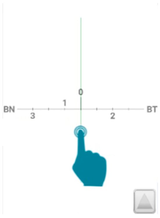
The apparent sizes of horizontal and vertical lines show an anisotropy known as the horizontal vertical illusion (HVI) wherein vertical lines appear to be longer than their horizontal counterparts. Whereas a typical HVI comparing vertical and horizontal lines in a plane produces a
[...] Read more.
The apparent sizes of horizontal and vertical lines show an anisotropy known as the horizontal vertical illusion (HVI) wherein vertical lines appear to be longer than their horizontal counterparts. Whereas a typical HVI comparing vertical and horizontal lines in a plane produces a 5–10% illusion, a much larger-scale illusion (15–25%) is often found for large objects in the real world, and this has been related to differential angular exaggerations in perceived elevation (vertical) and azimuthal (horizontal) direction. Recently supine observers in virtual environments were found to show larger exaggerations in perceived azimuth than upright observers. Here, 48 participants were tested in both supine and upright postures in an outdoor environment while matching fairly small physical extents in the real world. They adjusted the magnitude of the horizontal extent to perceptually match fairly small vertical poles (0.7–1.3 m tall) that were either presented at the same viewing distance as the matching extent or in a different depth plane, so that size at a distance had to be compared. Supine observers viewed the scene, as though upright, through a large mirror mounted overhead at 45° that was adjusted to approximate their normal eye height. When the matcher extent was at a different distance than the pole, horizontal extent matches typically exceeded the actual pole height by about 15% or more, whether the viewer was upright or supine. The average overestimation was only about 10% when the matching extent was at the same distance. Despite the similarity in performance across different postures for spatial matching, supine observers gave much higher explicit estimates of azimuthal direction than upright observers. However, although the observation of exaggeration in perceived azimuth for supine observers was replicated in a second study with 24 additional participants using a mirror with a smaller (more normal) aspect ratio, the magnitude of the exaggeration seemed to be greatly reduced when the field of view of the apparatus had a more typical aspect ratio. This suggests that the unusually large exaggeration of azimuth found in a previous report with supine observers may have been caused by the unusually large aspect ratio of the viewing apparatus used.
Full article

Figure 1


