Next Issue
Volume 16, April-2
Previous Issue
Volume 16, March-2


Cancers, Volume 16, Issue 7 (April-1 2024) – 202 articles

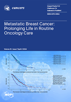
Cover Story (view full-size image): Overall survival (OS) of patients with metastatic breast cancer (MBC) has improved within controlled clinical trials. Whether these advances translate into improved survival in routine care remains controversial. A retrospective analysis of 1610 patients showed a continuous prolongation of median OS from 31.6 months (1995–2000) to 48.4 months (2018–2022). The median OS for the entire cohort of 1995-2022 was 38.0 months. Patients with triple-positive tumors lived the longest (52.3 months), followed by hormone-receptor-positive/HER2-negative (41.1 months) patients and HER2-positive/hormone-receptor-negative (36.6 months) patients. Triple-negative patients had the shortest survival time of 19.9 months. The OS of MBC has improved in a statistically significant and clinically relevant manner in routine oncology care. View this paper
  • Issues are regarded as officially published after their release is announced to the table of contents alert mailing list.
  • You may sign up for e-mail alerts to receive table of contents of newly released issues.
  • PDF is the official format for papers published in both, html and pdf forms. To view the papers in pdf format, click on the "PDF Full-text" link, and use the free Adobe Reader to open them.
Order results
Result details
Section
Select all
Export citation of selected articles as:
20 pages, 3913 KiB  
Article
scRNAseq and High-Throughput Spatial Analysis of Tumor and Normal Microenvironment in Solid Tumors Reveal a Possible Origin of Circulating Tumor Hybrid Cells
by Abdullah Mahmood Ali and Azra Raza
Cancers 2024, 16(7), 1444; https://doi.org/10.3390/cancers16071444 - 8 Apr 2024
Cited by 2 | Viewed by 2275
Abstract
Metastatic cancer is a leading cause of death in cancer patients worldwide. While circulating hybrid cells (CHCs) are implicated in metastatic spread, studies documenting their tissue origin remain sparse, with limited candidate approaches using one–two markers. Utilizing high-throughput single-cell and spatial transcriptomics, we [...] Read more.
Metastatic cancer is a leading cause of death in cancer patients worldwide. While circulating hybrid cells (CHCs) are implicated in metastatic spread, studies documenting their tissue origin remain sparse, with limited candidate approaches using one–two markers. Utilizing high-throughput single-cell and spatial transcriptomics, we identified tumor hybrid cells (THCs) co-expressing epithelial and macrophage markers and expressing a distinct transcriptome. Rarely, normal tissue showed these cells (NHCs), but their transcriptome was easily distinguishable from THCs. THCs with unique transcriptomes were observed in breast and colon cancers, suggesting this to be a generalizable phenomenon across cancer types. This study establishes a framework for HC identification in large datasets, providing compelling evidence for their tissue residence and offering comprehensive transcriptomic characterization. Furthermore, it sheds light on their differential function and identifies pathways that could explain their newly acquired invasive capabilities. THCs should be considered as potential therapeutic targets. Full article
(This article belongs to the Section Cancer Informatics and Big Data)
Show Figures

Figure 1

18 pages, 3487 KiB  
Systematic Review
Performance of Commercial Dermatoscopic Systems That Incorporate Artificial Intelligence for the Identification of Melanoma in General Practice: A Systematic Review
by Ian Miller, Nedeljka Rosic, Michael Stapelberg, Jeremy Hudson, Paul Coxon, James Furness, Joe Walsh and Mike Climstein
Cancers 2024, 16(7), 1443; https://doi.org/10.3390/cancers16071443 - 8 Apr 2024
Viewed by 2317
Abstract
Background: Cutaneous melanoma remains an increasing global public health burden, particularly in fair-skinned populations. Advancing technologies, particularly artificial intelligence (AI), may provide an additional tool for clinicians to help detect malignancies with a more accurate success rate. This systematic review aimed to report [...] Read more.
Background: Cutaneous melanoma remains an increasing global public health burden, particularly in fair-skinned populations. Advancing technologies, particularly artificial intelligence (AI), may provide an additional tool for clinicians to help detect malignancies with a more accurate success rate. This systematic review aimed to report the performance metrics of commercially available convolutional neural networks (CNNs) tasked with detecting MM. Methods: A systematic literature search was performed using CINAHL, Medline, Scopus, ScienceDirect and Web of Science databases. Results: A total of 16 articles reporting MM were included in this review. The combined number of melanomas detected was 1160, and non-melanoma lesions were 33,010. The performance of market-approved technology and clinician performance for classifying melanoma was highly heterogeneous, with sensitivity ranging from 16.4 to 100.0%, specificity between 40.0 and 98.3% and accuracy between 44.0 and 92.0%. Less heterogeneity was observed when clinicians worked in unison with AI, with sensitivity ranging between 83.3 and 100.0%, specificity between 83.7 and 87.3%, and accuracy between 86.4 and 86.9%. Conclusion: Instead of focusing on the performance of AI versus clinicians for classifying melanoma, more consistent performance has been obtained when clinicians’ work is supported by AI, facilitating management decisions and improving health outcomes. Full article
(This article belongs to the Collection Artificial Intelligence in Oncology)
Show Figures

Figure 1

11 pages, 657 KiB  
Article
Efficacy and Safety of Tinzaparin Thromboprophylaxis in Lung Cancer Patients with High Thromboembolic Risk: A Prospective, Observational, Single-Center Cohort Study
by Marousa Kouvela, Maria Effrosyni Livanou, Dimitra T. Stefanou, Ioannis A. Vathiotis, Fotini Sarropoulou, Maria Grammoustianou, Evangelos Dimakakos and Nikolaos Syrigos
Cancers 2024, 16(7), 1442; https://doi.org/10.3390/cancers16071442 - 8 Apr 2024
Viewed by 1071
Abstract
Background: The aim of this study was to record and assess the efficacy and safety ofthromboprophylaxis with an intermediate dose of Tinzaparin in lung cancer patients with high thrombotic risk. Methods: This was a non-interventional, single-arm, prospective cohort study of lung cancer patients [...] Read more.
Background: The aim of this study was to record and assess the efficacy and safety ofthromboprophylaxis with an intermediate dose of Tinzaparin in lung cancer patients with high thrombotic risk. Methods: This was a non-interventional, single-arm, prospective cohort study of lung cancer patients who received thromboprophylaxis with Tinzaparin 10.000 Anti-Xa IU in 0.5 mL, OD, used in current clinical practice. Enrolled ambulatory patients signed informed consent. Anti-Xa levels were tested. Results: In total, 140 patients were included in the study, of which 81.4% were males. The histology of the tumor was mainly adenocarcinoma. Lung cancer patients with high thrombotic risk based on tumor, patient, treatment, and laboratory-related factors were enrolled. Only one patient experienced a thrombotic event (0.7%), and 10 patients had bleeding events (7.1%), including only one major event. Anti-Xa levels measured at 10 days and 3 months did not differ significantly between patients who developed hemorrhagic events and those who did not (p = 0.26 and p = 0.32, respectively). Conclusion: Thromboprophylaxis with an intermediate Tinzaparin dose in high thrombotic-risk lung cancer patients is a safe and effective choice for the prevention of VTE. Full article
(This article belongs to the Special Issue Recent Advances in Trachea, Bronchus and Lung Cancer Management)
Show Figures

Figure 1

20 pages, 4005 KiB  
Article
Pathological Insights: Enhanced Vision Transformers for the Early Detection of Colorectal Cancer
by Gelan Ayana, Hika Barki and Se-woon Choe
Cancers 2024, 16(7), 1441; https://doi.org/10.3390/cancers16071441 - 8 Apr 2024
Viewed by 1690
Abstract
Endoscopic pathological findings of the gastrointestinal tract are crucial for the early diagnosis of colorectal cancer (CRC). Previous deep learning works, aimed at improving CRC detection performance and reducing subjective analysis errors, are limited to polyp segmentation. Pathological findings were not considered and [...] Read more.
Endoscopic pathological findings of the gastrointestinal tract are crucial for the early diagnosis of colorectal cancer (CRC). Previous deep learning works, aimed at improving CRC detection performance and reducing subjective analysis errors, are limited to polyp segmentation. Pathological findings were not considered and only convolutional neural networks (CNNs), which are not able to handle global image feature information, were utilized. This work introduces a novel vision transformer (ViT)-based approach for early CRC detection. The core components of the proposed approach are ViTCol, a boosted vision transformer for classifying endoscopic pathological findings, and PUTS, a vision transformer-based model for polyp segmentation. Results demonstrate the superiority of this vision transformer-based CRC detection method over existing CNN and vision transformer models. ViTCol exhibited an outstanding performance in classifying pathological findings, with an area under the receiver operating curve (AUC) value of 0.9999 ± 0.001 on the Kvasir dataset. PUTS provided outstanding results in segmenting polyp images, with mean intersection over union (mIoU) of 0.8673 and 0.9092 on the Kvasir-SEG and CVC-Clinic datasets, respectively. This work underscores the value of spatial transformers in localizing input images, which can seamlessly integrate into the main vision transformer network, enhancing the automated identification of critical image features for early CRC detection. Full article
(This article belongs to the Special Issue The Role of Colonoscopy in the Diagnosis of Colorectal Cancer)
Show Figures

Figure 1

30 pages, 2350 KiB  
Review
Immune-Related Adverse Events Due to Cancer Immunotherapy: Immune Mechanisms and Clinical Manifestations
by Silvia Casagrande, Giulia Boscato Sopetto, Giovanni Bertalot, Roberto Bortolotti, Vito Racanelli, Orazio Caffo, Bruno Giometto, Alvise Berti and Antonello Veccia
Cancers 2024, 16(7), 1440; https://doi.org/10.3390/cancers16071440 - 8 Apr 2024
Cited by 4 | Viewed by 3569
Abstract
The landscape of cancer treatment has undergone a significant transformation with the introduction of Immune Checkpoint Inhibitors (ICIs). Patients undergoing these treatments often report prolonged clinical and radiological responses, albeit with a potential risk of developing immune-related adverse events (irAEs). Here, we reviewed [...] Read more.
The landscape of cancer treatment has undergone a significant transformation with the introduction of Immune Checkpoint Inhibitors (ICIs). Patients undergoing these treatments often report prolonged clinical and radiological responses, albeit with a potential risk of developing immune-related adverse events (irAEs). Here, we reviewed and discussed the mechanisms of action of ICIs and their pivotal role in regulating the immune system to enhance the anti-tumor immune response. We scrutinized the intricate pathogenic mechanisms responsible for irAEs, arising from the evasion of self-tolerance checkpoints due to drug-induced immune modulation. We also summarized the main clinical manifestations due to irAEs categorized by organ types, detailing their incidence and associated risk factors. The occurrence of irAEs is more frequent when ICIs are combined; with neurological, cardiovascular, hematological, and rheumatic irAEs more commonly linked to PD1/PD-L1 inhibitors and cutaneous and gastrointestinal irAEs more prevalent with CTLA4 inhibitors. Due to the often-nonspecific signs and symptoms, the diagnosis of irAEs (especially for those rare ones) can be challenging. The differential with primary autoimmune disorders becomes sometimes intricate, given the clinical and pathophysiological similarities. In conclusion, considering the escalating use of ICIs, this area of research necessitates additional clinical studies and practical insights, especially the development of biomarkers for predicting immune toxicities. In addition, there is a need for heightened education for both clinicians and patients to enhance understanding and awareness. Full article
Show Figures

Figure 1

9 pages, 3563 KiB  
Article
Simultaneous Visualization of Lung Tumor and Intersegmental Plane during Pulmonary Segmentectomy by Intravenous Injection of Indocyanine Green
by Kyungsu Kim, Ok Hwa Jeon, Byeong Hyeon Choi, Jiyun Rho, Jun Hee Lee, Jae Seon Eo, Beop-Min Kim and Hyun Koo Kim
Cancers 2024, 16(7), 1439; https://doi.org/10.3390/cancers16071439 - 8 Apr 2024
Viewed by 1296
Abstract
Segmentectomy is a targeted surgical approach tailored for patients with compromised health and early-stage lung cancer. The key to successful segmentectomy lies in precisely identifying the tumor and intersegmental planes to ensure adequate resection margins. In this study, we aimed to enhance this [...] Read more.
Segmentectomy is a targeted surgical approach tailored for patients with compromised health and early-stage lung cancer. The key to successful segmentectomy lies in precisely identifying the tumor and intersegmental planes to ensure adequate resection margins. In this study, we aimed to enhance this process by simultaneously visualizing the tumor and intersegmental planes through the intravenous injection of indocyanine green (ICG) at different time points and doses. Lung tumors were detected by intravenous injection of ICG at a dose of 2 mg/kg 12 h before surgery in a rabbit model. Following the dissection of the pulmonary artery, vein, and bronchi of the target segment, 0.6 mg/kg of ICG was injected intravenously to detect the intersegmental plan. Fluorescent images of the lung tumors and segments were acquired, and the fluorescent signal was quantified using the signal-to-background ratio (SBR). Finally, a pilot study of this method was conducted in three patients with lung cancer. In a preclinical study, the SBR of the tumor (4.4 ± 0.1) and nontargeted segments (10.5 ± 0.8) were significantly higher than that of the targeted segment (1.6 ± 0.2) (targeted segment vs. nontarget segment, p < 0.0001; target segment vs. tumor, p < 0.01). Consistent with preclinical results, lung tumors and the intersegmental plane were successfully detected in patients with lung cancer. Consequently, adequate resection margins were identified during the surgery, and segmentectomy was successfully performed in patients with lung cancer. This study is the first to use intravenous ICG injections at different time points and doses to simultaneously detect lung cancer and intersegmental planes, thereby achieving segmentectomy for lung cancer. Full article
Show Figures

Figure 1

2 pages, 173 KiB  
Correction
Correction: Soda et al. Electrochemical Detection of Global DNA Methylation Using Biologically Assembled Polymer Beads. Cancers 2021, 13, 3787
by Narshone Soda, Zennia Jean Gonzaga, Amandeep Singh Pannu, Navid Kashaninejad, Richard Kline, Carlos Salomon, Nam-Trung Nguyen, Prashant Sonar, Bernd H. A. Rehm and Muhammad J. A. Shiddiky
Cancers 2024, 16(7), 1438; https://doi.org/10.3390/cancers16071438 - 8 Apr 2024
Viewed by 1042
Abstract
In the original publication [...] Full article
13 pages, 4124 KiB  
Article
Clinical [18F]FSPG Positron Emission Tomography Imaging Reveals Heterogeneity in Tumor-Associated System xc Activity
by Amy R. Sharkey, Norman Koglin, Erik S. Mittra, Sangwon Han, Gary J. R. Cook and Timothy H. Witney
Cancers 2024, 16(7), 1437; https://doi.org/10.3390/cancers16071437 - 8 Apr 2024
Cited by 1 | Viewed by 1688
Abstract
Background: (4S)-4-(3-[18F]fluoropropyl)-L-glutamic acid ([18F]FSPG) positron emission tomography/computed tomography (PET/CT) provides a readout of system xc transport activity and has been used for cancer detection in clinical studies of different cancer types. As system xc [...] Read more.
Background: (4S)-4-(3-[18F]fluoropropyl)-L-glutamic acid ([18F]FSPG) positron emission tomography/computed tomography (PET/CT) provides a readout of system xc transport activity and has been used for cancer detection in clinical studies of different cancer types. As system xc provides the rate-limiting precursor for glutathione biosynthesis, an abundant antioxidant, [18F]FSPG imaging may additionally provide important prognostic information. Here, we performed an analysis of [18F]FSPG radiotracer distribution between primary tumors, metastases, and normal organs from cancer patients. We further assessed the heterogeneity of [18F]FSPG retention between cancer types, and between and within individuals. Methods: This retrospective analysis of prospectively collected data compared [18F]FSPG PET/CT in subjects with head and neck squamous cell cancer (HNSCC, n = 5) and non-small-cell lung cancer (NSCLC, n = 10), scanned at different institutions. Using semi-automated regions of interest drawn around tumors and metastases, the maximum standardized uptake value (SUVmax), SUVmean, SUV standard deviation and SUVpeak were measured. [18F]FSPG time–activity curves (TACs) for normal organs, primary tumors and metastases were subsequently compared to 18F-2-fluoro-2-deoxy-D-glucose ([18F]FDG) PET/CT at 60 min post injection (p.i.). Results: The mean administered activity of [18F]FSPG was 309.3 ± 9.1 MBq in subjects with NSCLC and 285.1 ± 11.3 MBq in those with HNSCC. The biodistribution of [18F]FSPG in both cohorts showed similar TACs in healthy organs from cancer patients. There was no statistically significant overall difference in the average SUVmax of tumor lesions at 60 min p.i. for NSCLC (8.1 ± 7.1) compared to HNSCC (6.0 ± 4.1; p = 0.29) for [18F]FSPG. However, there was heterogeneous retention between and within cancer types; the SUVmax at 60 min p.i. ranged from 1.4 to 23.7 in NSCLC and 3.1–12.1 in HNSCC. Conclusion: [18F]FSPG PET/CT imaging from both NSCLC and HNSCC cohorts showed the same normal-tissue biodistribution, but marked tumor heterogeneity across subjects and between lesions. Despite rapid elimination through the urinary tract and low normal-background tissue retention, the diagnostic potential of [18F]FSPG was limited by variability in tumor retention. As [18F]FSPG retention is mediated by the tumor’s antioxidant capacity and response to oxidative stress, this heterogeneity may provide important insights into an individual tumor’s response or resistance to therapy. Full article
(This article belongs to the Special Issue Molecular Imaging in Oncology: Recent Advances)
Show Figures

Figure 1

15 pages, 4666 KiB  
Article
Exploring The Prognostic Significance of SET-Domain Containing 2 (SETD2) Expression in Advanced and Castrate-Resistant Prostate Cancer
by Yaser Gamallat, Joema Felipe Lima, Sima Seyedi, Qiaowang Li, Jon George Rokne, Reda Alhajj, Sunita Ghosh and Tarek A. Bismar
Cancers 2024, 16(7), 1436; https://doi.org/10.3390/cancers16071436 - 8 Apr 2024
Viewed by 1612
Abstract
SET-domain containing 2 (SETD2) is a histone methyltransferase and an epigenetic modifier with oncogenic functionality. In the current study, we investigated the potential prognostic role of SETD2 in prostate cancer. A cohort of 202 patients’ samples was assembled on tissue microarrays (TMAs) containing [...] Read more.
SET-domain containing 2 (SETD2) is a histone methyltransferase and an epigenetic modifier with oncogenic functionality. In the current study, we investigated the potential prognostic role of SETD2 in prostate cancer. A cohort of 202 patients’ samples was assembled on tissue microarrays (TMAs) containing incidental, advanced, and castrate-resistant CRPCa cases. Our data showed significant elevated SETD2 expression in advanced and castrate-resistant disease (CRPCa) compared to incidental cases (2.53 ± 0.58 and 2.21 ± 0.63 vs. 1.9 ± 0.68; p < 0.001, respectively). Interestingly, the mean intensity of SETD2 expression in deceased vs. alive patients was also significantly different (2.31 ± 0.66 vs. 2 ± 0.68; p = 0.003, respectively). Overall, high SETD2 expression was found to be considered high risk and was significantly associated with poor prognosis and worse overall survival (OS) (HR 1.80; 95% CI: 1.28–2.53, p = 0.001) and lower cause specific survival (CSS) (HR 3.14; 95% CI: 1.94–5.08, p < 0.0001). Moreover, combining high-intensity SETD2 with PTEN loss resulted in lower OS (HR 2.12; 95% CI: 1.22–3.69, p = 0.008) and unfavorable CSS (HR 3.74; 95% CI: 1.67–8.34, p = 0.001). Additionally, high SETD2 intensity with ERG positive expression showed worse prognosis for both OS (HR 1.99, 95% CI 0.87–4.59; p = 0.015) and CSS (HR 2.14, 95% CI 0.98–4.68, p = 0.058). We also investigated the protein expression database TCPA, and our results showed that high SETD2 expression is associated with a poor prognosis. Finally, we performed TCGA PRAD gene set enrichment analysis (GSEA) data for SETD2 overexpression, and our data revealed a potential association with pathways involved in tumor progression such as the AMPK signaling pathway, the cAMP signaling pathway, and the PI3K-Akt signaling pathway, which are potentially associated with tumor progression, chemoresistance, and a poor prognosis. Full article
(This article belongs to the Collection Biomarkers for Detection and Prognosis of Prostate Cancer)
Show Figures

Figure 1

19 pages, 1422 KiB  
Review
The Exacerbating Effects of the Tumor Necrosis Factor in Cardiovascular Stenosis: Intimal Hyperplasia
by Chandra Shekhar Boosani and Laxminarayana Burela
Cancers 2024, 16(7), 1435; https://doi.org/10.3390/cancers16071435 - 8 Apr 2024
Cited by 1 | Viewed by 1361
Abstract
TNF-α functions as a master regulator of inflammation, and it plays a prominent role in several immunological diseases. By promoting important cellular mechanisms, such as cell proliferation, migration, and phenotype switch, TNF-α induces its exacerbating effects, which are the underlying cause of many [...] Read more.
TNF-α functions as a master regulator of inflammation, and it plays a prominent role in several immunological diseases. By promoting important cellular mechanisms, such as cell proliferation, migration, and phenotype switch, TNF-α induces its exacerbating effects, which are the underlying cause of many proliferative diseases such as cancer and cardiovascular disease. TNF-α primarily alters the immune component of the disease, which subsequently affects normal functioning of the cells. Monoclonal antibodies and synthetic drugs that can target TNF-α and impair its effects have been developed and are currently used in the treatment of a few select human diseases. Vascular restenosis is a proliferative disorder that is initiated by immunological mechanisms. In this review, the role of TNF-α in exacerbating restenosis resulting from neointimal hyperplasia, as well as molecular mechanisms and cellular processes affected or induced by TNF-α, are discussed. As TNF-α-targeting drugs are currently not approved for the treatment of restenosis, the summation of the topics discussed here is anticipated to provide information that can emphasize on the use of TNF-α-targeting drug candidates to prevent vascular restenosis. Full article
Show Figures

Figure 1

15 pages, 5141 KiB  
Article
Identifying the Spatial Architecture That Restricts the Proximity of CD8+ T Cells to Tumor Cells in Pancreatic Ductal Adenocarcinoma
by Yihan Xia, Junrui Ma, Xiaobao Yang, Danping Liu, Yujie Zhu, Yanan Zhao, Xuefeng Fei, Dakang Xu and Jing Dai
Cancers 2024, 16(7), 1434; https://doi.org/10.3390/cancers16071434 - 7 Apr 2024
Viewed by 1720
Abstract
The anti-tumor function of CD8+ T cells is dependent on their proximity to tumor cells. Current studies have focused on the infiltration level of CD8+ T cells in the tumor microenvironment, while further spatial information, such as spatial localization and inter-cellular [...] Read more.
The anti-tumor function of CD8+ T cells is dependent on their proximity to tumor cells. Current studies have focused on the infiltration level of CD8+ T cells in the tumor microenvironment, while further spatial information, such as spatial localization and inter-cellular communication, have not been defined. In this study, co-detection by indexing (CODEX) was designed to characterize PDAC tissue regions with seven protein markers in order to identify the spatial architecture that regulates CD8+ T cells in human pancreatic ductal adenocarcinoma (PDAC). The cellular neighborhood algorithm was used to identify a total of six conserved and distinct cellular neighborhoods. Among these, one unique spatial architecture of CD8+ T and CD4+ T cell-enriched neighborhoods enriched the majority of CD8+ T cells, but heralded a poor prognosis. The proximity analysis revealed that the CD8+ T cells in this spatial architecture were significantly closer to themselves and the CD4+ T cells than to the tumor cells. Collectively, we identified a unique spatial architecture that restricted the proximity of CD8+ T cells to tumor cells in the tumor microenvironment, indicating a novel immune evasion mechanism of pancreatic ductal adenocarcinoma in a topologically regulated manner and providing new insights into the biology of PDAC. Full article
(This article belongs to the Special Issue Advanced Research in Pancreatic Ductal Adenocarcinoma)
Show Figures

Figure 1

21 pages, 6410 KiB  
Review
Translational Aspects in Metaplastic Breast Carcinoma
by Elizve Nairoby Barrientos-Toro, Qingqing Ding and Maria Gabriela Raso
Cancers 2024, 16(7), 1433; https://doi.org/10.3390/cancers16071433 - 7 Apr 2024
Viewed by 1831
Abstract
Breast cancer is the most common cancer among women. Metaplastic breast carcinoma (MpBC) is a rare, heterogeneous group of invasive breast carcinomas, which are classified as predominantly triple-negative breast carcinomas (TNBCs; HR-negative/HER2-negative). Histologically, MpBC is classified into six subtypes. Two of these are [...] Read more.
Breast cancer is the most common cancer among women. Metaplastic breast carcinoma (MpBC) is a rare, heterogeneous group of invasive breast carcinomas, which are classified as predominantly triple-negative breast carcinomas (TNBCs; HR-negative/HER2-negative). Histologically, MpBC is classified into six subtypes. Two of these are considered low-grade and the others are high-grade. MpBCs seem to be more aggressive, less responsive to neoadjuvant chemotherapy, and have higher rates of chemoresistance than other TNBCs. MpBCs have a lower survival rate than expected for TNBCs. MpBC treatment represents a challenge, leading to a thorough exploration of the tumor immune microenvironment, which has recently opened the possibility of new therapeutic strategies. The epithelial–mesenchymal transition in MpBC is characterized by the loss of intercellular adhesion, downregulation of epithelial markers, underexpression of genes with biological epithelial functions, upregulation of mesenchymal markers, overexpression of genes with biological mesenchymal functions, acquisition of fibroblast-like (spindle) morphology, cytoskeleton reorganization, increased motility, invasiveness, and metastatic capabilities. This article reviews and summarizes the current knowledge and translational aspects of MpBC. Full article
Show Figures

Figure 1

2 pages, 719 KiB  
Correction
Correction: Garza-Morales et al. Temozolomide Enhances Triple-Negative Breast Cancer Virotherapy In Vitro. Cancers 2018, 10, 144
by Rodolfo Garza-Morales, Roxana Gonzalez-Ramos, Akiko Chiba, Roberto Montes de Oca-Luna, Lacey R. McNally, Kelly M. McMasters and Jorge G. Gomez-Gutierrez
Cancers 2024, 16(7), 1432; https://doi.org/10.3390/cancers16071432 - 7 Apr 2024
Viewed by 848
Abstract
Error in Figure [...] Full article
Show Figures

Figure 1

21 pages, 692 KiB  
Perspective
A Plausible Framework Reveals Potential Similarities in the Regulation of Immunity against Some Cancers and Some Infectious Agents: Implications for Prevention and Treatment
by Peter A. Bretscher
Cancers 2024, 16(7), 1431; https://doi.org/10.3390/cancers16071431 - 7 Apr 2024
Viewed by 1167
Abstract
Different frameworks, which are currently employed to understand how immune responses are regulated, can account for different observations reported in the classical literature. I have argued that the predominant frameworks, employed over the last two/three decades to analyze the circumstances that determine whether [...] Read more.
Different frameworks, which are currently employed to understand how immune responses are regulated, can account for different observations reported in the classical literature. I have argued that the predominant frameworks, employed over the last two/three decades to analyze the circumstances that determine whether an immune response is generated or this potential is ablated, and that determine the class of immunity an antigen induces, are inconsistent with diverse classical observations. These observations are “paradoxical” within the context of these frameworks and, consequently, tend to be ignored by most contemporary researchers. One such observation is that low and high doses of diverse types of antigen result, respectively, in cell-mediated and IgG antibody responses. I suggest these paradoxes render these frameworks implausible. An alternative framework, The Threshold Hypothesis, accounts for the paradoxical observations. Some frameworks are judged more plausible when found to be valuable in understanding findings in fields beyond their original compass. I explore here how the Threshold Hypothesis, initially based on studies with chemically well-defined and “simple antigens”, most often a purified protein, can nevertheless shed light on diverse classical and more recent observations in the fields of immunity against cancer and against infectious agents, thus revealing common, immune mechanisms. Most cancers and some pathogens are best contained by cell-mediated immunity. The success of the Threshold Hypothesis has encouraged me to employ it as a basis for proposing strategies to prevent and to treat cancer and those infectious diseases caused by pathogens best contained by a cell-mediated attack. Full article
(This article belongs to the Section Infectious Agents and Cancer)
Show Figures

Figure 1

12 pages, 3026 KiB  
Article
PAX1/JAM3 Methylation and HPV Viral Load in Women with Persistent HPV Infection
by Mingzhu Li, Chao Zhao, Xiaobo Zhang, Jingran Li, Yun Zhao, Wei Zhang, Lihua Ren and Lihui Wei
Cancers 2024, 16(7), 1430; https://doi.org/10.3390/cancers16071430 - 7 Apr 2024
Cited by 2 | Viewed by 1681
Abstract
The relationship of PAX1/JAM3 methylation as well as HPV viral load (VL) with cervical lesions has been reported, but their role in persistent HPV infection without cervical high-grade lesions has not been fully elucidated. A total of 231 females diagnosed with persistent HPV [...] Read more.
The relationship of PAX1/JAM3 methylation as well as HPV viral load (VL) with cervical lesions has been reported, but their role in persistent HPV infection without cervical high-grade lesions has not been fully elucidated. A total of 231 females diagnosed with persistent HPV infection and pathologically confirmed absence of high-grade cervical lesions were selected from the Colposcopy Outpatient Clinic of Peking University People’s Hospital, from March 2023 to December 2023. They were categorized into two groups based on the duration of HPV infection: the HPV persistent less than 3 years group and the more than 3 years group. PAX1/JAM3 methylation and HPV VL were determined by real-time PCR and BioPerfectus Multiplex Real-Time (BMRT)-HPV reports type-specific VL/10,000 cells, respectively. The average age of individuals with HPV infection lasting more than 3 years was higher compared to those with less than 3 years (48.9 vs. 45.1 years), with a statistically significant difference. Among the participants, 81.8% (189/231) had no previous screening. The methylation levels of JAM3 and PAX1 were significantly higher in individuals with HPV infection persisting for more than 3 years compared to those with less than 3 years, with a statistically significant difference (p < 0.05). There was a significant correlation between PAX1 and JAM3 methylation (p < 0.001), which could be used as cumulative evidence of HPV infection duration before the occurrence of precancerous lesions. The incidence of vaginal intraepithelial lesions was higher in individuals with HPV infection persisting for more than 3 years compared to those with less than 3 years, and HPV VL can be used as an indicative biomarker for concurrent cervical–vaginal lesions, especially for HPV other than 16/18 genotypes. Full article
(This article belongs to the Section Infectious Agents and Cancer)
Show Figures

Figure 1

15 pages, 432 KiB  
Article
Exploring the Link between BMI and Aggressive Histopathological Subtypes in Differentiated Thyroid Carcinoma—Insights from a Multicentre Retrospective Study
by Giacomo Di Filippo, Gian Luigi Canu, Giovanni Lazzari, Dorin Serbusca, Eleonora Morelli, Paolo Brazzarola, Leonardo Rossi, Benard Gjeloshi, Mariangela Caradonna, George Kotsovolis, Ioannis Pliakos, Efthymios Poulios, Theodosios Papavramidis, Federico Cappellacci, Pier Francesco Nocini, Pietro Giorgio Calò, Gabriele Materazzi and Fabio Medas
Cancers 2024, 16(7), 1429; https://doi.org/10.3390/cancers16071429 - 7 Apr 2024
Cited by 1 | Viewed by 1302
Abstract
Obesity’s role in thyroid cancer development is still debated, as well as its association with aggressive histopathological subtypes (AHSs). To clarify the link between Body Mass Index (BMI) and AHS of differentiated thyroid carcinoma (DTC), we evaluated patients who underwent thyroidectomy for DTC [...] Read more.
Obesity’s role in thyroid cancer development is still debated, as well as its association with aggressive histopathological subtypes (AHSs). To clarify the link between Body Mass Index (BMI) and AHS of differentiated thyroid carcinoma (DTC), we evaluated patients who underwent thyroidectomy for DTC from 2020 to 2022 at four European referral centres for endocrine surgery. Based on BMI, patients were classified as normal-underweight, overweight, or obese. AHSs were defined according to 2022 WHO guidelines. Among 3868 patients included, 34.5% were overweight and 19.6% obese. Histological diagnoses were: 93.6% papillary (PTC), 4.8% follicular (FTC), and 1.6% Hürthle cell (HCC) thyroid carcinoma. Obese and overweight patients with PTC had a higher rate of AHSs (p = 0.03), bilateral, multifocal tumours (p = 0.014, 0.049), and larger nodal metastases (p = 0.017). In a multivariate analysis, BMI was an independent predictor of AHS of PTC, irrespective of gender (p = 0.028). In younger patients (<55 years old) with PTC > 1 cm, BMI predicted a higher ATA risk class (p = 0.036). Overweight and obese patients with FTC had larger tumours (p = 0.036). No difference was found in terms of AHS of FTC and HCC based on BMI category. Overweight and obese patients with PTC appear to be at an increased risk for AHS and aggressive clinico-pathological characteristics. Full article
(This article belongs to the Special Issue Thyroid Cancer: Incidence and Risk Factors)
Show Figures

Figure 1

14 pages, 2546 KiB  
Article
Effect of Neoadjuvant Chemotherapy on Tumor-Infiltrating Lymphocytes in Resectable Gastric Cancer: Analysis from a Western Academic Center
by Elliott J. Yee, Danielle Gilbert, Jeffrey Kaplan, Sachin Wani, Sunnie S. Kim, Martin D. McCarter and Camille L. Stewart
Cancers 2024, 16(7), 1428; https://doi.org/10.3390/cancers16071428 - 7 Apr 2024
Viewed by 1940
Abstract
Tumor-infiltrating lymphocytes (TILs) are an emerging biomarker predictive of response to immunotherapy across a spectrum of solid organ malignancies. The characterization of TILs in gastric cancer (GC) treated with contemporary, multiagent neoadjuvant chemotherapy (NAC) is understudied. In this retrospective investigation, we analyzed the [...] Read more.
Tumor-infiltrating lymphocytes (TILs) are an emerging biomarker predictive of response to immunotherapy across a spectrum of solid organ malignancies. The characterization of TILs in gastric cancer (GC) treated with contemporary, multiagent neoadjuvant chemotherapy (NAC) is understudied. In this retrospective investigation, we analyzed the degree of infiltration, phenotype, and spatial distribution of TILs via immunohistochemistry within resected GC specimens treated with or without NAC at a Western center. We hypothesized that NAC executes immunostimulatory roles, as evidenced by an increased number of anti-tumor TILs in the tumor microenvironment. We found significantly elevated levels of conventional and memory CD8+ T cells, as well as total TILs (CD4+, CD8+, Treg, B cells), within chemotherapy-treated tumors compared with chemotherapy-naïve specimens. We also revealed important associations between survival and pathologic responses with enhanced TIL infiltration. Taken together, our findings advocate for an immunostimulatory role of chemotherapy and underscore the potential synergistic effect of combining chemotherapy with immunotherapy in resectable gastric cancer. Full article
(This article belongs to the Special Issue Developments in the Management of Gastrointestinal Malignancies)
Show Figures

Figure 1

15 pages, 1214 KiB  
Article
Prognostic Indicators of Overall Survival in Hepatocellular Carcinoma Patients Undergoing Liver Resection
by Cristina-Paula Ursu, Andra Ciocan, Ștefan Ursu, Răzvan Alexandru Ciocan, Claudia Diana Gherman, Ariana-Anamaria Cordoș, Dan Vălean, Rodica Sorina Pop, Luminița Elena Furcea, Bogdan Procopeț, Horia Ștefănescu, Emil Ioan Moiș, Nadim Al Hajjar and Florin Graur
Cancers 2024, 16(7), 1427; https://doi.org/10.3390/cancers16071427 - 7 Apr 2024
Cited by 1 | Viewed by 1478
Abstract
Hepatocellular carcinoma (HCC) is the predominant form of primary liver cancer and the third contributor to malignancy-related deaths worldwide. The hepatic venous pressure gradient (HVPG), transient elastography-liver stiffness measurement (TE-LSM), and the association between TBS (tumor burden score), alpha-fetoprotein levels, and the Child–Pugh [...] Read more.
Hepatocellular carcinoma (HCC) is the predominant form of primary liver cancer and the third contributor to malignancy-related deaths worldwide. The hepatic venous pressure gradient (HVPG), transient elastography-liver stiffness measurement (TE-LSM), and the association between TBS (tumor burden score), alpha-fetoprotein levels, and the Child–Pugh classification (TAC score) can serve as valuable prognostic indicators for these patients. Therefore, the main objective of our research was to analyze the prognostic value of the HVPG, TE-LSM, TBS, and TAC scores. An observational and survival study was conducted on 144 subjects. Our findings indicated that HVPG greater than 10 mmHg, AFP surpassing 400 ng/mL, an advanced C–P class, and low TAC score are independent predictors of overall survival. During the multivariate analysis, AFP serum levels and C–P class proved statistically significant. The present study revealed significant differences in overall survival between the two groups divided upon HVPG values and settled by the cutoff of 10 mmHg (p = 0.02). Moreover, by dividing the cohort into three groups based on the TAC score (very low, low, and moderate), statistically significant differences in overall survival were observed across the groups (p = 0.004). Full article
(This article belongs to the Special Issue New Insights into Surgical Treatment of Hepatocellular Carcinoma)
Show Figures

Figure 1

21 pages, 4054 KiB  
Review
Spinal Meningiomas: A Comprehensive Review and Update on Advancements in Molecular Characterization, Diagnostics, Surgical Approach and Technology, and Alternative Therapies
by Danielle D. Dang, Luke A. Mugge, Omar K. Awan, Andrew D. Gong and Andrew A. Fanous
Cancers 2024, 16(7), 1426; https://doi.org/10.3390/cancers16071426 - 7 Apr 2024
Cited by 5 | Viewed by 2446
Abstract
Spinal meningiomas are the most common intradural, extramedullary tumor in adults, yet the least common entity when accounting for all meningiomas spanning the neuraxis. While traditionally considered a benign recapitulation of their intracranial counterpart, a paucity of knowledge exists regarding the differences between [...] Read more.
Spinal meningiomas are the most common intradural, extramedullary tumor in adults, yet the least common entity when accounting for all meningiomas spanning the neuraxis. While traditionally considered a benign recapitulation of their intracranial counterpart, a paucity of knowledge exists regarding the differences between meningiomas arising from these two anatomic compartments in terms of histopathologic subtypes, molecular tumor biology, surgical principles, long-term functional outcomes, and recurrence rates. To date, advancements at the bench have largely been made for intracranial meningiomas, including the discovery of novel gene targets, DNA methylation profiles, integrated diagnoses, and alternative systemic therapies, with few exceptions reserved for spinal pathology. Likewise, evolving clinical research offers significant updates to our understanding of guiding surgical principles, intraoperative technology, and perioperative patient management for intracranial meningiomas. Nonetheless, spinal meningiomas are predominantly relegated to studies considering non-specific intradural extramedullary spinal tumors of all histopathologic types. The aim of this review is to comprehensively report updates in both basic science and clinical research regarding intraspinal meningiomas and to provide illustrative case examples thereof, thereby lending a better understanding of this heterogenous class of central nervous system tumors. Full article
(This article belongs to the Special Issue Meningioma: From Bench to Bedside)
Show Figures

Figure 1

18 pages, 2076 KiB  
Systematic Review
The Impact of Adjunct Medical Therapy on Survival after Spine Metastasis: A Systematic Review and Pooled Data Analysis
by Lilly Groszman, Jonathan A. Hubermann, Paul Kooner, Nawaf Alamiri, Anthony Bozzo and Ahmed Aoude
Cancers 2024, 16(7), 1425; https://doi.org/10.3390/cancers16071425 - 7 Apr 2024
Viewed by 1731
Abstract
Targeted therapy has greatly improved the outlook for patients with spinal metastatic cancers. Scoring systems like the Tokuhashi or Tomita scores are commonly used to predict prognosis and inform surgical decisions, but they are outdated and fail to consider recent advancements. We aimed [...] Read more.
Targeted therapy has greatly improved the outlook for patients with spinal metastatic cancers. Scoring systems like the Tokuhashi or Tomita scores are commonly used to predict prognosis and inform surgical decisions, but they are outdated and fail to consider recent advancements. We aimed to investigate the current state of the literature and treatment options pertaining to advancements in targeted therapy compared to other forms of medical management for metastatic spinal tumors. This study represents the first comprehensive systematic review that encompasses the most common primary cancers that metastasize to the spine and evaluates the median overall survival (mOS) across five different medical treatment modalities as well as surgical intervention. Additionally, our study analyzes the tumor receptor status in conjunction with these treatments. A PubMed search was conducted, and according to the PRISMA guidelines, 28 articles out of 1834 met the inclusion criteria. The pooled data analysis highlighted the superior efficacy of targeted therapy, evidenced by a significant improvement in the mOS and lower hazard ratios in patients with lung and breast cancers who received targeted therapy compared to those who did not. Our study provides valuable insights into the recent advancements in the medical management of metastatic spinal tumors. Future indications include incorporating this literature into personalized treatment approaches for metastatic spinal tumors. Full article
(This article belongs to the Special Issue Bone and Spine Metastases)
Show Figures

Figure 1

14 pages, 3265 KiB  
Review
MRI–Ultrasound Fused Approach for Prostate Biopsy—How It Is Performed
by Jacob Lang, Timothy Dale McClure and Daniel J. A. Margolis
Cancers 2024, 16(7), 1424; https://doi.org/10.3390/cancers16071424 - 7 Apr 2024
Viewed by 2705
Abstract
The use of MRI–ultrasound image fusion targeted biopsy of the prostate in the face of an elevated serum PSA is now recommended by multiple societies, and results in improved detection of clinically significant cancer and, potentially, decreased detection of indolent disease. This combines [...] Read more.
The use of MRI–ultrasound image fusion targeted biopsy of the prostate in the face of an elevated serum PSA is now recommended by multiple societies, and results in improved detection of clinically significant cancer and, potentially, decreased detection of indolent disease. This combines the excellent sensitivity of MRI for clinically significant prostate cancer and the real-time biopsy guidance and confirmation of ultrasound. Both transperineal and transrectal approaches can be implemented using cognitive fusion, mechanical fusion with an articulated arm and electromagnetic registration, or pure software registration. The performance has been shown comparable to in-bore MRI biopsy performance. However, a number of factors influence the performance of this technique, including the quality and interpretation of the MRI, the approach used for biopsy, and experience of the practitioner, with most studies showing comparable performance of MRI–ultrasound fusion to in-bore targeted biopsy. Future improvements including artificial intelligence promise to refine the performance of all approaches. Full article
(This article belongs to the Special Issue Current Trends in Prostate Imaging and Its Management)
Show Figures

Figure 1

11 pages, 1194 KiB  
Article
Patterns of First Recurrence and Oncological Outcomes in Locally Advanced Cervical Cancer Patients: Does Surgical Staging Play a Role?
by Vicente Bebia, Berta Díaz-Feijoo, Álvaro Tejerizo, Aureli Torne, Virginia Benito, Alicia Hernández, Mikel Gorostidi, Santiago Domingo, Melissa Bradbury, Rocío Luna-Guibourg and Antonio Gil-Moreno
Cancers 2024, 16(7), 1423; https://doi.org/10.3390/cancers16071423 - 6 Apr 2024
Viewed by 1432
Abstract
Background: We aimed to determine whether surgical aortic staging by minimally invasive paraaortic lymphadenectomy (PALND) affects the pattern of first recurrence and survival in treated locally advanced cervical cancer (LACC) patients when compared to patients staged by imaging (noPALND). Methods: This study was [...] Read more.
Background: We aimed to determine whether surgical aortic staging by minimally invasive paraaortic lymphadenectomy (PALND) affects the pattern of first recurrence and survival in treated locally advanced cervical cancer (LACC) patients when compared to patients staged by imaging (noPALND). Methods: This study was a multicenter observational retrospective cohort study of patients with LACC treated at tertiary care hospitals throughout Spain. The inclusion criteria were histological diagnosis of squamous carcinoma, adenosquamous carcinoma, and/or adenocarcinoma; FIGO stages IB2, IIA2-IVA (FIGO 2009); and planned treatment with primary chemoradiotherapy between 2000 and 2016. Propensity score matching (PSM) was performed before the analysis. Results: After PSM and sample replacement, 1092 patients were included for analysis (noPALND n = 546, PALND n = 546). Twenty-one percent of patients recurred during follow-up, with the PALND group having almost double the recurrences of the noPALND group (noPALND: 15.0%, PALND: 28.0%, p < 0.001). Nodal (regional) recurrences were more frequently observed in PALND patients (noPALND:2.4%, PALND: 11.2%, p < 0.001). Among those who recurred regionally, 57.1% recurred at the pelvic nodes, 37.1% recurred at the aortic nodes, and 5.7% recurred simultaneously at both the pelvic and aortic nodes. Patients who underwent a staging PALND were more frequently diagnosed with a distant recurrence (noPALND: 7.0%, PALND: 15.6%, p < 0.001). PALND patients presented poorer overall, cancer-specific, and disease-free survival when compared to patients in the noPALND group. Conclusion: After treatment, surgically staged patients with LACC recurred more frequently and showed worse survival rates. Full article
Show Figures

Figure 1

15 pages, 252 KiB  
Review
The Current and Prospective Adjuvant Therapies for Hepatocellular Carcinoma
by Jeng-Shiun Du, Shih-Hsien Hsu and Shen-Nien Wang
Cancers 2024, 16(7), 1422; https://doi.org/10.3390/cancers16071422 - 6 Apr 2024
Cited by 1 | Viewed by 2022
Abstract
Hepatocellular carcinoma (HCC) stands as the most prevalent form of primary liver cancer and is highly invasive and easily recurs. For HCC, chemotherapy shows limited effect. The gold standard for HCC treatment includes curative surgical resection or liver transplantation. However, the recurrence rate [...] Read more.
Hepatocellular carcinoma (HCC) stands as the most prevalent form of primary liver cancer and is highly invasive and easily recurs. For HCC, chemotherapy shows limited effect. The gold standard for HCC treatment includes curative surgical resection or liver transplantation. However, the recurrence rate at 5 years after liver resection is estimated at approximately 70% and even at 5 years after liver transplantation, it is 20%. Therefore, improving survival outcomes after curative surgical resection of liver cancer is crucial. This review highlights the importance of identifying risk factors for HCC recurrence following radical surgical resection and adjuvant therapy options that may reduce the recurrence risk and improve overall survival, including local adjuvant therapy (e.g., transcatheter arterial chemoembolization and radiotherapy), adjuvant systemic therapy (e.g., small molecule targeted therapy and immunotherapy), and other adjuvant therapies (e.g., chemotherapy). However, further research is needed to refine the use of these therapies and optimize their effectiveness in preventing HCC recurrence. Full article
24 pages, 2736 KiB  
Article
The Micro-Immunotherapy Medicine 2LPAPI® Displays Immune-Modulatory Effects in a Model of Human Papillomavirus Type-16 L1-Protein Capsid-Treated Human Peripheral Blood Mononuclear Cells and Antiproliferative Effects in a Model of Cervical Cancer Cells
by Camille Jacques, Flora Marchand, Mathias Chatelais, Virginie Albinet, Claire Coustal and Ilaria Floris
Cancers 2024, 16(7), 1421; https://doi.org/10.3390/cancers16071421 - 5 Apr 2024
Cited by 1 | Viewed by 2213
Abstract
Human papillomavirus (HPV) is the second most common infectious agent causing cancer. Persistent infection with high-risk (HR)-HPV can lead to cervical intra-epithelial neoplasia and cervical carcinomas (CC). While host immune response is necessary for viral clearance, chronic immune activation contributes to a low-grade [...] Read more.
Human papillomavirus (HPV) is the second most common infectious agent causing cancer. Persistent infection with high-risk (HR)-HPV can lead to cervical intra-epithelial neoplasia and cervical carcinomas (CC). While host immune response is necessary for viral clearance, chronic immune activation contributes to a low-grade inflammation that can ultimately lead to carcinogenesis. The micro-immunotherapy medicine (MIM) 2LPAPI® could be a valuable tool to manage the clearance of the virus and reduce the risk of developing CC. In this in vitro study, we aimed to investigate its mode of action. We showed that actives from the MIM increased the IL-6, IFN-γ, and IP-10 secretion in human peripheral blood mononuclear cells (PBMCs) exposed to peptides derived from the HPV-16 capsid (HPV16(L1)). This could reflect an increase in the immune activity toward HPV-16. At the same time, some active substances reduced the lympho-proliferation and the expression of T-cell activation markers. Finally, some of the MIM actives displayed antiproliferative effects in CC-derived HeLa cells under serum-starvation conditions. Altogether, this body of data highlighted for the first time the dual effect of MIM in the framework of HR-HPV infections as a potential (i) immune modulator of HPV16(L1)-treated PBMCs and (ii) antiproliferative agent of HPV-positive CC cells. Full article
Show Figures

Figure 1

10 pages, 2280 KiB  
Communication
Growth Charts for Shwachman–Diamond Syndrome at Ages 0 to 18 Years
by Anna Pegoraro, Valentino Bezzerri, Gloria Tridello, Cecilia Brignole, Francesca Lucca, Emily Pintani, Cesare Danesino, Simone Cesaro, Francesca Fioredda and Marco Cipolli
Cancers 2024, 16(7), 1420; https://doi.org/10.3390/cancers16071420 - 5 Apr 2024
Viewed by 1729
Abstract
Shwachman–Diamond syndrome (SDS) is one of the most common inherited bone marrow failure syndromes. SDS is characterized by hypocellular bone marrow, with a severe impairment of the myeloid lineage, resulting in neutropenia, thrombocytopenia, and, more rarely, anemia. Almost 15% of patients with SDS [...] Read more.
Shwachman–Diamond syndrome (SDS) is one of the most common inherited bone marrow failure syndromes. SDS is characterized by hypocellular bone marrow, with a severe impairment of the myeloid lineage, resulting in neutropenia, thrombocytopenia, and, more rarely, anemia. Almost 15% of patients with SDS develop myelodysplastic syndrome or acute myeloid leukemia as early as childhood or young adulthood. Exocrine pancreatic insufficiency is another common feature of SDS. Almost all patients with SDS show failure to thrive, which is associated with skeletal abnormalities due to defective ossification. Considering these observations, it remains unfeasible to use the common growth charts already available for the general population. To address this issue, we report how we drew up growth charts of patients with SDS aged 0 to 18 years. We analyzed height, weight, and body max index (BMI) in 121 Italian patients with SDS. Results indicated that the 50th and 3rd percentiles of weight and height of the pediatric general population correspond to the 97th and 50th percentiles of patients with SDS aged 0–18 years, respectively. In addition, the percentage increment in weight of subjects aged 14–18 years was higher in patients with SDS than in the general population. SDS-specific growth charts, such as those described here, afford a new tool, which is potentially useful for both clinical and research purposes in SDS. Full article
Show Figures

Figure 1

11 pages, 821 KiB  
Article
Late Relapse after Allogeneic Stem Cell Transplantation in Patients Treated for Acute Myeloid Leukemia: Relapse Incidence, Characteristics, Role of Conditioning Regimen, and Outcome
by Chloé Antier, Maxime Jullien, Benoît Tessoulin, Marion Loirat, Pierre Peterlin, Alice Garnier, Amandine Le Bourgeois, Patrice Chevallier and Thierry Guillaume
Cancers 2024, 16(7), 1419; https://doi.org/10.3390/cancers16071419 - 5 Apr 2024
Viewed by 3262
Abstract
Late relapse, beyond 2 years following alloHSCT for AML, is rare. Among the 376 patients allografted for AML in our center between 1990 and 2016, 142 (38%) relapsed. The majority (68%) of relapses occurred during the first year following transplantation. Beyond 2 years [...] Read more.
Late relapse, beyond 2 years following alloHSCT for AML, is rare. Among the 376 patients allografted for AML in our center between 1990 and 2016, 142 (38%) relapsed. The majority (68%) of relapses occurred during the first year following transplantation. Beyond 2 years after alloHSCT, relapse was observed in 26 patients, representing 6.9% of the whole transplanted cohort and 18.3% of the relapsing patients. Cytogenetics at relapse was available in 21 patients and remained for 15 of them concordant to that at diagnosis. The majority (85.7%) of the patients were in CR prior to transplant. Thirteen patients had grade 1–2 acute GvHD, while 13 other patients had grade 3–4 acute GvHD. None of these patients subsequently developed chronic GvHD. In multivariate analyses, a predictive factor of the absence of relapse 2 years after transplantation was the development of extensive chronic GVHD. Salvage therapy achieved new CR in 77% of these patients. We conclude that late relapse can affect a significant minority of patients allografted for AML, and the intensity of the conditioning regimen does not seem to have an impact on these relapses. Moreover, we were able to show that those patients can receive effective salvage therapy. Full article
Show Figures

Figure 1

23 pages, 5413 KiB  
Article
Cisplatin-Resistant Urothelial Bladder Cancer Cells Undergo Metabolic Reprogramming beyond the Warburg Effect
by Julieta Afonso, Catarina Barbosa-Matos, Ricardo Silvestre, Joana Pereira-Vieira, Samuel Martins Gonçalves, Camille Mendes-Alves, Pier Parpot, Joana Pinto, Ângela Carapito, Paula Guedes de Pinho, Lúcio Santos, Adhemar Longatto-Filho and Fátima Baltazar
Cancers 2024, 16(7), 1418; https://doi.org/10.3390/cancers16071418 - 5 Apr 2024
Cited by 1 | Viewed by 1808
Abstract
Advanced urothelial bladder cancer (UBC) patients are tagged by a dismal prognosis and high mortality rates, mostly due to their poor response to standard-of-care platinum-based therapy. Mediators of chemoresistance are not fully elucidated. This work aimed to study the metabolic profile of advanced [...] Read more.
Advanced urothelial bladder cancer (UBC) patients are tagged by a dismal prognosis and high mortality rates, mostly due to their poor response to standard-of-care platinum-based therapy. Mediators of chemoresistance are not fully elucidated. This work aimed to study the metabolic profile of advanced UBC, in the context of cisplatin resistance. Three isogenic pairs of parental cell lines (T24, HT1376 and KU1919) and the matching cisplatin-resistant (R) sublines were used. A set of functional assays was used to perform a metabolic screening on the cells. In comparison to the parental sublines, a tendency was observed towards an exacerbated glycolytic metabolism in the cisplatin-resistant T24 and HT1376 cells; this glycolytic phenotype was particularly evident for the HT1376/HT1376R pair, for which the cisplatin resistance ratio was higher. HT1376R cells showed decreased basal respiration and oxygen consumption associated with ATP production; in accordance, the extracellular acidification rate was also higher in the resistant subline. Glycolytic rate assay confirmed that these cells presented higher basal glycolysis, with an increase in proton efflux. While the results of real-time metabolomics seem to substantiate the manifestation of the Warburg phenotype in HT1376R cells, a shift towards distinct metabolic pathways involving lactate uptake, lipid biosynthesis and glutamate metabolism occurred with time. On the other hand, KU1919R cells seem to engage in a metabolic rewiring, recovering their preference for oxidative phosphorylation. In conclusion, cisplatin-resistant UBC cells seem to display deep metabolic alterations surpassing the Warburg effect, which likely depend on the molecular signature of each cell line. Full article
(This article belongs to the Section Tumor Microenvironment)
Show Figures

Graphical abstract

14 pages, 1051 KiB  
Article
Safety of the Breast Cancer Adjuvant Radiotherapy in Ataxia–Telangiectasia Mutated Variant Carriers
by Rayan Bensenane, Arnaud Beddok, Fabienne Lesueur, Alain Fourquet, Mathilde Warcoin, Marine Le Mentec, Eve Cavaciuti, Dorothée Le Gal, Séverine Eon-Marchais, Nadine Andrieu, Dominique Stoppa-Lyonnet and Youlia Kirova
Cancers 2024, 16(7), 1417; https://doi.org/10.3390/cancers16071417 - 5 Apr 2024
Cited by 1 | Viewed by 1431
Abstract
The Ataxia–Telangiectasia Mutated (ATM) gene is implicated in DNA double-strand break repair. Controversies in clinical radiosensitivity remain known for monoallelic carriers of the ATM pathogenic variant (PV). An evaluation of the single-nucleotide polymorphism (SNP) rs1801516 (G-A) showed different results regarding late subcutaneous fibrosis [...] Read more.
The Ataxia–Telangiectasia Mutated (ATM) gene is implicated in DNA double-strand break repair. Controversies in clinical radiosensitivity remain known for monoallelic carriers of the ATM pathogenic variant (PV). An evaluation of the single-nucleotide polymorphism (SNP) rs1801516 (G-A) showed different results regarding late subcutaneous fibrosis after breast radiation therapy (RT). The main objective of this study was to evaluate acute and late toxicities in carriers of a rare ATM PV or predicted PV and in carriers of minor allele A of rs1801516 facing breast RT. Fifty women with localized breast cancer treated with adjuvant RT between 2000 and 2014 at Institut Curie were selected. Acute and late toxicities in carriers of a rare PV or predicted PV (n= 9), in noncarriers (n = 41) and in carriers of SNP rs1801516 (G-A) (n = 8), were examined. The median age at diagnosis was 53 years old and 82% of patients had an invasive ductal carcinoma and 84% were at clinical stage I–IIB. With a median follow-up of 13 years, no significant difference between carriers and noncarriers was found for acute toxicities (p > 0.05). The same results were observed for late toxicities without an effect from the rs1801516 genotype on toxicities. No significant difference in acute or late toxicities was observed between rare ATM variant carriers and noncarriers after breast RT for localized breast cancer. Full article
(This article belongs to the Special Issue Genetics and Epigenetics of Gynecological Cancer)
Show Figures

Figure 1

12 pages, 1111 KiB  
Article
Real-World Electronic Medical Records Data Identify Risk Factors for Myelofibrosis and Can Be Used to Validate Established Prognostic Scores
by Max Kappenstein and Nikolas von Bubnoff
Cancers 2024, 16(7), 1416; https://doi.org/10.3390/cancers16071416 - 5 Apr 2024
Viewed by 1487
Abstract
Myelofibrosis (MF) is a myeloproliferative neoplasia arising de novo as primary myelofibrosis (PMF) or secondary to polycythemia vera or essential thrombocythemia. Patients experience a high symptom burden and a marked reduction in life expectancy. Despite progress in molecular understanding and treatment, the clinical [...] Read more.
Myelofibrosis (MF) is a myeloproliferative neoplasia arising de novo as primary myelofibrosis (PMF) or secondary to polycythemia vera or essential thrombocythemia. Patients experience a high symptom burden and a marked reduction in life expectancy. Despite progress in molecular understanding and treatment, the clinical and prognostic heterogeneity of MF complicates treatment decisions. The International Prognostic Scoring System (IPSS) integrates clinical factors for risk stratification in MF. This study leverages the TriNetX database with more than 64,000 MF patients to assess the impact of accessible parameters on survival and complicating events, including AML transformation, cachexia, increased systemic inflammation, thrombosis and hemorrhage. Age over 65 years correlated with increased risks of death, AML transformation, thrombosis and hemorrhage. Anemia (Hb < 10 g/dL), leukocytosis (>25 × 103/µL) and thrombocytopenia (<150 × 103/µL) reduced survival and increased risks across all assessed events. Monocytosis is associated with decreased survival, whereas eosinophilia and basophilia were linked to improved survival. Further, as proof of concept for the applicability of TriNetX for clinical scores, we devised a simplified IPSS, and confirmed its value in predicting outcomes. This comprehensive study underscores the importance of age, anemia, leukocytosis and thrombocytopenia in predicting disease trajectory and contributes to refining prognostic models, addressing the challenges posed by the disease’s heterogeneity. Full article
(This article belongs to the Section Clinical Research of Cancer)
Show Figures

Figure 1

22 pages, 1067 KiB  
Review
Extracellular Vesicle-Related Non-Coding RNAs in Hepatocellular Carcinoma: An Overview
by Giuseppa Augello, Alessandra Cusimano, Melchiorre Cervello and Antonella Cusimano
Cancers 2024, 16(7), 1415; https://doi.org/10.3390/cancers16071415 - 4 Apr 2024
Cited by 2 | Viewed by 1845
Abstract
Hepatocellular carcinoma (HCC) is the most common primary liver cancer. It is a major public health problem worldwide, and it is often diagnosed at advanced stages, when no effective treatment options are available. Extracellular vesicles (EVs) are nanosized double-layer lipid vesicles containing various [...] Read more.
Hepatocellular carcinoma (HCC) is the most common primary liver cancer. It is a major public health problem worldwide, and it is often diagnosed at advanced stages, when no effective treatment options are available. Extracellular vesicles (EVs) are nanosized double-layer lipid vesicles containing various biomolecule cargoes, such as lipids, proteins, and nucleic acids. EVs are released from nearly all types of cells and have been shown to play an important role in cell-to-cell communication. In recent years, many studies have investigated the role of EVs in cancer, including HCC. Emerging studies have shown that EVs play primary roles in the development and progression of cancer, modulating tumor growth and metastasis formation. Moreover, it has been observed that non-coding RNAs (ncRNAs) carried by tumor cell-derived EVs promote tumorigenesis, regulating the tumor microenvironment (TME) and playing critical roles in the progression, angiogenesis, metastasis, immune escape, and drug resistance of HCC. EV-related ncRNAs can provide information regarding disease status, thus encompassing a role as biomarkers. In this review, we discuss the main roles of ncRNAs present in HCC-derived EVs, including micro(mi) RNAs, long non-coding (lnc) RNAs, and circular (circ) RNAs, and their potential clinical value as biomarkers and therapeutic targets. Full article
Show Figures

Figure 1

Previous Issue
Next Issue
Back to TopTop