Next Issue
Volume 11, July
Previous Issue
Volume 11, May


Dent. J., Volume 11, Issue 6 (June 2023) – 15 articles

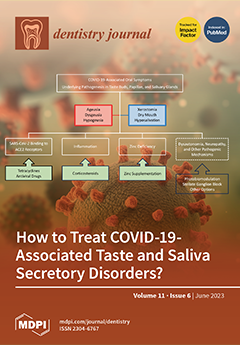
Cover Story (view full-size image): Treating taste and saliva secretory disorders associated with COVID-19 has become a critical issue since the worldwide spread of SARS-CoV-2 infection. Different treatment options are available on the basis of multiple modes of action on cellular entry and intracellular replication of cytopathic SARS-CoV-2; inflammation, zinc deficiency, dysautonomia, and neuropathy induced by SARS-CoV-2 infection; and gustatory and saliva secretory functions impaired by SARS-CoV-2. They include the use of repurposing tetracyclines, antiviral drugs, corticosteroids, zinc supplementation, photobiomodulation, stellate ganglion block, phytochemicals, acupuncture, etc., which are effectively applicable to COVID-19-associated ageusia, dysgeusia, or hypogeusia, and xerostomia, dry mouth, or hyposalivation. View this paper
  • Issues are regarded as officially published after their release is announced to the table of contents alert mailing list.
  • You may sign up for e-mail alerts to receive table of contents of newly released issues.
  • PDF is the official format for papers published in both, html and pdf forms. To view the papers in pdf format, click on the "PDF Full-text" link, and use the free Adobe Reader to open them.
Order results
Result details
Section
Select all
Export citation of selected articles as:
14 pages, 900 KiB  
Article
The Impact of the COVID-19 Pandemic on Pediatric Dentistry: Insights from an Italian Cross-Sectional Survey
by Giulio Conti, Francesca Amadori, Alessandra Bordanzi, Alessandra Majorana and Elena Bardellini
Dent. J. 2023, 11(6), 154; https://doi.org/10.3390/dj11060154 - 19 Jun 2023
Cited by 1 | Viewed by 1634
Abstract
Background. The COVID-19 pandemic has had a significant impact on pediatric dentistry and also dental education. This study aimed to assess the observed changes in children’s oral health by pediatric dentists and also served as an educational tool for dentistry students during the [...] Read more.
Background. The COVID-19 pandemic has had a significant impact on pediatric dentistry and also dental education. This study aimed to assess the observed changes in children’s oral health by pediatric dentists and also served as an educational tool for dentistry students during the pandemic. Methods. Postgraduate students in pediatric dentistry prepared a survey addressed to Italian pediatric dentists. Over 5476 dentists were invited to participate, and collaboration among the students took place through virtual meetings and electronic platforms. The online questionnaire was composed of 29 questions focusing on the management of pediatric patients during and after the lockdown period. A descriptive statistic was used for data analysis, and chi-square tests were performed (p < 0.05). Results. A total of 1752 pediatric dentists participated in the survey. During the lockdown, 68.3% of dentists exclusively handled dental emergencies. In the subsequent semester, a significant decrease in the number of pediatric treatments was reported. Pediatric dentists also noted a decline in children’s oral hygiene practices, a deterioration in dietary habits, and an increase in anxiety during dental procedures. Conclusions. This survey shed light on the diverse effects of the pandemic on children’s oral health and also provided valuable educational insights. Full article
(This article belongs to the Special Issue Oral Health Care in Paediatric Dentistry Volume 2)
Show Figures

Figure 1

9 pages, 2011 KiB  
Article
Regenerative and Protective Effects on Dental Tissues of a Fluoride–Silicon-Rich Toothpaste Associated with a Calcium Booster: An In Vitro Study
by Fabiano Vieira Vilhena, Simone dos Santos Grecco, Alejandra Hortencia Miranda González and Paulo Henrique Perlatti D’Alpino
Dent. J. 2023, 11(6), 153; https://doi.org/10.3390/dj11060153 - 14 Jun 2023
Cited by 4 | Viewed by 2313
Abstract
Calcium boosters have been used as a supplement for fluoride toothpastes to repair the dental tissues and reduce dentin permeability. This in vitro study aimed to characterize the regenerative and protective effects of the treatment of dental tissues with a fluoride–silicon-rich toothpaste associated [...] Read more.
Calcium boosters have been used as a supplement for fluoride toothpastes to repair the dental tissues and reduce dentin permeability. This in vitro study aimed to characterize the regenerative and protective effects of the treatment of dental tissues with a fluoride–silicon-rich toothpaste associated with a calcium booster. Bovine enamel and dentin blocks (n = 5) were obtained (4 × 4 × 6 mm). A fluoride–silicon-rich toothpaste and a calcium booster were used to brush the enamel and dentin both immediately and five days afterwards. The outcomes were then compared to those of the untreated control group. After that, the specimens were cross-sectioned. SEM was used to evaluate the micromorphology of the surface and cross-section. Energy-dispersive X-ray spectroscopy (EDS) was used to determine the elemental analyses (weight%). After treatment for 5 days with a booster/silicon-rich toothpaste, EDS analysis demonstrated that it induced a significant mineral change. It was also able to form a protective silicon-enriched mineral layer on both enamel and dentin surfaces. It was demonstrated in vitro that a fluoride–silicon-rich toothpaste associated with a calcium booster regenerates the dental tissues, remineralizing the enamel structure and occluding the dentin tubules. Full article
(This article belongs to the Special Issue Preventive Dentistry and Dental Public Health)
Show Figures

Figure 1

11 pages, 4668 KiB  
Article
A Novel Self-Assessment Method for Training Access Cavity on 3D Printed Endodontic Models
by Matteo Meglioli, Giovanni Mergoni, Francesco Artioli, Benedetta Ghezzi, Maddalena Manfredi, Guido Maria Macaluso and Simone Lumetti
Dent. J. 2023, 11(6), 152; https://doi.org/10.3390/dj11060152 - 13 Jun 2023
Cited by 1 | Viewed by 2149
Abstract
Background: New technologies can facilitate the transition from pre-clinical to clinical settings. We investigate students’ satisfaction with a novel learning method adopted in access cavity exercises. Methods: Students performed their access cavity on inexpensive, in-house 3D printed teeth. Their performances were evaluated by [...] Read more.
Background: New technologies can facilitate the transition from pre-clinical to clinical settings. We investigate students’ satisfaction with a novel learning method adopted in access cavity exercises. Methods: Students performed their access cavity on inexpensive, in-house 3D printed teeth. Their performances were evaluated by scanning the prepared teeth with an intraoral scanner and visualized using a mesh processing software. Then, the same software was used to align the tooth prepared by the student and the teacher’s one for self-assessment purposes. Students were asked to answer a questionnaire about their experiences with this new learning method. Results: From the teacher’s perspective, this novel learning approach was easy, straightforward and affordable. Overall, student feedback was positive: 73% found that access cavity assessment by scanning was more useful compared to a visual inspection under magnification and 57% reported that they had a better understanding of errors and mishaps. On the other hand, students pointed out that the material used to print teeth was too soft. Conclusion: The use of in-house 3D printed teeth in pre-clinical training is a simple way to overcome some of the drawbacks associated with extracted teeth, such as limited availability, variability, cross-infection control, and ethical constraints. The use of intraoral scanners and mesh processing software could improve student self-assessment. Full article
(This article belongs to the Special Issue Modern Endodontics)
Show Figures

Graphical abstract

22 pages, 3095 KiB  
Article
Characterization of SHH, SOX3, WNT3A and WNT9B Proteins in Human Non-Syndromic Cleft Lip and Palate Tissue
by Mārtiņš Vaivads, Ilze Akota and Māra Pilmane
Dent. J. 2023, 11(6), 151; https://doi.org/10.3390/dj11060151 - 9 Jun 2023
Cited by 2 | Viewed by 2849
Abstract
Orofacial clefts have been associated with specific cleft candidate genes which encode regulatory proteins required for orofacial region development. Cleft candidate genes encode proteins involved with the cleft morphopathogenesis process, but their exact interactions and roles are relatively unclear in human cleft tissue. [...] Read more.
Orofacial clefts have been associated with specific cleft candidate genes which encode regulatory proteins required for orofacial region development. Cleft candidate genes encode proteins involved with the cleft morphopathogenesis process, but their exact interactions and roles are relatively unclear in human cleft tissue. This study evaluates the presence and correlations of Sonic Hedgehog (SHH), SRY-Box Transcription Factor 3 (SOX3), Wingless-type Family Member 3A (WNT3A) and 9B (WNT9B) protein containing cells in different cleft tissue. Non-syndromic cleft-affected tissue was subdivided into three groups—unilateral cleft lip (UCL) (n = 36), bilateral cleft lip (BCL) (n = 13), cleft palate (CP) (n = 26). Control tissue was obtained from five individuals. Immunohistochemistry was implemented. The semi-quantitative method was used. Non-parametric statistical methods were applied. A significant decrease in SHH was found in BCL and CP tissue. SOX3, WNT3A and WNT9B had a significant decrease in all clefts. Statistically significant correlations were found. The significant decrease in SHH could be associated with BCL and CP pathogenesis. SOX3, WNT3A and WNT9B could have morphopathogenetic involvement in UCL, BCL, and CP. Similar correlations imply the presence of similar pathogenetic mechanisms in different cleft variations. Full article
Show Figures

Graphical abstract

20 pages, 2016 KiB  
Review
Dynamic Implant Surgery—An Accurate Alternative to Stereolithographic Guides—Systematic Review and Meta-Analysis
by Jordi Marques-Guasch, Anna Bofarull-Ballús, Maria Giralt-Hernando, Federico Hernández-Alfaro and Jordi Gargallo-Albiol
Dent. J. 2023, 11(6), 150; https://doi.org/10.3390/dj11060150 - 8 Jun 2023
Cited by 5 | Viewed by 3045
Abstract
(1) Background: Dynamic guided surgery is a computer-guided freehand technology that allows highly accurate procedures to be carried out in real time through motion-tracking instruments. The aim of this research was to compare the accuracy between dynamic guided surgery (DGS) and alternative implant [...] Read more.
(1) Background: Dynamic guided surgery is a computer-guided freehand technology that allows highly accurate procedures to be carried out in real time through motion-tracking instruments. The aim of this research was to compare the accuracy between dynamic guided surgery (DGS) and alternative implant guidance methods, namely, static guided surgery (SGS) and freehand (FH). (2) Methods: Searches were conducted in the Cochrane and Medline databases to identify randomized controlled clinical trials (RCTs) and prospective and retrospective case series and to answer the following focused question: “What implant guidance tool is more accurate and secure with regard to implant placement surgery?” The implant deviation coefficient was calculated for four different parameters: coronal and apical horizontal, angular, and vertical deviations. Statistical significance was set at a p-value of 0.05 following application of the eligibility criteria. (3) Results: Twenty-five publications were included in this systematic review. The results show a non-significant weighted mean difference (WMD) between the DGS and the SGS in all of the assessed parameters: coronal (n = 4 WMD = 0.02 mm; p = 0.903), angular (n = 4 WMD = −0.62°; p = 0.085), and apical (n = 3 WMD = 0.08 mm; p = 0.401). In terms of vertical deviation, not enough data were available for a meta-analysis. However, no significant differences were found among the techniques (p = 0.820). The WMD between DGS and FH demonstrated significant differences favoring DGS in three parameters as follows: coronal (n = 3 WMD = −0.66 mm; p =< 0.001), angular (n = 3 WMD = −3.52°; p < 0.001), and apical (n = 2 WMD = −0.73 mm; p =< 0.001). No WMD was observed regarding the vertical deviation analysis, but significant differences were seen among the different techniques (p = 0.038). (4) Conclusions: DGS is a valid alternative treatment achieving similar accuracy to SGS. DGS is also more accurate, secure, and precise than the FH method when transferring the presurgical virtual implant plan to the patient. Full article
Show Figures

Graphical abstract

11 pages, 835 KiB  
Article
Evaluation of Antibacterial Activity of a Bioactive Restorative Material Versus a Glass-Ionomer Cement on Streptococcus Mutans: In-Vitro Study
by Giulio Conti, Federica Veneri, Francesca Amadori, Alba Garzoni, Alessandra Majorana and Elena Bardellini
Dent. J. 2023, 11(6), 149; https://doi.org/10.3390/dj11060149 - 8 Jun 2023
Cited by 5 | Viewed by 2710
Abstract
Background: Dental caries management consists of both preventive and restorative approaches. Pediatric dentists can rely on many techniques and materials to restore decayed teeth, but a high failure rate is still observed, mainly due to secondary caries. New restorative bioactive materials combine the [...] Read more.
Background: Dental caries management consists of both preventive and restorative approaches. Pediatric dentists can rely on many techniques and materials to restore decayed teeth, but a high failure rate is still observed, mainly due to secondary caries. New restorative bioactive materials combine the mechanical and aesthetic characteristics of resinous materials with the capability to remineralize and the antimicrobial properties of glass ionomers, thus counteracting the occurrence of secondary caries. The aim of this study was to assess the antimicrobial activity against Streptococcus mutans of a bioactive restorative material (ACTIVA™ BioActive-Restorative™-Pulpdent©) and a glass ionomer cement with silver particles added (Ketac™ Silver—3M©), using agar diffusion assay. Methods: Each material was formed into disks of 4 mm in diameter, and four discs of each material were placed on nine agar plates. The analysis was repeated seven times. Results: Both materials showed statistically significant growth inhibition properties against S. mutans (p < 0.05). The difference in the effectiveness of the two materials was not statistically significant. Conclusion: Both ACTIVA™ and Ketac™ Silver can be recommended since both are similarly effective against S. mutans. However ACTIVA™, given its bioactivity and better aesthetics and mechanical properties compared to GICs, may provide better clinical performance. Full article
(This article belongs to the Special Issue Preventive Dentistry and Dental Public Health)
Show Figures

Figure 1

13 pages, 3072 KiB  
Article
SEM Evaluation of Thermal Effects Produced by a 445 nm Laser on Implant Surfaces
by Daniele Pergolini, Gaspare Palaia, Riccardo De Angelis, Federica Rocchetti, Gian Marco Podda, Gianluca Tenore, Alessandro Del Vecchio, Michela Relucenti and Umberto Romeo
Dent. J. 2023, 11(6), 148; https://doi.org/10.3390/dj11060148 - 7 Jun 2023
Cited by 3 | Viewed by 1433
Abstract
The aim of this in vitro study was to evaluate thermal effects on implant surfaces using a 445 nm diode laser (Eltech K-Laser Srl, Treviso, Italy) with different power settings and irradiation modalities. Fifteen new implants (Straumann, Basel, Switzerland) were irradiated to evaluate [...] Read more.
The aim of this in vitro study was to evaluate thermal effects on implant surfaces using a 445 nm diode laser (Eltech K-Laser Srl, Treviso, Italy) with different power settings and irradiation modalities. Fifteen new implants (Straumann, Basel, Switzerland) were irradiated to evaluate surface alteration. Each implant was divided into two zones: the anterior and posterior areas. The anterior coronal areas were irradiated with a distance of 1 mm between the optical fiber and the implant; the anterior apical ones were irradiated with the fiber in contact with the implant. Instead, the posterior surfaces of all of the implants were not irradiated and used as control surfaces. The protocol comprised two cycles of laser irradiation, lasting 30 s each, with a one-minute pause between them. Different power settings were tested: a 0.5 W pulsed beam (T-on 25 ms; T-off 25 ms), a 2 W continuous beam and a 3 W continuous beam. Lastly, through a scanning electron microscopy (SEM) analysis, dental implants’ surfaces were evaluated to investigate surface alterations. No surface alterations were detected using a 0.5 W laser beam with a pulsed mode at a distance of 1 mm. Using powers of irradiation of 2 W and 3 W with a continuous mode at 1 mm from the implant caused damage on the titanium surfaces. After the irradiation protocol was changed to using the fiber in contact with the implant, the surface alterations increased highly compared to the non-contact irradiation modality. The SEM results suggest that a power of irradiation of 0.5 W with a pulsed laser light emission mode, using an inactivated optical fiber placed 1 mm away from the implant, could be used in the treatment of peri-implantitis, since no implant surface alterations were detected. Full article
Show Figures

Figure 1

17 pages, 554 KiB  
Article
Oral Health-Related Quality of Life and Mental Health Impairment in Patients Affected by Medication-Related Osteonecrosis of the Jaws: A Case–Control Pilot Study
by Elena Calabria, Alessandro Antonelli, Selene Barone, Daniela Adamo, Marianna Salviati, Maria Giulia Cerra, Francesco Bennardo and Amerigo Giudice
Dent. J. 2023, 11(6), 147; https://doi.org/10.3390/dj11060147 - 7 Jun 2023
Cited by 4 | Viewed by 1694
Abstract
In the present case–control study, the impact of medication-related osteonecrosis of the jaws (MRONJ) on patients’ oral health-related quality of life (OHRQoL), overall quality of life (QoL), and psychological status was evaluated using a set of questionnaires. These questionnaires included the Oral Health [...] Read more.
In the present case–control study, the impact of medication-related osteonecrosis of the jaws (MRONJ) on patients’ oral health-related quality of life (OHRQoL), overall quality of life (QoL), and psychological status was evaluated using a set of questionnaires. These questionnaires included the Oral Health Impact Profile-14 (OHIP-14), the Short Form 36 Health Survey Questionnaire (SF-36), and the hospital anxiety and depression scale (HADS). A total of 25 MRONJ patients and 25 controls were included in the study. The results showed that MRONJ patients had a significantly poorer OHRQoL (OHIP-14 score p-value: 0.003) and lower general QoL, particularly in the domains of “physical functioning”, “physical role”, “body pain”, “general health”, and “vitality” in the SF-36 questionnaire (p-values: 0.001, 0.001, 0.013, 0.001, and 0.020). Although there were no significant differences between the groups in the SF-36 domains of “social functioning”, “emotional role”, and “mental health”, the mean sub-scores of the HADS, specifically the depression and anxiety scores (HADS-D and HADS-A), were significantly higher in MRONJ patients (p-values: 0.002 and 0.009). However, the “mental health” domain of the SF-36 questionnaire showed a correlation with both HADS-A and HADS-D scores (p-values: 0.003 and 0.031). Therefore, a comprehensive clinical examination of MRONJ patients should include the assessment of OHRQoL, overall QoL, and psychological profile using different questionnaires. This approach aims to gather detailed information about patients’ physical and psychological well-being, enabling the development of tailored treatments. Full article
(This article belongs to the Special Issue Dentistry Journal: 10th Anniversary)
Show Figures

Figure 1

20 pages, 1376 KiB  
Review
Do Systemic Diseases and Medications Influence Dental Implant Osseointegration and Dental Implant Health? An Umbrella Review
by Francesco D’Ambrosio, Alessandra Amato, Andrea Chiacchio, Laura Sisalli and Francesco Giordano
Dent. J. 2023, 11(6), 146; https://doi.org/10.3390/dj11060146 - 5 Jun 2023
Cited by 20 | Viewed by 5636
Abstract
The aim of this umbrella review is to evaluate what are the most common medications and systemic diseases that can affect bone–implant integration, the success rate and survival rate of dental implants, peri-implant tissue health, and implant loss. Systematic reviews, with meta-analysis or [...] Read more.
The aim of this umbrella review is to evaluate what are the most common medications and systemic diseases that can affect bone–implant integration, the success rate and survival rate of dental implants, peri-implant tissue health, and implant loss. Systematic reviews, with meta-analysis or not, about how systemic diseases and medications influence dental implant osseointegration, survival rate, success rate, and peri-implant diseases, published only in the English language, are electronically searched across the most important scientific databases. The present umbrella review includes eight systematic reviews, and osteoporosis and diabetes are the most investigated pathologies. Systemic diseases, such as neurologic disorders, HIV, hypothyroidism, cardiovascular diseases, and drugs, such as beta blockers, anti-hypertensives, or diuretics do not show a decreased rate of implant osseointegration. It seems that drugs, such as proton-pump inhibitors (PPIs) or serotonin reuptake inhibitors (SSRIs), negatively affect implant osseointegration. Few studies compare the effects of drugs and systemic diseases on the parameters considered in this overview. It is important to underline how the results of this review need to be validated with subsequent and more reviews. Full article
(This article belongs to the Special Issue Feature Review Papers in Dentistry)
Show Figures

Graphical abstract

11 pages, 927 KiB  
Protocol
The Caries-Arrest Effectiveness of Silver Diamine Fluoride Treatment with Different Post-Treatment Instructions in Preschool Children: A Study Protocol for a Randomized Controlled Trial
by Ivy Guofang Sun, Duangporn Duangthip, Edward Chin Man Lo and Chun Hung Chu
Dent. J. 2023, 11(6), 145; https://doi.org/10.3390/dj11060145 - 5 Jun 2023
Cited by 4 | Viewed by 2636
Abstract
In this 12-month randomized active-controlled clinical trial, we compare two post-treatment instructions for silver diamine fluoride (SDF) therapy in arresting dentine caries. The trial will include at least 254 kindergarten children with active dentine caries. The children will be randomized into two groups [...] Read more.
In this 12-month randomized active-controlled clinical trial, we compare two post-treatment instructions for silver diamine fluoride (SDF) therapy in arresting dentine caries. The trial will include at least 254 kindergarten children with active dentine caries. The children will be randomized into two groups and receive a 38% SDF solution applied topically to their carious lesions. Children in Group A will rinse immediately, whereas those in Group B will refrain from rinsing, eating, and drinking for 30 min. One trained examiner will perform the dental examination at baseline and every six months. The primary outcome measurement will be the proportion of caries lesions that become arrested at the 12-month examination. Potential confounding factors and parents’ satisfaction with SDF therapy at baseline and after 12 months will be collected using parental questionnaires. This trial will provide evidence-based information for clinical practitioners to give post-treatment instructions for SDF therapy. This study is registered at ClinicalTrial.gov (USA) (registration number: NCT05655286). Full article
(This article belongs to the Special Issue Oral Health Care in Paediatric Dentistry Volume 2)
Show Figures

Graphical abstract

14 pages, 615 KiB  
Review
The Use of Zirconia for Implant-Supported Fixed Complete Dental Prostheses: A Narrative Review
by Chiara Cinquini, Fortunato Alfonsi, Vincenzo Marchio, Francesco Gallo, Francesco Zingari, Alessandro Remigio Bolzoni, Stefano Romeggio and Antonio Barone
Dent. J. 2023, 11(6), 144; https://doi.org/10.3390/dj11060144 - 1 Jun 2023
Cited by 12 | Viewed by 4241
Abstract
The success of implant-supported fixed complete dental prostheses (ISFCDPs) depends on multiple factors: some are related to the fixtures, such as fixture material, surface characteristics, positioning, and type of connection to prosthetic components; others are related to the prostheses, such as design and [...] Read more.
The success of implant-supported fixed complete dental prostheses (ISFCDPs) depends on multiple factors: some are related to the fixtures, such as fixture material, surface characteristics, positioning, and type of connection to prosthetic components; others are related to the prostheses, such as design and materials used. Zirconia is a material widely used in fixed prosthodontics, whether on natural teeth or on implants, with excellent results over time. Regarding the use of zirconia for ISFCDPs, the 2018 ITI Consensus Report stated that “implant-supported monolithic zirconia prostheses may be a future option with more supporting evidence”. Since CAD/CAM technology and zirconia are being continuously innovated to achieve better results and performances over time, a narrative review of the literature seems necessary to focus research efforts towards effective and durable solutions for implant-supported, full-arch rehabilitations. The objective of the present narrative review was to search the literature for studies regarding the clinical performance of zirconia-based ISFCDPs. According to the results of this review, the use of zirconia for ISFCDPs showed good clinical outcomes, with high survival rates ranging from 88% to 100% and prosthetic complications that were restorable by the clinicians in most cases. Full article
(This article belongs to the Special Issue Oral Implantology and Rehabilitation)
Show Figures

Figure 1

21 pages, 951 KiB  
Systematic Review
Dental, Skeletal, and Soft Tissue Changes after Bone-Borne Surgically Assisted Rapid Maxillary Expansion: A Systematic Review and Meta-Analysis
by Christina Sekertzi, Maria-Marina Koukouviti, Athina Chatzigianni and Olga-Elpis Kolokitha
Dent. J. 2023, 11(6), 143; https://doi.org/10.3390/dj11060143 - 1 Jun 2023
Cited by 1 | Viewed by 1984
Abstract
(1) Background: For non-growing patients with marked transverse maxillary deficiency, bone-borne surgically assisted rapid maxillary expansion (SARME) has been proposed as an effective treatment option. Objective: To evaluate the dental, skeletal, and soft tissue changes following bone-borne SARME. (2) Methods: An unrestricted systematic [...] Read more.
(1) Background: For non-growing patients with marked transverse maxillary deficiency, bone-borne surgically assisted rapid maxillary expansion (SARME) has been proposed as an effective treatment option. Objective: To evaluate the dental, skeletal, and soft tissue changes following bone-borne SARME. (2) Methods: An unrestricted systematic electronic search of six databases, supplemented by manual searches, was performed up to April 2023. The eligibility criteria included prospective/retrospective clinical studies with outcomes pertaining to objective measurements of dental/skeletal/soft tissue effects of bone-borne SARME in healthy patients. (3) Results: Overall, 27 studies satisfied the inclusion criteria. The risk of bias of the non-randomized trials ranged between moderate (20) and serious (4). For the two RCTs, there were some concerns of bias. Trials with outcomes measured at the same landmarks within the scope of the prespecified timeframe were deemed eligible for quantitative synthesis. Eventually, five trials were included in the meta-analysis. SARME was associated with a statistically significant lengthening of the dental arch perimeter immediately after expansion, along with a marginally significant decrease in palatal depth during the post-SARME retention period. Post-treatment SNA values exhibited no statistically significant change. (4) Conclusion: Current evidence indicates that bone-borne SARME constitutes an effective treatment option for adult patients with maxillary transverse deficiency. Further long-term randomized clinical trials with robust methodology, large sample sizes, and 3D evaluation of the outcomes are needed. Full article
(This article belongs to the Special Issue Feature Review Papers in Dentistry)
Show Figures

Figure 1

16 pages, 28168 KiB  
Article
Effect of Different Silane Coupling Agents on the Bond Strength between Hydrogen Peroxide-Etched Epoxy-Based- Fiber-Reinforced Post and Composite Resin Core
by Sarah Adwani, Emad Elsubeihi, Ahmad Zebari, May Aljanahi, Keyvan Moharamzadeh and Haitham Elbishari
Dent. J. 2023, 11(6), 142; https://doi.org/10.3390/dj11060142 - 29 May 2023
Cited by 1 | Viewed by 2168
Abstract
The aim of the study was to evaluate the effect of various silane coupling agents on the micro-push-out bond strength between a hydrogen peroxide-etched epoxy-based fiber-reinforced post and composite resin core. Seventy-five cross-linked epoxy-based fiber-reinforced posts were etched with 24% hydrogen peroxide for [...] Read more.
The aim of the study was to evaluate the effect of various silane coupling agents on the micro-push-out bond strength between a hydrogen peroxide-etched epoxy-based fiber-reinforced post and composite resin core. Seventy-five cross-linked epoxy-based fiber-reinforced posts were etched with 24% hydrogen peroxide for 10 min. Then they were divided into five groups according to various silane coupling agents and bonded to a composite core. A Universal Testing Machine was utilized to evaluate the push-out bond strength. In addition, all groups’ modes of failure were assessed. The push-out bond strength data in MPa were analyzed using ANOVA and a Tukey HSD post hoc test to reveal any difference between the groups. Results revealed that the application of a two-bottle silane coupling agent exhibited the highest bond strength, while the application of a one-bottle silane coupling agent demonstrated the lowest bond strength for a hydrogen peroxide-etched fiber post bonded to a composite core material, which was statistically significant (p < 0.05). The strongest association with the highest bond strength was found with the two-bottle silane coupling agent when compared to the one-bottle. The study highlighted that the application of a silane-coupling agent may affect the bond strength between composite and epoxy-based fiber-reinforced posts. Full article
(This article belongs to the Special Issue Dental Materials Design and Innovative Treatment Approach)
Show Figures

Figure 1

12 pages, 283 KiB  
Article
Association between Malnutrition and Dental Caries in Iraqi Kurdish Children
by Mohammed Khalid Mahmood, Romain Lan, Herve Tassery and Delphine Tardivo
Dent. J. 2023, 11(6), 141; https://doi.org/10.3390/dj11060141 - 26 May 2023
Cited by 2 | Viewed by 2299
Abstract
Background: This paper aimed to study the association of serum Vitamin D level and Body Mass Index (BMI) as representatives of malnutrition at micro and macro levels, respectively, on dental caries. Method and Participants: A total of 333 randomly selected children aged 6–12 [...] Read more.
Background: This paper aimed to study the association of serum Vitamin D level and Body Mass Index (BMI) as representatives of malnutrition at micro and macro levels, respectively, on dental caries. Method and Participants: A total of 333 randomly selected children aged 6–12 years in Sulaimani, Kurdistan, Iraq were examined for three variables of the Decayed Missed Filled Tooth (DMFT) index, BMI, and Vitamin D serum levels in a single-time cross-sectional snapshot. Results: The majority of the population studied (70%) were Vitamin D deficient. In the linear regression analysis, both Vitamin D and BMI had insignificant effects on DMFT, with p-values of 0.22 and 0.55, respectively. After the categorization of the data, the risk estimate between normal (≥20 ng/mL) and deficient (<20 ng/mL) Vitamin D groups with regards to the caries and caries-free categories was 1.97 (95% CI: 0.91–4.24). According to the mean and median of the DMFT, which were both 4, the sample is categorized into the low-caries group (DMFT < 4) and high-caries group (DMFT > 4). When these groups are compared to Vitamin D levels and taking 20 and 15 as thresholds, the odds ratios were 1.19 (CI: 0.74–1.92) and 1.88 (CI: 1.20–2.94), respectively. Regarding the BMI, a normal BMI increases the chances of having a lower caries index (OR = 1.83, CI: 1.10–3.03). Conclusions: Our results show that having a serum Vitamin D level ≥15 ng/mL and having a normal BMI are associated with a lower caries index in children. Full article
(This article belongs to the Special Issue Updates and Highlights in Cariology)
29 pages, 422 KiB  
Review
Treatments of COVID-19-Associated Taste and Saliva Secretory Disorders
by Hironori Tsuchiya
Dent. J. 2023, 11(6), 140; https://doi.org/10.3390/dj11060140 - 25 May 2023
Cited by 4 | Viewed by 4748
Abstract
Since the worldwide spread of severe acute respiratory syndrome coronavirus 2 (SARS-CoV-2) infection, treating taste and saliva secretory disorders associated with coronavirus disease 2019 (COVID-19) has become a critical issue. The aim of the present study was to update information on treatments applicable [...] Read more.
Since the worldwide spread of severe acute respiratory syndrome coronavirus 2 (SARS-CoV-2) infection, treating taste and saliva secretory disorders associated with coronavirus disease 2019 (COVID-19) has become a critical issue. The aim of the present study was to update information on treatments applicable to such oral symptoms and discuss their pathogenic mechanisms. The literature search indicated that different treatments using tetracycline, corticosteroids, zinc, stellate ganglion block, phytochemical curcumin, traditional herbal medicine, nutraceutical vitamin D, photobiomodulation, antiviral drugs, malic acid sialagogue, chewing gum, acupuncture, and/or moxibustion have potential effects on COVID-19-associated ageusia/dysgeusia/hypogeusia and xerostomia/dry mouth/hyposalivation. These treatments have multiple modes of action on viral cellular entry and replication, cell proliferation and differentiation, immunity, and/or SARS-CoV-2 infection-induced pathological conditions such as inflammation, cytokine storm, pyroptosis, neuropathy, zinc dyshomeostasis, and dysautonomia. An understanding of currently available treatment options is required for dental professionals because they may treat patients who were infected with SARS-CoV-2 or who recovered from COVID-19, and become aware of their abnormal taste and salivary secretion. By doing so, dentists and dental hygienists could play a crucial role in managing COVID-19 oral symptoms and contribute to improving the oral health-related quality of life of the relevant patients. Full article
(This article belongs to the Special Issue Feature Review Papers in Dentistry)
Previous Issue
Next Issue
Back to TopTop