Next Issue
Volume 7, March
Previous Issue
Volume 7, January


J. Fungi, Volume 7, Issue 2 (February 2021) – 92 articles

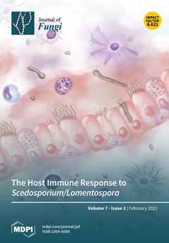
Cover Story (view full-size image): This review explains in a simplified way the information published in recent years on the immune response to the emerging fungi Scedosporium/Lomentospora. Fungal cell wall components, including peptidorhamnomannans, α-glucans and glucosylceramides, are involved in immune response activation following their recognition by TLR2, TLR4 and Dectin-1. After that, cytokine synthesis and antifungal activity of different phagocytes and epithelial cells against these fungi are species-specific. Furthermore, to evade host immune response, these fungi use different evasion mechanisms, including inducing host non-protective response, masking fungal molecular patterns and destructing host defense proteins. However, many aspects remain to be elucidated, and more research is necessary to shed light on the immune response to these fungi. View this paper
  • Issues are regarded as officially published after their release is announced to the table of contents alert mailing list.
  • You may sign up for e-mail alerts to receive table of contents of newly released issues.
  • PDF is the official format for papers published in both, html and pdf forms. To view the papers in pdf format, click on the "PDF Full-text" link, and use the free Adobe Reader to open them.
Order results
Result details
Section
Select all
Export citation of selected articles as:
23 pages, 15367 KiB  
Article
Development of Nano-Antifungal Therapy for Systemic and Endemic Mycoses
by Jorge H. Martínez-Montelongo, Iliana E. Medina-Ramírez, Yolanda Romo-Lozano, Antonio González-Gutiérrez and Jorge E. Macías-Díaz
J. Fungi 2021, 7(2), 158; https://doi.org/10.3390/jof7020158 - 23 Feb 2021
Cited by 9 | Viewed by 3057
Abstract
Fungal mycoses have become an important health and environmental concern due to the numerous deleterious side effects on the well-being of plants and humans. Antifungal therapy is limited, expensive, and unspecific (causes toxic effects), thus, more efficient alternatives need to be developed. In [...] Read more.
Fungal mycoses have become an important health and environmental concern due to the numerous deleterious side effects on the well-being of plants and humans. Antifungal therapy is limited, expensive, and unspecific (causes toxic effects), thus, more efficient alternatives need to be developed. In this work, Copper (I) Iodide (CuI) nanomaterials (NMs) were synthesized and fully characterized, aiming to develop efficient antifungal agents. The bioactivity of CuI NMs was evaluated using Sporothrix schenckii and Candida albicans as model organisms. CuI NMs were prepared as powders and as colloidal suspensions by a two-step reaction: first, the CuI2 controlled precipitation, followed by hydrazine reduction. Biopolymers (Arabic gum and chitosan) were used as surfactants to control the size of the CuI materials and to enhance its antifungal activity. The materials (powders and colloids) were characterized by SEM-EDX and AFM. The materials exhibit a hierarchical 3D shell morphology composed of ordered nanostructures. Excellent antifungal activity is shown by the NMs against pathogenic fungal strains, due to the simultaneous and multiple mechanisms of the composites to combat fungi. The minimum inhibitory concentration (MIC) and minimum fungicidal concentration (MFC) of CuI-AG and CuI-Chitosan are below 50 μg/mL (with 5 h of exposition). Optical and Atomic Force Microscopy (AFM) analyses demonstrate the capability of the materials to disrupt biofilm formation. AFM also demonstrates the ability of the materials to adhere and penetrate fungal cells, followed by their lysis and death. Following the concept of safe by design, the biocompatibility of the materials was tested. The hemolytic activity of the materials was evaluated using red blood cells. Our results indicate that the materials show an excellent antifungal activity at lower doses of hemolytic disruption. Full article
(This article belongs to the Special Issue Fungal Nanotechnology)
Show Figures

Figure 1

26 pages, 918 KiB  
Review
Paracoccidioidomycosis Diagnosed in Europe—A Systematic Literature Review
by Gernot Wagner, Deddo Moertl, Anna Glechner, Verena Mayr, Irma Klerings, Casey Zachariah, Miriam Van den Nest, Gerald Gartlehner and Birgit Willinger
J. Fungi 2021, 7(2), 157; https://doi.org/10.3390/jof7020157 - 23 Feb 2021
Cited by 15 | Viewed by 3281
Abstract
Paracoccidioidomycosis is a systemic mycosis that is endemic in geographical regions of Central and South America. Cases that occur in nonendemic regions of the world are imported through migration and travel. Due to the limited number of cases in Europe, most physicians are [...] Read more.
Paracoccidioidomycosis is a systemic mycosis that is endemic in geographical regions of Central and South America. Cases that occur in nonendemic regions of the world are imported through migration and travel. Due to the limited number of cases in Europe, most physicians are not familiar with paracoccidioidomycosis and its close clinical and histopathological resemblance to other infectious and noninfectious disease. To increase awareness of this insidious mycosis, we conducted a systematic review to summarize the evidence on cases diagnosed and reported in Europe. We searched PubMed and Embase to identify cases of paracoccidioidomycosis diagnosed in European countries. In addition, we used Scopus for citation tracking and manually screened bibliographies of relevant articles. We conducted dual abstract and full-text screening of references yielded by our searches. To identify publications published prior to 1985, we used the previously published review by Ajello et al. Overall, we identified 83 cases of paracoccidioidomycosis diagnosed in 11 European countries, published in 68 articles. Age of patients ranged from 24 to 77 years; the majority were male. Time from leaving the endemic region and first occurrence of symptoms considerably varied. Our review illustrates the challenges of considering systemic mycosis in the differential diagnosis of people returning or immigrating to Europe from endemic areas. Travel history is important for diagnostic-workup, though it might be difficult to obtain due to possible long latency period of the disease. Full article
Show Figures

Figure 1

13 pages, 405 KiB  
Review
Invasive Fungal Infections after Anti-CD19 Chimeric Antigen Receptor-Modified T-Cell Therapy: State of the Evidence and Future Directions
by Will Garner, Palash Samanta and Ghady Haidar
J. Fungi 2021, 7(2), 156; https://doi.org/10.3390/jof7020156 - 23 Feb 2021
Cited by 30 | Viewed by 3326
Abstract
Studies describing invasive fungal infections (IFIs) after chimeric antigen receptor-modified T-cell (CAR-T-cell) therapy are limited. Although post-CAR-T-cell IFIs appear to be uncommon, they are associated with significant morbidity and mortality. Specific risk factors for IFIs in CAR-T-cell recipients have not been fully characterized [...] Read more.
Studies describing invasive fungal infections (IFIs) after chimeric antigen receptor-modified T-cell (CAR-T-cell) therapy are limited. Although post-CAR-T-cell IFIs appear to be uncommon, they are associated with significant morbidity and mortality. Specific risk factors for IFIs in CAR-T-cell recipients have not been fully characterized and are often extrapolated from variables contributing to IFIs in patients with other hematologic malignancies or those undergoing hematopoietic cell transplant. Optimal prophylaxis strategies, including the use of yeast versus mold-active azoles, also remain ill-defined. Further research should investigate key risk factors for IFIs and establish an evidence-based approach to antifungal prophylaxis in these patients in order to improve clinical outcomes. Full article
(This article belongs to the Special Issue Fungal Infections in Immunocompromised Hosts 2.0)
Show Figures

Figure 1

15 pages, 2114 KiB  
Article
Preliminary Examination of the Toxicity of Spalting Fungal Pigments: A Comparison between Extraction Methods
by Badria H. Almurshidi, R.C. Van Court, Sarath M. Vega Gutierrez, Stacey Harper, Bryan Harper and Seri C. Robinson
J. Fungi 2021, 7(2), 155; https://doi.org/10.3390/jof7020155 - 22 Feb 2021
Cited by 6 | Viewed by 2833
Abstract
Spalting fungal pigments have shown potential in technologies ranging from green energy generation to natural colorants. However, their unknown toxicity has been a barrier to industrial adoption. In order to gain an understanding of the safety of the pigments, zebrafish embryos were exposed [...] Read more.
Spalting fungal pigments have shown potential in technologies ranging from green energy generation to natural colorants. However, their unknown toxicity has been a barrier to industrial adoption. In order to gain an understanding of the safety of the pigments, zebrafish embryos were exposed to multiple forms of liquid media and solvent-extracted pigments with concentrations of purified pigment ranging from 0 to 50 mM from Chlorociboria aeruginosa, Chlorociboria aeruginascens, and Scytalidium cuboideum. Purified xylindein from Chlorociboria sp. did not show toxicity at any tested concentration, while the red pigment dramada from S. cuboideum was only associated with significant toxicity above 23.2 uM. However, liquid cultures and pigment extracted into dichloromethane (DCM) showed toxicity, suggesting the co-production of bioactive secondary metabolites. Future research on purification and the bioavailability of the red dramada pigment will be important to identify appropriate use; however, purified forms of the blue-green pigment xylindein are likely safe for use across industries. This opens the door to the adoption of green technologies based on these pigments, with potential to replace synthetic colorants and less stable natural pigments. Full article
(This article belongs to the Special Issue Fungal Pigments 2021)
Show Figures

Figure 1

7 pages, 449 KiB  
Article
Comparison of Two Commercially Available qPCR Kits for the Detection of Candida auris
by Janko Sattler, Janina Noster, Anne Brunke, Georg Plum, Pia Wiegel, Oliver Kurzai, Jacques F. Meis and Axel Hamprecht
J. Fungi 2021, 7(2), 154; https://doi.org/10.3390/jof7020154 - 22 Feb 2021
Cited by 15 | Viewed by 3915
Abstract
Candida auris is an emerging pathogen with resistance to many commonly used antifungal agents. Infections with C. auris require rapid and reliable detection methods to initiate successful medical treatment and contain hospital outbreaks. Conventional identification methods are prone to errors and can lead [...] Read more.
Candida auris is an emerging pathogen with resistance to many commonly used antifungal agents. Infections with C. auris require rapid and reliable detection methods to initiate successful medical treatment and contain hospital outbreaks. Conventional identification methods are prone to errors and can lead to misidentifications. PCR-based assays, in turn, can provide reliable results with low turnaround times. However, only limited data are available on the performance of commercially available assays for C. auris detection. In the present study, the two commercially available PCR assays AurisID (OLM, Newcastle Upon Tyne, UK) and Fungiplex Candida Auris RUO Real-Time PCR (Bruker, Bremen, Germany) were challenged with 29 C. auris isolates from all five clades and eight other Candida species as controls. AurisID reliably detected C. auris with a limit of detection (LoD) of 1 genome copies/reaction. However, false positive results were obtained with high DNA amounts of the closely related species C. haemulonii, C. duobushaemulonii and C. pseudohaemulonii. The Fungiplex Candida Auris RUO Real-Time PCR kit detected C. auris with an LoD of 9 copies/reaction. No false positive results were obtained with this assay. In addition, C. auris could also be detected in human blood samples spiked with pure fungal cultures by both kits. In summary, both kits could detect C. auris-DNA at low DNA concentrations but differed slightly in their limits of detection and specificity. Full article
(This article belongs to the Special Issue Candida auris 2.0)
Show Figures

Figure 1

17 pages, 1990 KiB  
Article
The Emericellipsins A–E from an Alkalophilic Fungus Emericellopsis alkalina Show Potent Activity against Multidrug-Resistant Pathogenic Fungi
by Anastasia E. Kuvarina, Irina A. Gavryushina, Alexander B. Kulko, Igor A. Ivanov, Eugene A. Rogozhin, Marina L. Georgieva and Vera S. Sadykova
J. Fungi 2021, 7(2), 153; https://doi.org/10.3390/jof7020153 - 21 Feb 2021
Cited by 24 | Viewed by 3085
Abstract
Novel antimicrobial peptides with antifungal and cytotoxic activity were derived from the alkalophilic fungus Emericellopsis alkalina VKPM F1428. We previously reported that this strain produced emericellipsin A (EmiA), which has strong antifungal and cytotoxic properties. Further analyses of the metabolites obtained under a [...] Read more.
Novel antimicrobial peptides with antifungal and cytotoxic activity were derived from the alkalophilic fungus Emericellopsis alkalina VKPM F1428. We previously reported that this strain produced emericellipsin A (EmiA), which has strong antifungal and cytotoxic properties. Further analyses of the metabolites obtained under a special alkaline medium resulted in the isolation of four new homologous (Emi B–E). In this work, we report the complete primary structure and detailed biological activity for the newly synthesized nonribosomal antimicrobial peptides called emericellipsins B–E. The inhibitory activity of themajor compound, EmiA, against drug-resistant pathogenic fungi was similar to that of amphotericin B (AmpB). At the same time, EmiA had no hemolytic activity towards human erythrocytes. In addition, EmiA demonstrated low cytotoxic activity towards the normal HPF line, but possessed cancer selectivity to the K-562 and HCT-116 cell lines. Emericillipsins from the alkalophilic fungus Emericellopsis alkaline are promising treatment alternatives to licensed antifungal drugs for invasive mycosis therapy, especially for multidrug-resistant aspergillosis and cryptococcosis. Full article
(This article belongs to the Special Issue Fungi for Biotechnological Application and Environmental Cleanup)
Show Figures

Figure 1

19 pages, 1076 KiB  
Review
Molecular Epidemiology of Aspergillus fumigatus in Chronic Pulmonary Aspergillosis Patients
by Mireille H. van der Torre, Hongwei Shen, Riina Rautemaa-Richardson, Malcolm D. Richardson and Lilyann Novak-Frazer
J. Fungi 2021, 7(2), 152; https://doi.org/10.3390/jof7020152 - 20 Feb 2021
Cited by 9 | Viewed by 3675
Abstract
Molecular fungal genotyping techniques developed and employed for epidemiological studies have understandably concentrated on establishing the genetic diversity of Aspergillus fumigatus in invasive aspergillosis due to its severity, the urgency for treatment, and the need to demonstrate possible sources. Some early studies suggested [...] Read more.
Molecular fungal genotyping techniques developed and employed for epidemiological studies have understandably concentrated on establishing the genetic diversity of Aspergillus fumigatus in invasive aspergillosis due to its severity, the urgency for treatment, and the need to demonstrate possible sources. Some early studies suggested that these strains were phenotypically, if not genotypically, different from others. However, with improved discrimination and evaluations, incorporating environmental as well as clinical isolates from other Aspergillus conditions (e.g., chronic pulmonary aspergillosis and cystic fibrosis), this premise is no longer upheld. Moreover, with the onset of increased global triazole resistance, there has been a concerted effort to incorporate resistance profiling into genotyping studies and the realisation that the wider population of non-immunocompromised aspergillosis patients are at risk. This review summarises the developments in molecular genotyping studies that incorporate resistance profiling with attention to chronic pulmonary aspergillosis and an example of our UK experience. Full article
(This article belongs to the Special Issue Chronic Pulmonary Aspergillosis)
Show Figures

Figure 1

14 pages, 339 KiB  
Review
Endemic and Other Dimorphic Mycoses in The Americas
by Shawn R. Lockhart, Mitsuru Toda, Kaitlin Benedict, Diego H. Caceres and Anastasia P. Litvintseva
J. Fungi 2021, 7(2), 151; https://doi.org/10.3390/jof7020151 - 20 Feb 2021
Cited by 36 | Viewed by 4657
Abstract
Endemic fungi are thermally dimorphic fungi that have a limited geographic range and can cause both primary disease and opportunistic infections. The Americas are home to more genera of endemic fungi than anywhere else on earth. These include Coccidioides, Histoplasma, Blastomyces [...] Read more.
Endemic fungi are thermally dimorphic fungi that have a limited geographic range and can cause both primary disease and opportunistic infections. The Americas are home to more genera of endemic fungi than anywhere else on earth. These include Coccidioides, Histoplasma, Blastomyces, Paracoccidioides, and Sporothrix. Endemic fungi are found across the Americas and the Caribbean, from Blastomyces gilchristi, which extends into the northeast corners of North America, to Histoplasma capsulatum, which occurs all the way down in the southern regions of South America and into the Caribbean Islands. Symptoms of endemic fungal infection, when present, mimic those of many other diseases and are often diagnosed only after initial treatment for a bacterial or viral disease has failed. Endemic fungi place a significant medical burden on the populations they affect, especially in immunocompromised individuals and in resource-limited settings. This review summarizes the ecology, geographical range, epidemiology, and disease forms of the endemic fungi found in the Americas. An emphasis is placed on new and proposed taxonomic changes, including the assignment of new species names in Histoplasma, Blastomyces, and Paracoccidioides. Full article
(This article belongs to the Special Issue Emerging and Endemic Mycoses)
11 pages, 1149 KiB  
Article
Transcriptome Profiles of Sporisorium reilianum during the Early Infection of Resistant and Susceptible Maize Isogenic Lines
by Boqi Zhang, Nan Zhang, Qianqian Zhang, Qianya Xu, Tao Zhong, Kaiyue Zhang and Mingliang Xu
J. Fungi 2021, 7(2), 150; https://doi.org/10.3390/jof7020150 - 19 Feb 2021
Cited by 7 | Viewed by 2536
Abstract
The biotrophic fungus Sporisorium reilianum causes destructive head smut disease in maize (Zea mays L.). To explore the pathogenicity arsenal of this fungus, we tracked its transcriptome changes during infection of the maize seedling mesocotyls of two near-isogenic lines, HZ4 and HZ4R, [...] Read more.
The biotrophic fungus Sporisorium reilianum causes destructive head smut disease in maize (Zea mays L.). To explore the pathogenicity arsenal of this fungus, we tracked its transcriptome changes during infection of the maize seedling mesocotyls of two near-isogenic lines, HZ4 and HZ4R, differing solely in the disease resistance gene ZmWAK. Parasitic growth of S. reilianum resulted in thousands of differentially expressed genes (DEGs) compared with growth in axenic culture. The protein synthesis and energy metabolism of S. reilianum were predominantly enriched with down-regulated DEGs, consistent with the arrested hyphal growth observed following colonization. Nutrition-related metabolic processes were enriched with both up- and down-regulated DEGs, which, together with activated transmembrane transport, reflected a potential transition in nutrition uptake of S. reilianum once it invaded maize. Notably, genes encoding secreted proteins of S. reilianum were mostly up-regulated during biotrophy. ZmWAK-mediated resistance to head smut disease reduced the number of DEGs of S. reilianum, particularly those related to the secretome. These observations deepen our understanding of the mechanisms underlying S. reilianum pathogenicity and ZmWAK-induced innate immunity. Full article
(This article belongs to the Special Issue Smut Fungi)
Show Figures

Figure 1

12 pages, 2190 KiB  
Article
Versatile CRISPR/Cas9 Systems for Genome Editing in Ustilago maydis
by Sarah-Maria Wege, Katharina Gejer, Fabienne Becker, Michael Bölker, Johannes Freitag and Björn Sandrock
J. Fungi 2021, 7(2), 149; https://doi.org/10.3390/jof7020149 - 18 Feb 2021
Cited by 14 | Viewed by 3334
Abstract
The phytopathogenic smut fungus Ustilago maydis is a versatile model organism to study plant pathology, fungal genetics, and molecular cell biology. Here, we report several strategies to manipulate the genome of U. maydis by the CRISPR/Cas9 technology. These include targeted gene deletion via [...] Read more.
The phytopathogenic smut fungus Ustilago maydis is a versatile model organism to study plant pathology, fungal genetics, and molecular cell biology. Here, we report several strategies to manipulate the genome of U. maydis by the CRISPR/Cas9 technology. These include targeted gene deletion via homologous recombination of short double-stranded oligonucleotides, introduction of point mutations, heterologous complementation at the genomic locus, and endogenous N-terminal tagging with the fluorescent protein mCherry. All applications are independent of a permanent selectable marker and only require transient expression of the endonuclease Cas9hf and sgRNA. The techniques presented here are likely to accelerate research in the U. maydis community but can also act as a template for genome editing in other important fungi. Full article
(This article belongs to the Special Issue Smut Fungi)
Show Figures

Figure 1

29 pages, 4434 KiB  
Article
Comparison of Plant Metabolites in Root Exudates of Lolium perenne Infected with Different Strains of the Fungal Endophyte Epichloë festucae var. lolii
by Aurora Patchett and Jonathan A. Newman
J. Fungi 2021, 7(2), 148; https://doi.org/10.3390/jof7020148 - 18 Feb 2021
Cited by 25 | Viewed by 3593
Abstract
Lolium perenne infected with the fungal endophyte Epichloë festucae var. lolii have specific, endophyte strain-dependent, chemical phenotypes in their above-ground tissues. Differences in these chemical phenotypes have been largely associated with classes of fungal-derived alkaloids which protect the plant against many insect pests. [...] Read more.
Lolium perenne infected with the fungal endophyte Epichloë festucae var. lolii have specific, endophyte strain-dependent, chemical phenotypes in their above-ground tissues. Differences in these chemical phenotypes have been largely associated with classes of fungal-derived alkaloids which protect the plant against many insect pests. However, the use of new methodologies, such as various omic techniques, has demonstrated that many other chemical changes occur in both primary and secondary metabolites. Few studies have investigated changes in plant metabolites exiting the plant in the form of root exudates. As root exudates play an essential role in the acquisition of nutrients, microbial associations, and defense in the below-ground environment, it is of interest to understand how plant root exudate chemistry is influenced by the presence of strains of a fungal endophyte. In this study, we tested the influence of four strains of E. festucae var. lolii (E+ (also known as Lp19), AR1, AR37, NEA2), and uninfected controls (E−), on L. perenne growth and the composition of root exudate metabolites. Root exudates present in the hydroponic water were assessed by untargeted metabolomics using Accurate-Mass Quadrupole Time-of-Flight (Q–TOF) liquid chromatography–mass spectrometry (LC–MS). The NEA2 endophyte strain resulted in the greatest plant biomass and the lowest endophyte concentration. We found 84 metabolites that were differentially expressed in at least one of the endophyte treatments compared to E− plants. Two compounds were strongly associated with one endophyte treatment, one in AR37 (m/z 135.0546 RT 1.17), and one in E+ (m/z 517.1987 RT 9.26). These results provide evidence for important changes in L. perenne physiology in the presence of different fungal endophyte strains. Further research should aim to connect changes in root exudate chemical composition with soil ecosystem processes. Full article
(This article belongs to the Special Issue Systems Biology in Fungal Research)
Show Figures

Figure 1

16 pages, 323 KiB  
Review
Pharmacological Potential of Fungal Endophytes Associated with Medicinal Plants: A Review
by Bartholomew Saanu Adeleke and Olubukola Oluranti Babalola
J. Fungi 2021, 7(2), 147; https://doi.org/10.3390/jof7020147 - 17 Feb 2021
Cited by 88 | Viewed by 8740
Abstract
Endophytic microbes are microorganisms that colonize the intracellular spaces within the plant tissues without exerting any adverse or pathological effects. Currently, the world population is facing devastating chronic diseases that affect humans. The resistance of pathogens to commercial antibiotics is increasing, thus limiting [...] Read more.
Endophytic microbes are microorganisms that colonize the intracellular spaces within the plant tissues without exerting any adverse or pathological effects. Currently, the world population is facing devastating chronic diseases that affect humans. The resistance of pathogens to commercial antibiotics is increasing, thus limiting the therapeutic potential and effectiveness of antibiotics. Consequently, the need to search for novel, affordable and nontoxic natural bioactive compounds from endophytic fungi in developing new drugs with multifunction mechanisms to meet human needs is essential. Fungal endophytes produce invaluable bioactive metabolic compounds beneficial to humans with antimicrobial, anticancer, antidiabetic, anti-inflammatory, antitumor properties, etc. Some of these bioactive compounds include pestacin, taxol, camptothecin, ergoflavin, podophyllotoxin, benzopyran, isopestacin, phloroglucinol, tetrahydroxy-1-methylxanthone, salidroside, borneol, dibenzofurane, methyl peniphenone, lipopeptide, peniphenone etc. Despite the aforementioned importance of endophytic fungal metabolites, less information is available on their exploration and pharmacological importance. Therefore, in this review, we shall elucidate the fungal bioactive metabolites from medicinal plants and their pharmacological potential. Full article
14 pages, 12522 KiB  
Article
Characterization of Kazachstania slooffiae, a Proposed Commensal in the Porcine Gut
by Katie Lynn Summers, Juli Foster Frey and Ann M. Arfken
J. Fungi 2021, 7(2), 146; https://doi.org/10.3390/jof7020146 - 17 Feb 2021
Cited by 14 | Viewed by 3991
Abstract
Kazachstania slooffiae is a fungus commonly isolated from the gastrointestinal tract and feces of post-weaning pigs. Studies have implicated its ability to positively alter piglet gut health through potential symbioses with beneficial bacteria, including Lactobacillus and Prevotella, in providing amino acids as [...] Read more.
Kazachstania slooffiae is a fungus commonly isolated from the gastrointestinal tract and feces of post-weaning pigs. Studies have implicated its ability to positively alter piglet gut health through potential symbioses with beneficial bacteria, including Lactobacillus and Prevotella, in providing amino acids as an energy source for microbial and piglet growth, and it has been found to be positively correlated with short-chain fatty acids in the piglet gut. However, basic mycological information remains limited, hampering in vitro studies. In this study, we characterized the growth parameters, biofilm formation ability, susceptibility to antimicrobials, and genetic relatedness of K. slooffiae to other fungal isolates. Optimal fungal growth conditions were determined, no antifungal resistance was found against multiple classes of antifungal drugs (azoles, echinocandins, polyenes, or pyrimidine analogues), and dimorphic growth was observed. K. slooffiae produced biofilms that became more complex in the presence of Lactobacillus acidophilus supernatant, suggesting positive interactions with this bacterium in the gut, while Enterococcus faecalis supernatant decreased density, suggesting an antagonistic interaction. This study characterizes the in vitro growth conditions that are optimal for further studies of K. slooffiae, which is an important step in defining the role and interactions of K. slooffiae in the porcine gut environment. Full article
Show Figures

Figure 1

16 pages, 2638 KiB  
Article
Proteomics Reveals Octyl Gallate as an Environmentally Friendly Wood Preservative Leading to Reactive Oxygen Species-Driven Metabolic Inflexibility and Growth Inhibition in White-Rot Fungi (Lenzites betulina and Trametes versicolor)
by Jin-Wei Xu, Chen-Chung Liao, Ke-Chang Hung, Zhong-Yao Wang, Yu-Tang Tung and Jyh-Horng Wu
J. Fungi 2021, 7(2), 145; https://doi.org/10.3390/jof7020145 - 17 Feb 2021
Cited by 4 | Viewed by 2543
Abstract
The most commonly applied wood preservatives are based on creosote, pentachlorophenol, and waterborne chromate copper arsenate, which negatively affect the environment. Thus, environmentally friendly wood preservatives are required. This study investigated the antifungal activity and mechanism of several long-chain alkyl gallates (3,4,5-trihydroxybenzoates) against [...] Read more.
The most commonly applied wood preservatives are based on creosote, pentachlorophenol, and waterborne chromate copper arsenate, which negatively affect the environment. Thus, environmentally friendly wood preservatives are required. This study investigated the antifungal activity and mechanism of several long-chain alkyl gallates (3,4,5-trihydroxybenzoates) against white-rot fungi, Lenzites betulina and Trametes versicolor. The results revealed that octyl gallate (OG) had the best antifungal activity. Additionally, OG may have a mechanism of action similar to surfactants and inhibit ATPase activity, causing mitochondrial dysfunction and endogenous reactive oxygen species (ROS) production. Upon exposure to endogenous ROS, cells rapidly inhibit the synthesis of 60S ribosomal subunits, thus reducing the mycelial growth rate. L. betulina and T. versicolor also remodeled their energy metabolism in response to low ATP levels and endogenous ROS. After OG treatment, ATP citrate synthase activity was downregulated and glycolytic activity was upregulated in L. betulina. However, the activity of aerobic pathways was decreased and the oxidative branch of the pentose phosphate pathway was redirected form nicotinamide adenine dinucleotide phosphate (NADPH) to minimize endogenous ROS-mediated damage in T. versicolor. Taken together, these observations reveal that OG is a potent inhibitor of white-rot fungus. Further structural optimization research and pharmacological investigations are warranted. Full article
Show Figures

Figure 1

14 pages, 1855 KiB  
Article
Natural Killer Cell Line NK-92-Mediated Damage of Medically Important Fungi
by Stanislaw Schmidt, Marie Luckowitsch, Michael Hogardt and Thomas Lehrnbecher
J. Fungi 2021, 7(2), 144; https://doi.org/10.3390/jof7020144 - 17 Feb 2021
Cited by 6 | Viewed by 2939
Abstract
Invasive fungal disease (IFD) in hematopoietic stem cell transplantation is associated with high morbidity and mortality. As the antifungal host response determines risk and outcome of IFD, there is growing interest in adoptive immunotherapy using T cells or natural killer (NK) cells. Although [...] Read more.
Invasive fungal disease (IFD) in hematopoietic stem cell transplantation is associated with high morbidity and mortality. As the antifungal host response determines risk and outcome of IFD, there is growing interest in adoptive immunotherapy using T cells or natural killer (NK) cells. Although the NK-92 cell line has been tested as anticancer therapy in clinical trials, data on the antifungal activity of NK-92 cells are lacking. Here, we show that the NK-92 cell line exhibits considerable fungal damage on all medically important fungi tested, such as different species of Aspergillus, Candida, mucormycetes, and Fusarium. The extent of fungal damage differs across various species of mucormycetes and Fusarium, whereas it is comparable across different species of Aspergillus and Candida. Interferon (IFN)-γ levels in the supernatant were lower when NK-92 cells are co-incubated with Aspergillus fumigatus, Candida albicans, or Rhizopus arrhizus compared to the levels when NK-92 cells are incubated alone. Different to primary human NK cells, no increase of perforin levels in the supernatant was observed when the fungi were added to NK-92 cells. Our in vitro data demonstrated that the NK-92 cell line could be a feasible tool for antifungal immunotherapy, but data of animal models are warranted prior to clinical trials. Full article
(This article belongs to the Special Issue Antifungal Immunity and Fungal Vaccine Development)
Show Figures

Figure 1

15 pages, 2297 KiB  
Article
Characterization of a Yellow Laccase from Botrytis cinerea 241
by Ingrida Radveikienė, Regina Vidžiūnaitė, Rita Meškienė, Rolandas Meškys and Vida Časaitė
J. Fungi 2021, 7(2), 143; https://doi.org/10.3390/jof7020143 - 17 Feb 2021
Cited by 20 | Viewed by 3310
Abstract
Typical laccases have four copper atoms, which form three different copper centers, of which the T1 copper is responsible for the blue color of the enzyme and gives it a characteristic absorbance around 610 nm. Several laccases have unusual spectral properties and are [...] Read more.
Typical laccases have four copper atoms, which form three different copper centers, of which the T1 copper is responsible for the blue color of the enzyme and gives it a characteristic absorbance around 610 nm. Several laccases have unusual spectral properties and are referred to as yellow or white laccases. Only two yellow laccases from the Ascomycota phylum have been described previously, and only one amino acid sequence of those enzymes is available. A yellow laccase Bcl1 from Botrytis cinerea strain 241 has been identified, purified and characterized in this work. The enzyme appears to be a dimer with a molecular mass of 186 kDa. The gene encoding the Bcl1 protein has been cloned, and the sequence analysis shows that the yellow laccase Bcl1 is phylogenetically distinct from other known yellow laccases. In addition, a comparison of amino acid sequences, and 3D modeling shows that the Bcl1 laccase lacks a conservative tyrosine, which is responsible for absorption quenching at 610 nm in another yellow asco-laccase from Sclerotinia sclerotiorum. High thermostability, high salt tolerance, broad substrate specificity, and the ability to decolorize dyes without the mediators suggest that the Bcl1 laccase is a potential enzyme for various industrial applications. Full article
(This article belongs to the Section Fungal Genomics, Genetics and Molecular Biology)
Show Figures

Figure 1

14 pages, 1278 KiB  
Article
Beauveria bassiana Endophytic Strain as Plant Growth Promoter: The Case of the Grape Vine Vitis vinifera
by Spiridon Mantzoukas, Ioannis Lagogiannis, Dionusia Mpousia, Aristeidis Ntoukas, Katerina Karmakolia, Panagiotis A. Eliopoulos and Konstantinos Poulas
J. Fungi 2021, 7(2), 142; https://doi.org/10.3390/jof7020142 - 16 Feb 2021
Cited by 27 | Viewed by 4265
Abstract
The common grape vine, Vitis vinifera, is a widely known plant with commercial and pharmacological value. The plant hosts a variety of microorganisms known as endophytes that can live within the tissues of the plant for a considerable time period, or even [...] Read more.
The common grape vine, Vitis vinifera, is a widely known plant with commercial and pharmacological value. The plant hosts a variety of microorganisms known as endophytes that can live within the tissues of the plant for a considerable time period, or even their whole life cycle. The fungus Beauveria bassiana is a well-studied endophyte which can colonize a variety of plants in many ways and in different parts of the plant. In this study, we examined the effect of the endophytic fungus B. bassiana on the growth of V. vinifera. The results demonstrated not only a successful colonization of the endophyte, but also a noteworthy impact on the growth of the V. vinifera root without harming the plant in any way. The fungus was also re-isolated from the parts of the plant using inst bait method. Overall, the study demonstrates the capability of B. bassiana to colonize V. vinifera plants, adding to the already existing knowledge of its endophytic activity, and highlighting its beneficial impact on the root growth. Full article
(This article belongs to the Special Issue Fungal Endophytes in Agriculture and Ecosystems)
Show Figures

Figure 1

5 pages, 1648 KiB  
Communication
Does Structural Color Exist in True Fungi?
by Juliet Brodie, Colin J. Ingham and Silvia Vignolini
J. Fungi 2021, 7(2), 141; https://doi.org/10.3390/jof7020141 - 16 Feb 2021
Cited by 6 | Viewed by 4404
Abstract
Structural color occurs by the interaction of light with regular structures and so generates colors by completely different optical mechanisms to dyes and pigments. Structural color is found throughout the tree of life but has not, to date, been reported in the fungi. [...] Read more.
Structural color occurs by the interaction of light with regular structures and so generates colors by completely different optical mechanisms to dyes and pigments. Structural color is found throughout the tree of life but has not, to date, been reported in the fungi. Here we give an overview of structural color across the tree of life and provide a brief guide aimed at stimulating the search for this phenomenon in fungi. Full article
(This article belongs to the Special Issue Fungal Pigments 2021)
Show Figures

Graphical abstract

11 pages, 1928 KiB  
Article
Molecular Epidemiological Investigation of a Nosocomial Cluster of C. auris: Evidence of Recent Emergence in Italy and Ease of Transmission during the COVID-19 Pandemic
by Vincenzo Di Pilato, Giulia Codda, Lorenzo Ball, Daniele Roberto Giacobbe, Edward Willison, Malgorzata Mikulska, Laura Magnasco, Francesca Crea, Antonio Vena, Paolo Pelosi, Matteo Bassetti and Anna Marchese
J. Fungi 2021, 7(2), 140; https://doi.org/10.3390/jof7020140 - 15 Feb 2021
Cited by 62 | Viewed by 5433
Abstract
Candida auris is an emerging MDR pathogen raising major concerns worldwide. In Italy, it was first and only identified in July 2019 in our hospital (San Martino Hospital, Genoa), where infection or colonization cases have been increasingly recognized during the following months. To [...] Read more.
Candida auris is an emerging MDR pathogen raising major concerns worldwide. In Italy, it was first and only identified in July 2019 in our hospital (San Martino Hospital, Genoa), where infection or colonization cases have been increasingly recognized during the following months. To gain insights into the introduction, transmission dynamics, and resistance traits of this fungal pathogen, consecutive C. auris isolates collected from July 2019 to May 2020 (n = 10) were subjected to whole-genome sequencing (WGS) and antifungal susceptibility testing (AST); patients’ clinical and trace data were also collected. WGS resolved all isolates within the genetic clade I (South Asian) and showed that all but one were part of a cluster likely stemming from the index case. Phylogenetic molecular clock analyses predicted a recent introduction (May 2019) in the hospital setting and suggested that most transmissions were associated with a ward converted to a COVID-19-dedicated ICU during the pandemic. All isolates were resistant to amphotericin B, voriconazole, and fluconazole at high-level, owing to mutations in ERG11(K143R) and TACB1(A640V). Present data demonstrated that the introduction of MDR C. auris in Italy was a recent event and suggested that its spread could have been facilitated by the COVID-19 pandemic. Continued efforts to implement stringent infection prevention and control strategies are warranted to limit the spread of this emerging pathogen within the healthcare system. Full article
(This article belongs to the Special Issue Candida auris 2.0)
Show Figures

Figure 1

22 pages, 3994 KiB  
Review
Fusarium as a Novel Fungus for the Synthesis of Nanoparticles: Mechanism and Applications
by Mahendra Rai, Shital Bonde, Patrycja Golinska, Joanna Trzcińska-Wencel, Aniket Gade, Kamel A. Abd-Elsalam, Sudhir Shende, Swapnil Gaikwad and Avinash P. Ingle
J. Fungi 2021, 7(2), 139; https://doi.org/10.3390/jof7020139 - 15 Feb 2021
Cited by 122 | Viewed by 8433
Abstract
Nanotechnology is a new and developing branch that has revolutionized the world by its applications in various fields including medicine and agriculture. In nanotechnology, nanoparticles play an important role in diagnostics, drug delivery, and therapy. The synthesis of nanoparticles by fungi is a [...] Read more.
Nanotechnology is a new and developing branch that has revolutionized the world by its applications in various fields including medicine and agriculture. In nanotechnology, nanoparticles play an important role in diagnostics, drug delivery, and therapy. The synthesis of nanoparticles by fungi is a novel, cost-effective and eco-friendly approach. Among fungi, Fusarium spp. play an important role in the synthesis of nanoparticles and can be considered as a nanofactory for the fabrication of nanoparticles. The synthesis of silver nanoparticles (AgNPs) from Fusarium, its mechanism and applications are discussed in this review. The synthesis of nanoparticles from Fusarium is the biogenic and green approach. Fusaria are found to be a versatile biological system with the ability to synthesize nanoparticles extracellularly. Different species of Fusaria have the potential to synthesise nanoparticles. Among these, F. oxysporum has demonstrated a high potential for the synthesis of AgNPs. It is hypothesised that NADH-dependent nitrate reductase enzyme secreted by F. oxysporum is responsible for the reduction of aqueous silver ions into AgNPs. The toxicity of nanoparticles depends upon the shape, size, surface charge, and the concentration used. The nanoparticles synthesised by different species of Fusaria can be used in medicine and agriculture. Full article
(This article belongs to the Special Issue Fungal Nanotechnology)
Show Figures

Figure 1

10 pages, 1988 KiB  
Article
Establishment of a Genetic Transformation System in Guanophilic Fungus Amphichorda guana
by Min Liang, Wei Li, Landa Qi, Guocan Chen, Lei Cai and Wen-Bing Yin
J. Fungi 2021, 7(2), 138; https://doi.org/10.3390/jof7020138 - 14 Feb 2021
Cited by 10 | Viewed by 2581
Abstract
Fungi from unique environments exhibit special physiological characters and plenty of bioactive natural products. However, the recalcitrant genetics or poor transformation efficiencies prevent scientists from systematically studying molecular biological mechanisms and exploiting their metabolites. In this study, we targeted a guanophilic fungus Amphichorda [...] Read more.
Fungi from unique environments exhibit special physiological characters and plenty of bioactive natural products. However, the recalcitrant genetics or poor transformation efficiencies prevent scientists from systematically studying molecular biological mechanisms and exploiting their metabolites. In this study, we targeted a guanophilic fungus Amphichorda guana LC5815 and developed a genetic transformation system. We firstly established an efficient protoplast preparing method by conditional optimization of sporulation and protoplast regeneration. The regeneration rate of the protoplast is up to about 34.6% with 0.8 M sucrose as the osmotic pressure stabilizer. To develop the genetic transformation, we used the polyethylene glycol-mediated protoplast transformation, and the testing gene AG04914 encoding a major facilitator superfamily transporter was deleted in strain LC5815, which proves the feasibility of this genetic manipulation system. Furthermore, a uridine/uracil auxotrophic strain was created by using a positive screening protocol with 5-fluoroorotic acid as a selective reagent. Finally, the genetic transformation system was successfully established in the guanophilic fungus strain LC5815, which lays the foundation for the molecular genetics research and will facilitate the exploitation of bioactive secondary metabolites in fungi. Full article
Show Figures

Figure 1

25 pages, 402 KiB  
Review
Paracoccidioidomycosis Protective Immunity
by Eva Burger
J. Fungi 2021, 7(2), 137; https://doi.org/10.3390/jof7020137 - 13 Feb 2021
Cited by 15 | Viewed by 3233
Abstract
Protective immunity against Paracoccidioides consists of a stepwise activation of numerous effector mechanisms that comprise many cellular and soluble components. At the initial phase of non-specific innate immunity, resistance against Paracoccidioides comes from phagocytic polymorphonuclear neutrophils, natural killer (NK) cells and monocytes, supplemented [...] Read more.
Protective immunity against Paracoccidioides consists of a stepwise activation of numerous effector mechanisms that comprise many cellular and soluble components. At the initial phase of non-specific innate immunity, resistance against Paracoccidioides comes from phagocytic polymorphonuclear neutrophils, natural killer (NK) cells and monocytes, supplemented by soluble factors such as cytokines and complement system components. Invariant receptors (Toll-like receptors (TLRs), Dectins) which are present in cells of the immune system, detect patterns present in Paracoccidioides (but not in the host) informing the hosts cells that there is an infection in progress, and that the acquired immunity must be activated. The role of components involved in the innate immunity of paracoccidioidomycosis is herein presented. Humoral immunity, represented by specific antibodies which control the fungi in the blood and body fluids, and its role in paracoccidioidomycosis (which was previously considered controversial) is also discussed. The protective mechanisms (involving various components) of cellular immunity are also discussed, covering topics such as: lysis by activated macrophages and cytotoxic T lymphocytes, the participation of lytic products, and the role of cytokines secreted by T helper lymphocytes in increasing the efficiency of Paracoccidioides, lysis. Full article
(This article belongs to the Special Issue Paracoccidioides and Paracoccidioidomycosis)
18 pages, 2773 KiB  
Article
Direct Visualization of Fungal Burden in Filamentous Fungus-Infected Silkworms
by Yidong Yu, Ann-Katrin Wolf, Sina Thusek, Thorsten Heinekamp, Michael Bromley, Sven Krappmann, Ulrich Terpitz, Kerstin Voigt, Axel A. Brakhage and Andreas Beilhack
J. Fungi 2021, 7(2), 136; https://doi.org/10.3390/jof7020136 - 13 Feb 2021
Cited by 10 | Viewed by 3925
Abstract
Invasive fungal infections (IFIs) are difficult to diagnose and to treat and, despite several available antifungal drugs, cause high mortality rates. In the past decades, the incidence of IFIs has continuously increased. More recently, SARS-CoV-2-associated lethal IFIs have been reported worldwide in critically [...] Read more.
Invasive fungal infections (IFIs) are difficult to diagnose and to treat and, despite several available antifungal drugs, cause high mortality rates. In the past decades, the incidence of IFIs has continuously increased. More recently, SARS-CoV-2-associated lethal IFIs have been reported worldwide in critically ill patients. Combating IFIs requires a more profound understanding of fungal pathogenicity to facilitate the development of novel antifungal strategies. Animal models are indispensable for studying fungal infections and to develop new antifungals. However, using mammalian animal models faces various hurdles including ethical issues and high costs, which makes large-scale infection experiments extremely challenging. To overcome these limitations, we optimized an invertebrate model and introduced a simple calcofluor white (CW) staining protocol to macroscopically and microscopically monitor disease progression in silkworms (Bombyx mori) infected with the human pathogenic filamentous fungi Aspergillus fumigatus and Lichtheimia corymbifera. This advanced silkworm A. fumigatus infection model could validate knockout mutants with either attenuated, strongly attenuated or unchanged virulence. Finally, CW staining allowed us to efficiently visualize antifungal treatment outcomes in infected silkworms. Conclusively, we here present a powerful animal model combined with a straightforward staining protocol to expedite large-scale in vivo research of fungal pathogenicity and to investigate novel antifungal candidates. Full article
(This article belongs to the Section Fungal Pathogenesis and Disease Control)
Show Figures

Figure 1

16 pages, 1292 KiB  
Article
Hungry for Sex: Differential Roles for Ustilago maydisb Locus Components in Haploid Cells vis à vis Nutritional Availability
by R. Margaret Wallen, Kirsten Richardson, Madison Furnish, Hector Mendoza, Allison Dentinger, Sunita Khanal and Michael H. Perlin
J. Fungi 2021, 7(2), 135; https://doi.org/10.3390/jof7020135 - 12 Feb 2021
Cited by 7 | Viewed by 3755
Abstract
Mating-types allow single-celled eukaryotic organisms to distinguish self from non-self in preparation for sexual reproduction. The components of mating-type loci provide initial self/non-self-recognition through pheromone and receptor interactions that control early cell fusion events. However, they may also provide a second level of [...] Read more.
Mating-types allow single-celled eukaryotic organisms to distinguish self from non-self in preparation for sexual reproduction. The components of mating-type loci provide initial self/non-self-recognition through pheromone and receptor interactions that control early cell fusion events. However, they may also provide a second level of scrutiny that requires differences in alleles leading to production of a transcription factor required for successful downstream developmental pathways after initial cell fusion. Interestingly, the protein subunits of these transcription factors have not been thoroughly examined for their roles, if any, in the haploid cells themselves. In Ustilago maydis, the causative agent of galls in maize plants, the b locus, encoding bEast (bE) and bWest (bW), components of the eventual requisite transcription factor, has been extensively studied for its role in formation of the stable dikaryon after mating and subsequent pathogenic program. Little is known, however, about any roles for bE or bW in haploid cells. Since mating in fungi is often induced under conditions of nitrogen starvation, we have explored connections between the b locus and the nitrogen-sensing and response pathways in U. maydis. We previously identified a connection in haploid cells between the b locus and Ump2, the high-affinity transceptor, a protein that both transports ammonium and triggers filamentous growth as a response to nitrogen starvation. Deletion of the entire b locus abrogates the filamentous response to low ammonium, a phenotype that is rescued by overexpression of Ump2. Here we further investigated the individual roles of bE and bW in haploid cells. We show that bE and bW are expressed differentially in haploid cells starved for ammonium. Their respective deletion elicits different effects on transcription of mating and pathogenic-related genes and, importantly, on the degree of pathogenic development in host plants. This is the first demonstration of a role for these mating locus components on haploid development and the first to demonstrate a connection to the ammonium transceptors. Full article
(This article belongs to the Special Issue Smut Fungi)
Show Figures

Figure 1

8 pages, 966 KiB  
Communication
Summary of Guidelines for Managing Histoplasmosis among People Living with HIV
by Freddy Perez, Diego H. Caceres, Nathan Ford, Giovanni Ravasi, Beatriz L. Gomez, Alessandro C. Pasqualotto, Paul Hine, Antoine A. Adenis, Mathieu Nacher, Tom Chiller, John Baddley and for the Guideline Development Group for diagnosing and managing disseminated histoplasmosis among people living with HIV
J. Fungi 2021, 7(2), 134; https://doi.org/10.3390/jof7020134 - 12 Feb 2021
Cited by 24 | Viewed by 5985
Abstract
Histoplasmosis is a frequent fungal opportunistic infection in people living with HIV (PLHIV), associated every year to a total of 5% to 15% of AIDS-related deaths among this population. In 2020, the first global guidelines for diagnosing and managing disseminated histoplasmosis among PLHIV [...] Read more.
Histoplasmosis is a frequent fungal opportunistic infection in people living with HIV (PLHIV), associated every year to a total of 5% to 15% of AIDS-related deaths among this population. In 2020, the first global guidelines for diagnosing and managing disseminated histoplasmosis among PLHIV was published. This document recommends (1) detection of circulating Histoplasma antigens as the recommended laboratory assay to diagnose histoplasmosis among PLHIV; (2) the use of liposomal amphotericin for induction therapy in severe or moderately severe disease, followed by a maintenance therapy with itraconazole for 12 months; a shorter maintenance therapy could be considered if the patient is clinically stable and if immune status has improved; (3) antiretroviral therapy initiation as soon as possible among patients with histoplasmosis without involvement of central nervous system; and (4) that for the treatment of co-infection with histoplasmosis and tuberculosis (TB), treatment of TB should be initiated according to the World Health Organization treatment guidelines. Appropriate health education of providers, supportive supervision, and policy guidance for the care of PLHIV are required. Full article
(This article belongs to the Special Issue Histoplasma and Histoplasmosis 2020)
Show Figures

Figure 1

22 pages, 4765 KiB  
Article
Base Excision Repair AP-Endonucleases-Like Genes Modulate DNA Damage Response and Virulence of the Human Pathogen Cryptococcus neoformans
by Rayssa Karla de Medeiros Oliveira, Fabián Andrés Hurtado, Pedro Henrique Gomes, Luiza Lassi Puglia, Fernanda Fonsêca Ferreira, Kunal Ranjan, Patrícia Albuquerque, Márcio José Poças-Fonseca, Ildinete Silva-Pereira and Larissa Fernandes
J. Fungi 2021, 7(2), 133; https://doi.org/10.3390/jof7020133 - 12 Feb 2021
Cited by 3 | Viewed by 2845
Abstract
Pathogenic microbes are exposed to a number of potential DNA-damaging stimuli during interaction with the host immune system. Microbial survival in this situation depends on a fine balance between the maintenance of DNA integrity and the adaptability provided by mutations. In this study, [...] Read more.
Pathogenic microbes are exposed to a number of potential DNA-damaging stimuli during interaction with the host immune system. Microbial survival in this situation depends on a fine balance between the maintenance of DNA integrity and the adaptability provided by mutations. In this study, we investigated the association of the DNA repair response with the virulence of Cryptococcus neoformans, a basidiomycete that causes life-threatening meningoencephalitis in immunocompromised individuals. We focused on the characterization of C. neoformansAPN1 and APN2 putative genes, aiming to evaluate a possible role of the predicted Apurinic/apyrimidinic (AP) endonucleases 1 and 2 of the base excision repair (BER) pathway on C. neoformans response to stress conditions and virulence. Our results demonstrated the involvement of the putative AP-endonucleases Apn1 and Apn2 in the cellular response to DNA damage induced by alkylation and by UV radiation, in melanin production, in tolerance to drugs and in virulence of C. neoformans in vivo. We also pointed out the potential use of DNA repair inhibitor methoxy-amine in combination with conventional antifungal drugs, for the development of new therapeutic approaches against this human fungal pathogen. This work provides new information about the DNA damage response of the highly important pathogenic fungus C. neoformans. Full article
(This article belongs to the Section Fungal Genomics, Genetics and Molecular Biology)
Show Figures

Figure 1

10 pages, 440 KiB  
Article
Evaluation of Three Commercial PCR Assays for the Detection of Azole-Resistant Aspergillus fumigatus from Respiratory Samples of Immunocompromised Patients
by Ulrike Scharmann, Lisa Kirchhoff, Andrea Hain, Jan Buer, Michael Koldehoff, Joerg Steinmann and Peter-Michael Rath
J. Fungi 2021, 7(2), 132; https://doi.org/10.3390/jof7020132 - 11 Feb 2021
Cited by 15 | Viewed by 2833
Abstract
This is the first study comparing three commercially available PCR assays for the detection of Aspergillus DNA from respiratory specimen of immunocompromised patients and the presence of cyp51A gene mutations. Bronchoalveolar lavages (BALs, N = 103) from patients with haematological/oncological underlying diseases were [...] Read more.
This is the first study comparing three commercially available PCR assays for the detection of Aspergillus DNA from respiratory specimen of immunocompromised patients and the presence of cyp51A gene mutations. Bronchoalveolar lavages (BALs, N = 103) from patients with haematological/oncological underlying diseases were retrospectively investigated. The performance of three PCR assays, namely MycoGENIE®Aspergillus fumigatus Real-Time PCR Kit (Adamtech), Fungiplex®Aspergillus Azole-R IVD Real-Time PCR Kit (Bruker Daltonik GmbH) and AsperGenius® (PathoNostics B.V.), were evaluated. All patients were categorised following current EORTC/MSG criteria, with exclusion of the PCR-results. From the 11 invasive pulmonary aspergillosis (IPA) probable samples, eight were detected with MycoGENIE®, resulting in a sensitivity of 80% and a specificity of 73%. Furthermore, Fungiplex® resulted in six positive BALs with a sensitivity of 60% and a specificity of 91% and AsperGenius® in seven positive BAL samples, with a sensitivity of 64% and a specificity of 97%. No proven IPA was detected. One isolate showed phenotypically an azole-resistance, which was also detected in each of the tested PCR assays with the mutation in TR34. The here tested PCR assays were capable of reliably detecting A. fumigatus DNA, as well as differentiation of the common cyp51A gene mutations. However, evaluation on the AsperGenius® assay revealed a low risk of false positive results. Full article
Show Figures

Figure 1

25 pages, 34291 KiB  
Article
Resolution of the Hypoxylon fuscum Complex (Hypoxylaceae, Xylariales) and Discovery and Biological Characterization of Two of Its Prominent Secondary Metabolites
by Christopher Lambert, Mohammad Javad Pourmoghaddam, Marjorie Cedeño-Sanchez, Frank Surup, Seyed Akbar Khodaparast, Irmgard Krisai-Greilhuber, Hermann Voglmayr, Theresia E. B. Stradal and Marc Stadler
J. Fungi 2021, 7(2), 131; https://doi.org/10.3390/jof7020131 - 11 Feb 2021
Cited by 24 | Viewed by 4505
Abstract
Hypoxylon, a large, cosmopolitan genus of Ascomycota is in the focus of our current poly-thetic taxonomic studies, and served as an excellent source for bioactive secondary metabolites at the same time. The present work concerns a survey of the Hypoxylon fuscum species complex [...] Read more.
Hypoxylon, a large, cosmopolitan genus of Ascomycota is in the focus of our current poly-thetic taxonomic studies, and served as an excellent source for bioactive secondary metabolites at the same time. The present work concerns a survey of the Hypoxylon fuscum species complex based on specimens from Iran and Europe by morphological studies and high performance liquid chromatography coupled to mass spectrometry and diode array detection (HPLC-MS-DAD). Apart from known chemotaxonomic markers like binaphthalene tetrol (BNT) and daldinin F, two unprece-dented molecules were detected and subsequently isolated to purity by semi preparative HPLC. Their structures were established by nuclear-magnetic resonance (NMR) spectroscopy as 3′-malonyl-daldinin F (6) and pseudofuscochalasin A (4). The new daldinin derivative 6 showed weak cytotoxicity towards mammalian cells but bactericidal activity. The new cytochalasin 4 was compared to cytochalasin C in an actin disruption assay using fluorescence microscopy of human osteo-sarcoma U2OS cells, revealing comparable activity towards F-actin but being irreversible compared to cytochalasin C. Concurrently, a multilocus molecular phylogeny based on ribosomal and proteinogenic nucleotide sequences of Hypoxylon species resulted in a well-supported clade for H. fuscum and its allies. From a comparison of morphological, chemotaxonomic and phylogenetic evidence, we introduce the new species H. eurasiaticum and H. pseudofuscum. Full article
Show Figures

Figure 1

9 pages, 387 KiB  
Review
Review of T-2307, an Investigational Agent That Causes Collapse of Fungal Mitochondrial Membrane Potential
by Nathan P. Wiederhold
J. Fungi 2021, 7(2), 130; https://doi.org/10.3390/jof7020130 - 11 Feb 2021
Cited by 18 | Viewed by 3369
Abstract
Invasive infections caused by Candida that are resistant to clinically available antifungals are of increasing concern. Increasing rates of fluconazole resistance in non-albicans Candida species have been documented in multiple countries on several continents. This situation has been further exacerbated over the [...] Read more.
Invasive infections caused by Candida that are resistant to clinically available antifungals are of increasing concern. Increasing rates of fluconazole resistance in non-albicans Candida species have been documented in multiple countries on several continents. This situation has been further exacerbated over the last several years by Candida auris, as isolates of this emerging pathogen that are often resistant to multiple antifungals. T-2307 is an aromatic diamidine currently in development for the treatment of invasive fungal infections. This agent has been shown to selectively cause the collapse of the mitochondrial membrane potential in yeasts when compared to mammalian cells. In vitro activity has been demonstrated against Candida species, including C. albicans, C. glabrata, and C. auris strains, which are resistant to azole and echinocandin antifungals. Activity has also been reported against Cryptococcus species, and this has translated into in vivo efficacy in experimental models of invasive candidiasis and cryptococcosis. However, little is known regarding the clinical efficacy and safety of this agent, as published data from studies involving humans are not currently available. Full article
(This article belongs to the Special Issue Mitochondria: Gene Lineage, Antifungal Targets and Pathogenesis)
Show Figures

Figure 1

19 pages, 6469 KiB  
Article
In Vitro and In Vivo Anti-Candida Activity and Structural Analysis of Killer Peptide (KP)-Derivatives
by Tecla Ciociola, Thelma A. Pertinhez, Tiziano De Simone, Walter Magliani, Elena Ferrari, Silvana Belletti, Tiziana D’Adda, Stefania Conti and Laura Giovati
J. Fungi 2021, 7(2), 129; https://doi.org/10.3390/jof7020129 - 10 Feb 2021
Cited by 9 | Viewed by 3513
Abstract
The previously described decapeptide AKVTMTCSAS (killer peptide, KP), derived from the variable region of a recombinant yeast killer toxin-like anti-idiotypic antibody, proved to exert a variety of antimicrobial, antiviral, and immunomodulatory activities. It also showed a peculiar self-assembly ability, likely responsible for the [...] Read more.
The previously described decapeptide AKVTMTCSAS (killer peptide, KP), derived from the variable region of a recombinant yeast killer toxin-like anti-idiotypic antibody, proved to exert a variety of antimicrobial, antiviral, and immunomodulatory activities. It also showed a peculiar self-assembly ability, likely responsible for the therapeutic effect in animal models of systemic and mucosal candidiasis. The present study analyzed the biological and structural properties of peptides derived from KP by substitution or deletion of the first residue, leaving unchanged the remaining amino acids. The investigated peptides proved to exert differential in vitro and/or in vivo anti-Candida activity without showing toxic effects on mammalian cells. The change of the first residue in KP amino acidic sequence affected the conformation of the resulting peptides in solution, as assessed by circular dichroism spectroscopy. KP-derivatives, except one, were able to induce apoptosis in yeast cells, like KP itself. ROS production and changes in mitochondrial transmembrane potential were also observed. Confocal and transmission electron microscopy studies allowed to establish that selected peptides could penetrate within C. albicans cells and cause gross morphological alterations. Overall, the physical and chemical properties of the first residue were found to be important for peptide conformation, candidacidal activity and possible mechanism of action. Small antimicrobial peptides could be exploited for the development of a new generation of antifungal drugs, given their relative low cost and ease of production as well as the possibility of devising novel delivery systems. Full article
(This article belongs to the Special Issue Antifungal Peptides 2020)
Show Figures

Graphical abstract

Previous Issue
Next Issue
Back to TopTop