The Biomarkers for the Diagnosis and Prognosis in Cancer (Closed)

A topical collection in Cancers (ISSN 2072-6694). This collection belongs to the section "Cancer Biomarkers".

Viewed by 157546

Editor


E-Mail Website
Collection Editor
Hillman Cancer Center, University of Pittsburgh, Pittsburg, PA 15219, USA
Interests: cancer biomarkers; ovarian and pancreatic cancers; pre-tumoral microenvironment

Topical Collection Information

Dear Colleagues,

This Topical Collection is dedicated to cancer biomarkers, with a special emphasis on biomarkers obtained from biological fluids, such as blood, urine, stool, saliva, etc., also known as a liquid biopsy. Liquid biopsy has several advantages over tissue biopsy: it is rapid, non-invasive, represents the tumor in its entirety, and allows for the longitudinal evaluation of cancer evolution. Cancer biomarkers offer a broad range of biological analytes and molecules (proteins, nucleic acids, metabolites), as well as protein modifications (glycosylation and methylation), circulating tumor cells, exosomes, tumor-educated platelets, etc.

Biomarkers have many potential applications in oncology, including estimating the risk of disease, screening for occult primary tumors, distinguishing benign from malignant disease, determining prognosis/prediction for cancer patients, and monitoring the status of the disease, either to detect recurrence or to determine the response or progression to therapy. Circulating biomarkers offer an opportunity for the understanding of tumorigenesis and metastasis, which could provide an insight into the evolution of the tumor dynamics during treatment and disease progression.

In this Topical Collection, we will highlight the current state and recent discoveries in the field of cancer biomarkers.

Prof. Dr. Anna E. Lokshin
Collection Editor

Manuscript Submission Information

Manuscripts should be submitted online at www.mdpi.com by registering and logging in to this website. Once you are registered, click here to go to the submission form. Manuscripts can be submitted until the deadline. All submissions that pass pre-check are peer-reviewed. Accepted papers will be published continuously in the journal (as soon as accepted) and will be listed together on the collection website. Research articles, review articles as well as communications are invited. For planned papers, a title and short abstract (about 100 words) can be sent to the Editorial Office for announcement on this website.

Submitted manuscripts should not have been published previously, nor be under consideration for publication elsewhere (except conference proceedings papers). All manuscripts are thoroughly refereed through a single-blind peer-review process. A guide for authors and other relevant information for submission of manuscripts is available on the Instructions for Authors page. Cancers is an international peer-reviewed open access semimonthly journal published by MDPI.

Please visit the Instructions for Authors page before submitting a manuscript. The Article Processing Charge (APC) for publication in this open access journal is 2900 CHF (Swiss Francs). Submitted papers should be well formatted and use good English. Authors may use MDPI's English editing service prior to publication or during author revisions.

Keywords

  • biomarkers
  • cancer
  • liquid biopsy
  • early detection
Published Papers (52 papers)

2024

Jump to: 2023, 2022, 2021, 2020

31 pages, 893 KiB  
Review
Protein Biomarkers of Gastric Preneoplasia and Cancer Lesions in Blood: A Comprehensive Review
by Thomas Bazin, Karine Nozeret, Catherine Julié, Dominique Lamarque and Eliette Touati
Cancers 2024, 16(17), 3019; https://doi.org/10.3390/cancers16173019 - 29 Aug 2024
Viewed by 932
Abstract
Gastric cancer (GC) is a major cause of cancer-related mortality worldwide. It is often associated with a bad prognosis because of its asymptomatic phenotype until advanced stages, highlighting the need for its prevention and early detection. GC development is preceded by the emergence [...] Read more.
Gastric cancer (GC) is a major cause of cancer-related mortality worldwide. It is often associated with a bad prognosis because of its asymptomatic phenotype until advanced stages, highlighting the need for its prevention and early detection. GC development is preceded by the emergence of gastric preneoplasia lesions (GPNLs), namely atrophic gastritis (AG), intestinal metaplasia (IM), and dysplasia (DYS). GC is currently diagnosed by endoscopy, which is invasive and costly and has limited effectiveness for the detection of GPNLs. Therefore, the discovery of non-invasive biomarkers in liquid biopsies, such as blood samples, in order to identify the presence of gastric preneoplasia and/or cancer lesions at asymptomatic stages is of paramount interest. This comprehensive review provides an overview of recently identified plasma/serum proteins and their diagnostic performance for the prediction of GPNLs and early cancer lesions. Autoantibodies appear to be promising biomarkers for AG, IM and early gastric cancer detection, along with inflammation and immunity-related proteins and antibodies against H. pylori virulence factors. There is a lack of specific protein biomarkers with which to detect DYS. Despite the need for further investigation and validation, some emerging candidates could pave the way for the development of reliable, non-invasive diagnostic tests for the detection and prevention of GC. Full article
Show Figures

Figure 1

2023

Jump to: 2024, 2022, 2021, 2020

11 pages, 1689 KiB  
Article
Immunogenic Biomarkers HMGB1 and sRAGE Are Potential Diagnostic Tools for Ovarian Malignancies
by Lars Schröder, Alexander B. A. Rupp, Kathrin M. E. Gihr, Makbule Kobilay, Christian M. Domroese, Michael R. Mallmann and Stefan Holdenrieder
Cancers 2023, 15(20), 5081; https://doi.org/10.3390/cancers15205081 - 20 Oct 2023
Viewed by 1193
Abstract
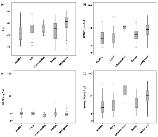
Background: High mobility group box 1 (HMGB1), soluble receptor of advanced glycation end products (sRAGE) and programmed cell death markers PD-1 and PD-L1 are immunogenic serum biomarkers that may serve as novel diagnostic tools for cancer diagnosis. Methods: We investigated the four markers [...] Read more.
Background: High mobility group box 1 (HMGB1), soluble receptor of advanced glycation end products (sRAGE) and programmed cell death markers PD-1 and PD-L1 are immunogenic serum biomarkers that may serve as novel diagnostic tools for cancer diagnosis. Methods: We investigated the four markers in sera of 231 women, among them 76 with ovarian cancer, 87 with benign diseases and 68 healthy controls, using enzyme immunoassays. Discrimination between groups was calculated using receiver operating characteristic (ROC) curves and sensitivities at fixed 90% and 95% specificities. Results: HMGB1 levels were significantly elevated and sRAGE levels were decreased in cancer patients as compared to benign and healthy controls. In consequence, the ratio of HMGB1 and sRAGE discriminated best between diagnostic groups. The areas under the curve (AUCs) of the ROC curves for differentiation of cancer vs. healthy were 0.77 for HMGB1, 0.65 for sRAGE and 0.78 for the HMGB1/sRAGE ratio, and slightly lower for the differentiation of cancer vs. benigns with 0.72 for HMGB1, 0.61 for sRAGE and 0.74 for the ratio of both. The highest sensitivities for cancer detection at 90% specificity versus benign diseases were achieved using HMGB1 with 41.3% and the HMGB1/sRAGE ratio with 39.2%, followed by sRAGE with 18.9%. PD-1 showed only minor and PD-L1 no power for discrimination between ovarian cancer and benign diseases. Conclusion: HMGB1 and sRAGE have differential diagnostic potential for ovarian cancer detection and warrant inclusion in further validation studies. Full article
Show Figures

Figure 1

9 pages, 868 KiB  
Article
Clinical Relevance of Red Blood Cell Distribution Width (RDW) in Endometrial Cancer: A Retrospective Single-Center Experience from Korea
by Kyung-Jin Eoh, Tae-Kyung Lee, Eun-Ji Nam, Sang-Wun Kim and Young-Tae Kim
Cancers 2023, 15(15), 3984; https://doi.org/10.3390/cancers15153984 - 5 Aug 2023
Cited by 6 | Viewed by 1542
Abstract
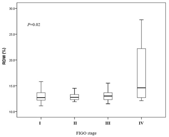
Background: Red blood cell distribution width (RDW) is a standard parameter of complete blood count and indicates the variability in red blood cell size. This study aimed to determine whether preoperative RDW can be used to predict the recurrence and prognosis of endometrial [...] Read more.
Background: Red blood cell distribution width (RDW) is a standard parameter of complete blood count and indicates the variability in red blood cell size. This study aimed to determine whether preoperative RDW can be used to predict the recurrence and prognosis of endometrial carcinoma. Methods: The medical records of 431 patients diagnosed with endometrial carcinoma were retrospectively reviewed between May 2006 and June 2018. In addition to RDW, the clinicopathological factors, survival curves, and prognoses of the patients with endometrial carcinoma were compared between the high (n = 213) and low (n = 218) groups according to the median RDW value (12.8%). Results: The patients with high RDW had significantly advanced-stage (p = 0.00) pelvic lymph node metastasis (p = 0.01) and recurrence (p = 0.01) compared to those in the low-RDW group. In univariate analysis with DFS as the endpoint, surgical stage, type II histology, grade, RDW, and lymph node metastasis were independently associated with survival. Patients with high RDW values had significantly shorter disease-free survival and overall survival than those with low RDW values (log-rank p = 0.03, log-rank p = 0.04, respectively). Conclusion: Our results demonstrate that RDW is a simple and convenient indicator of endometrial carcinoma recurrence. Prospective studies are needed to validate the findings of the current study. Full article
Show Figures

Figure 1

20 pages, 4200 KiB  
Article
Co-Expression of Immunohistochemical Markers MRP2, CXCR4, and PD-L1 in Gallbladder Tumors Is Associated with Prolonged Patient Survival
by Andrés Tittarelli, Omar Barría, Evy Sanders, Anna Bergqvist, Daniel Uribe Brange, Mabel Vidal, María Alejandra Gleisner, Jorge Ramón Vergara, Ignacio Niechi, Iván Flores, Cristián Pereda, Cristian Carrasco, Claudia Quezada-Monrás and Flavio Salazar-Onfray
Cancers 2023, 15(13), 3440; https://doi.org/10.3390/cancers15133440 - 30 Jun 2023
Cited by 2 | Viewed by 2237
Abstract
Gallbladder cancer (GBC) is a rare pathology in Western countries. However, it constitutes a relevant health problem in Asia and Latin America, with a high mortality in middle-aged Chilean women. The limited therapeutic options for GBC require the identification of targetable proteins with [...] Read more.
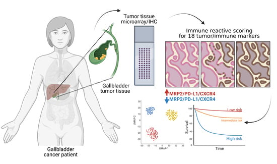
Gallbladder cancer (GBC) is a rare pathology in Western countries. However, it constitutes a relevant health problem in Asia and Latin America, with a high mortality in middle-aged Chilean women. The limited therapeutic options for GBC require the identification of targetable proteins with prognostic value for improving clinical management support. We evaluated the expression of targetable proteins, including three epithelial tumor markers, four proteins associated with multidrug and apoptosis resistance, and eleven immunological markers in 241 primary gallbladder adenocarcinomas. We investigated correlations between tumor marker expression, the primary tumor staging, and GBC patients’ survival using automated immunohistochemistry, a semi-automatic method for image analysis, univariate and multivariate statistical analyses, and machine learning algorithms. Our data show a significant association between the expression of MRP2 (p = 0.0028), CXCR4 (p = 0.0423), and PD-L1 (p = 0.0264), and a better prognosis for patients with late-stage primary tumors. The expression of the MRP2/CXCR4/PD-L1 cluster of markers discriminates among short-, medium-, and long-term patient survival, with an ROC of significant prognostic value (AUC = 0.85, p = 0.0012). Moreover, a high MRP2/CXCR4/PD-L1 co-expression is associated with increased survival time (30 vs. 6 months, p = 0.0025) in GBC patients, regardless of tumor stage. Hence, our results suggest that the MRP2/CXCR4/PD-L1 cluster could potentially be a prognostic marker for GBC. Full article
Show Figures

Graphical abstract

31 pages, 8010 KiB  
Article
In Vitro and In Silico Analysis of Epithelial-Mesenchymal Transition and Cancer Stemness as Prognostic Markers of Clear Cell Renal Cell Carcinoma
by Revati Sharma, Showan Balta, Ali Raza, Ruth M. Escalona, George Kannourakis, Prashanth Prithviraj and Nuzhat Ahmed
Cancers 2023, 15(9), 2586; https://doi.org/10.3390/cancers15092586 - 1 May 2023
Cited by 4 | Viewed by 2528
Abstract
The process of epithelial-mesenchymal transition (EMT) involves the phenotypic transformation of cells from epithelial to mesenchymal status. The cells exhibiting EMT contain features of cancer stem cells (CSC), and the dual processes are responsible for progressive cancers. Activation of hypoxia-inducible factors (HIF) is [...] Read more.
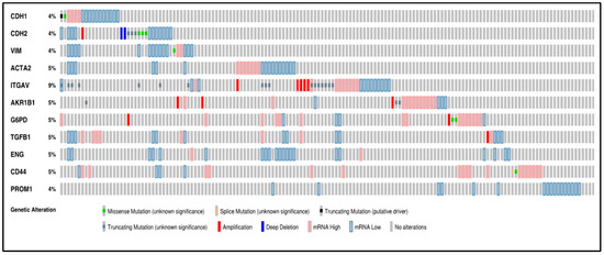
The process of epithelial-mesenchymal transition (EMT) involves the phenotypic transformation of cells from epithelial to mesenchymal status. The cells exhibiting EMT contain features of cancer stem cells (CSC), and the dual processes are responsible for progressive cancers. Activation of hypoxia-inducible factors (HIF) is fundamental to the pathogenesis of clear cell renal cell carcinoma (ccRCC), and their role in promoting EMT and CSCs is crucial for ccRCC tumour cell survival, disease progression, and metastatic spread. In this study, we explored the status of HIF genes and their downstream targets, EMT and CSC markers, by immunohistochemistry on in-house accrued ccRCC biopsies and adjacent non-tumorous tissues from patients undergoing partial or radical nephrectomy. In combination, we comprehensively analysed the expression of HIF genes and its downstream EMT and CSC-associated targets relevant to ccRCC by using publicly available datasets, the cancer genome atlas (TCGA) and the clinical proteome tumour analysis consortium (CPTAC). The aim was to search for novel biological prognostic markers that can stratify high-risk patients likely to experience metastatic disease. Using the above two approaches, we report the development of novel gene signatures that may help to identify patients at a high risk of developing metastatic and progressive disease. Full article
Show Figures

Figure 1

15 pages, 7172 KiB  
Article
High Expression of IRS-1, RUNX3 and SMAD4 Are Positive Prognostic Factors in Stage I–III Colon Cancer
by Hallgeir Selven, Lill-Tove Rasmussen Busund, Sigve Andersen, Mona Irene Pedersen, Ana Paola Giometti Lombardi and Thomas Karsten Kilvaer
Cancers 2023, 15(5), 1448; https://doi.org/10.3390/cancers15051448 - 24 Feb 2023
Cited by 2 | Viewed by 1916
Abstract
Colon cancer is a common malignancy and a major contributor to human morbidity and mortality. In this study, we explore the expression and prognostic impact of IRS-1, IRS-2, RUNx3, and SMAD4 in colon cancer. Furthermore, we elucidate their correlations with miRs 126, 17-5p, [...] Read more.
Colon cancer is a common malignancy and a major contributor to human morbidity and mortality. In this study, we explore the expression and prognostic impact of IRS-1, IRS-2, RUNx3, and SMAD4 in colon cancer. Furthermore, we elucidate their correlations with miRs 126, 17-5p, and 20a-5p, which are identified as potential regulators of these proteins. Tumor tissue from 452 patients operated for stage I–III colon cancer was retrospectively collected and assembled into tissue microarrays. Biomarkers’ expressions were examined by immunohistochemistry and analyzed using digital pathology. In univariate analyses, high expression levels of IRS1 in stromal cytoplasm, RUNX3 in tumor (nucleus and cytoplasm) and stroma (nucleus and cytoplasm), and SMAD4 in tumor (nucleus and cytoplasm) and stromal cytoplasm were related to increased disease-specific survival (DSS). In multivariate analyses, high expression of IRS1 in stromal cytoplasm, RUNX3 in tumor nucleus and stromal cytoplasm, and high expression of SMAD4 in tumor and stromal cytoplasm remained independent predictors of improved DSS. Surprisingly, with the exception of weak correlations (0.2 < r < 0.25) between miR-126 and SMAD4, the investigated markers were mostly uncorrelated with the miRs. However, weak to moderate/strong correlations (0.3 < r < 0.6) were observed between CD3 and CD8 positive lymphocyte density and stromal RUNX3 expression. High expression levels of IRS1, RUNX3, and SMAD4 are positive prognostic factors in stage I–III colon cancer. Furthermore, stromal expression of RUNX3 is associated with increased lymphocyte density, suggesting that RUNX3 is an important mediator during recruitment and activation of immune cells in colon cancer. Full article
Show Figures

Figure 1

17 pages, 5724 KiB  
Article
CD39-Expressing CD8+ T Cells as a New Molecular Marker for Diagnosis and Prognosis of Esophageal Squamous Cell Carcinoma
by Meitong Liu, Yaning Zhao, Zhuoyun Xiao, Rongmiao Zhou, Xiaodong Chen, Saijin Cui, Shiru Cao, Xi Huang, Tianyu Chen, Xiangran Huo, Guoqiang Zhang, Ziqiang Tian and Na Wang
Cancers 2023, 15(4), 1184; https://doi.org/10.3390/cancers15041184 - 13 Feb 2023
Cited by 2 | Viewed by 2307
Abstract
We aimed to explore the effect of CD39 expression on CD8+ T cells and on the diagnosis and prognosis of esophageal squamous cell carcinoma (ESCC). The independent prognostic factors for the surgical specimens of the 95 ESCC patients were screened by multivariate [...] Read more.
We aimed to explore the effect of CD39 expression on CD8+ T cells and on the diagnosis and prognosis of esophageal squamous cell carcinoma (ESCC). The independent prognostic factors for the surgical specimens of the 95 ESCC patients were screened by multivariate Cox regression analysis. Differential gene expression analysis was performed by the NetworkAnalyst platform based on data from the Gene Expression Omnibus (GEO). The expression of CD39 on CD8+ T cells in the CK+ region was higher in cancer tissue than in paracancerous tissue (p = 0.011), and high CD39-expressing CD8+ T cells in the CK+ region (HR, 2.587; p = 0.033) and high CD39-expressing CD8+ T cells in the CK region (HR, 3.090; p = 0.008) were independent risk factors for prognosis in ESCC patients; the expression of ENTPD1 was upregulated in ESCC tissues compared to normal tissues (adjusted p < 0.001; log2 fold change = 1.99), and its expression was significantly positively correlated with the expression of PDCD1, CTLA4, and HAVCR2. High CD39-expressing CD8+ T cells can be used as a new molecular marker for the diagnosis and prognosis of ESCC, and the restoration of partially exhausted CD8+ T cells by inhibiting CD39 may be a new strategy for treating ESCC. Full article
Show Figures

Graphical abstract

2022

Jump to: 2024, 2023, 2021, 2020

16 pages, 1079 KiB  
Article
Blood Plasma Metabolome Profiling at Different Stages of Renal Cell Carcinoma
by Dmitry L. Maslov, Oxana P. Trifonova, Steven Lichtenberg, Elena E. Balashova, Zaman Z. Mamedli, Aleksandr A. Alferov, Ivan S. Stilidi, Petr G. Lokhov, Nikolay E. Kushlinskii and Alexander I. Archakov
Cancers 2023, 15(1), 140; https://doi.org/10.3390/cancers15010140 - 26 Dec 2022
Cited by 6 | Viewed by 2015
Abstract
Early diagnostics significantly improves the survival of patients with renal cell carcinoma (RCC), which is the prevailing type of adult kidney cancer. However, the absence of clinically obvious symptoms and effective screening strategies at the early stages result to disease progression and survival [...] Read more.
Early diagnostics significantly improves the survival of patients with renal cell carcinoma (RCC), which is the prevailing type of adult kidney cancer. However, the absence of clinically obvious symptoms and effective screening strategies at the early stages result to disease progression and survival rate reducing. The study was focused on revealing of potential low molecular biomarkers for early-stage RCC. The untargeted direct injection mass spectrometry-based metabolite profiling of blood plasma samples from 51 non-cancer volunteers (control) and 78 patients with different RCC subtypes and stages (early stages of clear cell RCC (ccRCC), papillary RCC (pRCC), chromophobe RCC (chrRCC) and advanced stages of ccRCC) was performed. Comparative analysis of the blood plasma metabolites between the control and cancer groups provided the detection of metabolites associated with different tumor stages. The designed model based on the revealed metabolites demonstrated high diagnostic power and accuracy. Overall, using the metabolomics approach the study revealed the metabolites demonstrating a high value for design of plasma-based test to improve early ccRCC diagnosis. Full article
Show Figures

Graphical abstract

14 pages, 6617 KiB  
Article
LRRC8A Is a Promising Prognostic Biomarker and Therapeutic Target for Pancreatic Adenocarcinoma
by Rong Xu, Yaohua Hu, Qinghua Xie, Caiqin Zhang, Yong Zhao, He Zhang, Hailong Shi, Xiaoming Wang and Changhong Shi
Cancers 2022, 14(22), 5526; https://doi.org/10.3390/cancers14225526 - 10 Nov 2022
Cited by 5 | Viewed by 2114
Abstract
Pancreatic adenocarcinoma (PAAD) is a highly malignant tumor of the digestive system with increasing morbidity and mortality. The lack of sensitive and reliable biomarkers is one of the main reasons for the poor prognosis. Volume-regulated anion channels (VRAC), which are ubiquitously expressed in [...] Read more.
Pancreatic adenocarcinoma (PAAD) is a highly malignant tumor of the digestive system with increasing morbidity and mortality. The lack of sensitive and reliable biomarkers is one of the main reasons for the poor prognosis. Volume-regulated anion channels (VRAC), which are ubiquitously expressed in the vertebrate cell membrane, are composed of leucine-rich repeat-containing 8A (LRRC8A) and four other homologous family members (LRRC8B–E). VRAC heterogeneous complex is implicated in each of the six “hallmarks of cancer” and represents a novel therapeutic target for cancer. In this study, LRRC8A was speculated to be a promising prognostic biomarker and therapeutic target for PAAD based on a series of bioinformatics analyses. Additional cell experiments and immunohistochemical assays demonstrated that LRRC8A can affect the prognosis of PAAD and is correlated to cell proliferation, cell migration, drug resistance, and immune infiltration. Functional analysis indicated that LRRC8A influences the progression and prognosis of patients with PAAD by the regulation of CD8+ T cells immune infiltration. Taken together, these results can help in the design of new therapeutic drugs for patients with PAAD. Full article
Show Figures

Figure 1

21 pages, 29044 KiB  
Article
Integrative Analysis Identifies TCIRG1 as a Potential Prognostic and Immunotherapy-Relevant Biomarker Associated with Malignant Cell Migration in Clear Cell Renal Cell Carcinoma
by Chao Xu, Bolin Jia, Zhan Yang, Zhenwei Han, Zhu Wang, Wuyao Liu, Yilong Cao, Yao Chen, Junfei Gu and Yong Zhang
Cancers 2022, 14(19), 4583; https://doi.org/10.3390/cancers14194583 - 21 Sep 2022
Cited by 5 | Viewed by 2231
Abstract
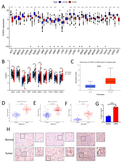
Background: TCIRG1, also known as V-ATPase-a3, is critical for cellular life activities through its dependent acidification. Prior to the present research, its relationship with prognostic and tumor immunity in clear cell renal cell carcinoma (ccRCC) had not yet been investigated. Methods: We assessed [...] Read more.
Background: TCIRG1, also known as V-ATPase-a3, is critical for cellular life activities through its dependent acidification. Prior to the present research, its relationship with prognostic and tumor immunity in clear cell renal cell carcinoma (ccRCC) had not yet been investigated. Methods: We assessed TCIRG1 expression in normal and tumor tissues using data from TCGA, GEO, GTEX, and IHC. We also analyzed the relationship between TCIRG1 and somatic mutations, TMB, DNA methylation, cancer stemness, and immune infiltration. We evaluated the relevance of TCIRG1 to immunotherapy and potential drugs. Finally, we explored the effect of TCIRG1 knockdown on tumor cells. Results: TCIRG1 was overexpressed in tumor tissue and predicted a significantly unfavorable clinical outcome. High TCIRG1 expression may be associated with fewer PBRM1 and more BAP1 mutations and may reduce DNA methylation, thus leading to a poor prognosis. TCIRG1 was strongly associated with CD8+ T-cell, Treg, and CD4+ T-cell infiltration. Moreover, TCIRG1 was positively correlated with TIDE scores and many drug sensitivities. Finally, experiments showed that the knockdown of TCIRG1 inhibited the migration of ccRCC cells. Conclusions: TCIRG1 may have great potential in identifying prognostic and immunomodulatory mechanisms in tumor patients and may provide a new therapeutic strategy for ccRCC. Full article
Show Figures

Figure 1

12 pages, 1902 KiB  
Article
Dynamic Expression of EpCAM in Primary and Metastatic Lung Cancer Is Controlled by Both Genetic and Epigenetic Mechanisms
by Yeting Cui, Jiapeng Li, Xiyu Liu, Lixing Gu, Mengqing Lyu, Jingjiao Zhou, Xiaoyu Zhang, Yu Liu, Haichuan Zhu, Tongcun Zhang and Fan Sun
Cancers 2022, 14(17), 4121; https://doi.org/10.3390/cancers14174121 - 25 Aug 2022
Cited by 5 | Viewed by 2698
Abstract
Although great progress has been achieved in cancer treatment in the past decades, lung cancer remains the leading cause of cancer death, which is partially caused by the fact that most lung cancers are diagnosed at advanced stages. To improve the sensitivity and [...] Read more.
Although great progress has been achieved in cancer treatment in the past decades, lung cancer remains the leading cause of cancer death, which is partially caused by the fact that most lung cancers are diagnosed at advanced stages. To improve the sensitivity and specificity of lung cancer diagnosis, the underlying mechanisms of current diagnosis methods are in urgent need to be explored. Herein, we find that the expression of EpCAM, the widely used molecular marker for tumor cell characterization and isolation, is strongly upregulated in primary lung tumors, which is caused by both gene amplification and promoter hypomethylation. In contrast, EpCAM expression is severely repressed in metastatic lung tumors, which can be reversed by epigenetic drugs, DNMT inhibitor 5-aza-dC and HDAC inhibitor MS-275. Moreover, tumor-associated macrophages (TAMs) impede EpCAM expression probably through TGFβ-induced EMT signaling. These findings unveil the dynamic expression patterns of EpCAM and differential roles of epigenetic modification in EpCAM expression in primary and metastatic lung tumors, providing novel insights into tumor cell isolation and lung cancer diagnosis. Full article
Show Figures

Figure 1

16 pages, 1261 KiB  
Article
Implication of Hepsin from Primary Tumor in the Prognosis of Colorectal Cancer Patients
by David Zaragoza-Huesca, Andrés Nieto-Olivares, Francisco García-Molina, Guillermo Ricote, Sofía Montenegro, Manuel Sánchez-Cánovas, Pedro Garrido-Rodríguez, Julia Peñas-Martínez, Vicente Vicente, Francisco Martínez, María Luisa Lozano, Alberto Carmona-Bayonas and Irene Martínez-Martínez
Cancers 2022, 14(13), 3106; https://doi.org/10.3390/cancers14133106 - 24 Jun 2022
Cited by 7 | Viewed by 2264
Abstract
Hepsin is a type II transmembrane serine protease whose deregulation promotes tumor invasion by proteolysis of the pericellular components. In colorectal cancer, the implication of hepsin is unknown. Consequently, we aimed to study the correlations between hepsin expression and different clinical-histopathological variables in [...] Read more.
Hepsin is a type II transmembrane serine protease whose deregulation promotes tumor invasion by proteolysis of the pericellular components. In colorectal cancer, the implication of hepsin is unknown. Consequently, we aimed to study the correlations between hepsin expression and different clinical-histopathological variables in 169 patients with localized colorectal cancer and 118 with metastases. Tissue microarrays were produced from samples at diagnosis of primary tumors and stained with an anti-hepsin antibody. Hepsin expression was correlated with clinical-histopathological variables by using the chi-square and Kruskal–Wallis tests, Kaplan–Meier and Aalen–Johansen estimators, and Cox and Fine and Gray multivariate models. In localized cancer patients, high-intensity hepsin staining was associated with reduced 5-year disease-free survival (p-value = 0.16). Medium and high intensity of hepsin expression versus low expression was associated with an increased risk of metastatic relapse (hazard ratio 2.83, p-value = 0.035 and hazard ratio 3.30, p-value = 0.012, respectively), being a better prognostic factor than classic histological variables. Additionally, in patients with localized tumor, 5-year thrombosis cumulative incidence increased with the increment of hepsin expression (p-value = 0.038). Medium and high intensities of hepsin with respect to low intensity were associated with an increase in thrombotic risk (hazard ratio 7.71, p-value = 0.043 and hazard ratio 9.02, p-value = 0.028, respectively). This relationship was independent of previous tumor relapse (p-value = 0.036). Among metastatic patients, low hepsin expression was associated with a low degree of tumor differentiation (p-value < 0.001) and with major metastatic dissemination (p-value = 0.023). Hepsin is a potential thrombotic and metastatic biomarker in patients with localized colorectal cancer. In metastatic patients, hepsin behaves in a paradoxical way with respect to differentiation and invasion processes. Full article
Show Figures

Graphical abstract

15 pages, 2184 KiB  
Article
Enhancer of Zeste Homolog 2 (EZH2) Is a Marker of High-Grade Neuroendocrine Neoplasia in Gastroenteropancreatic and Pulmonary Tract and Predicts Poor Prognosis
by Sebastian C. B. Bremer, Gabi Bittner, Omar Elakad, Helen Dinter, Jochen Gaedcke, Alexander O. König, Ahmad Amanzada, Volker Ellenrieder, Alexander Freiherr von Hammerstein-Equord, Philipp Ströbel and Hanibal Bohnenberger
Cancers 2022, 14(12), 2828; https://doi.org/10.3390/cancers14122828 - 8 Jun 2022
Cited by 2 | Viewed by 1983
Abstract
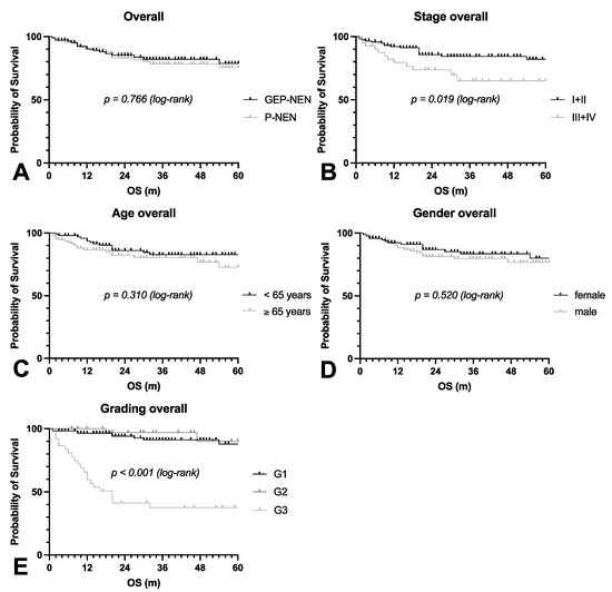
Tumor grading is a robust prognostic predictor in patients with neuroendocrine neoplasms (NEN) and guides therapy, especially in tumors with high proliferation. NEN can be separated into well-differentiated and poorly differentiated types. The more aggressive NEN have been further separated into neuroendocrine tumors [...] Read more.
Tumor grading is a robust prognostic predictor in patients with neuroendocrine neoplasms (NEN) and guides therapy, especially in tumors with high proliferation. NEN can be separated into well-differentiated and poorly differentiated types. The more aggressive NEN have been further separated into neuroendocrine tumors (NET G3) with a better prognosis and neuroendocrine carcinomas (NEC) with a worse prognosis. Despite this distinction’s tremendous clinical and therapeutic relevance, optimal diagnostic biomarkers are still lacking. In this study, we analyzed the protein expression and prognostic impact of Enhancer of Zeste Homolog 2 (EZH2) by immunohistochemistry in 219 tissue samples of gastroenteropancreatic (GEP-NEN) and pulmonary NEN (P-NEN). EZH2 was almost exclusively expressed in NEN with a proliferation rate above 20% (G3), while all low-grade tumors were nearly negative. Among high-grade NEN, 65% showed high and 35% low expression of EZH2. In this group, the high expression of EZH2 was significantly associated with poor overall survival and NEC histology. Interestingly, EZH2 seems to act independently of Polycomb Repressive Complex 2 (PRC2) in NEN. In conclusion, we propose EZH2 as a robust biomarker for distinguishing between NET G3 and NEC among gastroenteropancreatic and pulmonary NEN. Full article
Show Figures

Figure 1

13 pages, 2605 KiB  
Article
Characterization of Poorly Cohesive and Signet Ring Cell Carcinomas and Identification of PTPRM as a Diagnostic Marker
by Go Eun Bae, Sun Hyung Kang, Ju Seok Kim, Seok-Hwan Kim, Kyung-Hee Kim, Jin-Man Kim, Kwang-Sun Suh, Hyung Kyu Park, Dong-Wook Kang, Hyunjung Lee and Min-Kyung Yeo
Cancers 2022, 14(10), 2502; https://doi.org/10.3390/cancers14102502 - 19 May 2022
Cited by 3 | Viewed by 4093
Abstract
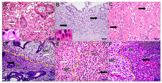
Background and aims. Signet ring cell (SRC) and poorly cohesive (PC) gastric carcinomas are morphologically similar but exhibit different biological behavior. We compared the clinical and molecular characteristics of SRC and PC carcinomas. Methods. Diffuse-type gastric cancer (GC) cases were classified into SRC [...] Read more.
Background and aims. Signet ring cell (SRC) and poorly cohesive (PC) gastric carcinomas are morphologically similar but exhibit different biological behavior. We compared the clinical and molecular characteristics of SRC and PC carcinomas. Methods. Diffuse-type gastric cancer (GC) cases were classified into SRC carcinomas (>90% of SRCs), PC carcinomas (<10% of SRCs), and combined PC/SRC carcinomas (≤90% but ≥10% of SRCs). The gene expression patterns in SRC and PC carcinomas were examined by transcriptome and protein immunohistochemistry analyses, and diagnostic and prognostic biomarkers were identified. Results. SRC and PC carcinomas showed significantly different clinical behaviors but shared common RNA expression patterns. PC carcinomas showed an increased expression of genes related to cancer progression. Among genes differentially expressed between PC and SRC carcinomas, protein tyrosine phosphatase receptor type M (PTPRM) was overexpressed in PC and related to unfavorable clinical factors. Conclusion. We found that PC and SRC carcinomas had distinct clinical characteristics and should be classified as different carcinoma types. PTPRM was identified as a potential diagnostic and prognostic biomarker for PC carcinomas and could represent a potential therapeutic target. Full article
Show Figures

Figure 1

20 pages, 2877 KiB  
Article
Putative Clinical Potential of ERBB2 Amplification Assessment by ddPCR in FFPE-DNA and cfDNA of Gastroesophageal Adenocarcinoma Patients
by Elisa Boldrin, Marcodomenico Mazza, Maria Assunta Piano, Rita Alfieri, Isabella Monia Montagner, Giovanna Magni, Maria Chiara Scaini, Loretta Vassallo, Antonio Rosato, Pierluigi Pilati, Antonio Scapinello and Matteo Curtarello
Cancers 2022, 14(9), 2180; https://doi.org/10.3390/cancers14092180 - 27 Apr 2022
Cited by 7 | Viewed by 3248
Abstract
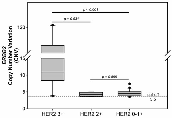
Anti-HER2 monoclonal antibody trastuzumab improves the survival of those patients with advanced gastroesophageal adenocarcinoma (GEA) exhibiting HER2/ERBB2 overexpression/amplification. The current gold standard methods used to diagnose the HER2 status in GEA are immunohistochemistry (IHC) and silver or fluorescence in situ hybridization (SISH [...] Read more.
Anti-HER2 monoclonal antibody trastuzumab improves the survival of those patients with advanced gastroesophageal adenocarcinoma (GEA) exhibiting HER2/ERBB2 overexpression/amplification. The current gold standard methods used to diagnose the HER2 status in GEA are immunohistochemistry (IHC) and silver or fluorescence in situ hybridization (SISH or FISH). However, they do not permit spatial and temporal tumor monitoring, nor do they overcome intra-cancer heterogeneity. Droplet digital PCR (ddPCR) was used to implement the assessment of HER2 status in formalin-fixed paraffin-embedded (FFPE) tumor DNA from a retrospective cohort (86 patients) and in cell-free DNA (cfDNA) samples from a prospective cohort (28 patients). In comparison to IHC/SISH, ddPCR assay revealed ERBB2 amplification in a larger patient fraction, including HER2 2+ and 0–1+ of the retrospective cohort (45.3% vs. 15.1%). In addition, a considerable number of HER2 2+ and 0–1+ prospective patients who were negative in FFPE by both IHC/SISH and ddPCR, showed ERBB2 amplification in the cfDNA collected just before surgery. cfDNA analysis in a few longitudinal cases revealed an increasing ERBB2 trend at progression. In conclusion, ddPCR in liquid biopsy may improve the detection rate of HER2 positive patients, preventing those patients who could benefit from targeted therapy from being incorrectly excluded. Full article
Show Figures

Figure 1

21 pages, 858 KiB  
Review
Evaluation of the TCR Repertoire as a Predictive and Prognostic Biomarker in Cancer: Diversity or Clonality?
by Andrea Aran, Laia Garrigós, Giuseppe Curigliano, Javier Cortés and Mercè Martí
Cancers 2022, 14(7), 1771; https://doi.org/10.3390/cancers14071771 - 31 Mar 2022
Cited by 26 | Viewed by 6251
Abstract
T cells play a vital role in the anti-tumoural response, and the presence of tumour-infiltrating lymphocytes has shown to be directly correlated with a good prognosis in several cancer types. Nevertheless, some patients presenting tumour-infiltrating lymphocytes do not have favourable outcomes. The TCR [...] Read more.
T cells play a vital role in the anti-tumoural response, and the presence of tumour-infiltrating lymphocytes has shown to be directly correlated with a good prognosis in several cancer types. Nevertheless, some patients presenting tumour-infiltrating lymphocytes do not have favourable outcomes. The TCR determines the specificities of T cells, so the analysis of the TCR repertoire has been recently considered to be a potential biomarker for patients’ progression and response to therapies with immune checkpoint inhibitors. The TCR repertoire is one of the multiple elements comprising the immune system and is conditioned by several factors, including tissue type, tumour mutational burden, and patients’ immunogenetics. Its study is crucial to understanding the anti-tumoural response, how to beneficially modulate the immune response with current or new treatments, and how to better predict the prognosis. Here, we present a critical review including essential studies on TCR repertoire conducted in patients with cancer with the aim to draw the current conclusions and try to elucidate whether it is better to encounter higher clonality with few TCRs at higher frequencies, or higher diversity with many different TCRs at lower frequencies. Full article
Show Figures

Graphical abstract

12 pages, 1837 KiB  
Article
Stacking Machine Learning Algorithms for Biomarker-Based Preoperative Diagnosis of a Pelvic Mass
by Reid Shaw, Anna E. Lokshin, Michael C. Miller, Geralyn Messerlian-Lambert and Richard G. Moore
Cancers 2022, 14(5), 1291; https://doi.org/10.3390/cancers14051291 - 2 Mar 2022
Cited by 8 | Viewed by 3101
Abstract
Objective: To identify the most predictive parameters of ovarian malignancy and develop a machine learning (ML) based algorithm to preoperatively distinguish between a benign and malignant pelvic mass. Methods: Retrospective study of 70 predictive parameters collected from 140 women with a pelvic mass. [...] Read more.
Objective: To identify the most predictive parameters of ovarian malignancy and develop a machine learning (ML) based algorithm to preoperatively distinguish between a benign and malignant pelvic mass. Methods: Retrospective study of 70 predictive parameters collected from 140 women with a pelvic mass. The women were split into a 3:1 “training” to “testing” dataset. Feature selection was performed using Gini impurity through an embedded random forest model and principal component analysis. Nine unique ML classifiers were assessed across a variety of model-specific hyperparameters using 25 bootstrap resamples of the training data. Model predictions were then combined into an ensemble stack by LASSO regression. The final ensemble stack and individual classifiers were then applied to the testing dataset to assess model performance. Results: Feature selection identified HE4, CA125, and transferrin as three predictive parameters of malignancy. Assessment of the ensemble stack on the testing dataset outperformed all individual ML classifiers in predicting malignancy. The ensemble stack demonstrated an accuracy of 97.1%, a receiver operating characteristic (ROC) area under the curve (AUC) of 0.951, and a sensitivity of 93.3% with a specificity of 100%. Conclusions: Combining the measurement of three distinct biomarkers with the stacking of multiple ML classifiers into an ensemble can provide valuable preoperative diagnostic predictions for patients with a pelvic mass. Full article
Show Figures

Figure 1

16 pages, 1818 KiB  
Article
Identification of Prognostic Markers of Gynecologic Cancers Utilizing Patient-Derived Xenograft Mouse Models
by Ha-Yeon Shin, Eun-ju Lee, Wookyeom Yang, Hyo Sun Kim, Dawn Chung, Hanbyoul Cho and Jae-Hoon Kim
Cancers 2022, 14(3), 829; https://doi.org/10.3390/cancers14030829 - 6 Feb 2022
Cited by 13 | Viewed by 3099
Abstract
Patient-derived xenografts (PDXs) are important in vivo models for the development of precision medicine. However, challenges exist regarding genetic alterations and relapse after primary treatment. Thus, PDX models are required as a new approach for preclinical and clinical studies. We established PDX models [...] Read more.
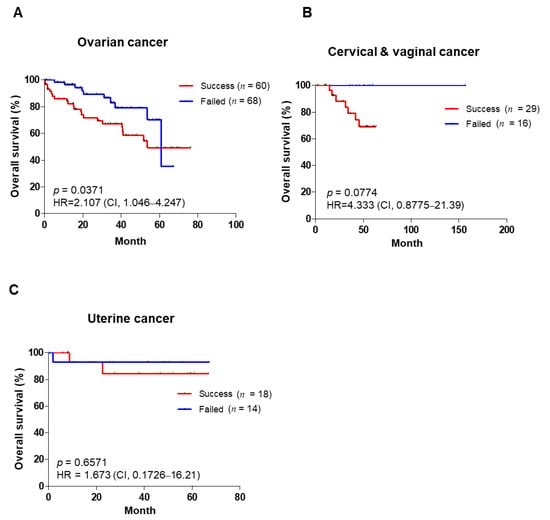
Patient-derived xenografts (PDXs) are important in vivo models for the development of precision medicine. However, challenges exist regarding genetic alterations and relapse after primary treatment. Thus, PDX models are required as a new approach for preclinical and clinical studies. We established PDX models of gynecologic cancers and analyzed their clinical information. We subcutaneously transplanted 207 tumor tissues from patients with gynecologic cancer into nude mice from 2014 to 2019. The successful engraftment rate of ovarian, cervical, and uterine cancer was 47%, 64%, and 56%, respectively. The subsequent passages (P2 and P3) showed higher success and faster growth rates than the first passage (P1). Using gynecologic cancer PDX models, the tumor grade is a common clinical factor affecting PDX establishment. We found that the PDX success rate correlated with the patient’s prognosis, and also that ovarian cancer patients with a poor prognosis had a faster PDX growth rate (p < 0.0001). Next, the gene sets associated with inflammation and immune responses were shown in high-ranking successful PDX engraftment through gene set enrichment analysis and RNA sequencing. Up-regulated genes in successful engraftment were found to correlate with ovarian clear cell cancer patient outcomes via Gene Expression Omnibus dataset analysis. Full article
Show Figures

Figure 1

2021

Jump to: 2024, 2023, 2022, 2020

13 pages, 1539 KiB  
Article
Clinicopathological and Molecular Prognostic Classifier for Intermediate/High-Risk Clear Cell Renal Cell Carcinoma
by Fiorella L. Roldán, Juan J. Lozano, Mercedes Ingelmo-Torres, Raquel Carrasco, Esther Díaz, Miguel Ramirez-Backhaus, José Rubio, Oscar Reig, Antonio Alcaraz, Lourdes Mengual and Laura Izquierdo
Cancers 2021, 13(24), 6338; https://doi.org/10.3390/cancers13246338 - 17 Dec 2021
Cited by 2 | Viewed by 2575
Abstract
The probability of tumor progression in intermediate/high-risk clear cell renal cell carcinoma (ccRCC) is highly variable, underlining the lack of predictive accuracy of the current clinicopathological factors. To develop an accurate prognostic classifier for these patients, we analyzed global gene expression patterns in [...] Read more.
The probability of tumor progression in intermediate/high-risk clear cell renal cell carcinoma (ccRCC) is highly variable, underlining the lack of predictive accuracy of the current clinicopathological factors. To develop an accurate prognostic classifier for these patients, we analyzed global gene expression patterns in 13 tissue samples from progressive and non-progressive ccRCC using Illumina Hi-seq 4000. Expression levels of 22 selected differentially expressed genes (DEG) were assessed by nCounter analysis in an independent series of 71 ccRCCs. A clinicopathological-molecular model for predicting tumor progression was developed and in silico validated in a total of 202 ccRCC patients using the TCGA cohort. A total of 1202 DEGs were found between progressive and non-progressive intermediate/high-risk ccRCC in RNAseq analysis, and seven of the 22 DEGs selected were validated by nCounter. Expression of HS6ST2, pT stage, tumor size, and ISUP grade were found to be independent prognostic factors for tumor progression. A risk score generated using these variables was able to distinguish patients at higher risk of tumor progression (HR 7.27; p < 0.001), consistent with the results obtained from the TCGA cohort (HR 2.74; p < 0.002). In summary, a combined prognostic algorithm was successfully developed and validated. This model may aid physicians to select high-risk patients for adjuvant therapy. Full article
Show Figures

Figure 1

12 pages, 1057 KiB  
Article
Pretreatment Albumin-to-Alkaline Phosphatase Ratio Is a Prognostic Marker in Lung Cancer Patients: A Registry-Based Study of 7077 Lung Cancer Patients
by Birgitte Sandfeld-Paulsen, Ninna Aggerholm-Pedersen and Anne Winther-Larsen
Cancers 2021, 13(23), 6133; https://doi.org/10.3390/cancers13236133 - 6 Dec 2021
Cited by 11 | Viewed by 2986
Abstract
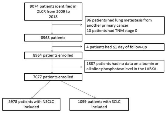
The albumin-to-alkaline phosphatase ratio (AAPR) is a novel promising prognostic marker in cancer patients. However, the evidence for its significance in lung cancer is scarce. Therefore, we assessed the prognostic value of the AAPR in a large cohort of lung cancer patients. Data [...] Read more.
The albumin-to-alkaline phosphatase ratio (AAPR) is a novel promising prognostic marker in cancer patients. However, the evidence for its significance in lung cancer is scarce. Therefore, we assessed the prognostic value of the AAPR in a large cohort of lung cancer patients. Data on lung cancer patients diagnosed from January 2009 to June 2018 were extracted from the Danish Lung Cancer Registry and combined with data on the pretreatment serum AAPR level extracted from the clinical laboratory information system (LABKA). AAPR tertiles were applied as cutoffs. Cox proportional hazard models assessed the prognostic value of the AAPR. In total, 5978 non-small cell lung cancer (NSCLC) patients and 1099 small cell lung cancer (SCLC) patients were included. Decreasing AAPR level was significantly associated with declining median overall survival (OS) in NSCLC patients (medium vs. low AAPR, adjusted HR = 0.73 (95% confidence interval (CI) 0.68–0.79); high vs. low AAPR, adjusted HR = 0.68 (95% CI 0.62–0.73)) and in SCLC patients (medium vs. low AAPR, adjusted HR = 0.62 (95% CI 0.52–0.74); high vs. low, adjusted HR = 0.59 (95% CI 0.50–0.70)). In conclusion, the AAPR was an independent prognostic factor in NSCLC and SCLC patients. The correlation seems to be level dependent, with reducing survival found to be associated with decreasing AAPR level. Full article
Show Figures

Figure 1

18 pages, 2971 KiB  
Article
Characterization of Recurrent Relevant Genes Reveals a Novel Role of RPL36A in Radioresistant Oral Squamous Cell Carcinoma
by Ting-Wen Chen, Kai-Ping Chang, Chun-Chia Cheng, Cheng-Yi Chen, Shu-Wen Hong, Zong-Lin Sie, Hsing-Wen Cheng, Wei-Chen Yen, Yenlin Huang, Shu-Chen Liu and Chun-I Wang
Cancers 2021, 13(22), 5623; https://doi.org/10.3390/cancers13225623 - 10 Nov 2021
Cited by 7 | Viewed by 2458
Abstract
Radioresistance is one of the major factors that contributes to radiotherapy failure in oral cavity squamous cell carcinoma (OSCC). By comparing the prognostic values of 20,502 genes expressed in patients in The Cancer Genome Atlas (TCGA)-OSCC cohort with (n = 162) and [...] Read more.
Radioresistance is one of the major factors that contributes to radiotherapy failure in oral cavity squamous cell carcinoma (OSCC). By comparing the prognostic values of 20,502 genes expressed in patients in The Cancer Genome Atlas (TCGA)-OSCC cohort with (n = 162) and without radiotherapy (n = 118), herein identified 297 genes positively correlated with poor disease-free survival in OSCC patients with radiotherapy as the potential radioresistance-associated genes. Among the potential radioresistance-associated genes, 36 genes were upregulated in cancerous tissues relative to normal tissues. The bioinformatics analysis revealed that 60S ribosomal protein L36a (RPL36A) was the most frequently detected gene involved in radioresistance-associated gene-mediated biological pathways. Then, two independent cohorts (n = 162 and n = 136) were assessed to confirm that higher RPL36A transcript levels were significantly associated with a poor prognosis only in OSCC patients with radiotherapy. Mechanistically, we found that knockdown of RPL36A increased radiosensitivity via sensitizing cells to DNA damage and promoted G2/M cell cycle arrest followed by augmenting the irradiation-induced apoptosis pathway in OSCC cells. Taken together, our study supports the use of large-scale genomic data for identifying specific radioresistance-associated genes and suggests a regulatory role for RPL36A in the development of radioresistance in OSCC. Full article
Show Figures

Figure 1

11 pages, 1013 KiB  
Article
Development and Validation of a Novel Serum Prognostic Marker for Patients with Metastatic Colorectal Cancer on Regorafenib Treatment
by Yu-Li Su, Kai-Lung Tsai, Tai-Jan Chiu, Yueh-Ming Lin, Ko-Chao Lee, Chien-Chang Lu, Hong-Hwa Chen, Chia-Che Wu and Hung-Chih Hsu
Cancers 2021, 13(20), 5080; https://doi.org/10.3390/cancers13205080 - 11 Oct 2021
Cited by 8 | Viewed by 2386
Abstract
(1) Background: To investigate the prognostic value of cancer-inflammation prognostic index (CIPI) in patients with metastatic colorectal cancer (mCRC) on regorafenib treatment; (2) Methods: Patients with mCRC who were given regorafenib as later-line treatment at Kaohsiung and Linkou Chang-Gung Memorial Hospital between November [...] Read more.
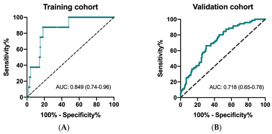
(1) Background: To investigate the prognostic value of cancer-inflammation prognostic index (CIPI) in patients with metastatic colorectal cancer (mCRC) on regorafenib treatment; (2) Methods: Patients with mCRC who were given regorafenib as later-line treatment at Kaohsiung and Linkou Chang-Gung Memorial Hospital between November 2014 and January 2021 were consecutively enrolled. All relevant clinicopathologic, laboratory data and survival status were recorded. Independent prognostic factors were determined by the multivariate Cox regression method; (3) Results: In total, 106 patients in the training cohort and 250 in the validation cohort were enrolled. The median OS for patients with CIPI ≥ 300 and < 300 in the training cohort was 3.8 and 9.0 months, respectively (hazard ratio (HR) 2.78, 95% confidence interval (CI) 1.82–4.23; p < 0.0001). Time to regorafenib, liver metastasis and CIPI were independent factors by multivariate Cox regression analyses. A new scoring model demonstrated a good discriminatory ability to risk stratification of a patient’s survival; (4) Conclusions: We identified CIPI as a novel serum marker highly associated with overall survival in patients with mCRC receiving regorafenib treatment. Further confirmatory studies are warranted. Full article
Show Figures

Figure 1

13 pages, 11381 KiB  
Article
Level of Combined Estrogen and Progesterone Receptor Expression Determines the Eligibility for Adjuvant Endocrine Therapy in Breast Cancer Patients
by Jee Hyun Ahn, Soon Bo Choi, Jung Min Park, Jee Ye Kim, Hyung Seok Park, Seung Il Kim, Byeong-Woo Park and Seho Park
Cancers 2021, 13(19), 5007; https://doi.org/10.3390/cancers13195007 - 6 Oct 2021
Cited by 2 | Viewed by 2442
Abstract
Hormone receptor (HR)-positive breast cancer has a heterogeneous pattern according to the level of receptor expression. Patients whose breast cancers express low levels of estrogen receptor (ER) or progesterone receptor (PgR) may be eligible for adjuvant endocrine therapy, but limited data are available [...] Read more.
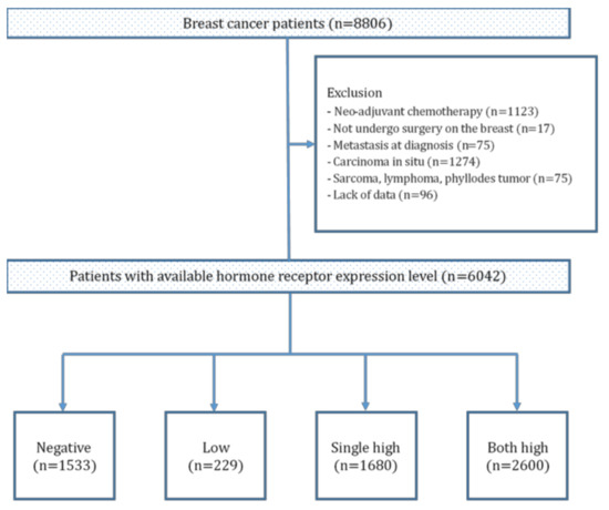
Hormone receptor (HR)-positive breast cancer has a heterogeneous pattern according to the level of receptor expression. Patients whose breast cancers express low levels of estrogen receptor (ER) or progesterone receptor (PgR) may be eligible for adjuvant endocrine therapy, but limited data are available to support this notion. We aimed to determine whether HR expression level is related to prognosis. Tumors from 6042 patients with breast cancer were retrospectively analyzed for combined HR levels of ER and PgR. Low expression was defined as ER 1–10% and PgR 1–20%. Four HR groups were identified by combining ER and PgR expression levels. Patients whose tumors expressed high levels of a single receptor showed the worst survival outcomes, and their risk continuously increased even after the 10-year follow-up. Endocrine therapy had a significant benefit for patients whose tumors expressed high HR levels and a favorable tendency for patients with tumors expressing low HR levels. We established the possible benefit of endocrine therapy for patients whose breast tumors expressed low HR levels. Thus, HR level was a prognostic factor and might be a determinant of extended therapy, especially for patients with high expression of a single receptor. Full article
Show Figures

Figure 1

13 pages, 1568 KiB  
Article
Early Changes in DCE-MRI Biomarkers May Predict Survival Outcomes in Patients with Advanced Hepatocellular Carcinoma after Sorafenib Failure: Two Prospective Phase II Trials
by Bang-Bin Chen, Zhong-Zhe Lin, Yu-Yun Shao, Chiun Hsu, Chih-Hung Hsu, Ann-Lii Cheng, Po-Chin Liang and Tiffany Ting-Fang Shih
Cancers 2021, 13(19), 4962; https://doi.org/10.3390/cancers13194962 - 1 Oct 2021
Cited by 4 | Viewed by 2228
Abstract
In this paper, our main objective was to predict survival outcomes using DCE-MRI biomarkers in patients with advanced hepatocellular carcinoma (HCC) after progression from 1st-line sorafenib treatment in two prospective phase II trials. This study included 74 participants (men/women = 64/10, mean age [...] Read more.
In this paper, our main objective was to predict survival outcomes using DCE-MRI biomarkers in patients with advanced hepatocellular carcinoma (HCC) after progression from 1st-line sorafenib treatment in two prospective phase II trials. This study included 74 participants (men/women = 64/10, mean age 60 ± 11.8 years) with advanced HCC who received 2nd-line targeted therapy (n = 41 with lenalidomide in one clinical trial; n = 33 with axitinib in another clinical trial) after sorafenib failure from two prospective phase II studies. Among them, all patients underwent DCE-MRI at baseline, and on days 3 and 14 of treatment. The relative changes (Δ) in the DCE-MRI parameters, including ΔPeak, ΔAUC, and ΔKtrans, were derived from the largest hepatic tumor. The treatment response was evaluated using the Response Evaluation Criteria in Solid Tumors (RECIST 1.1). The Cox model was used to investigate the associations of the clinical variables and DCE-MRI biomarkers with progression-free survival (PFS) and overall survival (OS). The objective response rate (ORR) was 10.8% (8/74) and the disease control rate (DCR) was 58.1% (43/74). The median PFS and OS values were 1.9 and 7.8 months, respectively. On day 3 (D3), participants with high reductions in ΔPeak_D3 (hazard ratio (HR) 0.4, 95% confidence interval (CI) 0.17–0.93, p = 0.017) or ΔAUC_D3 (HR 0.51, 95% CI 0.25–1.04, p = 0.043) were associated with better PFS. On day 14, participants with high reductions in ΔPeak_D14 (HR 0.51, 95% CI 0.26–1.01, p = 0.032), ΔAUC_D14 (HR 0.54, 95% CI 0.33–0.9, p = 0.009), or ΔKtrans_D14 (HR 0.26, 95% CI 0.12–0.56, p < 0.001) had a higher PFS than those with lower reduction values. In addition, high reductions in ΔAUC_D14 (HR 0.53, 95% CI 0.32–0.9, p = 0.016) or ΔKtrans_D14 (HR 0.47, 95% CI 0.23–0.98, p = 0.038) were associated with a better OS. Among the clinical variables, ORR was associated with both PFS (p = 0.001) and OS (p = 0.005). DCR was associated with PFS (p = 0.002), but not OS (p = 0.089). Cox multivariable analysis revealed that ΔKtrans_D14 (p = 0.002) remained an independent predictor of PFS after controlling for ORR and DCR. An early reduction in tumor perfusion detected by DCE-MRI biomarkers, especially on day 14, may predict favorable survival outcomes in participants with HCC receiving 2nd-line targeted therapy after sorafenib failure. Full article
Show Figures

Figure 1

14 pages, 1949 KiB  
Article
18F-Fluorodeoxyglucose Positron Emission Tomography Is Useful in the Evaluation of Prognosis in Retroperitoneal Sarcoma
by Toru Wakamatsu, Yoshinori Imura, Hironari Tamiya, Toshinari Yagi, Naohiro Yasuda, Sho Nakai, Takaaki Nakai, Hidetatsu Outani, Kenichiro Hamada, Shigeki Kakunaga, Nobuhito Araki, Takafumi Ueda and Satoshi Takenaka
Cancers 2021, 13(18), 4611; https://doi.org/10.3390/cancers13184611 - 14 Sep 2021
Cited by 7 | Viewed by 1993
Abstract
Background: Retroperitoneal sarcomas are rare neoplasms that occur in the retroperitoneum. Complete surgical resection is the only effective treatment option. The prediction of prognosis by histological diagnosis has not yet been established. The purpose of this study was to identify the usefulness of [...] Read more.
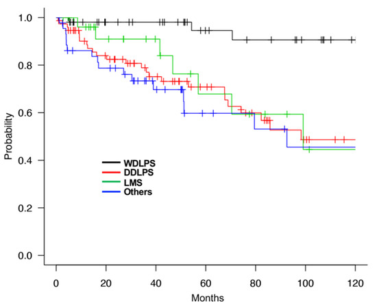
Background: Retroperitoneal sarcomas are rare neoplasms that occur in the retroperitoneum. Complete surgical resection is the only effective treatment option. The prediction of prognosis by histological diagnosis has not yet been established. The purpose of this study was to identify the usefulness of [18-F] fluorodeoxyglucose (FDG) positron emission tomography (PET) imaging for validating the prognosis of retroperitoneal sarcoma (RPS) established by histological diagnosis. Methods: We retrospectively reviewed 201 patients with RPS treated at the Osaka International Cancer Institute between 2010 and 2021. We extracted the clinical data, including standardized uptake values (SUVs), evaluated with FDG-PET, and statistically analyzed the data. Results: The median age of patients was 64 years (range, 31–85 years). A total of 101 (50.2%) patients were men, and 100 (49.8%) were women. Surgical resection was performed in 155 (77.1%) patients. On histological analysis, 75 (37.3%), 52 (25.9%), and 29 (14.4%) patients were diagnosed with dedifferentiated liposarcoma, well-differentiated liposarcoma, and leiomyosarcoma, respectively. The median survival time for patients with high maximum SUV (SUVmax) (≥4) or low SUVmax (<4) was 275.8 months and 79.5 months, respectively. Furthermore, among the patients with dedifferentiated liposarcoma, the overall survival rate for patients with high SUVmax (≥4) was significantly lower than that of those with low SUVmax (<4). Conclusions: The present study demonstrated that SUVmax calculated with FDG-PET was useful as a prognostic factor in RPS, especially in dedifferentiated liposarcoma and Grade2 RPS. To devise a treatment strategy for RPS, SUVmax during FDG-PET scan may be considered for clinical assessment. Full article
Show Figures

Figure 1

17 pages, 5220 KiB  
Article
Necroptosis in Esophageal Squamous Cell Carcinoma: An Independent Prognostic Factor and Its Correlation with Tumor-Infiltrating Lymphocytes
by Takuro Yamauchi, Fumiyoshi Fujishima, Masatoshi Hashimoto, Junichi Tsunokake, Ryujiro Akaishi, Yusuke Gokon, Shunsuke Ueki, Yohei Ozawa, Toshiaki Fukutomi, Hiroshi Okamoto, Chiaki Sato, Yusuke Taniyama, Tomohiro Nakamura, Naoki Nakaya, Takashi Kamei and Hironobu Sasano
Cancers 2021, 13(17), 4473; https://doi.org/10.3390/cancers13174473 - 5 Sep 2021
Cited by 12 | Viewed by 2930
Abstract
Necroptosis is a pivotal process in cancer biology; however, the clinical significance of necroptosis in esophageal squamous cell carcinoma (ESCC) has remained unknown. Therefore, in this study, we aimed to verify the potential involvement of necroptosis in the clinical outcome, chemotherapeutic resistance, and [...] Read more.
Necroptosis is a pivotal process in cancer biology; however, the clinical significance of necroptosis in esophageal squamous cell carcinoma (ESCC) has remained unknown. Therefore, in this study, we aimed to verify the potential involvement of necroptosis in the clinical outcome, chemotherapeutic resistance, and tumor microenvironment of ESCC. Mixed lineage kinase domain-like protein (MLKL) and phosphorylated MLKL (pMLKL) were immunohistochemically examined in 88 surgically resected specimens following neoadjuvant chemotherapy (NAC) and 53 pre-therapeutic biopsy specimens, respectively. Tumor-infiltrating lymphocytes (TILs) were also evaluated by immunolocalizing CD3, CD8, and forkhead box protein 3 (FOXP3) in the residual tumors after NAC. High pMLKL status in the post-NAC resected specimens was significantly correlated with worse prognosis in ESCC patients. Multivariate analysis demonstrated that a high pMLKL status was an independent prognostic factor. In pre-NAC biopsy specimens, a high pMLKL status was significantly associated with a lower therapeutic efficacy. CD8+ TILs were significantly lower in the high-pMLKL group. FOXP3+ TILs were significantly higher in both high-MLKL and high-pMLKL groups. We first demonstrated pMLKL status as an independent prognostic factor in ESCC patients. Our study revealed the possible involvement of necroptosis in the immunosuppressive microenvironment, resulting in the attenuated therapeutic efficacy of NAC and eventual adverse clinical outcomes in ESCC. Full article
Show Figures

Figure 1

18 pages, 1853 KiB  
Systematic Review
A Clinical Update on the Prognostic Effect of microRNA Biomarkers for Survival Outcome in Nasopharyngeal Carcinoma: A Systematic Review and Meta-Analysis
by Peter Shaw, Raghul Senthilnathan, Sunil Krishnan, Deepa Suresh, Sameep Shetty, Gothandam Kodiveri Muthukaliannan, Ravishankar Ram Mani, Palanisamy Sivanandy, Harish Chinna Konda Chandramoorthy, Madan Mohan Gupta, Siddhartha Baxi and Rama Jayaraj
Cancers 2021, 13(17), 4369; https://doi.org/10.3390/cancers13174369 - 29 Aug 2021
Cited by 7 | Viewed by 4716
Abstract
Background: Nasopharyngeal carcinoma (NPC), a relatively uncommon malignancy in the Western world, is highly prevalent in Southeast Asia where the treatment outcomes are poor. Despite recent improvements in diagnosis and treatment locoregional control, distant metastasis and chemoresistance continue to be a significant [...] Read more.
Background: Nasopharyngeal carcinoma (NPC), a relatively uncommon malignancy in the Western world, is highly prevalent in Southeast Asia where the treatment outcomes are poor. Despite recent improvements in diagnosis and treatment locoregional control, distant metastasis and chemoresistance continue to be a significant cause of mortality. Identification of a reliable and comprehensive prognostic biomarker is highly desirable. The potential relevance of microRNAs (miRNAs) as prognostic markers in NPC is assessed in this systematic review and meta-analysis. Methods: A systematic review was performed using the PubMed and Science Direct databases. The search was limited to search results between 2018 and 2020 with the keywords and search strings developed as per the Preferred Reporting Items for Systematic Review and Meta-analysis (PRISMA) guidelines. The recovered articles were carefully screened based on the selection criteria. In the meta-analysis study, high and low expression levels of miRNAs were measured using the hazard ratio (HR) and 95 percent confidence interval (CI) for patients’ survival outcomes. Egger’s bias indicator test and funnel plot symmetry were used to assess the risk of bias. Results: Amongst the 25 studies, 13 fulfilled the conditions of inclusion in this meta-analysis. The researchers further delved into the 21 miRNA expression levels from 3015 NPC patients to ascertain a link between miRNA’s predictive role and survival outcomes. The majority of the articles retrieved during this study were from China, with two studies from Canada and Malaysia. The overall pooled effect size estimation (HR) for dysregulated miRNAs was 1.590 (95% CI: 1.253–2.017), displaying that miRNA marker expression increased the risk of mortality in NPC patients by 59%. Conclusions: This meta-analysis is novel and looks at the prognostic significance of miRNAs as biomarkers in NPC patients using a continuous version pooled meta-analysis. Although our findings are ambiguous, they do show that greater miRNA expression in NPC may be associated with a lower overall survival rate. To acquire clear conclusions, more prospective studies with large cohorts are required to determine the clinical utility of miRNAs as prognostic biomarkers. Full article
Show Figures

Figure 1

16 pages, 2219 KiB  
Article
Pri-miR526b and Pri-miR655 Are Potential Blood Biomarkers for Breast Cancer
by Mousumi Majumder, Kingsley Chukwunonso Ugwuagbo, Sujit Maiti, Peeyush K Lala and Muriel Brackstone
Cancers 2021, 13(15), 3838; https://doi.org/10.3390/cancers13153838 - 30 Jul 2021
Cited by 6 | Viewed by 3620
Abstract
We reported that two microRNAs, miR526b and miR655, are oncogenic in breast cancer (BC). Overexpression of these two miRNAs in poorly metastatic BC cells promotes aggressive BC phenotypes in vitro and in vivo. High expression of each miRNA was associated with poor patient [...] Read more.
We reported that two microRNAs, miR526b and miR655, are oncogenic in breast cancer (BC). Overexpression of these two miRNAs in poorly metastatic BC cells promotes aggressive BC phenotypes in vitro and in vivo. High expression of each miRNA was associated with poor patient survival. In this pilot biomarker study, we report for the first time that miRNA precursor RNAs (pri-miRNAs) are robust and sensitive biomarkers for BC, detectable in both human blood plasma and biopsy tissues. Pri-miRNA detection and quantification do not require a special enrichment procedure, thus reducing specimen quantity. Blood plasma samples from 90 malignant tumor-bearing patients and 20 benign lesion-bearing participants (control) were analyzed for pri-miRNA expression with a quantitative real-time polymerase chain reaction. Results revealed that normalized expressions of plasma pri-miR526b and pri-miR655 are significantly upregulated in malignancy compared to benign plasmas (p = 0.002 and p = 0.03, respectively). Both pri-miRNAs showed more prominent results to distinguish stage I plasmas from benign plasmas (p = 0.001 for pri-miR526b and p = 0.0001 for pri-miR655). We have also validated pri-miRNA expression in independent tumor bank tissues, showing significant upregulation of both pri-miRNAs in BC; thus, pri-miRNAs are robust markers. The diagnostic relevance of pri-miRNAs was computed with the area under the curve (AUC). Pri-miR526b is a sensitive biomarker to distinguish cancer from control plasmas (sensitivity of 86%; AUC = 71.47%, p = 0.0027) with a positive predictive value of 88.89%; however, pri-miR655 did not show significant sensitivity. Furthermore, pri-miR526b could also significantly distinguish tumors as early as stage I from control (sensitivity of 75%; AUC = 72.71%, p = 0.0037). Therefore, pri-miR526b can be used as an early diagnostic biomarker. The expression of both pri-miRNAs was significantly high in ER-positive and HER2-negative subgroups of BC; hence, these biomarkers might play a role in the management of endocrine therapy designs. Additionally, with a case–control cohort study, we identified that high expression of pri-miR526b in the blood is also a risk factor associated with breast cancer (OR = 4.3, CI = 1.39–13.34, p = 0.01). Pri-miRNAs could be considered novel breast cancer blood biomarkers. Full article
Show Figures

Graphical abstract

17 pages, 4949 KiB  
Article
Pre-Therapeutic VEGF Level in Plasma Is a Prognostic Bio-Marker in Head and Neck Squamous Cell Carcinoma (HNSCC)
by Julia Siemert, Theresa Wald, Marlen Kolb, Isolde Pettinella, Ulrike Böhm, Markus Pirlich, Susanne Wiegand, Andreas Dietz and Gunnar Wichmann
Cancers 2021, 13(15), 3781; https://doi.org/10.3390/cancers13153781 - 27 Jul 2021
Cited by 12 | Viewed by 2198
Abstract
Vascular endothelial growth factor (VEGF) is centrally involved in cancer angiogenesis. We hypothesized that pre-therapeutic VEGF levels in serum and plasma differ in their potential as biomarkers for outcomes in head and neck squamous cell carcinoma (HNSCC) patients. As prospectively defined in the [...] Read more.
Vascular endothelial growth factor (VEGF) is centrally involved in cancer angiogenesis. We hypothesized that pre-therapeutic VEGF levels in serum and plasma differ in their potential as biomarkers for outcomes in head and neck squamous cell carcinoma (HNSCC) patients. As prospectively defined in the study protocols of TRANSCAN-DietINT and NICEI-CIH, we measured VEGF in pretreatment serum and plasma of 75 HNSCC test cohort (TC) patients. We analyzed the prognostic value of VEGF concentrations in serum (VEGFSerum) and plasma (VEGFPlasma) for event-free survival (EFS) utilizing receiver-operating characteristics (ROC). Mean VEGF concentrations in plasma (34.6, 95% CI 26.0–43.3 ng/L) were significantly lower (p = 3.35 × 10⁻¹⁸) than in serum (214.8, 95% CI 179.6–250.0 ng/L) but, based on ROC (area under the curve, AUCPlasma = 0.707, 95% CI 0.573–0.840; p = 0.006 versus AUCSerum = 0.665, 95% CI 0.528–0.801; p = 0.030), superiorly correlated with event-free survival (EFS) of TC patients. Youden indices revealed optimum binary classification with VEGFPlasma 26 ng/L and VEGFSerum 264 ng/L. Kaplan–Meier plots demonstrated superiority of VEGFPlasma in discriminating patients regarding outcome. Patients with VEGFPlasma < 26 ng/L had superior nodal (NC), local (LC) and loco-regional control (LRC) leading to significant prolonged progression-free survival (PFS) and EFS. We successfully validated VEGFPlasma according the cut-off <26 ng/L as predictive for superior outcome in an independent validation cohort (iVC) of 104 HNSCC patients from the studies DeLOS-II and LIFE and found better outcomes including prolonged tumor-specific (TSS) and overall survival (OS). Outcomes in TC and iVC combined again was related to VEGFPlasma, and multivariate Cox regression revealed that VEGFPlasma was an independent outcome predictor. In HNSCC, pre-therapeutic VEGFPlasma is prognostic for outcomes. Full article
Show Figures

Figure 1

21 pages, 3072 KiB  
Article
Proteomics of Primary Uveal Melanoma: Insights into Metastasis and Protein Biomarkers
by Geeng-Fu Jang, Jack S. Crabb, Bo Hu, Belinda Willard, Helen Kalirai, Arun D. Singh, Sarah E. Coupland and John W. Crabb
Cancers 2021, 13(14), 3520; https://doi.org/10.3390/cancers13143520 - 14 Jul 2021
Cited by 10 | Viewed by 4441
Abstract
Uveal melanoma metastases are lethal and remain incurable. A quantitative proteomic analysis of 53 metastasizing and 47 non-metastasizing primary uveal melanoma (pUM) was pursued for insights into UM metastasis and protein biomarkers. The metastatic status of the pUM specimens was defined based on [...] Read more.
Uveal melanoma metastases are lethal and remain incurable. A quantitative proteomic analysis of 53 metastasizing and 47 non-metastasizing primary uveal melanoma (pUM) was pursued for insights into UM metastasis and protein biomarkers. The metastatic status of the pUM specimens was defined based on clinical data, survival histories, prognostic analyses, and liver histopathology. LC MS/MS iTRAQ technology, the Mascot search engine, and the UniProt human database were used to identify and quantify pUM proteins relative to the normal choroid excised from UM donor eyes. The determined proteomes of all 100 tumors were very similar, encompassing a total of 3935 pUM proteins. Proteins differentially expressed (DE) between metastasizing and non-metastasizing pUM (n = 402) were employed in bioinformatic analyses that predicted significant differences in the immune system between metastasizing and non-metastasizing pUM. The immune proteins (n = 778) identified in this study support the immune-suppressive nature and low abundance of immune checkpoint regulators in pUM, and suggest CDH1, HLA-DPA1, and several DE immune kinases and phosphatases as possible candidates for immune therapy checkpoint blockade. Prediction modeling identified 32 proteins capable of predicting metastasizing versus non-metastasizing pUM with 93% discriminatory accuracy, supporting the potential for protein-based prognostic methods for detecting UM metastasis. Full article
Show Figures

Figure 1

14 pages, 1560 KiB  
Article
A Novel Automated Immunoassay Platform to Evaluate the Association of Adiponectin and Leptin Levels with Breast Cancer Risk
by Debora Macis, Valentina Aristarco, Harriet Johansson, Aliana Guerrieri-Gonzaga, Sara Raimondi, Matteo Lazzeroni, Ivana Sestak, Jack Cuzick, Andrea DeCensi, Bernardo Bonanni and Sara Gandini
Cancers 2021, 13(13), 3303; https://doi.org/10.3390/cancers13133303 - 30 Jun 2021
Cited by 9 | Viewed by 3251
Abstract
Adiponectin and leptin are adipokines secreted by the adipose tissue that are associated with several chronic diseases including cancer. We aimed to compare the immunoassay platform ELLA with an enzyme-linked immunosorbent assay (ELISA) kit and to assess whether the results of the association [...] Read more.
Adiponectin and leptin are adipokines secreted by the adipose tissue that are associated with several chronic diseases including cancer. We aimed to compare the immunoassay platform ELLA with an enzyme-linked immunosorbent assay (ELISA) kit and to assess whether the results of the association analyses with breast cancer risk were dependent on the assay used. We measured adiponectin and leptin with ELLA and ELISA on baseline serum samples of 116 Italian postmenopausal women enrolled in two international breast cancer prevention trials. Results were compared with Deming, Passing–Bablok regression and Bland–Altman plots. Disease-free survival was analyzed with the Cox model. There was a good correlation between the methods for adiponectin and leptin (r > 0.96). We found an increased breast cancer risk for very low adiponectin levels (HR for ELLA = 3.75; 95% CI: 1.37;10.25, p = 0.01), whereas no significant association was found for leptin levels. The disease-free survival curves were almost identical for values obtained with the two methods, for both biomarkers. The ELLA platform showed a good concordance with ELISA for adiponectin and leptin measurements. Our results support the association of very low adiponectin levels with postmenopausal breast cancer risk, irrespective of the method used. The ELLA platform is a time-saving system with high reproducibility, therefore we recommend its use for biomarker assessment. Full article
Show Figures

Graphical abstract

14 pages, 1475 KiB  
Article
Establishment of a Pretreatment Nomogram to Predict the 6-Month Mortality Rate of Patients with Advanced Biliary Tract Cancers Undergoing Gemcitabine-Based Chemotherapy
by Chiao-En Wu, Wen-Kuan Huang, Wen-Chi Chou, Chia-Hsun Hsieh, John Wen-Cheng Chang, Cheng-Yu Lin, Chun-Nan Yeh and Jen-Shi Chen
Cancers 2021, 13(13), 3139; https://doi.org/10.3390/cancers13133139 - 23 Jun 2021
Cited by 6 | Viewed by 1928
Abstract
Background: The estimation of mortality risk among patients diagnosed with advanced cancer provides important information for clinicians and patients in clinical practice. Currently, gemcitabine-based chemotherapy regimens are the standard treatment for patients with advanced biliary tract cancer (BTC). We aimed to develop a [...] Read more.
Background: The estimation of mortality risk among patients diagnosed with advanced cancer provides important information for clinicians and patients in clinical practice. Currently, gemcitabine-based chemotherapy regimens are the standard treatment for patients with advanced biliary tract cancer (BTC). We aimed to develop a nomogram to predict the 6-month mortality rate among patients with advanced BTC to help physicians evaluate treatment options and outcomes. Patients: We conducted a retrospective analysis to evaluate the 6-month mortality rate among patients with advanced BTC who underwent gemcitabine-based chemotherapy from 2012 to 2018. Data regarding pretreatment factors and the clinical response to treatment were collected. Univariate and multivariate analyses were performed to identify independent factors for nomogram creation. Results: A total of 202 advanced BTC patients who were treated with gemcitabine-based chemotherapy were included in this analysis. No difference in survival was identified between patients undergoing gemcitabine monotherapy and those treated with gemcitabine combined with other cytotoxic agents. The univariate analysis revealed 10 significant factors, while the multivariate analysis identified four independent factors, including gender, monocyte to lymphocyte ratio (MLR), alkaline phosphatase (ALP), and liver metastasis, which were used to establish the nomogram. The performance of this nomogram for the prediction of 6-month mortality risk was found to be promising and feasible based on logistic regression. Conclusion: A nomogram based on four independent pretreatment factors, including gender, MLR, ALP, and liver metastasis, was established to predict the 6-month mortality risk in patients with advanced BTC; it can provide clinicians and patients with additional information when evaluating treatment outcomes. Full article
Show Figures

Figure 1

12 pages, 2452 KiB  
Article
Identification of Candidate Protein Biomarkers for CIN2+ Lesions from Self-Sampled, Dried Cervico–Vaginal Fluid Using LC-MS/MS
by Ariadna Lara Gutiérrez, Julia Hedlund Lindberg, Ganna Shevchenko, Inger Gustavsson, Jonas Bergquist, Ulf Gyllensten and Stefan Enroth
Cancers 2021, 13(11), 2592; https://doi.org/10.3390/cancers13112592 - 25 May 2021
Cited by 7 | Viewed by 3350
Abstract
Molecular screening programs for cervical cancer detect the presence of human papilloma virus (HPV) in cell material or vaginal fluids. Persistent infection with high-risk HPV is a necessary pre-requisite, but the majority of infections do not lead to pathological states. Additional biomarkers are [...] Read more.
Molecular screening programs for cervical cancer detect the presence of human papilloma virus (HPV) in cell material or vaginal fluids. Persistent infection with high-risk HPV is a necessary pre-requisite, but the majority of infections do not lead to pathological states. Additional biomarkers are needed to increase the specificity of the molecular tests. Here, we have investigated the possibility of detecting protein biomarkers using mass spectrometry from dried self-sampled cervico–vaginal fluid deposited on FTA cards. We found significant intra-individual correlations (p < 2.2 × 10⁻¹⁶), although heterogenous protein profiles were obtained between individuals. Out of 3699 proteins found in total, 169 were detected in at least 95% of the samples. Using a discovery/replication design, 18 proteins were found to be significant in the discovery cohort, with higher values in those cases compared to controls. All of these were found to also have higher levels among the cases in the replication cohort, with one protein (DEAD-Box Helicase) remaining statistically significant. Finally, a predictive 7-protein multivariate model was developed with a sensitivity and specificity of 0.90 and 0.55, respectively. Our results demonstrate that robust measurements of protein biomarkers can be obtained from self-sampled dried CVF and that these could be used to predict cervical cancer pre-stages. Full article
Show Figures

Graphical abstract

14 pages, 6181 KiB  
Article
The Mutational Landscape of Acute Myeloid Leukaemia Predicts Responses and Outcomes in Elderly Patients from the PETHEMA-FLUGAZA Phase 3 Clinical Trial
by Rosa Ayala, Inmaculada Rapado, Esther Onecha, David Martínez-Cuadrón, Gonzalo Carreño-Tarragona, Juan Miguel Bergua, Susana Vives, Jesus Lorenzo Algarra, Mar Tormo, Pilar Martinez, Josefina Serrano, Pilar Herrera, Fernando Ramos, Olga Salamero, Esperanza Lavilla, Cristina Gil, Jose Luis López Lorenzo, María Belén Vidriales, Jorge Labrador, José Francisco Falantes, María José Sayas, Bruno Paiva, Eva Barragán, Felipe Prosper, Miguel Ángel Sanz, Joaquín Martínez-López, Pau Montesinos and on behalf of the Programa para el Estudio de la Terapeutica en Hemopatias Malignas (PETHEMA) Cooperative Study Groupadd Show full author list remove Hide full author list
Cancers 2021, 13(10), 2458; https://doi.org/10.3390/cancers13102458 - 18 May 2021
Cited by 7 | Viewed by 3563
Abstract
We sought to predict treatment responses and outcomes in older patients with newly diagnosed acute myeloid leukemia (AML) from our FLUGAZA phase III clinical trial (PETHEMA group) based on mutational status, comparing azacytidine (AZA) with fludarabine plus low-dose cytarabine (FLUGA). Mutational profiling using [...] Read more.
We sought to predict treatment responses and outcomes in older patients with newly diagnosed acute myeloid leukemia (AML) from our FLUGAZA phase III clinical trial (PETHEMA group) based on mutational status, comparing azacytidine (AZA) with fludarabine plus low-dose cytarabine (FLUGA). Mutational profiling using a custom 43-gene next-generation sequencing panel revealed differences in profiles between older and younger patients, and several prognostic markers that were useful in young patients were ineffective in older patients. We examined the associations between variables and overall responses at the end of the third cycle. Patients with mutated DNMT3A or EZH2 were shown to benefit from azacytidine in the treatment-adjusted subgroup analysis. An analysis of the associations with tumor burden using variant allele frequency (VAF) quantification showed that a higher overall response was associated with an increase in TET2 VAF (odds ratio (OR), 1.014; p = 0.030) and lower TP53 VAF (OR, 0.981; p = 0.003). In the treatment-adjusted multivariate survival analyses, only the NRAS (hazard ratio (HR), 1.9, p = 0.005) and TP53 (HR, 2.6, p = 9.8 × 10⁻⁷) variants were associated with shorter overall survival (OS), whereas only mutated BCOR (HR, 3.6, p = 0.0003) was associated with a shorter relapse-free survival (RFS). Subgroup analyses of OS according to biological and genomic characteristics showed that patients with low–intermediate cytogenetic risk (HR, 1.51, p = 0.045) and mutated NRAS (HR, 3.66, p = 0.047) benefited from azacytidine therapy. In the subgroup analyses, patients with mutated TP53 (HR, 4.71, p = 0.009) showed a better RFS in the azacytidine arm. In conclusion, differential mutational profiling might anticipate the outcomes of first-line treatment choices (AZA or FLUGA) in older patients with AML. The study is registered at ClinicalTrials.gov as NCT02319135. Full article
Show Figures

Figure 1

12 pages, 1413 KiB  
Article
Glycolysis on F-18 FDG PET/CT Is Superior to Amino Acid Metabolism on C-11 Methionine PET/CT in Identifying Advanced Renal Cell Carcinoma at Staging
by Suk-Hyun Lee, Jee-Soo Park, Hyunjeong Kim, Dongwoo Kim, Seung-Hwan Lee, Won-Sik Ham, Woong-Kyu Han, Young-Deuk Choi and Mijin Yun
Cancers 2021, 13(10), 2381; https://doi.org/10.3390/cancers13102381 - 14 May 2021
Cited by 7 | Viewed by 2642
Abstract
We evaluated the value of F-18 fluorodeoxyglucose (FDG) and C-11 methionine positron emission tomography/computed tomography (PET/CT) to predict high-Fuhrman grade and advanced-stage tumours in patients with renal cell carcinoma (RCC). Forty patients with RCC underwent F-18 FDG and C-11 methionine PET/CT between September [...] Read more.
We evaluated the value of F-18 fluorodeoxyglucose (FDG) and C-11 methionine positron emission tomography/computed tomography (PET/CT) to predict high-Fuhrman grade and advanced-stage tumours in patients with renal cell carcinoma (RCC). Forty patients with RCC underwent F-18 FDG and C-11 methionine PET/CT between September 2016 and September 2018. They were classified into limited (stages I and II, n = 15) or advanced stages (stages III and IV, n = 25) according to pathological staging. Logistic regressions were used to predict the advanced stage using various parameters, including maximum standardised uptake value (SUVmax) and metabolic tumour volume (MTV). Receiver operating characteristic analyses were performed to predict high-grade tumours (Fuhrman 3 and 4). On univariate analysis, tumour size, SUVmax and MTV of F-18 FDG and C-11 methionine, and Fuhrman grades were significant predictors for the advanced stage. On multivariate analysis, F-18 FDG MTV > 21.3 cm3 was the most significant predictor (p < 0.001). The area under the curve for predicting high-grade tumours was 0.830 for F-18 FDG (p < 0.001) and 0.726 for C-11 methionine PET/CT (p = 0.014). In conclusion, glycolysis on F-18 FDG PET/CT and amino acid metabolism on C-11 methionine PET/CT were variable but increased in high-grade RCCs. Increased MTV on F-18 FDG PET/CT is a powerful predictor of advanced-stage tumours. Full article
Show Figures

Figure 1

12 pages, 1691 KiB  
Article
Biomarkers of Oncogenesis, Adipose Tissue Dysfunction and Systemic Inflammation for the Detection of Hepatocellular Carcinoma in Patients with Nonalcoholic Fatty Liver Disease
by Gian Paolo Caviglia, Angelo Armandi, Chiara Rosso, Silvia Gaia, Serena Aneli, Emanuela Rolle, Maria Lorena Abate, Antonella Olivero, Aurora Nicolosi, Marta Guariglia, Davide Giuseppe Ribaldone, Patrizia Carucci, Giorgio Maria Saracco and Elisabetta Bugianesi
Cancers 2021, 13(10), 2305; https://doi.org/10.3390/cancers13102305 - 11 May 2021
Cited by 30 | Viewed by 3328
Abstract
Current surveillance strategy for patients with nonalcoholic fatty liver disease (NAFLD) at risk of hepatocellular carcinoma (HCC) development is unsatisfactory. We aimed to investigate the diagnostic accuracy of alpha-fetoprotein (AFP), protein induced by vitamin K absence or antagonist-II (PIVKA-II), glypican-3 (GPC-3), adiponectin, leptin [...] Read more.
Current surveillance strategy for patients with nonalcoholic fatty liver disease (NAFLD) at risk of hepatocellular carcinoma (HCC) development is unsatisfactory. We aimed to investigate the diagnostic accuracy of alpha-fetoprotein (AFP), protein induced by vitamin K absence or antagonist-II (PIVKA-II), glypican-3 (GPC-3), adiponectin, leptin and interleukin-6 (IL-6), alone or in combination, for the discrimination between NAFLD patients with or without HCC. The biomarkers were investigated in a cohort of 191 NAFLD patients (median age 62, 54–68 years; 121 males and 70 females) with advanced fibrosis/cirrhosis, 72 of whom had a diagnosis of HCC. PIVKA-II showed the best performance for the detection of HCC with an area under the curve (AUC) of 0.853, followed by adiponectin (AUC = 0.770), AFP (AUC = 0.763), GPC-3 (AUC = 0.759) and by IL-6 (AUC = 0.731), while the leptin values were not different between patients with and without HCC. The accuracy of the biomarkers’ combination was assessed by a stratified cross-validation approach. The combination of age, gender, PIVKA-II, GPC-3 and adiponectin further improved the diagnostic accuracy (AUC = 0.948); the model correctly identified the 87% of the patients. In conclusion, we developed a model with excellent accuracy for the detection of HCC that may be useful to improve the surveillance of NAFLD patients at risk of tumor development. Full article
Show Figures

Figure 1

12 pages, 18574 KiB  
Article
Monocarboxylate Transporters 1 and 4 and MTCO1 in Gastric Cancer
by Maarit Eskuri, Niko Kemi and Joonas H. Kauppila
Cancers 2021, 13(9), 2142; https://doi.org/10.3390/cancers13092142 - 29 Apr 2021
Cited by 8 | Viewed by 2337
Abstract
Background: Monocarboxylate transporters (MCTs) appear to play an important role in tumor development and aggressiveness. The present study aimed to evaluate associations between cytoplasmic MCT1, MCT4, and mitochondrial cytochrome c oxidase (MTCO1) expression and clinicopathological variables or survival in gastric cancer. Material and [...] Read more.
Background: Monocarboxylate transporters (MCTs) appear to play an important role in tumor development and aggressiveness. The present study aimed to evaluate associations between cytoplasmic MCT1, MCT4, and mitochondrial cytochrome c oxidase (MTCO1) expression and clinicopathological variables or survival in gastric cancer. Material and methods: A total of 568 gastric adenocarcinoma patients were included in this retrospective cohort study. Protein expressions were detected by immunohistochemical staining. The patients were divided into low expression and high expression groups by median value. The Chi-squared test was used to compare categorical variables. The T-test was used to compare continuous variables. Expressions were analyzed in relation to 5-year survival and overall survival. Cox regression provided HRs and 95% CIs, adjusted for confounders. Results: High cytoplasmic MCT1 expression was associated statistically significantly with higher T-class (p = 0.020). High cytoplasmic MCT4 expression was associated statistically significantly with positive lymph node status (p = 0.005) and was more common in Lauren’s intestinal type (p < 0.001). Low cytoplasmic MTCO1 expression was associated statistically significantly with positive distant metastases (p = 0.030), and high cytoplasmic MTCO1 expression was associated more often with intestinal type (p = 0.044). However, MCT1, MCT4, and MTCO1 were not associated with survival. Conclusions: Monocarboxylate receptors seem to be associated with gastric cancer progression but have no independent prognostic relevance. Full article
Show Figures

Figure 1

15 pages, 1591 KiB  
Article
PRKCA Overexpression Is Frequent in Young Oral Tongue Squamous Cell Carcinoma Patients and Is Associated with Poor Prognosis
by Thomas Parzefall, Julia Schnoell, Laura Monschein, Elisabeth Foki, David Tianxiang Liu, Alexandra Frohne, Stefan Grasl, Johannes Pammer, Trevor Lucas, Lorenz Kadletz and Markus Brunner
Cancers 2021, 13(9), 2082; https://doi.org/10.3390/cancers13092082 - 25 Apr 2021
Cited by 10 | Viewed by 2607
Abstract
Oral tongue squamous cell carcinomas (OTSCCs) have an increasing incidence in young patients, and many have an aggressive course of disease. The objective of this study was to identify candidate prognostic protein markers associated with early-onset OTSCC. We performed an exploratory screening for [...] Read more.
Oral tongue squamous cell carcinomas (OTSCCs) have an increasing incidence in young patients, and many have an aggressive course of disease. The objective of this study was to identify candidate prognostic protein markers associated with early-onset OTSCC. We performed an exploratory screening for differential protein expression in younger (≤45 years) versus older (>45 years) OTSCC patients in The Cancer Genome Atlas (TCGA) cohort (n = 97). Expression of candidate markers was then validated in an independent Austrian OTSCC patient group (n = 34) by immunohistochemistry. Kaplan–Meier survival estimates were computed, and genomic and mRNA enrichment in silico analyses were performed. Overexpression of protein kinase C alpha (PRKCA) was significantly more frequent among young patients of both the TCGA (p = 0.0001) and the Austrian cohort (p = 0.02), associated with a negative anamnesis for alcohol consumption (p = 0.009) and tobacco smoking (p = 0.02) and poorer overall survival (univariate p = 0.02, multivariate p< 0.01). Within the young subgroup, both overall and disease-free survival were significantly decreased in patients with PRKCA overexpression (both p < 0.001). TCGA mRNA enrichment analysis revealed 332 mRNAs with significant differential expression in PRKCA-upregulated versus PRKCA-downregulated OTSCC (all FDR ≤ 0.01). Our findings suggest that PRKCA overexpression may be a hallmark of a novel molecular subtype of early-onset alcohol- and tobacco-negative high-risk OTSCC. Further analysis of the molecular PRKCA interactome may decipher the underlying mechanisms of carcinogenesis and clinicopathological behavior of PRKCA-overexpressing OTSCC. Full article
Show Figures

Figure 1

16 pages, 300 KiB  
Review
Liquid Biopsies in Head and Neck Cancer: Current State and Future Challenges
by Lingyi Kong and Andrew C. Birkeland
Cancers 2021, 13(8), 1874; https://doi.org/10.3390/cancers13081874 - 14 Apr 2021
Cited by 36 | Viewed by 3918
Abstract
Head and neck cancers are the seventh most frequent malignancy worldwide, consisting of a heterogeneous group of cancers that develop in the oral cavity, pharynx, and larynx, with head and neck squamous cell carcinoma (HNSCC) being the most common pathology. Due to limitations [...] Read more.
Head and neck cancers are the seventh most frequent malignancy worldwide, consisting of a heterogeneous group of cancers that develop in the oral cavity, pharynx, and larynx, with head and neck squamous cell carcinoma (HNSCC) being the most common pathology. Due to limitations with screening and physical examination, HNSCC often presents in advanced disease states and is thus associated with poor survival. In this setting, liquid biopsies, or obtaining patient bodily fluid samples for cancer diagnosis and prognosis, may play a dramatic role in optimizing care for HNSCC patients. In recent years, there have been dramatic advancements in investigations focused on optimizing and implementing liquid biopsies in general, and specifically for HNSCC patients. Moving forward, there remain significant challenges in liquid biopsy technological development, as well as opportunities for the development of HNSCC liquid biopsy clinical trials and treatment paradigms. In this review, we discuss the current state of liquid biopsy technologies via circulating tumor cells, circulating tumor DNA and exosomes, approaches in head and neck cancer, challenges to optimization and application of liquid biopsies for clinical study, and future prospects for this field of research as it applies to head and neck cancer. Full article
11 pages, 1021 KiB  
Article
Serum Dickkopf-1 in Combined with CA 19-9 as a Biomarker of Intrahepatic Cholangiocarcinoma
by Si-Young Kim, Hee-Seung Lee, Seung-Min Bang, Dai-Hoon Han, Ho-Kyoung Hwang, Gi-Hong Choi, Moon-Jae Chung and Seung-Up Kim
Cancers 2021, 13(8), 1828; https://doi.org/10.3390/cancers13081828 - 12 Apr 2021
Cited by 12 | Viewed by 2313
Abstract
Dickkopf-related protein 1 (DKK-1) has a diagnostic and prognostic value in various malignant tumors. We investigated the diagnostic and prognostic performance of DKK-1 in combination with carbohydrate antigen 19-9 (CA 19-9) in cholangiocarcinoma (CCC) patients. Serum DKK-1 levels were measured using enzyme-linked immunosorbent [...] Read more.
Dickkopf-related protein 1 (DKK-1) has a diagnostic and prognostic value in various malignant tumors. We investigated the diagnostic and prognostic performance of DKK-1 in combination with carbohydrate antigen 19-9 (CA 19-9) in cholangiocarcinoma (CCC) patients. Serum DKK-1 levels were measured using enzyme-linked immunosorbent assay. The receiver operating characteristic (ROC) curve, area under ROC (AUROC) analyses, Kaplan–Meier method, and Cox proportional hazard model were used to evaluate the diagnostic and prognostic performance of DKK-1 in combination with CA 19-9. We checked DKK-1 levels in 356 CCC patients and found that DKK-1 was significantly elevated only in 79 intrahepatic CCC (ICC) patients compared to controls (340.5 vs. 249.8 pg/mL, p = 0.002). The optimal cutoff level of DKK-1 used to identify ICC patients was 258.0 pg/mL (AUROC = 0.637, sensitivity = 59.5%, specificity = 56.9%, positive predictive value (PPV) = 40.5%, negative predictive value (NPV) = 74.0%, positive likelihood ratio (LR) = 1.38, and negative LR = 0.71). Using this cutoff, 47 (59.5%) patients were correctly diagnosed with ICC. DKK-1 in combination with CA 19-9 showed a better diagnostic performance (AUROC = 0.793, sensitivity = 74.7%, specificity = 56.3%, PPV = 45.7, NPV = 81.8, positive LR = 1.71, and negative LR = 0.45) than CA 19-9 alone. The low DKK-1 and CA 19-9 expression group had a significantly longer overall survival (OS) than the high expression group (p = 0.006). The higher level of DKK-1 and CA 19-9 was independently associated with shorter OS (hazard ratio = 3.077, 95% confidence interval 1.389–6.819, p = 0.006). The diagnostic and prognostic performance of DKK-1 in combination with CA 19-9 might be better than those of CA 19-9 alone in ICC patients. Full article
Show Figures

Figure 1

19 pages, 2892 KiB  
Article
Is Carboxypeptidase B1 a Prognostic Marker for Ductal Carcinoma In Situ?
by Charu Kothari, Alisson Clemenceau, Geneviève Ouellette, Kaoutar Ennour-Idrissi, Annick Michaud, Caroline Diorio and Francine Durocher
Cancers 2021, 13(7), 1726; https://doi.org/10.3390/cancers13071726 - 6 Apr 2021
Cited by 5 | Viewed by 3537
Abstract
Ductal carcinoma in situ (DCIS) is considered a non-obligatory precursor for invasive ductal carcinoma (IDC). Around 70% of women with atypical ductal hyperplasia (ADH) undergo unnecessary surgery due to the difficulty in differentiating ADH from low-grade DCIS. If untreated, 14–60% of DCIS progress [...] Read more.
Ductal carcinoma in situ (DCIS) is considered a non-obligatory precursor for invasive ductal carcinoma (IDC). Around 70% of women with atypical ductal hyperplasia (ADH) undergo unnecessary surgery due to the difficulty in differentiating ADH from low-grade DCIS. If untreated, 14–60% of DCIS progress to IDC, highlighting the importance of identifying a DCIS gene signature. Human transcriptome data of breast tissue samples representing each step of BC progression were analyzed and high expression of carboxypeptidase B1 (CPB1) expression strongly correlated with DCIS. This was confirmed by quantitative PCR in breast tissue samples and cell lines model. High CPB1 expression correlated with better survival outcome, and mRNA level was highest in DCIS than DCIS adjacent to IDC and IDC. Moreover, loss of CPB1 in a DCIS cell line led to invasive properties associated with activation of HIF1α, FN1, STAT3 and SPP1 and downregulation of SFRP1 and OS9. The expression of CPB1 could predict 90.1% of DCIS in a cohort consisting of DCIS and IDC. We identified CPB1, a biomarker that helps differentiate DCIS from ADH or IDC and in predicting if a DCIS is likely to progress to IDC, thereby helping clinicians in their decisions. Full article
Show Figures

Figure 1

21 pages, 4350 KiB  
Systematic Review
Prognostic Significance of Glucocorticoid Receptor Expression in Cancer: A Systematic Review and Meta-Analysis
by Noor Bakour, Frank Moriarty, Gillian Moore, Tracy Robson and Stephanie L. Annett
Cancers 2021, 13(7), 1649; https://doi.org/10.3390/cancers13071649 - 1 Apr 2021
Cited by 19 | Viewed by 3952
Abstract
In solid malignancies, the glucocorticoid receptor (GR) signalling axis is associated with tumour progression and GR antagonists are in clinical development. Therefore, GR expression may be a useful potential prognostic or predictive biomarker for GR antagonist therapy in cancer. The aim of this [...] Read more.
In solid malignancies, the glucocorticoid receptor (GR) signalling axis is associated with tumour progression and GR antagonists are in clinical development. Therefore, GR expression may be a useful potential prognostic or predictive biomarker for GR antagonist therapy in cancer. The aim of this review is to investigate if GR expression in tumours is predictive of overall survival or progression free survival. Twenty-five studies were identified through systematic searches of three databases and a meta-analysis conducted using a random effects model, quantifying statistical heterogeneity. Subgroup analysis was conducted for cancer types and publication bias was assessed via funnel plots. There was high heterogeneity in meta-analysis of the studies in all cancer types, which found no association between high GR expression with overall survival (pooled unadjusted HR 1.16, 95% CI (0.89–1.50), n = 2814; pooled adjusted HR 1.02, 95% CI (0.77–1.37), n = 2355) or progression-free survival (pooled unadjusted HR 1.12, 95% CI (0.88–1.42), n = 3365; pooled adjusted HR 1.04, 95% CI (0.6–1.81), n = 582) across all cancer types. However, subgroup meta-analyses showed that high GR expression in gynaecological cancers (endometrial and ovarian) (unadjusted HR 1.83, 95% CI (1.31–2.56), n = 664) and early stage, untreated triple negative breast cancers (TNBCs) (unadjusted HR 1.73, 95% CI (1.35–2.23), n = 687) is associated with disease progression. GR expression in late stage, chemotherapy treated TNBC was not prognostic (unadjusted HR 0.76, 95% CI (0.44, 1.32), n = 287). In conclusion, high GR expression is associated with an increased risk of disease progression in gynaecological and early stage, untreated TNBC. Additional studies are required to elucidate the tumour specific function of the GR receptor in order to ensure GR antagonists target the correct patient groups. Full article
Show Figures

Figure 1

12 pages, 1177 KiB  
Article
IGF2BP3 Associates with Proliferative Phenotype and Prognostic Features in B-Cell Acute Lymphoblastic Leukemia
by Artturi Mäkinen, Atte Nikkilä, Teppo Haapaniemi, Laura Oksa, Juha Mehtonen, Matti Vänskä, Merja Heinäniemi, Timo Paavonen and Olli Lohi
Cancers 2021, 13(7), 1505; https://doi.org/10.3390/cancers13071505 - 25 Mar 2021
Cited by 12 | Viewed by 4164
Abstract
The oncofetal protein insulin-like growth factor 2 mRNA-binding protein 3 (IGF2BP3) belongs to a family of RNA-binding proteins involved in localization, stability, and translational regulation of target RNAs. IGF2BP3 is used as a diagnostic and prognostic marker in several malignancies. Although [...] Read more.
The oncofetal protein insulin-like growth factor 2 mRNA-binding protein 3 (IGF2BP3) belongs to a family of RNA-binding proteins involved in localization, stability, and translational regulation of target RNAs. IGF2BP3 is used as a diagnostic and prognostic marker in several malignancies. Although the prognosis of pediatric B-cell acute lymphoblastic leukemia (B-ALL) has improved, a subgroup of patients exhibits high-risk features and suffer from disease recurrence. We sought to identify additional biomarkers to improve diagnostics, and we assessed expression of IGF2BP3 in a population-based pediatric cohort of B-ALL using a tissue microarray platform. The majority of pediatric B-ALL cases were positive for IGF2BP3 immunohistochemistry and were associated with an increased proliferative phenotype and activated STAT5 signaling pathway. Two large gene expression data sets were probed for the expression of IGF2BP3—the highest levels were seen among the B-cell lymphomas of a germinal center origin and well-established (KMT2A-rearranged and ETV6-RUNX1) and novel subtypes of B-ALL (e.g., NUTM1 and ETV6-RUNX1-like). A high mRNA for IGF2BP3 was associated with a proliferative “metagene” signature and a high expression of CDK6 in B-ALL. A low expression portended inferior survival in a high-risk cohort of pediatric B-ALL. Overall, our results show that IGF2BP3 shows subtype-specificity in expression and provides prognostic utility in high-risk B-ALL. Full article
Show Figures

Figure 1

23 pages, 9320 KiB  
Article
Multi-Omics Data Analysis of Gene Expressions and Alterations, Cancer-Associated Fibroblast and Immune Infiltrations, Reveals the Onco-Immune Prognostic Relevance of STAT3/CDK2/4/6 in Human Malignancies
by Bashir Lawal, Li-Ching Lin, Jih-Chin Lee, Jia-Hong Chen, Tanios S. Bekaii-Saab, Alexander T. H. Wu and Ching-Liang Ho
Cancers 2021, 13(5), 954; https://doi.org/10.3390/cancers13050954 - 25 Feb 2021
Cited by 31 | Viewed by 3630
Abstract
Signal transducer and activator of transcription 3 (STAT3)/Cyclin-dependent kinases are multifunctional proteins that play an important implicative role in cancer initiations, progression, drug resistance, and metastasis, and has been extensively explored in cancer therapy. However, the genetic alterations of STAT3/CDK2/4/6 and its role [...] Read more.
Signal transducer and activator of transcription 3 (STAT3)/Cyclin-dependent kinases are multifunctional proteins that play an important implicative role in cancer initiations, progression, drug resistance, and metastasis, and has been extensively explored in cancer therapy. However, the genetic alterations of STAT3/CDK2/4/6 and its role in predicting immune infiltration and immunotherapeutic response are yet to be well exploited. In this study, we use in silico methods to analyze differential expression, prognostic value, genetic and epigenetic alterations, association with tumor-infiltrating immune cells, and cancer-associated fibroblast (CAF) infiltrations of STAT3/CDK2/4/6 in multiple cancer types. Our results revealed that the expression of STAT3/CDK2/4/6 was altered in various cancers and is associated with poor overall and disease-free survival of the cohorts. Moreover, genetic alterations in STAT3/CDK2/4/6 co-occurred with a number of other genetic alterations and are associated with poorer prognoses of the cohorts. The protein-protein interaction (PPI) network analysis suggests CDK2/4/6/STAT3 may directly interact with factors that promote tumorigenesis and immune response. We found that STAT3/CDK2/4/6 expressions were associated with infiltrations of CAF and the various immune cells in multiple cancers and it’s associated with poor response to immunotherapy. Collectively, our study suggested that STAT3/CDK2/4/6 are important onco-immune signatures that play central roles in tumor immune invasion, poor prognoses and, immune therapy response. Findings from the present study may therefore be clinically useful in prognosis assessment and follow-up management of immunotherapy. Full article
Show Figures

Figure 1

5 pages, 205 KiB  
Editorial
Circulating Cancer Biomarkers
by Anna Lokshin, Robert C. Bast and Karin Rodland
Cancers 2021, 13(4), 802; https://doi.org/10.3390/cancers13040802 - 15 Feb 2021
Cited by 19 | Viewed by 3023
Abstract
Cancer is among the major public health problems worldwide, representing the leading cause of morbidity and mortality in industrialized countries [...] Full article
14 pages, 684 KiB  
Article
The Immunoscore in Localized Urothelial Carcinoma Treated with Neoadjuvant Chemotherapy: Clinical Significance for Pathologic Responses and Overall Survival
by Elise F. Nassif, Bernhard Mlecnik, Constance Thibault, Marie Auvray, Daniela Bruni, Alexandre Colau, Eva Compérat, Gabriela Bindea, Aurélie Catteau, Aurélie Fugon, Isabelle Boquet, Marine Martel, Philippe Camparo, Pierre Colin, Roubini Zakopoulou, Aristotelis Bamias, Mostefa Bennamoun, Xavier Barthere, Bruno D’acremont, Marine Lefevre, Francois Audenet, Arnaud Mejean, Virginie Verkarre, Stéphane Oudard and Jérôme Galonadd Show full author list remove Hide full author list
Cancers 2021, 13(3), 494; https://doi.org/10.3390/cancers13030494 - 28 Jan 2021
Cited by 13 | Viewed by 2809
Abstract
(1) Background—The five-year overall survival (OS) of muscle-invasive bladder cancer (MIBC) with neoadjuvant chemotherapy and cystectomy is around 50%. There is no validated biomarker to guide the treatment decision. We investigated whether the Immunoscore (IS) could predict the pathologic response to neoadjuvant chemotherapy [...] Read more.
(1) Background—The five-year overall survival (OS) of muscle-invasive bladder cancer (MIBC) with neoadjuvant chemotherapy and cystectomy is around 50%. There is no validated biomarker to guide the treatment decision. We investigated whether the Immunoscore (IS) could predict the pathologic response to neoadjuvant chemotherapy and survival outcomes. (2) Methods—This retrospective study evaluated the IS in 117 patients treated using neoadjuvant chemotherapy for localized MIBC from six centers (France and Greece). Pre-treatment tumor samples were immunostained for CD3+ and CD8+ T cells and quantified to determine the IS. The results were associated with the response to neoadjuvant chemotherapy, time to recurrence (TTR), and OS. (3) Results—Low (IS-0), intermediate (IS-1–2), and high (IS-3–4) ISs were observed in 36.5, 43.7, and 19.8% of the cohort, respectively. IS was positively associated with a pathologic complete response (pCR; p-value = 0.0096). A high IS was found in 35.7% of patients with a pCR, whereas it was found in 11.3% of patients without a pCR. A low IS was observed in 48.4% of patients with no pCR and in 21.4% of patients with a pCR. Low-, intermediate-, and high-IS patients had five-year recurrence-free rates of 37.2%, 36.5%, and 72.6%, respectively. In the multivariable analysis, a high IS was associated with a prolonged TTR (high vs. low: p = 0.0134) and OS (high vs. low: p = 0.011). (4) Conclusions—This study showed the significant prognostic and predictive roles of IS regarding localized MIBC. Full article
Show Figures

Figure 1

19 pages, 1132 KiB  
Review
Biomarkers for Precision Urothelial Carcinoma Diagnosis: Current Approaches and the Application of Single-Cell Technologies
by Michelle Hong, George He, Siting Goh, Alvin Wei Xiang Low, Kae Jack Tay, Tony Kiat Hon Lim, Joe Yeong, Li Yan Khor and Tong Seng Lim
Cancers 2021, 13(2), 260; https://doi.org/10.3390/cancers13020260 - 12 Jan 2021
Cited by 21 | Viewed by 4543
Abstract
Urothelial carcinoma (UC) is the most frequent malignancy of the urinary system and is ranked the sixth most diagnosed cancer in men worldwide. Around 70–75% of newly diagnosed UC manifests as the non-muscle invasive bladder cancer (NMIBC) subtype, which can be treated by [...] Read more.
Urothelial carcinoma (UC) is the most frequent malignancy of the urinary system and is ranked the sixth most diagnosed cancer in men worldwide. Around 70–75% of newly diagnosed UC manifests as the non-muscle invasive bladder cancer (NMIBC) subtype, which can be treated by a transurethral resection of the tumor. However, patients require life-long monitoring due to its high rate of recurrence. The current gold standard for UC diagnosis, prognosis, and disease surveillance relies on a combination of cytology and cystoscopy, which is invasive, costly, and associated with comorbidities. Hence, there is considerable interest in the development of highly specific and sensitive urinary biomarkers for the non-invasive early detection of UC. In this review, we assess the performance of current diagnostic assays for UC and highlight some of the most promising biomarkers investigated to date. We also highlight some of the recent advances in single-cell technologies that may offer a paradigm shift in the field of UC biomarker discovery and precision diagnostics. Full article
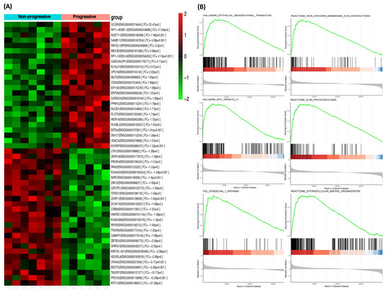
Show Figures

Figure 1

13 pages, 1236 KiB  
Review
From Allergy to Cancer—Clinical Usefulness of Eotaxins
by Monika Zajkowska and Barbara Mroczko
Cancers 2021, 13(1), 128; https://doi.org/10.3390/cancers13010128 - 3 Jan 2021
Cited by 20 | Viewed by 3181
Abstract
Eotaxins are proteins which belong to the group of cytokines. These small molecules are secreted by cells that are mainly involved in immune-mediated reactions in the course of allergic diseases. Eotaxins were discovered in 1994 and their main role was considered to be [...] Read more.
Eotaxins are proteins which belong to the group of cytokines. These small molecules are secreted by cells that are mainly involved in immune-mediated reactions in the course of allergic diseases. Eotaxins were discovered in 1994 and their main role was considered to be the selective recruitment of eosinophils. As those blood cells are involved in the course of all inflammatory diseases, including cancer, we decided to perform an extensive search of the literature pertaining to our investigation via the MEDLINE/PubMed database. On the basis of available literature, we can assume that eotaxins can be used as markers for the detection and determination of origin or type of allergic disease. Many publications also confirm that eotaxins can be used in the determination of allergic disease treatment. Moreover, there are also studies indicating a connection between eotaxins and cancer. Some researchers revealed that CCL11 (C-C motif chemokine ligand 11, eotaxin-1) concentrations differed between the control and tested groups indicating their possible usefulness in cancer detection. Furthermore, some papers showed usefulness of eotaxins in determining the treatment efficacy as markers of decreasing inflammation. Therefore, in this paper we present the current knowledge on eotaxins in the course of allergic and cancerous diseases. Full article
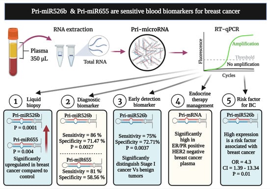
Show Figures

Graphical abstract

2020

Jump to: 2024, 2023, 2022, 2021

13 pages, 4145 KiB  
Article
Global Autozygosity Is Associated with Cancer Risk, Mutational Signature and Prognosis
by Limin Jiang, Fei Guo, Jijun Tang, Shuguan Leng, Scott Ness, Fei Ye, Huining Kang, David C. Samuels and Yan Guo
Cancers 2020, 12(12), 3646; https://doi.org/10.3390/cancers12123646 - 4 Dec 2020
Cited by 2 | Viewed by 2272
Abstract
Global autozygosity quantifies the genome-wide levels of homozygous and heterozygous variants. It is the signature of non-random reproduction, though it can also be driven by other factors, and has been used to assess risk in various diseases. However, the association between global autozygosity [...] Read more.
Global autozygosity quantifies the genome-wide levels of homozygous and heterozygous variants. It is the signature of non-random reproduction, though it can also be driven by other factors, and has been used to assess risk in various diseases. However, the association between global autozygosity and cancer risk has not been studied. From 4057 cancer subjects and 1668 healthy controls, we found strong associations between global autozygosity and risk in ten different cancer types. For example, the heterozygosity ratio was found to be significantly associated with breast invasive carcinoma in Blacks and with male skin cutaneous melanoma in Caucasians. We also discovered eleven associations between global autozygosity and mutational signatures which can explain a portion of the etiology. Furthermore, four significant associations for heterozygosity ratio were revealed in disease-specific survival analyses. This study demonstrates that global autozygosity is effective for cancer risk assessment. Full article
Show Figures

Graphical abstract

11 pages, 335 KiB  
Article
Prognostic Role of the Red Blood Cell Distribution Width (RDW) in Hodgkin Lymphoma
by Ines Herraez, Leyre Bento, Raquel Del Campo, Adriana Sas, Rafael Ramos, Javier Ibarra, Francesc Mestre, Regina Alemany, Joan Bargay, Antonia Sampol and Antonio Gutierrez
Cancers 2020, 12(11), 3262; https://doi.org/10.3390/cancers12113262 - 4 Nov 2020
Cited by 21 | Viewed by 3592
Abstract
The red blood cell distribution width (RDW) is a parameter available from an automated blood count, which measures the degree of heterogeneity of erythrocyte volume and increases in inflammatory conditions. The prognostic role of RDW has been described in different types of cancers. [...] Read more.
The red blood cell distribution width (RDW) is a parameter available from an automated blood count, which measures the degree of heterogeneity of erythrocyte volume and increases in inflammatory conditions. The prognostic role of RDW has been described in different types of cancers. Hodgkin lymphoma (HL) is a hematological malignancy, known to have a proinflammatory background. We aim to study the prognostic role of RDW in HL. We retrospectively analyzed 264 patients with HL from two hospitals in the Balearic Islands between 1990 and 2018. Higher levels of RDW were independently related to anemia, B-symptoms, and low albumin. In age ≥45 years, the presence of lymphopenia and higher RDW were independently associated with worse event-free survival (EFS) and overall survival (OS). Long-term incidence of secondary malignancies was significantly higher in patients with higher RDW, particularly lung cancer. In conclusion, we report for the first time that RDW is a simple, cheap, and easily available prognostic factor in HL that identifies a group with worse EFS, OS, and a higher potential incidence of secondary malignancies. RDW seems to be related to most adverse prognostic factors in HL, making RDW an excellent candidate to be included in prognostic scores for HL. Full article
Show Figures

Figure 1

13 pages, 20609 KiB  
Article
Impact of Genetic Ancestry on Prognostic Biomarkers in Uveal Melanoma
by Daniel A. Rodriguez, Margaret I. Sanchez, Christina L. Decatur, Zelia M. Correa, Eden R. Martin and J. William Harbour
Cancers 2020, 12(11), 3208; https://doi.org/10.3390/cancers12113208 - 31 Oct 2020
Cited by 6 | Viewed by 2971
Abstract
Uveal melanoma (UM) is the most common cancer of the eye and leads to metastatic death in up to half of patients. Genomic prognostic biomarkers play an important role in clinical management in UM. However, research has been conducted almost exclusively in patients [...] Read more.
Uveal melanoma (UM) is the most common cancer of the eye and leads to metastatic death in up to half of patients. Genomic prognostic biomarkers play an important role in clinical management in UM. However, research has been conducted almost exclusively in patients of European descent, such that the association between genetic admixture and prognostic biomarkers is unknown. In this study, we compiled 1381 control genomes from West African, European, East Asian, and Native American individuals, assembled a bioinformatic pipeline for assessing global and local ancestry, and performed an initial pilot study of 141 UM patients from our international referral center that manages many admixed individuals. Global and local estimates were associated with genomic prognostic determinants. Expression quantitative trait loci (eQTL) analysis was performed on variants found in segments. Globally, after correction for multiple testing, no prognostic variable was significantly enriched in a given ancestral group. However, there was a trend suggesting an increased proportion of European ancestry associated with expression of the PRAME oncogene (q = 0.06). Locally enriched European haplotypes were associated with the poor prognosis class 2 gene expression profile and with genes involved in immune regulation (q = 4.7 × 10⁻¹¹). These findings reveal potential influences of genetic ancestry on prognostic variables, implicate immune genes in prognostic differences based on ancestry, and provide a basis for future studies of admixed patients with UM using rigorous genetic ancestry methodology. Full article
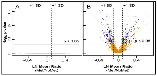
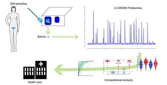
Show Figures

Figure 1

10 pages, 636 KiB  
Article
Validating fPSA Glycoprofile as a Prostate Cancer Biomarker to Avoid Unnecessary Biopsies and Re-Biopsies
by Tomas Bertok, Eduard Jane, Aniko Bertokova, Lenka Lorencova, Peter Zvara, Bozena Smolkova, Radek Kucera, Helmut Klocker and Jan Tkac
Cancers 2020, 12(10), 2988; https://doi.org/10.3390/cancers12102988 - 15 Oct 2020
Cited by 20 | Viewed by 2953
Abstract
Background: To compare the clinical performance of a new PCa serum biomarker based on fPSA glycoprofiling to fPSA% and PHI. Methods: Serum samples from men who underwent prostate biopsy due to increased PSA were used. A comparison between two equal groups (with histologically [...] Read more.
Background: To compare the clinical performance of a new PCa serum biomarker based on fPSA glycoprofiling to fPSA% and PHI. Methods: Serum samples from men who underwent prostate biopsy due to increased PSA were used. A comparison between two equal groups (with histologically confirmed PCa or benign, non-cancer condition) was used for the clinical validation of a new glycan-based PCa oncomarker. SPSS and R software packages were used for the multiparametric analyses of the receiver operating curve (ROC) and for genetic algorithm metaheuristics. Results: When comparing the non-cancer and PCa cohorts, the combination of four fPSA glycoforms with two clinical parameters (PGI, prostate glycan index (PGI)) showed an area under receiver operating curve (AUC) value of 0.821 (95% CI 0.754–0.890). AUC values were 0.517 for PSA, 0.683 for fPSA%, and 0.737 for PHI. A glycan analysis was also applied to discriminate low-grade tumors (GS = 6) from significant tumors (GS ≥ 7). Conclusions: Compared to PSA on its own, or fPSA% and the PHI, PGI showed improved discrimination between presence and absence of PCa and in predicting clinically significant PCa. In addition, the use of PGI would help practitioners avoid 63.5% of unnecessary biopsies, while the use of fPSA% and PHI would help avoid 17.5% and 33.3% of biopsies, respectively, while missing four significant tumors (9.5%). Full article
Show Figures

Graphical abstract

Back to TopTop