Mast Cells in Health and Diseases

A topical collection in Cells (ISSN 2073-4409). This collection belongs to the section "Cellular Immunology".

Viewed by 142457

Editor


E-Mail Website
Collection Editor
Director of Center for Allergy, Associate Professor, Allergy and Immunology Research Project Team, Research Institute of Medical Science, Nihon University School of Medicine, Tokyo 173-8610, Japan
Interests: human mast cell heterogeneity; the roles of mast cell-derived exosomes in health and diseases; the roles of Mas-related G-protein recepror X2 (MRGPRX2) in diseases; the activation mechanism of mast cells in chronic spontaneous urticaria and rheumatoid arthritis

Topical Collection Information

Dear Colleagues,

Mast cells leave the bone marrow as progenitors and reach their final maturation within peripheral tissues. Mast cells play a central role in IgE-dependent allergic diseases such as asthma and rhinoconjunctivitis through the release of a variety of vasoactive and bronchospastic autacoid mediators and functionally diverse proteases, chemokines and cytokines. Mast cells play an important role in innate immunity, angiogenesis and tissue repairing through non-IgE-dependent stimulation. In this Topical Collection of Cells, I invite you to contribute, either in the form of original research articles, reviews or shorter perspective articles on all aspects related to the theme of “Mast Cells in Health and Diseases”. Expert articles describing new roles of mast cells in health and diseases, new evidence of mast cell differentiation and maturation, and mast cell heterogeneity are welcome.

Dr. Yoshimichi Okayama
Collection Editor

Manuscript Submission Information

Manuscripts should be submitted online at www.mdpi.com by registering and logging in to this website. Once you are registered, click here to go to the submission form. Manuscripts can be submitted until the deadline. All submissions that pass pre-check are peer-reviewed. Accepted papers will be published continuously in the journal (as soon as accepted) and will be listed together on the collection website. Research articles, review articles as well as short communications are invited. For planned papers, a title and short abstract (about 100 words) can be sent to the Editorial Office for announcement on this website.

Submitted manuscripts should not have been published previously, nor be under consideration for publication elsewhere (except conference proceedings papers). All manuscripts are thoroughly refereed through a single-blind peer-review process. A guide for authors and other relevant information for submission of manuscripts is available on the Instructions for Authors page. Cells is an international peer-reviewed open access semimonthly journal published by MDPI.

Please visit the Instructions for Authors page before submitting a manuscript. The Article Processing Charge (APC) for publication in this open access journal is 2700 CHF (Swiss Francs). Submitted papers should be well formatted and use good English. Authors may use MDPI's English editing service prior to publication or during author revisions.

Keywords

  • Mast cell;
  • IgE;
  • Cytokine;
  • Lipid mediator.
Published Papers (29 papers)

2024

Jump to: 2022, 2021

16 pages, 2434 KiB  
Article
CREB Is Critically Implicated in Skin Mast Cell Degranulation Elicited via FcεRI and MRGPRX2
by Zhuoran Li, Jean Schneikert, Shiva Raj Tripathi, Manqiu Jin, Gürkan Bal, Torsten Zuberbier and Magda Babina
Cells 2024, 13(20), 1681; https://doi.org/10.3390/cells13201681 - 11 Oct 2024
Viewed by 795
Abstract
Skin mast cells (MCs) mediate acute allergic reactions in the cutaneous environment and contribute to chronic dermatoses, including urticaria, and atopic or contact dermatitis. The cAMP response element binding protein (CREB), an evolutionarily well conserved transcription factor (TF) with over 4,000 binding sites [...] Read more.
Skin mast cells (MCs) mediate acute allergic reactions in the cutaneous environment and contribute to chronic dermatoses, including urticaria, and atopic or contact dermatitis. The cAMP response element binding protein (CREB), an evolutionarily well conserved transcription factor (TF) with over 4,000 binding sites in the genome, was recently found to form a feedforward loop with KIT, maintaining MC survival. The most selective MC function is degranulation with its acute release of prestored mediators. Herein, we asked whether CREB contributes to the expression and function of the degranulation-competent receptors FcεRI and MRGPRX2. Interference with CREB by pharmacological inhibition (CREBi, 666-15) or RNA interference only slightly affected the expression of these receptors, while KIT was strongly attenuated. Interestingly, MRGPRX2 surface expression moderately increased following CREB-knockdown, whereas MRGPRX2-dependent exocytosis simultaneously decreased. FcεRI expression and function were regulated consistently, although the effect was stronger at the functional level. Preformed MC mediators (tryptase, histamine, β-hexosaminidase) remained comparable following CREB attenuation, suggesting that granule synthesis did not rely on CREB function. Collectively, in contrast to KIT, FcεRI and MRGPRX2 moderately depend on unperturbed CREB function. Nevertheless, CREB is required to maintain MC releasability irrespective of stimulus, insinuating that CREB may operate by safeguarding the degranulation machinery. To our knowledge, CREB is the first factor identified to regulate MRGPRX2 expression and function in opposite direction. Overall, the ancient TF is an indispensable component of skin MCs, orchestrating not only survival and proliferation but also their secretory competence. Full article
Show Figures

Figure 1

14 pages, 1632 KiB  
Review
The Role Played by Autophagy in FcεRI-Dependent Activation of Mast Cells
by Anastasia N. Pavlyuchenkova, Maxim S. Smirnov, Boris V. Chernyak and Maria A. Chelombitko
Cells 2024, 13(8), 690; https://doi.org/10.3390/cells13080690 - 16 Apr 2024
Cited by 2 | Viewed by 1431
Abstract
The significant role of mast cells in the development of allergic and inflammatory diseases is well-established. Among the various mechanisms of mast cell activation, the interaction of antigens/allergens with IgE and the subsequent binding of this complex to the high-affinity IgE receptor FcεRI [...] Read more.
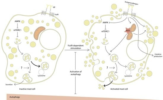
The significant role of mast cells in the development of allergic and inflammatory diseases is well-established. Among the various mechanisms of mast cell activation, the interaction of antigens/allergens with IgE and the subsequent binding of this complex to the high-affinity IgE receptor FcεRI stand out as the most studied and fundamental pathways. This activation process leads to the rapid exocytosis of granules containing preformed mediators, followed by the production of newly synthesized mediators, including a diverse array of cytokines, chemokines, arachidonic acid metabolites, and more. While conventional approaches to allergy control primarily focus on allergen avoidance and the use of antihistamines (despite their associated side effects), there is increasing interest in exploring novel methods to modulate mast cell activity in modern medicine. Recent evidence suggests a role for autophagy in mast cell activation, offering potential avenues for utilizing low-molecular-weight autophagy regulators in the treatment of allergic diseases. More specifically, mitochondria, which play an important role in the regulation of autophagy as well as mast cell activation, emerge as promising targets for drug development. This review examines the existing literature regarding the involvement of the molecular machinery associated with autophagy in FcεRI-dependent mast cell activation. Full article
Show Figures

Graphical abstract

15 pages, 1308 KiB  
Article
Coronavirus Receptor Expression Profiles in Human Mast Cells, Basophils, and Eosinophils
by Lina Degenfeld-Schonburg, Irina Sadovnik, Dubravka Smiljkovic, Barbara Peter, Gabriele Stefanzl, Clemens Gstoettner, Peter Jaksch, Konrad Hoetzenecker, Clemens Aigner, Christine Radtke, Michel Arock, Wolfgang R. Sperr and Peter Valent
Cells 2024, 13(2), 173; https://doi.org/10.3390/cells13020173 - 17 Jan 2024
Cited by 2 | Viewed by 2080
Abstract
A major problem in SARS-CoV-2-infected patients is the massive tissue inflammation in certain target organs, including the lungs. Mast cells (MC), basophils (BA), and eosinophils (EO) are key effector cells in inflammatory processes. These cells have recently been implicated in the pathogenesis of [...] Read more.
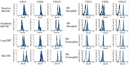
A major problem in SARS-CoV-2-infected patients is the massive tissue inflammation in certain target organs, including the lungs. Mast cells (MC), basophils (BA), and eosinophils (EO) are key effector cells in inflammatory processes. These cells have recently been implicated in the pathogenesis of SARS-CoV-2 infections. We explored coronavirus receptor (CoV-R) expression profiles in primary human MC, BA, and EO, and in related cell lines (HMC-1, ROSA, MCPV-1, KU812, and EOL-1). As determined using flow cytometry, primary MC, BA, and EO, and their corresponding cell lines, displayed the CoV-R CD13 and CD147. Primary skin MC and BA, as well as EOL-1 cells, also displayed CD26, whereas primary EO and the MC and BA cell lines failed to express CD26. As assessed using qPCR, most cell lines expressed transcripts for CD13, CD147, and ABL2, whereas ACE2 mRNA was not detectable, and CD26 mRNA was only identified in EOL-1 cells. We also screened for drug effects on CoV-R expression. However, dexamethasone, vitamin D, and hydroxychloroquine did not exert substantial effects on the expression of CD13, CD26, or CD147 in the cells. Together, MC, BA, and EO express distinct CoV-R profiles. Whether these receptors mediate virus–cell interactions and thereby virus-induced inflammation remains unknown at present. Full article
Show Figures

Figure 1

2022

Jump to: 2024, 2021

14 pages, 1984 KiB  
Article
Mast Cell Proteases Promote Diverse Effects on the Plasminogen Activation System and Wound Healing in A549 Alveolar Epithelial Cells
by Sofia Mogren, Frida Berlin, Lykke Eskilsson, Nicole Van Der Burg, Ellen Tufvesson and Cecilia K. Andersson
Cells 2022, 11(18), 2916; https://doi.org/10.3390/cells11182916 - 18 Sep 2022
Cited by 1 | Viewed by 2854
Abstract
Tissue damage, epithelial alterations, and intraepithelial presence of mast cells (MCs) are characteristics of asthma pathogenesis. Increased alveolar infiltration of MC populations has also been identified as a feature of asthma and other chronic respiratory diseases. The asthma associated receptor, urokinase plasminogen activator [...] Read more.
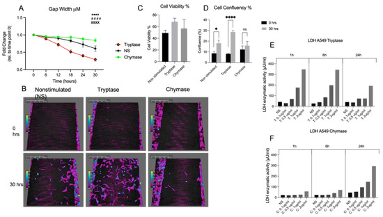
Tissue damage, epithelial alterations, and intraepithelial presence of mast cells (MCs) are characteristics of asthma pathogenesis. Increased alveolar infiltration of MC populations has also been identified as a feature of asthma and other chronic respiratory diseases. The asthma associated receptor, urokinase plasminogen activator receptor (uPAR), has been shown to regulate bronchial epithelial repair responses. However, the impact of MC tryptase and chymase on functional properties and expression of uPAR in alveolar epithelial cells have not been fully investigated. Alveolar epithelial cell migration and wound healing were investigated using holographic live cell imaging of A549 cells in a wound scratch model post stimulation with tryptase or chymase. The expression of uPAR was investigated on the protein and gene level from cellular supernatants and in bronchoalveolar lavage fluid fractions from allergic asthmatics. We found that tryptase improved wound healing capacity, cellular migration and membrane bound uPAR expression. Chymase reduced gap closure capacity, cellular migration and membrane bound uPAR expression but increased soluble uPAR release. Our data suggest a dual regulatory response from the MC proteases in events related to uPAR expression and wound healing which could be important features in asthmatic disease. Full article
Show Figures

Figure 1

14 pages, 3208 KiB  
Review
Mast Cells Meet Cytomegalovirus: A New Example of Protective Mast Cell Involvement in an Infectious Disease
by Sara Becker, Matthias J. Reddehase and Niels A. Lemmermann
Cells 2022, 11(9), 1402; https://doi.org/10.3390/cells11091402 - 21 Apr 2022
Cited by 1 | Viewed by 2366
Abstract
Cytomegaloviruses (CMVs) belong to the β-subfamily of herpesviruses. Their host-to-host transmission involves the airways. As primary infection of an immunocompetent host causes only mild feverish symptoms, human CMV (hCMV) is usually not considered in routine differential diagnostics of common airway infections. Medical relevance [...] Read more.
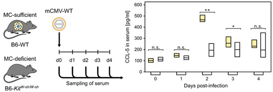
Cytomegaloviruses (CMVs) belong to the β-subfamily of herpesviruses. Their host-to-host transmission involves the airways. As primary infection of an immunocompetent host causes only mild feverish symptoms, human CMV (hCMV) is usually not considered in routine differential diagnostics of common airway infections. Medical relevance results from unrestricted tissue infection in an immunocompromised host. One risk group of concern are patients who receive hematopoietic cell transplantation (HCT) for immune reconstitution following hematoablative therapy of hematopoietic malignancies. In HCT patients, interstitial pneumonia is a frequent cause of death from hCMV strains that have developed resistance against antiviral drugs. Prevention of CMV pneumonia requires efficient reconstitution of antiviral CD8 T cells that infiltrate lung tissue. A role for mast cells (MC) in the immune control of lung infection by a CMV was discovered only recently in a mouse model. MC were shown to be susceptible for productive infection and to secrete the chemokine CCL-5, which recruits antiviral CD8 T cells to the lungs and thereby improves the immune control of pulmonary infection. Here, we review recent data on the mechanism of MC-CMV interaction, a field of science that is new for CMV virologists as well as for immunologists who have specialized in MC. Full article
Show Figures

Figure 1

17 pages, 3184 KiB  
Article
Insights into Early-Pregnancy Mechanisms: Mast Cells and Chymase CMA1 Shape the Phenotype and Modulate the Functionality of Human Trophoblast Cells, Vascular Smooth-Muscle Cells and Endothelial Cells
by Ningjuan Zhang, Anne Schumacher, Beate Fink, Mario Bauer, Ana Claudia Zenclussen and Nicole Meyer
Cells 2022, 11(7), 1158; https://doi.org/10.3390/cells11071158 - 29 Mar 2022
Cited by 4 | Viewed by 3090
Abstract
Spiral-artery (SA) remodeling is a fundamental process during pregnancy that involves the action of cells of the initial vessel, such as vascular smooth-muscle cells (VSMCs) and endothelial cells, but also maternal immune cells and fetal extravillous trophoblast cells (EVTs). Mast cells (MCs), and [...] Read more.
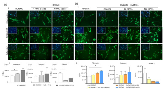
Spiral-artery (SA) remodeling is a fundamental process during pregnancy that involves the action of cells of the initial vessel, such as vascular smooth-muscle cells (VSMCs) and endothelial cells, but also maternal immune cells and fetal extravillous trophoblast cells (EVTs). Mast cells (MCs), and specifically chymase-expressing cells, have been identified as key to a sufficient SA-remodeling process in vivo. However, the mechanisms are still unclear. The purpose of this study is to evaluate the effects of the MC line HMC-1 and recombinant human chymase (rhuCMA1) on human primary uterine vascular smooth-muscle cells (HUtSMCs), a human trophoblast cell line (HTR8/SV-neo), and human umbilical-vein endothelial cells (HUVEC) in vitro. Both HMC-1 and rhuCMA1 stimulated migration, proliferation, and changed protein expression in HUtSMCs. HMC-1 increased proliferation, migration, and changed gene expression of HTR8/SVneo cells, while rhuCMA treatment led to increased migration and decreased expression of tissue inhibitors of matrix metalloproteinases. Additionally, rhuCMA1 enhanced endothelial-cell-tube formation. Collectively, we identified possible mechanisms by which MCs/rhuCMA1 promote SA remodeling. Our findings are relevant to the understanding of this crucial step in pregnancy and thus of the dysregulated pathways that can lead to pregnancy complications such as fetal growth restriction and preeclampsia. Full article
Show Figures

Figure 1

18 pages, 1937 KiB  
Article
Functional and Phenotypic Characterization of Siglec-6 on Human Mast Cells
by Piper A. Robida, Clayton H. Rische, Netali Ben-Baruch Morgenstern, Rethavathi Janarthanam, Yun Cao, Rebecca A. Krier-Burris, Wouter Korver, Alan Xu, Thuy Luu, Julia Schanin, John Leung, Marc E. Rothenberg, Joshua B. Wechsler, Bradford A. Youngblood, Bruce S. Bochner and Jeremy A. O’Sullivan
Cells 2022, 11(7), 1138; https://doi.org/10.3390/cells11071138 - 28 Mar 2022
Cited by 22 | Viewed by 6703
Abstract
Mast cells are tissue-resident cells that contribute to allergic diseases, among others, due to excessive or inappropriate cellular activation and degranulation. Therapeutic approaches to modulate mast cell activation are urgently needed. Siglec-6 is an immunoreceptor tyrosine-based inhibitory motif (ITIM)-bearing receptor selectively expressed by [...] Read more.
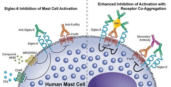
Mast cells are tissue-resident cells that contribute to allergic diseases, among others, due to excessive or inappropriate cellular activation and degranulation. Therapeutic approaches to modulate mast cell activation are urgently needed. Siglec-6 is an immunoreceptor tyrosine-based inhibitory motif (ITIM)-bearing receptor selectively expressed by mast cells, making it a promising target for therapeutic intervention. However, the effects of its engagement on mast cells are poorly defined. Siglec-6 expression and endocytosis on primary human mast cells and mast cell lines were assessed by flow cytometry. SIGLEC6 mRNA expression was examined by single-cell RNAseq in esophageal tissue biopsy samples. The ability of Siglec-6 engagement or co-engagement to prevent primary mast cell activation was determined based on assessments of mediator and cytokine secretion and degranulation markers. Siglec-6 was highly expressed by all mast cells examined, and the SIGLEC6 transcript was restricted to mast cells in esophageal biopsy samples. Siglec-6 endocytosis occurred with delayed kinetics relative to the related receptor Siglec-8. Co-crosslinking of Siglec-6 with FcεRIα enhanced the inhibition of mast cell activation and diminished downstream ERK1/2 and p38 phosphorylation. The selective, stable expression and potent inhibitory capacity of Siglec-6 on human mast cells are favorable for its use as a therapeutic target in mast cell-driven diseases. Full article
Show Figures

Graphical abstract

18 pages, 3656 KiB  
Article
MRGPRX2-Mediated Degranulation of Human Skin Mast Cells Requires the Operation of Gαi, Gαq, Ca++ Channels, ERK1/2 and PI3K—Interconnection between Early and Late Signaling
by Zhao Wang, Kristin Franke, Gürkan Bal, Zhuoran Li, Torsten Zuberbier and Magda Babina
Cells 2022, 11(6), 953; https://doi.org/10.3390/cells11060953 - 10 Mar 2022
Cited by 23 | Viewed by 5187
Abstract
The recent discovery of MRGPRX2 explains mast cell (MC)-dependent symptoms independently of FcεRI-activation. Because of its novelty, signaling cascades triggered by MRGPRX2 are rudimentarily understood, especially in cutaneous MCs, by which MRGPRX2 is chiefly expressed. Here, MCs purified from human skin were used [...] Read more.
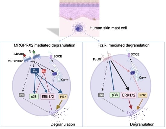
The recent discovery of MRGPRX2 explains mast cell (MC)-dependent symptoms independently of FcεRI-activation. Because of its novelty, signaling cascades triggered by MRGPRX2 are rudimentarily understood, especially in cutaneous MCs, by which MRGPRX2 is chiefly expressed. Here, MCs purified from human skin were used following preculture or ex vivo and stimulated by FcεRI-aggregation or MRGPRX2 agonists (compound 48/80, Substance P) in the presence/absence of inhibitors. Degranulation was assessed by β-hexosaminidase or histamine release. Phosphorylation events were studied by immunoblotting. As a G protein-coupled receptor, MRGPRX2 signals by activating G proteins; however, their nature has remained controversial. In skin MCs, Gαi and Gαq were required for degranulation, but Gαi was clearly more relevant. Ca++ channels were likewise crucial. Downstream, PI3K was essential for granule discharge initiated by MRGPRX2 or FcεRI. ERK1/2 and JNK were additional participants, especially in the allergic route. Addressing possible points of intersection between early and later events, pERK1/2 and pAKT were found to depend on Gαi, further highlighting its significance. Gαq and Ca++ channels made some contributions to the phosphorylation of ERK. Ca++ differentially affected PI3K activation in FcεRI- vis-à-vis MRGPRX2-signaling, as channel inhibition increased pAKT only when triggered via FcεRI. Collectively, our study significantly extends our understanding of the molecular framework behind granule secretion from skin MCs. Full article
Show Figures

Graphical abstract

20 pages, 2716 KiB  
Article
Fetal Tissue-Derived Mast Cells (MC) as Experimental Surrogate for In Vivo Connective Tissue MC
by Caterina Iuliano, Magdalena Absmaier-Kijak, Tobias Sinnberg, Nils Hoffard, Miriam Hils, Martin Köberle, Florian Wölbing, Ekaterina Shumilina, Nicole Heise, Birgit Fehrenbacher, Martin Schaller, Florian Lang, Susanne Kaesler and Tilo Biedermann
Cells 2022, 11(6), 928; https://doi.org/10.3390/cells11060928 - 8 Mar 2022
Cited by 4 | Viewed by 3945
Abstract
Bone-marrow-derived mast cells are matured from bone marrow cells in medium containing 20% fetal calf serum (FCS), interleukin (IL)-3 and stem-cell factor (SCF) and are used as in vitro models to study mast cells (MC) and their role in health and disease. In [...] Read more.
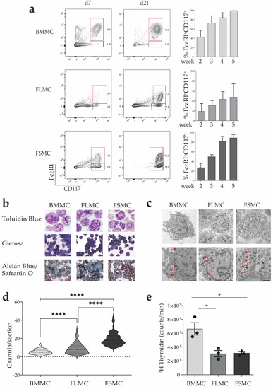
Bone-marrow-derived mast cells are matured from bone marrow cells in medium containing 20% fetal calf serum (FCS), interleukin (IL)-3 and stem-cell factor (SCF) and are used as in vitro models to study mast cells (MC) and their role in health and disease. In vivo, however, BM-derived hematopoietic stem cells account for only a fraction of MC; the majority of MC in vivo are and remain tissue resident. In this study we established a side-by-side culture with BMMC, fetal skin MC (FSMC) or fetal liver MC (FLMC) for comparative studies to identify the best surrogates for mature connective tissue MC (CTMC). All three MC types showed comparable morphology by histology and MC phenotype by flow cytometry. Heterogeneity was detected in the transcriptome with the most differentially expressed genes in FSMC compared to BMMC being Hdc and Tpsb2. Expression of ST2 was highly expressed in BMMC and FSMC and reduced in FLMC, diminishing their secretion of type 2 cytokines. Higher granule content, stronger response to FcεRI activation and significantly higher release of histamine from FSMC compared to FLMC and BMMC indicated differences in MC development in vitro dependent on the tissue of origin. Thus, tissues of origin imprint MC precursor cells to acquire distinct phenotypes and signatures despite identical culture conditions. Fetal-derived MC resemble mature CTMC, with FSMC being the most developed. Full article
Show Figures

Figure 1

13 pages, 5812 KiB  
Review
Mast Cells and Acupuncture Analgesia
by Yingchen Li, Yi Yu, Yuhang Liu and Wei Yao
Cells 2022, 11(5), 860; https://doi.org/10.3390/cells11050860 - 2 Mar 2022
Cited by 24 | Viewed by 7523
Abstract
Mast cells are widely distributed in various parts of the human body and play a vital role in the progression of many diseases. Recently, the close relationship between mast cells and acupoints was elucidated, and the role of mast cells in acupuncture analgesia [...] Read more.
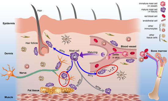
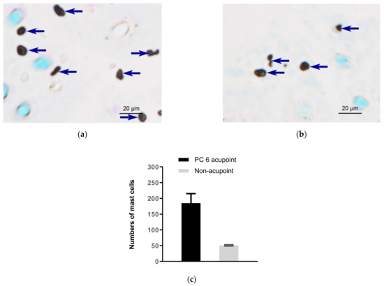
Mast cells are widely distributed in various parts of the human body and play a vital role in the progression of many diseases. Recently, the close relationship between mast cells and acupoints was elucidated, and the role of mast cells in acupuncture analgesia has attracted the attention of researchers worldwide. Using mast cells, acupuncture analgesia and acupoint as key words to search CNKI, PubMed, Web of Science and other databases, combining the representative articles in these databases with the published research papers of our group, we summarized: The enrichment of mast cells and the dense arrangement of collagen fibers, microvessels, and nerves form the basis for acupoints as the reaction sites of acupuncture; acupuncture can cause the deformation of collagen fibers and activate TRPV channels on mast cells membrane, so as to stimulate mast cells to release bioactive substances and activate nerve receptors to generate analgesic effect; system biology models are set up to explain the quantitative process of information initiation and transmission at acupuncture points, and indicate that the acupuncture effect depends on the local mast cells density. In a conclusion, this review will give a scientific explanation of acupuncture analgesia from the material basis of acupoints, the local initiation, and afferent biological mechanism. Full article
Show Figures

Figure 1

28 pages, 47685 KiB  
Review
Activation of Subcutaneous Mast Cells in Acupuncture Points Triggers Analgesia
by Li-Na Wang, Xue-Zhi Wang, Yu-Jia Li, Bing-Rong Li, Meng Huang, Xiao-Yu Wang, Ryszard Grygorczyk, Guang-Hong Ding and Wolfgang Schwarz
Cells 2022, 11(5), 809; https://doi.org/10.3390/cells11050809 - 25 Feb 2022
Cited by 30 | Viewed by 6190
Abstract
This review summarizes experimental evidence indicating that subcutaneous mast cells are involved in the trigger mechanism of analgesia induced by acupuncture, a traditional oriental therapy, which has gradually become accepted worldwide. The results are essentially based on work from our laboratories. Skin mast [...] Read more.
This review summarizes experimental evidence indicating that subcutaneous mast cells are involved in the trigger mechanism of analgesia induced by acupuncture, a traditional oriental therapy, which has gradually become accepted worldwide. The results are essentially based on work from our laboratories. Skin mast cells are present at a high density in acupuncture points where fine needles are inserted and manipulated during acupuncture intervention. Mast cells are sensitive to mechanical stimulation because they express multiple types of mechanosensitive channels, including TRPV1, TRPV2, TRPV4, receptors and chloride channels. Acupuncture manipulation generates force and torque that indirectly activate the mast cells via the collagen network. Subsequently, various mediators, for example, histamine, serotonin, adenosine triphosphate and adenosine, are released from activated mast cells to the interstitial space; they or their downstream products activate the corresponding receptors situated at local nerve terminals of sensory neurons in peripheral ganglia. The analgesic effects are thought to be generated via the reduced electrical activities of the primary sensory neurons. Alternatively, these neurons project such signals to pain-relevant regions in spinal cord and/or higher centers of the brain. Full article
Show Figures

Figure 1

26 pages, 4577 KiB  
Article
Potential Role of Intracranial Mast Cells in Neuroinflammation and Neuropathology Associated with Food Allergy
by Danielle L. Germundson and Kumi Nagamoto-Combs
Cells 2022, 11(4), 738; https://doi.org/10.3390/cells11040738 - 20 Feb 2022
Cited by 6 | Viewed by 5668
Abstract
Mast cells (MCs) are the major effector cells of allergic responses and reside throughout the body, including in the brain and meninges. Previously, we showed in a mouse model of subclinical cow’s milk allergy that brain MC numbers were elevated in sensitized mice. [...] Read more.
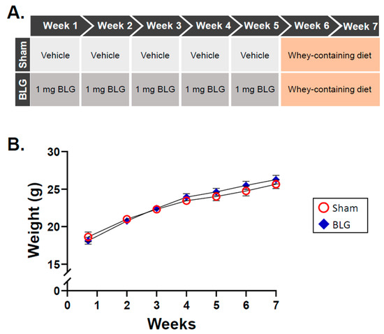
Mast cells (MCs) are the major effector cells of allergic responses and reside throughout the body, including in the brain and meninges. Previously, we showed in a mouse model of subclinical cow’s milk allergy that brain MC numbers were elevated in sensitized mice. However, the neurophysiological consequences of intracranial MC accumulation and activation are unclear. We hypothesized that centrally recruited MCs in sensitized mice could be activated by the allergen via the IgE/FcεRI mechanism and increase the blood–brain barrier (BBB) permeability to promote neuroinflammation. Furthermore, we suspected that repeated allergen exposure could sustain MC activation. To investigate our hypothesis, we sensitized C57BL6/J mice to a bovine whey allergen, β-lactoglobulin (BLG), and subsequently placed them on a whey-containing diet for two weeks. MC activity and associated changes in the brain were examined. BLG-sensitized mice showed mobility changes and depression-like behavior with significantly increased MC numbers and histamine levels in select brain regions. IgG extravasation and perivascular astrogliosis were also evident. Importantly, myelin staining revealed cortical demyelination in the BLG-sensitized mice, suggesting a potential neural substrate for their behavioral changes. Our findings support the ability of brain MCs to release histamine and other mediators to increase BBB permeability and facilitate neuroinflammatory responses in the brain. Full article
Show Figures

Figure 1

14 pages, 942 KiB  
Review
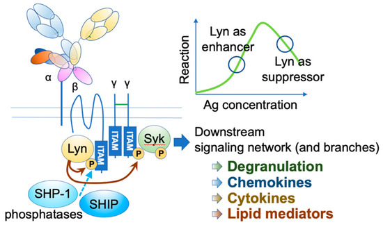
FcεRI: A Master Regulator of Mast Cell Functions
by Yuka Nagata and Ryo Suzuki
Cells 2022, 11(4), 622; https://doi.org/10.3390/cells11040622 - 11 Feb 2022
Cited by 29 | Viewed by 4738
Abstract
Mast cells (MCs) perform multiple functions thought to underlie different manifestations of allergies. Various aspects of antigens (Ags) and their interactions with immunoglobulin E (IgE) cause diverse responses in MCs. FcεRI, a high-affinity IgE receptor, deciphers the Ag–IgE interaction and drives allergic responses. [...] Read more.
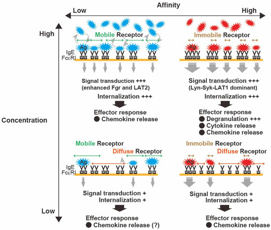
Mast cells (MCs) perform multiple functions thought to underlie different manifestations of allergies. Various aspects of antigens (Ags) and their interactions with immunoglobulin E (IgE) cause diverse responses in MCs. FcεRI, a high-affinity IgE receptor, deciphers the Ag–IgE interaction and drives allergic responses. FcεRI clustering is essential for signal transduction and, therefore, determines the quality of MC responses. Ag properties precisely regulate FcεRI dynamics, which consequently initiates differential outcomes by switching the intracellular-signaling pathway, suggesting that Ag properties can control MC responses, both qualitatively and quantitatively. Thus, the therapeutic benefits of FcεRI-targeting strategies have long been examined. Disrupting IgE–FcεRI interactions is a potential therapeutic strategy because the binding affinity between IgE and FcεRI is extremely high. Specifically, FcεRI desensitization, due to internalization, is also a potential therapeutic target that is involved in the mechanisms of allergen-specific immunotherapy. Several recent findings have suggested that silent internalization is strongly associated with FcεRI dynamics. A comprehensive understanding of the role of FcεRI may lead to the development of novel therapies for allergies. Here, we review the qualitatively diverse responses of MCs that impact the attenuation/development of allergies with a focus on the role of FcεRI toward Ag exposure. Full article
Show Figures

Figure 1

14 pages, 3690 KiB  
Article
Synergistic Cytokine Production by ATP and PGE2 via P2X4 and EP3 Receptors in Mouse Bone-Marrow-Derived Mast Cells
by Kosuke Obayashi, Kazuki Yoshida, Masa-aki Ito, Tetsuya Mori, Kimiko Yamamoto, Toshiyashu Imai and Isao Matsuoka
Cells 2022, 11(4), 616; https://doi.org/10.3390/cells11040616 - 10 Feb 2022
Cited by 4 | Viewed by 2725
Abstract
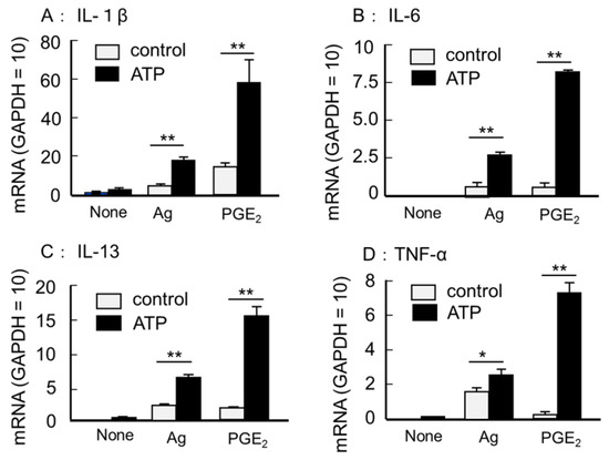
ATP is an important intercellular messenger in the extracellular space. In mast cells (MCs), ATP stimulates the ionotropic P2X4 receptor (P2X4R), resulting in enhanced degranulation and exacerbation of acute allergic reactions. In this study, we investigate whether ATP regulates inflammatory cytokine production in [...] Read more.
ATP is an important intercellular messenger in the extracellular space. In mast cells (MCs), ATP stimulates the ionotropic P2X4 receptor (P2X4R), resulting in enhanced degranulation and exacerbation of acute allergic reactions. In this study, we investigate whether ATP regulates inflammatory cytokine production in MCs. Gene expression was analyzed by quantitative RT-PCR, and cytokine production was measured using ELISA. The stimulation of mouse bone-marrow-derived MCs (BMMCs) with ATP alone had little effect on cytokine secretion. However, the co-stimulation with prostaglandin (PG) E2 resulted in a marked increase in the secretion of various cytokines, such as tumor necrosis factor-α, interleukin (IL)-6, and IL-13, accompanied by an increase in their mRNA levels. The effects of ATP were inhibited by P2X4R antagonists and diminished in BMMCs derived from P2X4R-deficient mice, suggesting that P2X4R mediated the reaction. The effects of PGE2 were mimicked by an EP3 receptor (EP3R) agonist and blocked by an EP3R antagonist. The synergistic cytokine mRNA elevations induced by ATP and PGE2 were blocked by nuclear factor-κB and Ca2+-calcineurin signaling inhibitors. Altogether, these results suggest that combining P2X4R and EP3R signaling enhances acute degranulation and the subsequent cytokine secretion, exacerbating allergic inflammation. Full article
Show Figures

Figure 1

14 pages, 1863 KiB  
Article
The Critical Role Played by Mitochondrial MITF Serine 73 Phosphorylation in Immunologically Activated Mast Cells
by Lakshmi Bhargavi Paruchuru, Sharmila Govindaraj and Ehud Razin
Cells 2022, 11(3), 589; https://doi.org/10.3390/cells11030589 - 8 Feb 2022
Cited by 7 | Viewed by 3032
Abstract
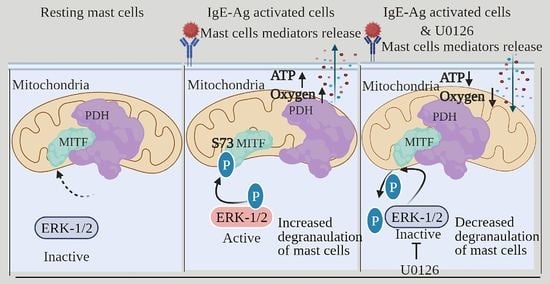
In recent years, growing evidence has indicated the pivotal role of mitochondria in mast cell immunological activation. We have previously reported a decrease in degranulation and cytokine secretion following the inhibition of pyruvate dehydrogenase (PDH) either by CPI-613 (PDH inhibitor/anti-cancer drug) or through [...] Read more.
In recent years, growing evidence has indicated the pivotal role of mitochondria in mast cell immunological activation. We have previously reported a decrease in degranulation and cytokine secretion following the inhibition of pyruvate dehydrogenase (PDH) either by CPI-613 (PDH inhibitor/anti-cancer drug) or through its interaction with mitochondrial microphthalmia-associated transcription factor (MITF). In the present study, we further explored the role played by mitochondrial MITF in mast cell exocytosis using rat basophil leukemia cells [RBL], as well as mouse bone marrow-derived mast cells (BMMCs). Here, we report that mast cell degranulation, cytokine secretion and oxidative phosphorylation (OXPHOS) activities were associated with phosphorylation of Serine 73 of mitochondrial MITF, controlled by extracellular signals regulated by protein kinase (ERK1/2) activity. Also, we report here that decreased OXPHOS activity following ERK1/2 inhibition (U0126 treatment) during IgE-Ag activation was mediated by the dephosphorylation of Serine 73 mitochondrial MITF, which inhibited its association with PDH. This led to a reduction in mast cell reactivity. In addition, a phosphorylation-mimicking mitochondrial MITF-S73D positively regulated the mitochondrial activity, thereby supporting mast cell degranulation. Thus, the present research findings highlight the prominence of mitochondrial MITF Serine 73 phosphorylation in immunologically activated mast cells. Full article
Show Figures

Graphical abstract

23 pages, 4332 KiB  
Review
Carboxypeptidase A3—A Key Component of the Protease Phenotype of Mast Cells
by Dmitri Atiakshin, Andrey Kostin, Ivan Trotsenko, Vera Samoilova, Igor Buchwalow and Markus Tiemann
Cells 2022, 11(3), 570; https://doi.org/10.3390/cells11030570 - 6 Feb 2022
Cited by 27 | Viewed by 3915
Abstract
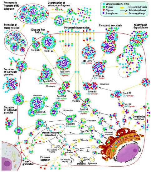
Carboxypeptidase A3 (CPA3) is a specific mast cell (MC) protease with variable expression. This protease is one of the preformed components of the secretome. During maturation of granules, CPA3 becomes an active enzyme with a characteristic localization determining the features of the cytological [...] Read more.
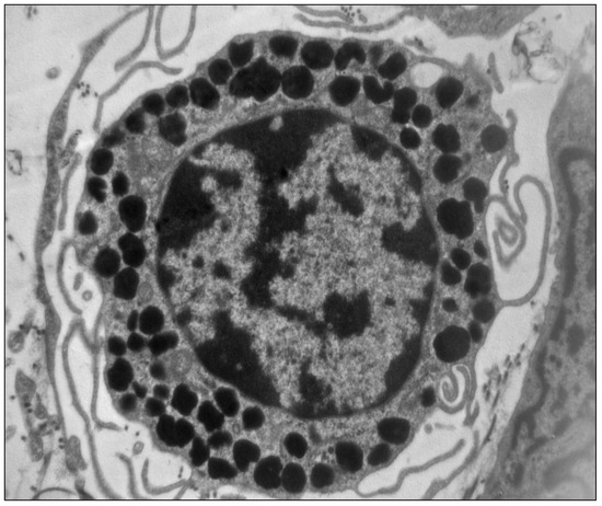
Carboxypeptidase A3 (CPA3) is a specific mast cell (MC) protease with variable expression. This protease is one of the preformed components of the secretome. During maturation of granules, CPA3 becomes an active enzyme with a characteristic localization determining the features of the cytological and ultrastructural phenotype of MC. CPA3 takes part in the regulation of a specific tissue microenvironment, affecting the implementation of innate immunity, the mechanisms of angiogenesis, the processes of remodeling of the extracellular matrix, etc. Characterization of CPA3 expression in MC can be used to refine the MC classification, help in a prognosis, and increase the effectiveness of targeted therapy. Full article
Show Figures

Figure 1

9 pages, 293 KiB  
Review
Mast Cells in Immune-Mediated Cholangitis and Cholangiocarcinoma
by Marisol I. González, Danielle T. Vannan, Bertus Eksteen, Irán Flores-Sotelo and José Luis Reyes
Cells 2022, 11(3), 375; https://doi.org/10.3390/cells11030375 - 22 Jan 2022
Cited by 8 | Viewed by 4381
Abstract
Cholestasis, which is impaired bile flow from the liver into the intestine, can be caused by cholangitis and/or bile duct obstruction. Cholangitis can arise from bacterial infections and cholelithiasis, however, immune-mediated cholangitis in primary biliary cholangitis (PBC) and primary sclerosing cholangitis (PSC) is [...] Read more.
Cholestasis, which is impaired bile flow from the liver into the intestine, can be caused by cholangitis and/or bile duct obstruction. Cholangitis can arise from bacterial infections and cholelithiasis, however, immune-mediated cholangitis in primary biliary cholangitis (PBC) and primary sclerosing cholangitis (PSC) is characterized by a strong immune response targeting the biliary epithelial cells (BECs). Persistent biliary inflammation further represents a risk for biliary neoplasia, cholangiocarcinoma (CCA) by driving chronic cellular stress in the BECs. Currently, immune-mediated cholangitis is considered a Th1-Th17-dominant disease, however, the presence of Th2-related mast cells (MCs) in tissue samples from PBC, PSC and CCA patients has been described, showing that these MCs are active players in these diseases. Here, we reviewed and discussed experimental and clinical data supporting a pro-fibrotic role for MCs in immune-mediated cholangitis as well as their participation in supporting tumor growth acting as angiogenesis promoters. Thus, although MCs have classically been identified as downstream effectors of Th2 responses in allergies and parasitic infections, evidence suggests that these MCs are relevant players in biliary inflammation and neoplasia. The availability of strategies to prevent MCs’ activation represents a therapeutic opportunity in biliary diseases. Full article
12 pages, 14778 KiB  
Article
Tumor-Derived Extracellular Vesicles Induce CCL18 Production by Mast Cells: A Possible Link to Angiogenesis
by Irit Shefler, Pazit Salamon, Tali Zitman-Gal and Yoseph A. Mekori
Cells 2022, 11(3), 353; https://doi.org/10.3390/cells11030353 - 21 Jan 2022
Cited by 5 | Viewed by 3217
Abstract
Mast cells (MCs) function as a component of the tumor microenvironment (TME) and have both pro- and anti-tumorigenic roles depending on the tumor type and its developmental stage. Several reports indicate the involvement of MCs in angiogenesis in the TME by releasing angiogenic [...] Read more.
Mast cells (MCs) function as a component of the tumor microenvironment (TME) and have both pro- and anti-tumorigenic roles depending on the tumor type and its developmental stage. Several reports indicate the involvement of MCs in angiogenesis in the TME by releasing angiogenic mediators. Tumor cells and other cells in the TME may interact by releasing extracellular vesicles (EVs) that affect the cells in the region. We have previously shown that tumor-derived microvesicles (TMVs) from non-small-cell lung cancer (NSCLC) cells interact with human MCs and activate them to release several cytokines and chemokines. In the present study, we characterized the MC expression of other mediators after exposure to TMVs derived from NSCLC. Whole-genome expression profiling disclosed the production of several chemokines, including CC chemokine ligand 18 (CCL18). This chemokine is expressed in various types of cancer, and was found to be associated with extensive angiogenesis, both in vitro and in vivo. We now show that CCL18 secreted from MCs activated by NSCLC-TMVs increased the migration of human umbilical cord endothelial cells (HUVECs), tube formation and endothelial- to-mesenchymal transition (EndMT), thus promoting angiogenesis. Our findings support the conclusion that TMVs have the potential to influence MC activity and may affect angiogenesis in the TME. Full article
Show Figures

Figure 1

38 pages, 4018 KiB  
Review
Mast Cell–Tumor Interactions: Molecular Mechanisms of Recruitment, Intratumoral Communication and Potential Therapeutic Targets for Tumor Growth
by Deisy Segura-Villalobos, Itzel G. Ramírez-Moreno, Magnolia Martínez-Aguilar, Alfredo Ibarra-Sánchez, J. Omar Muñoz-Bello, Isabel Anaya-Rubio, Alejandro Padilla, Marina Macías-Silva, Marcela Lizano and Claudia González-Espinosa
Cells 2022, 11(3), 349; https://doi.org/10.3390/cells11030349 - 20 Jan 2022
Cited by 31 | Viewed by 5433
Abstract
Mast cells (MCs) are tissue-resident immune cells that are important players in diseases associated with chronic inflammation such as cancer. Since MCs can infiltrate solid tumors and promote or limit tumor growth, a possible polarization of MCs to pro-tumoral or anti-tumoral phenotypes has [...] Read more.
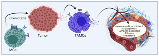
Mast cells (MCs) are tissue-resident immune cells that are important players in diseases associated with chronic inflammation such as cancer. Since MCs can infiltrate solid tumors and promote or limit tumor growth, a possible polarization of MCs to pro-tumoral or anti-tumoral phenotypes has been proposed and remains as a challenging research field. Here, we review the recent evidence regarding the complex relationship between MCs and tumor cells. In particular, we consider: (1) the multifaceted role of MCs on tumor growth suggested by histological analysis of tumor biopsies and studies performed in MC-deficient animal models; (2) the signaling pathways triggered by tumor-derived chemotactic mediators and bioactive lipids that promote MC migration and modulate their function inside tumors; (3) the possible phenotypic changes on MCs triggered by prevalent conditions in the tumor microenvironment (TME) such as hypoxia; (4) the signaling pathways that specifically lead to the production of angiogenic factors, mainly VEGF; and (5) the possible role of MCs on tumor fibrosis and metastasis. Finally, we discuss the novel literature on the molecular mechanisms potentially related to phenotypic changes that MCs undergo into the TME and some therapeutic strategies targeting MC activation to limit tumor growth. Full article
Show Figures

Figure 1

14 pages, 978 KiB  
Review
Mucosal Mast Cells as Key Effector Cells in Food Allergies
by Nobuhiro Nakano and Jiro Kitaura
Cells 2022, 11(3), 329; https://doi.org/10.3390/cells11030329 - 19 Jan 2022
Cited by 15 | Viewed by 4570
Abstract
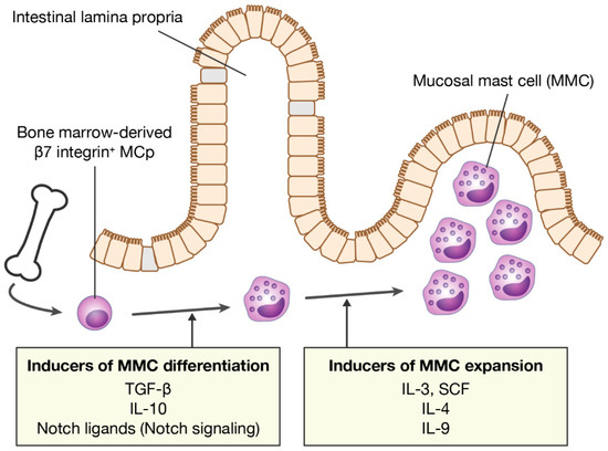
Mucosal mast cells (MMCs) localized in the intestinal mucosa play a key role in the development of IgE-mediated food allergies. Recent advances have revealed that MMCs are a distinctly different population from connective tissue mast cells localized in skin and other connective tissues. [...] Read more.
Mucosal mast cells (MMCs) localized in the intestinal mucosa play a key role in the development of IgE-mediated food allergies. Recent advances have revealed that MMCs are a distinctly different population from connective tissue mast cells localized in skin and other connective tissues. MMCs are inducible and transient cells that arise from bone marrow-derived mast cell progenitors, and their numbers increase rapidly during mucosal allergic inflammation. However, the mechanism of the dramatic expansion of MMCs and their cell functions are not well understood. Here, we review recent findings on the mechanisms of MMC differentiation and expansion, and we discuss the potential for the inducers of differentiation and expansion to serve as targets for food allergy therapy. In addition, we also discuss the mechanism by which oral immunotherapy, a promising treatment for food allergy patients, induces unresponsiveness to food allergens and the roles of MMCs in this process. Research focusing on MMCs should provide useful information for understanding the underlying mechanisms of food allergies in order to further advance the treatment of food allergies. Full article
Show Figures

Figure 1

2021

Jump to: 2024, 2022

21 pages, 1304 KiB  
Review
Therapeutic Potential of MRGPRX2 Inhibitors on Mast Cells
by Hiroyuki Ogasawara and Masato Noguchi
Cells 2021, 10(11), 2906; https://doi.org/10.3390/cells10112906 - 27 Oct 2021
Cited by 43 | Viewed by 11040
Abstract
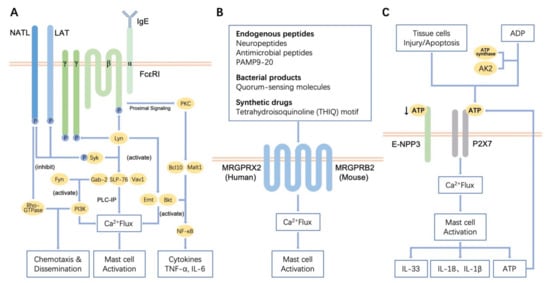
Mast cells (MCs) act as primary effectors in inflammatory and allergic reactions by releasing intracellularly-stored inflammatory mediators in diseases. The two major pathways for MC activation are known to be immunoglobulin E (IgE)-dependent and -independent. Although IgE-dependent signaling is the main pathway to [...] Read more.
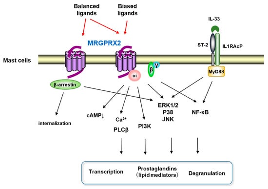
Mast cells (MCs) act as primary effectors in inflammatory and allergic reactions by releasing intracellularly-stored inflammatory mediators in diseases. The two major pathways for MC activation are known to be immunoglobulin E (IgE)-dependent and -independent. Although IgE-dependent signaling is the main pathway to MC activation, IgE-independent pathways have also been found to serve pivotal roles in the pathophysiology of various inflammatory conditions. Recent studies have shown that human and mouse MCs express several regulatory receptors such as toll-like receptors (TLRs), CD48, C300a, and GPCRs, including mas-related GPCR-X2 (MRGPRX2). MRGPRX2 has been reported as a novel GPCR that is expressed in MCs activated by basic secretagogues, neurokinin peptides, host defense antimicrobial peptides, and small molecule compounds (e.g., neuromuscular blocking agents) and leads to MC degranulation and eicosanoids release under in vitro experimental condition. Functional analyses of MRGPRX2 and Mrgprb2 (mouse ortholog) indicate that MRGPRX2 is involved in MC hypersensitivity reactions causing neuroinflammation such as postoperative pain, type 2 inflammation, non-histaminergic itch, and drug-induced anaphylactic-like reactions. In this review, we discuss the roles in innate immunity through functional studies on MRGPRX2-mediated IgE-independent MC activation and also the therapeutic potential of MRGPRX2 inhibitors on allergic and inflammatory diseases. Full article
Show Figures

Figure 1

16 pages, 1721 KiB  
Review
Roles of IgE and Histamine in Mast Cell Maturation
by Satoshi Tanaka and Kazuyuki Furuta
Cells 2021, 10(8), 2170; https://doi.org/10.3390/cells10082170 - 23 Aug 2021
Cited by 38 | Viewed by 11667
Abstract
Mast cells are activated upon immunoglobulin E (IgE)-mediated antigen stimulation, and release a wide variety of mediators, including histamine to trigger inflammatory responses. The surface expression levels of Fcε receptor I (FcεRI), a high affinity receptor of IgE, were found to be positively [...] Read more.
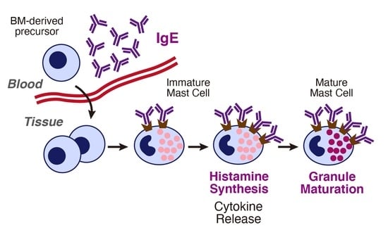
Mast cells are activated upon immunoglobulin E (IgE)-mediated antigen stimulation, and release a wide variety of mediators, including histamine to trigger inflammatory responses. The surface expression levels of Fcε receptor I (FcεRI), a high affinity receptor of IgE, were found to be positively regulated by IgE. IgE could protect murine cultured mast cells from apoptotic cell death induced by the deprivation of interleukin-3 and a certain kind of IgE could activate immature mast cells in the absence of antigens, leading to the release of pro-inflammatory cytokines and a transient increase in histamine synthesis. Histamine synthesis in mast cells was found to be required for the maturation of murine connective tissue-type mast cells, raising the possibility that IgE indirectly modulates local mast cell maturation. Although it remains controversial to what extent this concept of “monomeric IgE effects” could have relevance in the modulation of human mast cell functions, the therapeutic effects of anti-IgE antibodies might be accounted for in terms of the decreased serum IgE concentrations. Because drastic increases in serum IgE concentrations are often observed in patients with atopic dermatitis and chronic urticaria, a close investigation of the roles of IgE in mast cell maturation should contribute to development of novel therapeutic approaches for these inflammatory diseases. Full article
Show Figures

Graphical abstract

11 pages, 2432 KiB  
Review
Mast Cells and the Pancreas in Human Type 1 and Type 2 Diabetes
by Matilde Masini, Mara Suleiman, Michela Novelli, Lorella Marselli, Piero Marchetti and Vincenzo De Tata
Cells 2021, 10(8), 1875; https://doi.org/10.3390/cells10081875 - 23 Jul 2021
Cited by 6 | Viewed by 4512
Abstract
Mast cells are highly differentiated, widely distributed cells of the innate immune system, that are currently considered as key regulators of both innate and adaptive immunity. Mast cells play a key role in health and survival mechanisms, especially as sentinel cells that can [...] Read more.
Mast cells are highly differentiated, widely distributed cells of the innate immune system, that are currently considered as key regulators of both innate and adaptive immunity. Mast cells play a key role in health and survival mechanisms, especially as sentinel cells that can stimulate protective immune responses. On the other hand, it has been shown that mast cells are involved in the pathogenesis of several diseases, and recently a possible pathogenetic role of mast cells in diabetes has been proposed. In this review we summarize the evidence on the increased presence of mast cells in the pancreas of subjects with type 1 diabetes, which is due to the autoimmune destruction of insulin secreting beta cells, and discuss the differences with type 2 diabetes, the other major form of diabetes. In addition, we describe some of the pathophysiological mechanisms through which mast cells might exert their actions, which could be targeted to potentially protect the beta cells in autoimmune diabetes. Full article
Show Figures

Figure 1

10 pages, 1323 KiB  
Review
The Role of Coagulation and Complement Factors for Mast Cell Activation in the Pathogenesis of Chronic Spontaneous Urticaria
by Yuhki Yanase, Shunsuke Takahagi, Koichiro Ozawa and Michihiro Hide
Cells 2021, 10(7), 1759; https://doi.org/10.3390/cells10071759 - 12 Jul 2021
Cited by 42 | Viewed by 7605
Abstract
Chronic spontaneous urticaria (CSU) is a common skin disorder characterized by an almost daily recurrence of wheal and flare with itch for more than 6 weeks, in association with the release of stored inflammatory mediators, such as histamine, from skin mast cells and/or [...] Read more.
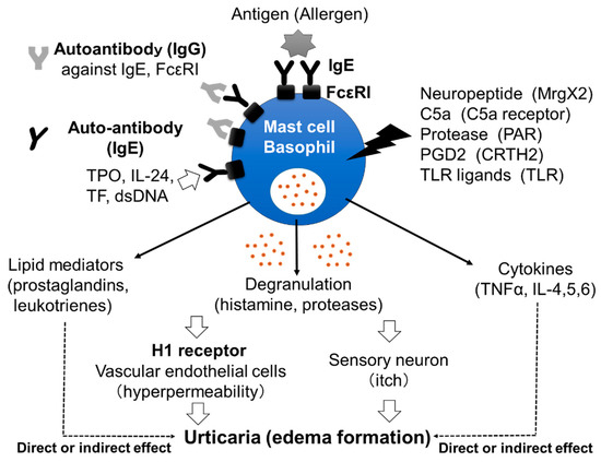
Chronic spontaneous urticaria (CSU) is a common skin disorder characterized by an almost daily recurrence of wheal and flare with itch for more than 6 weeks, in association with the release of stored inflammatory mediators, such as histamine, from skin mast cells and/or peripheral basophils. The involvement of the extrinsic coagulation cascade triggered by tissue factor (TF) and complement factors, such as C3a and C5a, has been implied in the pathogenesis of CSU. However, it has been unclear how the TF-triggered coagulation pathway and complement factors induce the activation of skin mast cells and peripheral basophils in patients with CSU. In this review, we focus on the role of vascular endothelial cells, leukocytes, extrinsic coagulation factors and complement components on TF-induced activation of skin mast cells and peripheral basophils followed by the edema formation clinically recognized as urticaria. These findings suggest that medications targeting activated coagulation factors and/or complement components may represent new and effective treatments for patients with severe and refractory CSU. Full article
Show Figures

Figure 1

18 pages, 3180 KiB  
Review
Tuning IgE: IgE-Associating Molecules and Their Effects on IgE-Dependent Mast Cell Reactions
by Tomoaki Ando and Jiro Kitaura
Cells 2021, 10(7), 1697; https://doi.org/10.3390/cells10071697 - 5 Jul 2021
Cited by 22 | Viewed by 6651
Abstract
The recent emergence of anti-immunoglobulin E (IgE) drugs and their candidates for humans has endorsed the significance of IgE-dependent pathways in allergic disorders. IgE is distributed locally in the tissues or systemically to confer a sensory mechanism in a domain of adaptive immunity [...] Read more.
The recent emergence of anti-immunoglobulin E (IgE) drugs and their candidates for humans has endorsed the significance of IgE-dependent pathways in allergic disorders. IgE is distributed locally in the tissues or systemically to confer a sensory mechanism in a domain of adaptive immunity to the otherwise innate type of effector cells, namely, mast cells and basophils. Bound on the high-affinity IgE receptor FcεRI, IgE enables fast memory responses against revisiting threats of venoms, parasites, and bacteria. However, the dysregulation of IgE-dependent reactions leads to potentially life-threatening allergic diseases, such as asthma and anaphylaxis. Therefore, reactivity of the IgE sensor is fine-tuned by various IgE-associating molecules. In this review, we discuss the mechanistic basis for how IgE-dependent mast cell activation is regulated by the IgE-associating molecules, including the newly developed therapeutic candidates. Full article
Show Figures

Figure 1

15 pages, 2617 KiB  
Article
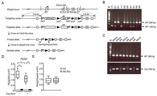
Mast Cell-Specific Deletion of Group III Secreted Phospholipase A2 Impairs Mast Cell Maturation and Functions
by Yoshitaka Taketomi, Yuki Endo, Takayoshi Higashi, Remi Murase, Tomio Ono, Choji Taya, Tetsuyuki Kobayashi and Makoto Murakami
Cells 2021, 10(7), 1691; https://doi.org/10.3390/cells10071691 - 4 Jul 2021
Cited by 8 | Viewed by 3604
Abstract
Tissue-resident mast cells (MCs) have important roles in IgE-associated and -independent allergic reactions. Although microenvironmental alterations in MC phenotypes affect the susceptibility to allergy, understanding of the regulation of MC maturation is still incomplete. We previously reported that group III secreted phospholipase A [...] Read more.
Tissue-resident mast cells (MCs) have important roles in IgE-associated and -independent allergic reactions. Although microenvironmental alterations in MC phenotypes affect the susceptibility to allergy, understanding of the regulation of MC maturation is still incomplete. We previously reported that group III secreted phospholipase A2 (sPLA2-III) released from immature MCs is functionally coupled with lipocalin-type prostaglandin D2 (PGD2) synthase in neighboring fibroblasts to supply a microenvironmental pool of PGD2, which in turn acts on the PGD2 receptor DP1 on MCs to promote their proper maturation. In the present study, we reevaluated the role of sPLA2-III in MCs using a newly generated MC-specific Pla2g3-deficient mouse strain. Mice lacking sPLA2-III specifically in MCs, like those lacking the enzyme in all tissues, had immature MCs and displayed reduced local and systemic anaphylactic responses. Furthermore, MC-specific Pla2g3-deficient mice, as well as MC-deficient KitW-sh mice reconstituted with MCs prepared from global Pla2g3-null mice, displayed a significant reduction in irritant contact dermatitis (ICD) and an aggravation of contact hypersensitivity (CHS). The increased CHS response by Pla2g3 deficiency depended at least partly on the reduced expression of hematopoietic PGD2 synthase and thereby reduced production of PGD2 due to immaturity of MCs. Overall, our present study has confirmed that MC-secreted sPLA2-III promotes MC maturation, thereby facilitating acute anaphylactic and ICD reactions and limiting delayed CHS response. Full article
Show Figures

Figure 1

19 pages, 1411 KiB  
Review
Two Sides of the Coin: Mast Cells as a Key Regulator of Allergy and Acute/Chronic Inflammation
by Zhongwei Zhang and Yosuke Kurashima
Cells 2021, 10(7), 1615; https://doi.org/10.3390/cells10071615 - 28 Jun 2021
Cited by 32 | Viewed by 5930
Abstract
It is well known that mast cells (MCs) initiate type I allergic reactions and inflammation in a quick response to the various stimulants, including—but not limited to—allergens, pathogen-associated molecular patterns (PAMPs), and damage-associated molecular patterns (DAMPs). MCs highly express receptors of these ligands [...] Read more.
It is well known that mast cells (MCs) initiate type I allergic reactions and inflammation in a quick response to the various stimulants, including—but not limited to—allergens, pathogen-associated molecular patterns (PAMPs), and damage-associated molecular patterns (DAMPs). MCs highly express receptors of these ligands and proteases (e.g., tryptase, chymase) and cytokines (TNF), and other granular components (e.g., histamine and serotonin) and aggravate the allergic reaction and inflammation. On the other hand, accumulated evidence has revealed that MCs also possess immune-regulatory functions, suppressing chronic inflammation and allergic reactions on some occasions. IL-2 and IL-10 released from MCs inhibit excessive immune responses. Recently, it has been revealed that allergen immunotherapy modulates the function of MCs from their allergic function to their regulatory function to suppress allergic reactions. This evidence suggests the possibility that manipulation of MCs functions will result in a novel approach to the treatment of various MCs-mediated diseases. Full article
Show Figures

Figure 1

15 pages, 3232 KiB  
Article
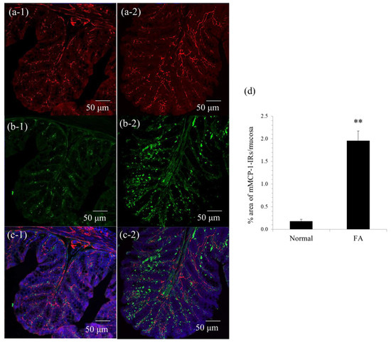
Pathophysiological Roles of Neuro-Immune Interactions between Enteric Neurons and Mucosal Mast Cells in the Gut of Food Allergy Mice
by Tomoe Yashiro, Hanako Ogata, Syed Faisal Zaidi, Jaemin Lee, Shusaku Hayashi, Takeshi Yamamoto and Makoto Kadowaki
Cells 2021, 10(7), 1586; https://doi.org/10.3390/cells10071586 - 23 Jun 2021
Cited by 15 | Viewed by 3257
Abstract
Recently, the involvement of the nervous system in the pathology of allergic diseases has attracted increasing interest. However, the precise pathophysiological role of enteric neurons in food allergies has not been elucidated. We report the presence of functional high-affinity IgE receptors (FcεRIs) in [...] Read more.
Recently, the involvement of the nervous system in the pathology of allergic diseases has attracted increasing interest. However, the precise pathophysiological role of enteric neurons in food allergies has not been elucidated. We report the presence of functional high-affinity IgE receptors (FcεRIs) in enteric neurons. FcεRI immunoreactivities were observed in approximately 70% of cholinergic myenteric neurons from choline acetyltransferase-eGFP mice. Furthermore, stimulation by IgE-antigen elevated intracellular Ca2+ concentration in isolated myenteric neurons from normal mice, suggesting that FcεRIs are capable of activating myenteric neurons. Additionally, the morphological investigation revealed that the majority of mucosal mast cells were in close proximity to enteric nerve fibers in the colonic mucosa of food allergy mice. Next, using a newly developed coculture system of isolated myenteric neurons and mucosal-type bone-marrow-derived mast cells (mBMMCs) with a calcium imaging system, we demonstrated that the stimulation of isolated myenteric neurons by veratridine caused the activation of mBMMCs, which was suppressed by the adenosine A3 receptor antagonist MRE 3008F20. Moreover, the expression of the adenosine A3 receptor gene was detected in mBMMCs. Therefore, in conclusion, it is suggested that, through interaction with mucosal mast cells, IgE-antigen-activated myenteric neurons play a pathological role in further exacerbating the pathology of food allergy. Full article
Show Figures

Figure 1

16 pages, 3654 KiB  
Article
Mast Cell Mediated Regulation of Small Intestinal Chloride Malabsorption in SAMP1/YitFc Mouse Model of Spontaneous Chronic Ileitis
by M Motiur Rahman, Sheuli Afroz, Subha Arthur and Uma Sundaram
Cells 2021, 10(3), 697; https://doi.org/10.3390/cells10030697 - 21 Mar 2021
Cited by 4 | Viewed by 3562
Abstract
In Inflammatory Bowel Disease (IBD), malabsorption of electrolytes (NaCl) results in diarrhea. Inhibition of coupled NaCl absorption, mediated by the dual operation of Na:H and Cl:HCO3 exchangers on the brush border membrane (BBM) of the intestinal villus cells has been reported in [...] Read more.
In Inflammatory Bowel Disease (IBD), malabsorption of electrolytes (NaCl) results in diarrhea. Inhibition of coupled NaCl absorption, mediated by the dual operation of Na:H and Cl:HCO3 exchangers on the brush border membrane (BBM) of the intestinal villus cells has been reported in IBD. In the SAMP1/YitFcs (SAMP1) mice model of spontaneous ileitis, representing Crohn’s disease, DRA (Downregulated in Adenoma) mediated Cl:HCO3 exchange was shown to be inhibited secondary to diminished affinity of the exchanger for Cl. However, NHE3 mediated Na:H exchange remained unaffected. Mast cells and their secreted mediators are known to be increased in the IBD mucosa and can affect intestinal electrolyte absorption. However, how mast cell mediators may regulate Cl:HCO3 exchange in SAMP1 mice is unknown. Therefore, the aim of this study was to determine the effect of mast cell mediators on the downregulation of DRA in SAMP1 mice. Mast cell numbers and their degranulation marker enzyme (β-hexosaminidase) levels were significantly increased in SAMP1 mice compared to control AKR mice. However, treatment of SAMP1 mice with a mast cell stabilizer, ketotifen, restored the β-hexosaminidase enzyme levels to normal in the intestine, demonstrating stabilization of mast cells by ketotifen. Moreover, downregulation of Cl:HCO3 exchange activity was restored in ketotifen treated SAMP1 mice. Kinetic studies showed that ketotifen restored the altered affinity of Cl:HCO3 exchange in SAMP1 mice villus cells thus reinstating its activity to normal. Further, RT-qPCR, Western blot and immunofluorescence studies showed that the expression levels of DRA mRNA and BBM protein, respectively remained unaltered in all experimental conditions, supporting the kinetic data. Thus, inhibition of Cl:HCO3 exchange resulting in chloride malabsorption leading to diarrhea in IBD is likely mediated by mast cell mediators. Full article
Show Figures

Figure 1

Back to TopTop