ijms-logo

Journal Browser

Journal Browser

Feature Papers in Molecular Pharmacology

A topical collection in International Journal of Molecular Sciences (ISSN 1422-0067). This collection belongs to the section "Molecular Pharmacology".

Viewed by 294673

Editor


E-Mail Website
Collection Editor
Rudolf-Boehm-Institut für Pharmakologie und Toxikologie, Universitaet Leipzig, Leipzig, Germany
Interests: neuropharmacology; neurotoxicology; purinergic mechanisms; modulation of synaptic transmission; glial cells; necrosis/apoptosis; analgesia; learning and memory; epileptic state
Special Issues, Collections and Topics in MDPI journals

Topical Collection Information

Dear Colleagues,

The Topical Collection entitled “Feature Papers in Molecular Pharmacology” aims to collect high-quality research articles and reviews in the field of molecular pharmacology. We encourage Editorial Board Members of this IJMS Section to submit manuscripts reporting on significant advances in molecular pharmacology research. Special emphasis will be placed on drug effects upon nucleic acids, proteins involved in nucleic acid metabolism, and interaction of nucleic acids with proteins, by using the whole plethora of molecular biology methods. Genetic and epigenetic factors determining drug effects will be accordingly dealt with.

Prof. Dr. Peter Illes
Guest Editor

Manuscript Submission Information

Manuscripts should be submitted online at www.mdpi.com by registering and logging in to this website. Once you are registered, click here to go to the submission form. Manuscripts can be submitted until the deadline. All submissions that pass pre-check are peer-reviewed. Accepted papers will be published continuously in the journal (as soon as accepted) and will be listed together on the collection website. Research articles, review articles as well as short communications are invited. For planned papers, a title and short abstract (about 100 words) can be sent to the Editorial Office for announcement on this website.

Submitted manuscripts should not have been published previously, nor be under consideration for publication elsewhere (except conference proceedings papers). All manuscripts are thoroughly refereed through a single-blind peer-review process. A guide for authors and other relevant information for submission of manuscripts is available on the Instructions for Authors page. International Journal of Molecular Sciences is an international peer-reviewed open access semimonthly journal published by MDPI.

Please visit the Instructions for Authors page before submitting a manuscript. There is an Article Processing Charge (APC) for publication in this open access journal. For details about the APC please see here. Submitted papers should be well formatted and use good English. Authors may use MDPI's English editing service prior to publication or during author revisions.

Published Papers (77 papers)

2024

Jump to: 2023, 2022, 2021, 2020, 2019

15 pages, 3249 KiB  
Article
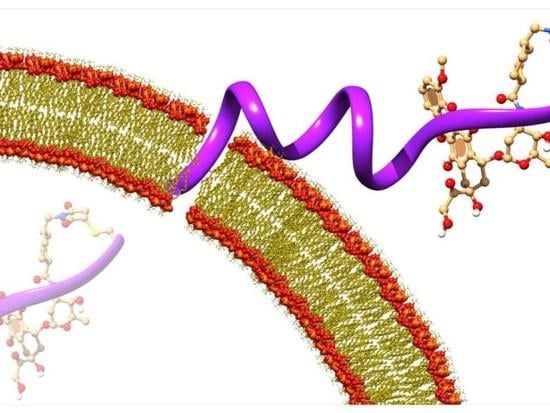
Antimicrobial Peptide Pro10-1D Exhibits Anti-Allergic Activity: A Promising Therapeutic Candidate
by Min Yeong Choi, Min Geun Jo, Keun Young Min, Byeongkwon Kim, Yangmee Kim and Wahn Soo Choi
Int. J. Mol. Sci. 2024, 25(22), 12138; https://doi.org/10.3390/ijms252212138 - 12 Nov 2024
Viewed by 390
Abstract
Although antimicrobial peptides (AMPs) exhibit a range of biological functions, reports on AMPs with therapeutic effects in allergic disorders are limited. In this study, we investigated the anti-allergic effects of Pro10-1D, a 10-meric AMP derived from insect defensin protaetiamycine. Our findings demonstrate that [...] Read more.
Although antimicrobial peptides (AMPs) exhibit a range of biological functions, reports on AMPs with therapeutic effects in allergic disorders are limited. In this study, we investigated the anti-allergic effects of Pro10-1D, a 10-meric AMP derived from insect defensin protaetiamycine. Our findings demonstrate that Pro10-1D effectively inhibits antigen-induced degranulation of mast cells (MCs) with IC50 values of approximately 11.6 μM for RBL-2H3 cells and 2.7 μM for bone marrow-derived MCs. Furthermore, Pro10-1D suppressed the secretion of cytokines with IC50 values of approximately 2.8 μM for IL-4 and approximately 8.6 μM for TNF-α. Mechanistically, Pro10-1D inhibited the Syk-LAT-PLCγ1 signaling pathway in MCs and decreased the activation of mitogen-activated protein kinases (MAPKs). Pro10-1D demonstrated a dose-dependent reduction in IgE-mediated passive cutaneous anaphylaxis in mice with an ED50 value of approximately 7.6 mg/kg. Further investigation revealed that Pro10-1D significantly reduced the activity of key kinases Fyn and Lyn, which are critical in the initial phase of the FcεRI-mediated signaling pathway, with IC50 values of approximately 22.6 μM for Fyn and approximately 1.5 μM for Lyn. Collectively, these findings suggest that Pro10-1D represents a novel therapeutic candidate for the treatment of IgE-mediated allergic disorders by targeting the Lyn/Fyn Src family kinases in MCs. Full article
Show Figures

Graphical abstract

26 pages, 3897 KiB  
Review
A Comprehensive Review of Xanthan Gum-Based Oral Drug Delivery Systems
by Buddhadev Layek
Int. J. Mol. Sci. 2024, 25(18), 10143; https://doi.org/10.3390/ijms251810143 - 21 Sep 2024
Viewed by 1583
Abstract
Xanthan gum (XG) is an exopolysaccharide synthesized by the aerobic fermentation of simple sugars using Xanthomonas bacteria. It comprises a cellulosic backbone with a trisaccharide side chain connected to alternative glucose residues in the main backbone through α (1→3) linkage. XG dissolves readily [...] Read more.
Xanthan gum (XG) is an exopolysaccharide synthesized by the aerobic fermentation of simple sugars using Xanthomonas bacteria. It comprises a cellulosic backbone with a trisaccharide side chain connected to alternative glucose residues in the main backbone through α (1→3) linkage. XG dissolves readily in cold and hot water to produce a viscous solution that behaves like a pseudoplastic fluid. It shows excellent resistance to enzymatic degradation and great stability throughout a broad temperature, pH, or salt concentration range. Additionally, XG is nontoxic, biocompatible, and biodegradable, making it a suitable carrier for drug delivery. Furthermore, the carboxylic functions of pyruvate and glucuronic acid offer a considerable opportunity for chemical modification to meet the desired criteria for a specific application. Therefore, XG or its derivatives in conjunction with other polymers have frequently been studied as matrices for tablets, nanoparticles, microparticles, and hydrogels. This review primarily focuses on the applications of XG in various oral delivery systems over the past decade, including sustained-release formulations, gastroretentive dosage forms, and colon-targeted drug delivery. Source, production methods, and physicochemical properties relevant to drug delivery applications of XG have also been discussed. Full article
Show Figures

Graphical abstract

19 pages, 4490 KiB  
Article
Drug–Target Interaction Prediction Based on an Interactive Inference Network
by Yuqi Chen, Xiaomin Liang, Wei Du, Yanchun Liang, Garry Wong and Liang Chen
Int. J. Mol. Sci. 2024, 25(14), 7753; https://doi.org/10.3390/ijms25147753 - 15 Jul 2024
Cited by 1 | Viewed by 1501
Abstract
Drug–target interactions underlie the actions of chemical substances in medicine. Moreover, drug repurposing can expand use profiles while reducing costs and development time by exploiting potential multi-functional pharmacological properties based upon additional target interactions. Nonetheless, drug repurposing relies on the accurate identification and [...] Read more.
Drug–target interactions underlie the actions of chemical substances in medicine. Moreover, drug repurposing can expand use profiles while reducing costs and development time by exploiting potential multi-functional pharmacological properties based upon additional target interactions. Nonetheless, drug repurposing relies on the accurate identification and validation of drug–target interactions (DTIs). In this study, a novel drug–target interaction prediction model was developed. The model, based on an interactive inference network, contains embedding, encoding, interaction, feature extraction, and output layers. In addition, this study used Morgan and PubChem molecular fingerprints as additional information for drug encoding. The interaction layer in our model simulates the drug–target interaction process, which assists in understanding the interaction by representing the interaction space. Our method achieves high levels of predictive performance, as well as interpretability of drug–target interactions. Additionally, we predicted and validated 22 Alzheimer’s disease-related targets, suggesting our model is robust and effective and thus may be beneficial for drug repurposing. Full article
Show Figures

Graphical abstract

17 pages, 3777 KiB  
Article
Prokaryotic Expression and Functional Verification of Antimicrobial Peptide LRGG
by Xiang Liu, Yining Ding, Yuhan Shen, Sizhuo Liu, Yuehua Liu, Yuting Wang, Shikun Wang, Claudio Orlando Gualerzi, Attilio Fabbretti, Lili Guan, Lingcong Kong, Haipeng Zhang, Hongxia Ma and Chengguang He
Int. J. Mol. Sci. 2024, 25(13), 7072; https://doi.org/10.3390/ijms25137072 - 27 Jun 2024
Cited by 1 | Viewed by 950
Abstract
The antimicrobial peptide LRGG (LLRLLRRGGRRLLRLL-NH2) was designed and chemically synthesized in a study conducted by Jia et al. Gram-negative bacteria were found to be sensitive to LRGG and exhibited a high therapeutic index. Genetic engineering methods were used to create the [...] Read more.
The antimicrobial peptide LRGG (LLRLLRRGGRRLLRLL-NH2) was designed and chemically synthesized in a study conducted by Jia et al. Gram-negative bacteria were found to be sensitive to LRGG and exhibited a high therapeutic index. Genetic engineering methods were used to create the prokaryotic fusion expression vector pQE-GFP-LRGG, and the resulting corresponding fusion protein GFP-LRGG was subsequently expressed and purified. The precursor GFP was then removed by TEV proteolysis, and pure LRGG was obtained after another round of purification and endotoxin removal. The prokaryotic-expressed antimicrobial peptide LRGG displays a broad-spectrum antibacterial effect on Gram-negative bacteria, and its minimum inhibitory activity (MIC) against Escherichia coli can reach 2 μg/mL. Compared to the chemically synthesized LRGG, the prokaryotic-expressed LRGG exhibits similar temperature, pH, salt ion, serum stability, and cell selectivity. Furthermore, prokaryotic-expressed LRGG showed excellent therapeutic effects in both the infection model of cell selectivity and no embryotoxicity in a Galleria mellonella infection model. The mechanism by which LRGG causes bacterial death was found to be the disruption of the Gram-negative cell membrane. Full article
Show Figures

Figure 1

26 pages, 1553 KiB  
Review
Peptides Are Cardioprotective Drugs of the Future: The Receptor and Signaling Mechanisms of the Cardioprotective Effect of Glucagon-like Peptide-1 Receptor Agonists
by Alla A. Boshchenko, Leonid N. Maslov, Alexander V. Mukhomedzyanov, Olga A. Zhuravleva, Alisa S. Slidnevskaya, Natalia V. Naryzhnaya, Arina S. Zinovieva and Philipp A. Ilinykh
Int. J. Mol. Sci. 2024, 25(9), 4900; https://doi.org/10.3390/ijms25094900 - 30 Apr 2024
Cited by 4 | Viewed by 2919
Abstract
The high mortality rate among patients with acute myocardial infarction (AMI) is one of the main problems of modern cardiology. It is quite obvious that there is an urgent need to create more effective drugs for the treatment of AMI than those currently [...] Read more.
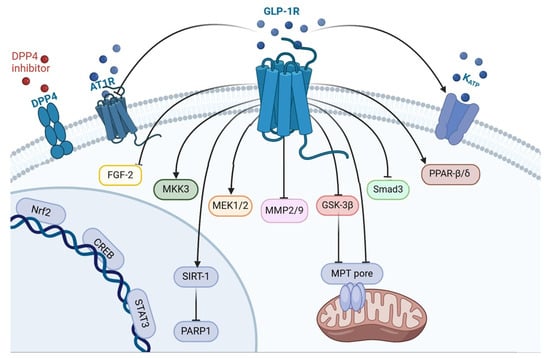
The high mortality rate among patients with acute myocardial infarction (AMI) is one of the main problems of modern cardiology. It is quite obvious that there is an urgent need to create more effective drugs for the treatment of AMI than those currently used in the clinic. Such drugs could be enzyme-resistant peptide analogs of glucagon-like peptide-1 (GLP-1). GLP-1 receptor (GLP1R) agonists can prevent ischemia/reperfusion (I/R) cardiac injury. In addition, chronic administration of GLP1R agonists can alleviate the development of adverse cardiac remodeling in myocardial infarction, hypertension, and diabetes mellitus. GLP1R agonists can protect the heart against oxidative stress and reduce proinflammatory cytokine (IL-1β, TNF-α, IL-6, and MCP-1) expression in the myocardium. GLP1R stimulation inhibits apoptosis, necroptosis, pyroptosis, and ferroptosis of cardiomyocytes. The activation of the GLP1R augments autophagy and mitophagy in the myocardium. GLP1R agonists downregulate reactive species generation through the activation of Epac and the GLP1R/PI3K/Akt/survivin pathway. The GLP1R, kinases (PKCε, PKA, Akt, AMPK, PI3K, ERK1/2, mTOR, GSK-3β, PKG, MEK1/2, and MKK3), enzymes (HO-1 and eNOS), transcription factors (STAT3, CREB, Nrf2, and FoxO3), KATP channel opening, and MPT pore closing are involved in the cardioprotective effect of GLP1R agonists. Full article
Show Figures

Figure 1

11 pages, 1793 KiB  
Article
Iridoid Glycosides and Coumarin Glycoside Derivatives from the Roots of Nymphoides peltata and Their In Vitro Wound Healing Properties
by Tae-Young Kim, Bum Soo Lee, Beom-Geun Jo, Seong Pil Heo, Young Suk Jung, Su-Nam Kim, Ki Hyun Kim and Min Hye Yang
Int. J. Mol. Sci. 2024, 25(2), 1268; https://doi.org/10.3390/ijms25021268 - 19 Jan 2024
Viewed by 1461
Abstract
Nymphoides peltata has been used as a medicinal herb in traditional medicines to treat strangury, polyuria, and swelling. The phytochemical investigation of the MeOH extract of N. peltata roots led to the isolation of three iridoid glycosides and three coumarin glycoside derivatives, which [...] Read more.
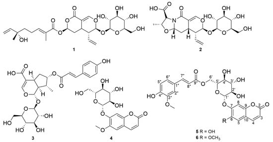
Nymphoides peltata has been used as a medicinal herb in traditional medicines to treat strangury, polyuria, and swelling. The phytochemical investigation of the MeOH extract of N. peltata roots led to the isolation of three iridoid glycosides and three coumarin glycoside derivatives, which were characterized as menthiafolin (1), threoninosecologanin (2), callicoside C (3), and scopolin (4), as well as two undescribed peltatamarins A (5) and B (6). The chemical structures of the undescribed compounds were determined by analyzing their 1 dimensional (D) and 2D nuclear magnetic resonance (NMR) spectra and using high-resolution (HR)-electrospray ionization mass spectroscopy (ESI-MS), along with the chemical reaction of acid hydrolysis. The wound healing activities of the isolated compounds 16 were evaluated using a HaCaT cell scratch test. Among the isolates, scopolin (4) and peltatamarin A (5) promoted HaCaT cell migration over scratch wounds, and compound 5 was the most effective. Furthermore, compound 5 significantly promoted cell migration without adversely affecting cell proliferation, even when treated at a high dose (100 μM). Our results demonstrate that peltatamarin A (5), isolated from N. peltata roots, has the potential for wound healing effects. Full article
Show Figures

Figure 1

2023

Jump to: 2024, 2022, 2021, 2020, 2019

27 pages, 2382 KiB  
Article
Existence of Quantum Pharmacology in Sartans: Evidence in Isolated Rabbit Iliac Arteries
by Laura Kate Gadanec, Jordan Swiderski, Vasso Apostolopoulos, Kostantinos Kelaidonis, Veroniki P. Vidali, Aleksander Canko, Graham J. Moore, John M. Matsoukas and Anthony Zulli
Int. J. Mol. Sci. 2023, 24(24), 17559; https://doi.org/10.3390/ijms242417559 - 16 Dec 2023
Cited by 3 | Viewed by 1661
Abstract
Quantum pharmacology introduces theoretical models to describe the possibility of ultra-high dilutions to produce biological effects, which may help to explain the placebo effect observed in hypertensive clinical trials. To determine this within physiology and to evaluate novel ARBs, we tested the ability [...] Read more.
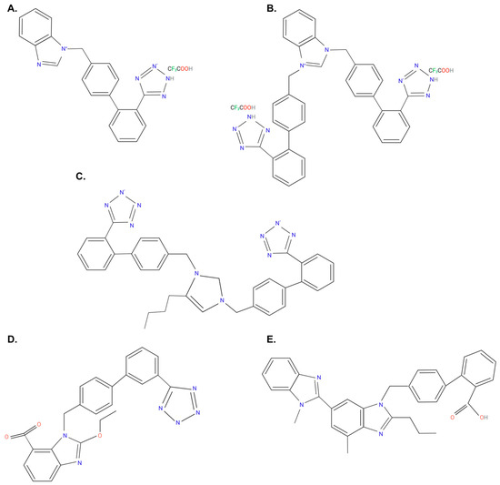
Quantum pharmacology introduces theoretical models to describe the possibility of ultra-high dilutions to produce biological effects, which may help to explain the placebo effect observed in hypertensive clinical trials. To determine this within physiology and to evaluate novel ARBs, we tested the ability of known angiotensin II receptor blockers (ARBs) (candesartan and telmisartan) used to treat hypertension and other cardiovascular diseases, as well as novel ARBs (benzimidazole-N-biphenyl tetrazole (ACC519T), benzimidazole-bis-N,N′-biphenyl tetrazole (ACC519T(2)) and 4-butyl-N,N0-bis[[20-2Htetrazol-5-yl)biphenyl-4-yl]methyl)imidazolium bromide (BV6(K+)2), and nirmatrelvir (the active ingredient in Paxlovid) to modulate vascular contraction in iliac rings from healthy male New Zealand White rabbits in responses to various vasopressors (angiotensin A, angiotensin II and phenylephrine). Additionally, the hemodynamic effect of ACC519T and telmisartan on mean arterial pressure in conscious rabbits was determined, while the ex vivo ability of BV6(K+)2 to activate angiotensin-converting enzyme-2 (ACE2) was also investigated. We show that commercially available and novel ARBs can modulate contraction responses at ultra-high dilutions to different vasopressors. ACC519T produced a dose-dependent reduction in rabbit mean arterial pressure while BV6(K+)2 significantly increased ACE2 metabolism. The ability of ARBs to inhibit contraction responses even at ultra-low concentrations provides evidence of the existence of quantum pharmacology. Furthermore, the ability of ACC519T and BV6(K+)2 to modulate blood pressure and ACE2 activity, respectively, indicates their therapeutic potential against hypertension. Full article
Show Figures

Figure 1

21 pages, 3907 KiB  
Article
Exploring Hydrophilic PD-L1 Radiotracers Utilizing Phosphonic Acids: Insights into Unforeseen Pharmacokinetics
by Fabian Krutzek, Cornelius K. Donat and Sven Stadlbauer
Int. J. Mol. Sci. 2023, 24(20), 15088; https://doi.org/10.3390/ijms242015088 - 11 Oct 2023
Cited by 1 | Viewed by 1401
Abstract
Immune checkpoint inhibitor therapy targeting the PD-1/PD-L1 axis in cancer patients, is a promising oncological treatment. However, the number of non-responders remains high, causing a burden for the patient and the healthcare system. Consequently, a diagnostic tool to predict treatment outcomes would help [...] Read more.
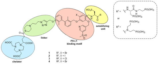
Immune checkpoint inhibitor therapy targeting the PD-1/PD-L1 axis in cancer patients, is a promising oncological treatment. However, the number of non-responders remains high, causing a burden for the patient and the healthcare system. Consequently, a diagnostic tool to predict treatment outcomes would help with patient stratification. Molecular imaging provides said diagnostic tool by offering a whole-body quantitative assessment of PD-L1 expression, hence supporting therapy decisions. Four PD-L1 radioligand candidates containing a linker-chelator system for radiometalation, along with three hydrophilizing units—one sulfonic and two phosphonic acids—were synthesized. After labeling with 64Cu, log D7.4 values of less than −3.03 were determined and proteolytic stability confirmed over 94% intact compound after 48 h. Binding affinity was determined using two different assays, revealing high affinities up to 13 nM. µPET/CT imaging was performed in tumor-bearing mice to investigate PD-L1-specific tumor uptake and the pharmacokinetic profile of radioligands. These results yielded an unexpected in vivo distribution, such as low tumor uptake in PD-L1 positive tumors, high liver uptake, and accumulation in bone/bone marrow and potentially synovial spaces. These effects are likely caused by Ca2+-affinity and/or binding to macrophages. Despite phosphonic acids providing high water solubility, their incorporation must be carefully considered to avoid compromising the pharmacokinetic behavior of radioligands. Full article
Show Figures

Figure 1

14 pages, 3190 KiB  
Article
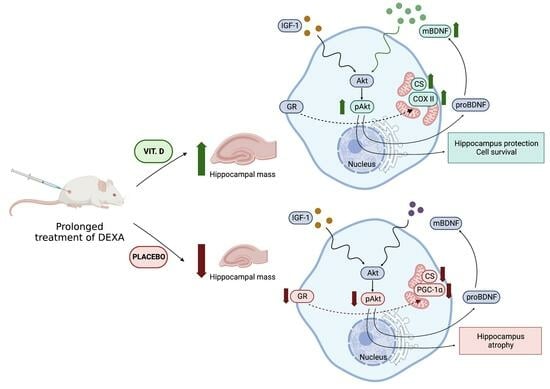
Supplementation with Vitamin D3 Protects against Mitochondrial Dysfunction and Loss of BDNF-Mediated Akt Activity in the Hippocampus during Long-Term Dexamethasone Treatment in Rats
by Daria Korewo-Labelle, Mateusz Jakub Karnia, Dorota Myślińska and Jan Jacek Kaczor
Int. J. Mol. Sci. 2023, 24(18), 13941; https://doi.org/10.3390/ijms241813941 - 11 Sep 2023
Cited by 2 | Viewed by 1868
Abstract
Dexamethasone (DEXA) is a commonly used steroid drug with immunosuppressive and analgesic properties. Unfortunately, long-term exposure to DEXA severely impairs brain function. This study aimed to investigate the effects of vitamin D3 supplementation during chronic DEXA treatment on neurogenesis, mitochondrial energy metabolism, [...] Read more.
Dexamethasone (DEXA) is a commonly used steroid drug with immunosuppressive and analgesic properties. Unfortunately, long-term exposure to DEXA severely impairs brain function. This study aimed to investigate the effects of vitamin D3 supplementation during chronic DEXA treatment on neurogenesis, mitochondrial energy metabolism, protein levels involved in the BDNF-mediated Akt activity, and specific receptors in the hippocampus. We found reduced serum concentrations of 25-hydroxyvitamin D3 (25(OH)D3), downregulated proBDNF and pAkt, dysregulated glucocorticosteroid and mineralocorticoid receptors, impaired mitochondrial biogenesis, and dysfunctional mitochondria energy metabolism in the DEXA-treated group. In contrast, supplementation with vitamin D3 restored the 25(OH)D3 concentration to a value close to that of the control group. There was an elevation in neurotrophic factor protein level, along with augmented activity of pAkt and increased citrate synthase activity in the hippocampus after vitamin D3 administration in long-term DEXA-treated rats. Our findings demonstrate that vitamin D3 supplementation plays a protective role in the hippocampus and partially mitigates the deleterious effects of long-term DEXA administration. The association between serum 25(OH)D3 concentration and BDNF level in the hippocampus indicates the importance of applying vitamin D3 supplementation to prevent and treat pathological conditions. Full article
Show Figures

Graphical abstract

28 pages, 2643 KiB  
Review
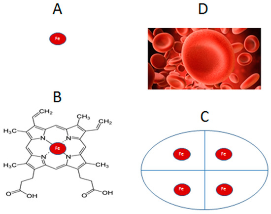
Iron Load Toxicity in Medicine: From Molecular and Cellular Aspects to Clinical Implications
by George J. Kontoghiorghes
Int. J. Mol. Sci. 2023, 24(16), 12928; https://doi.org/10.3390/ijms241612928 - 18 Aug 2023
Cited by 10 | Viewed by 4256
Abstract
Iron is essential for all organisms and cells. Diseases of iron imbalance affect billions of patients, including those with iron overload and other forms of iron toxicity. Excess iron load is an adverse prognostic factor for all diseases and can cause serious organ [...] Read more.
Iron is essential for all organisms and cells. Diseases of iron imbalance affect billions of patients, including those with iron overload and other forms of iron toxicity. Excess iron load is an adverse prognostic factor for all diseases and can cause serious organ damage and fatalities following chronic red blood cell transfusions in patients of many conditions, including hemoglobinopathies, myelodyspasia, and hematopoietic stem cell transplantation. Similar toxicity of excess body iron load but at a slower rate of disease progression is found in idiopathic haemochromatosis patients. Excess iron deposition in different regions of the brain with suspected toxicity has been identified by MRI T2* and similar methods in many neurodegenerative diseases, including Alzheimer’s disease and Parkinson’s disease. Based on its role as the major biological catalyst of free radical reactions and the Fenton reaction, iron has also been implicated in all diseases associated with free radical pathology and tissue damage. Furthermore, the recent discovery of ferroptosis, which is a cell death program based on free radical generation by iron and cell membrane lipid oxidation, sparked thousands of investigations and the association of iron with cardiac, kidney, liver, and many other diseases, including cancer and infections. The toxicity implications of iron in a labile, non-protein bound form and its complexes with dietary molecules such as vitamin C and drugs such as doxorubicin and other xenobiotic molecules in relation to carcinogenesis and other forms of toxicity are also discussed. In each case and form of iron toxicity, the mechanistic insights, diagnostic criteria, and molecular interactions are essential for the design of new and effective therapeutic interventions and of future targeted therapeutic strategies. In particular, this approach has been successful for the treatment of most iron loading conditions and especially for the transition of thalassemia from a fatal to a chronic disease due to new therapeutic protocols resulting in the complete elimination of iron overload and of iron toxicity. Full article
Show Figures

Figure 1

24 pages, 10108 KiB  
Article
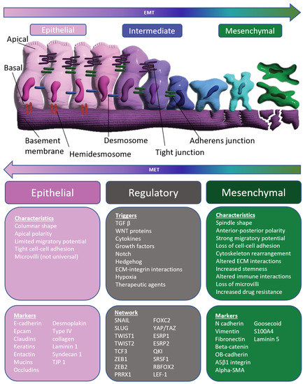
Extended Exposure Topotecan Significantly Improves Long-Term Drug Sensitivity by Decreasing Malignant Cell Heterogeneity and by Preventing Epithelial–Mesenchymal Transition
by Joshua T. Davis, Taraswi Mitra Ghosh, Suman Mazumder, Amit Mitra, Richard Curtis Bird and Robert D. Arnold
Int. J. Mol. Sci. 2023, 24(10), 8490; https://doi.org/10.3390/ijms24108490 - 9 May 2023
Cited by 1 | Viewed by 2390
Abstract
Maximum tolerable dosing (MTD) of chemotherapeutics has long been the gold standard for aggressive malignancies. Recently, alternative dosing strategies have gained traction for their improved toxicity profiles and unique mechanisms of action, such as inhibition of angiogenesis and stimulation of immunity. In this [...] Read more.
Maximum tolerable dosing (MTD) of chemotherapeutics has long been the gold standard for aggressive malignancies. Recently, alternative dosing strategies have gained traction for their improved toxicity profiles and unique mechanisms of action, such as inhibition of angiogenesis and stimulation of immunity. In this article, we investigated whether extended exposure (EE) topotecan could improve long-term drug sensitivity by preventing drug resistance. To achieve significantly longer exposure times, we used a spheroidal model system of castration-resistant prostate cancer. We also used state-of-the-art transcriptomic analysis to further elucidate any underlying phenotypic changes that occurred in the malignant population following each treatment. We determined that EE topotecan had a much higher barrier to resistance relative to MTD topotecan and was able to maintain consistent efficacy throughout the study period (EE IC50 of 54.4 nM (Week 6) vs. MTD IC50 of 2200 nM (Week 6) vs. 83.8 nM IC50 for control (Week 6) vs. 37.8 nM IC50 for control (Week 0)). As a possible explanation for these results, we determined that MTD topotecan stimulated epithelial–mesenchymal transition (EMT), upregulated efflux pumps, and produced altered topoisomerases relative to EE topotecan. Overall, EE topotecan resulted in a more sustained treatment response and maintained a less aggressive malignant phenotype relative to MTD topotecan. Full article
Show Figures

Figure 1

9 pages, 764 KiB  
Communication
Intestinal Permeability in Patients Early after Kidney Transplantation Treated with Two Different Formulations of Once-Daily Tacrolimus
by Aureliusz Kolonko, Natalia Słabiak-Błaż, Patrycja Pokora, Grzegorz Piecha and Andrzej Więcek
Int. J. Mol. Sci. 2023, 24(9), 8344; https://doi.org/10.3390/ijms24098344 - 6 May 2023
Cited by 1 | Viewed by 1680
Abstract
Adequate tacrolimus blood exposure is crucial in the early post-renal transplant period and a gut epithelial barrier integrity may play a role. We prospectively investigated several markers of intestinal permeability in recent kidney transplant recipients (KTRs) treated with different tacrolimus extended-release formulations. Within [...] Read more.
Adequate tacrolimus blood exposure is crucial in the early post-renal transplant period and a gut epithelial barrier integrity may play a role. We prospectively investigated several markers of intestinal permeability in recent kidney transplant recipients (KTRs) treated with different tacrolimus extended-release formulations. Within each of the 49 KTR pairs that received grafts from the same donor, an early randomized conversion was performed from twice-daily (Prograf) to once-daily tacrolimus formulation: Advagraf or Envarsus. Plasma zonulin, calprotectin, circulating lipopolysaccharide (LPS), LPS-binding protein (LBP), intestinal fatty acid binding protein (FABP-2), and CD-14 levels were measured. There was no difference in the recipient age, dialysis vintage, BMI, and residual diuresis between Advagraf and Envarsus groups. FABP-2 and LPS levels were significantly associated with tacrolimus trough level, 3-h level, and area under the curve (AUC) in the Envarsus but not in the Advagraf group. AUC was independently increased by LPS and decreased by age, FABP-2 concentration, and the use of Envarsus formulation as compared with Advagraf. Functional changes of gastrointestinal tract in patients treated with Envarsus may influence intestinal tacrolimus absorption to a greater extent than in Advagraf-treated KTRs and may lead to inadequate variability of tacrolimus exposure early after kidney transplantation. Full article
Show Figures

Figure 1

12 pages, 2220 KiB  
Article
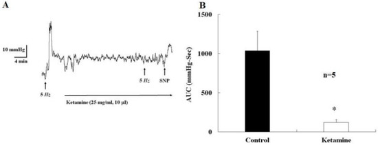
The Acute Effects and Mechanism of Ketamine on Nicotine-Induced Neurogenic Relaxation of the Corpus Cavernosum in Mice
by Ming-Wei Li, Tze-Chen Chao, Li-Yi Lim, Hsi-Hsien Chang and Stephen Shei-Dei Yang
Int. J. Mol. Sci. 2023, 24(8), 6976; https://doi.org/10.3390/ijms24086976 - 10 Apr 2023
Cited by 2 | Viewed by 2609
Abstract
The present study aimed to investigate the acute effects and the mechanism of ketamine on nicotine-induced relaxation of the corpus cavernosum (CC) in mice. This study measured the intra-cavernosal pressure (ICP) of male C57BL/6 mice and the CC muscle activities using an organ [...] Read more.
The present study aimed to investigate the acute effects and the mechanism of ketamine on nicotine-induced relaxation of the corpus cavernosum (CC) in mice. This study measured the intra-cavernosal pressure (ICP) of male C57BL/6 mice and the CC muscle activities using an organ bath wire myograph. Various drugs were used to investigate the mechanism of ketamine on nicotine-induced relaxation. Direct ketamine injection into the major pelvic ganglion (MPG) inhibited MPG-induced increases in ICP. D-serine/L-glutamate-induced relaxation of the CC was inhibited by MK-801 (N-methyl-D-aspartate (NMDA) receptor inhibitor), and nicotine-induced relaxation was enhanced by D-serine/L-glutamate. NMDA had no effect on CC relaxation. Nicotine-induced relaxation of the CC was suppressed by mecamylamine (a non-selective nicotinic acetylcholine receptor antagonist), lidocaine, guanethidine (an adrenergic neuronal blocker), Nw-nitro-L-arginine (a non-selective nitric oxide synthase inhibitor), MK-801, and ketamine. This relaxation was almost completely inhibited in CC strips pretreated with 6-hydroxydopamine (a neurotoxic synthetic organic compound). Ketamine inhibited cavernosal nerve neurotransmission via direct action on the ganglion and impaired nicotine-induced CC relaxation. The relaxation of the CC was dependent on the interaction of the sympathetic and parasympathetic nerves, which may be mediated by the NMDA receptor. Full article
Show Figures

Figure 1

11 pages, 1955 KiB  
Communication
A Lipidated Single-B-Chain Derivative of Relaxin Exhibits Improved In Vitro Serum Stability without Altering Activity
by Praveen Praveen, Chao Wang, Thomas N. G. Handley, Hongkang Wu, Chrishan S. Samuel, Ross A. D. Bathgate and Mohammed Akhter Hossain
Int. J. Mol. Sci. 2023, 24(7), 6616; https://doi.org/10.3390/ijms24076616 - 1 Apr 2023
Cited by 4 | Viewed by 2308
Abstract
Human relaxin-2 (H2 relaxin) is therapeutically very important due to its strong anti-fibrotic, vasodilatory, and cardioprotective effects. Therefore, relaxin’s receptor, relaxin family peptide receptor 1 (RXFP1), is a potential target for the treatment of fibrosis and related disorders, including heart failure. H2 relaxin [...] Read more.
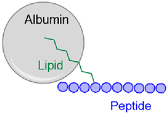
Human relaxin-2 (H2 relaxin) is therapeutically very important due to its strong anti-fibrotic, vasodilatory, and cardioprotective effects. Therefore, relaxin’s receptor, relaxin family peptide receptor 1 (RXFP1), is a potential target for the treatment of fibrosis and related disorders, including heart failure. H2 relaxin has a complex two-chain structure (A and B) and three disulfide bridges. Our laboratory has recently developed B7-33 peptide, a single-chain agonist based on the B-chain of H2 relaxin. However, the peptide B7-33 has a short circulation time in vitro in serum (t1/2 = ~6 min). In this study, we report structure-activity relationship studies on B7-33 utilizing different fatty-acid conjugations at different positions. We have shown that by fatty-acid conjugation with an appropriate spacer length, the in vitro half-life of B7-33 can be increased from 6 min to 60 min. In the future, the lead lipidated molecule will be studied in animal models to measure its PK/PD properties, which will lead to their pre-clinical applications. Full article
Show Figures

Figure 1

16 pages, 3362 KiB  
Article
Aconitum carmichaelii Debx. Attenuates Heart Failure through Inhibiting Inflammation and Abnormal Vascular Remodeling
by Ziwei Xing, Junren Chen, Tingting Yu, Xu Li, Wei Dong, Cheng Peng and Dan Li
Int. J. Mol. Sci. 2023, 24(6), 5838; https://doi.org/10.3390/ijms24065838 - 19 Mar 2023
Cited by 8 | Viewed by 2383
Abstract
Heart failure (HF) is the most common complication following myocardial infarction, closely associated with ventricular remodeling. Aconitum carmichaelii Debx., a traditional Chinese herb, possesses therapeutic effects on HF and related cardiac diseases. However, its effects and mechanisms on HF-associated cardiac diseases are still [...] Read more.
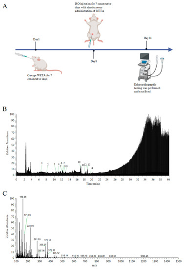
Heart failure (HF) is the most common complication following myocardial infarction, closely associated with ventricular remodeling. Aconitum carmichaelii Debx., a traditional Chinese herb, possesses therapeutic effects on HF and related cardiac diseases. However, its effects and mechanisms on HF-associated cardiac diseases are still unclear. In the present study, a water extraction of toasted Aconitum carmichaelii Debx. (WETA) was verified using UPLC-Q/TOF-MS. The heart function of HF rats was assessed by echocardiography and strain analysis, and myocardial injury was measured by serum levels of CK-MB, cTnT, and cTnI. The pathological changes of cardiac tissues were evaluated by 2,3,5-triphenyltetrazolium chloride (TTC) staining, hematoxylin and eosin (H&E) staining, and Masson’s trichrome staining. Additionally, the levels of inflammation-related genes and proteins and components related to vascular remodeling were detected by RT-qPCR, Western blot, and immunofluorescence. WETA significantly inhibited the changes in echocardiographic parameters and the increase in heart weight, cardiac infarction size, the myonecrosis, edema, and infiltration of inflammatory cells, collagen deposition in heart tissues, and also mitigated the elevated serum levels of CK-MB, cTnT, and cTnI in ISO-induced rats. Additionally, WETA suppressed the expressions of inflammatory genes, including IL-1β, IL-6, and TNF-α and vascular injury-related genes, such as VCAM1, ICAM1, ANP, BNP, and MHC in heart tissues of ISO-induced HF rats, which were further confirmed by Western blotting and immunofluorescence. In summary, the myocardial protective effect of WETA was conferred through inhibiting inflammatory responses and abnormal vascular remodeling in ISO-treated rats. Full article
Show Figures

Figure 1

21 pages, 7868 KiB  
Article
Structural Basis for Agonistic Activity and Selectivity toward Melatonin Receptors hMT1 and hMT2
by Mattia Cantarini, Dario Rusciano, Rosario Amato, Alessio Canovai, Maurizio Cammalleri, Massimo Dal Monte, Cristina Minnelli, Emiliano Laudadio, Giovanna Mobbili, Giorgia Giorgini and Roberta Galeazzi
Int. J. Mol. Sci. 2023, 24(3), 2863; https://doi.org/10.3390/ijms24032863 - 2 Feb 2023
Cited by 7 | Viewed by 2553
Abstract
Glaucoma, a major ocular neuropathy originating from a progressive degeneration of retinal ganglion cells, is often associated with increased intraocular pressure (IOP). Daily IOP fluctuations are physiologically influenced by the antioxidant and signaling activities of melatonin. This endogenous modulator has limited employment in [...] Read more.
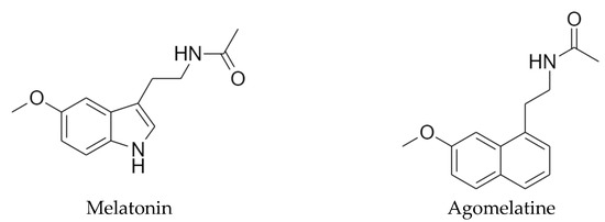
Glaucoma, a major ocular neuropathy originating from a progressive degeneration of retinal ganglion cells, is often associated with increased intraocular pressure (IOP). Daily IOP fluctuations are physiologically influenced by the antioxidant and signaling activities of melatonin. This endogenous modulator has limited employment in treating altered IOP disorders due to its low stability and bioavailability. The search for low-toxic compounds as potential melatonin agonists with higher stability and bioavailability than melatonin itself could start only from knowing the molecular basis of melatonergic activity. Thus, using a computational approach, we studied the melatonin binding toward its natural macromolecular targets, namely melatonin receptors 1 (MT1) and 2 (MT2), both involved in IOP signaling regulation. Besides, agomelatine, a melatonin-derivative agonist and, at the same time, an atypical antidepressant, was also included in the study due to its powerful IOP-lowering effects. For both ligands, we evaluated both stability and ligand positioning inside the orthosteric site of MTs, mapping the main molecular interactions responsible for receptor activation. Affinity values in terms of free binding energy (ΔGbind) were calculated for the selected poses of the chosen compounds after stabilization through a dynamic molecular docking protocol. The results were compared with experimental in vivo effects, showing a higher potency and more durable effect for agomelatine with respect to melatonin, which could be ascribed both to its higher affinity for hMT2 and to its additional activity as an antagonist for the serotonin receptor 5-HT2c, in agreement with the in silico results. Full article
Show Figures

Figure 1

11 pages, 1984 KiB  
Article
Tenebrio molitor as a Simple and Cheap Preclinical Pharmacokinetic and Toxicity Model
by Annalaura Brai, Federica Poggialini, Chiara Vagaggini, Claudia Pasqualini, Sauro Simoni, Valeria Francardi and Elena Dreassi
Int. J. Mol. Sci. 2023, 24(3), 2296; https://doi.org/10.3390/ijms24032296 - 24 Jan 2023
Cited by 18 | Viewed by 2978
Abstract
The progression of drugs into clinical phases requires proper toxicity assessment in animals and the correct identification of possible metabolites. Accordingly, different animal models are used to preliminarily evaluate toxicity and biotransformations. Rodents are the most common models used to preliminarily evaluate the [...] Read more.
The progression of drugs into clinical phases requires proper toxicity assessment in animals and the correct identification of possible metabolites. Accordingly, different animal models are used to preliminarily evaluate toxicity and biotransformations. Rodents are the most common models used to preliminarily evaluate the safety of drugs; however, their use is subject to ethical consideration and elevated costs, and strictly regulated by national legislations. Herein, we developed a novel, cheap and convenient toxicity model using Tenebrio molitor coleoptera (TMC). A panel of 15 drugs—including antivirals and antibacterials—with different therapeutic applications was administered to TMC and the LD50 was determined. The values are comparable with those already determined in mice and rats. In addition, a TMC model was used to determine the presence of the main metabolites and in vivo pharmacokinetics (PK), and results were compared with those available from in vitro assays and the literature. Taken together, our results demonstrate that TMC can be used as a novel and convenient preliminary toxicity model to preliminarily evaluate the safety of experimental compounds and the formation of main metabolites, and to reduce the costs and number of rodents, according to 3R principles. Full article
Show Figures

Figure 1

20 pages, 7424 KiB  
Article
Warburg Effect as a Novel Mechanism for Homocysteine-Induced Features of Age-Related Macular Degeneration
by Yara A. Samra, Yusra Zaidi, Pragya Rajpurohit, Raju Raghavan, Lun Cai, Ismail Kaddour-Djebbar and Amany Tawfik
Int. J. Mol. Sci. 2023, 24(2), 1071; https://doi.org/10.3390/ijms24021071 - 5 Jan 2023
Cited by 8 | Viewed by 2622
Abstract
Age-related macular degeneration (AMD) is a major cause of blindness. Recent studies have reported impaired glycolysis in AMD patients with a high lactate/pyruvate ratio. Elevated homocysteine (Hcy) (Hyperhomocysteinemia, HHcy) was observed in several clinical studies, reporting an association between HHcy and AMD. We [...] Read more.
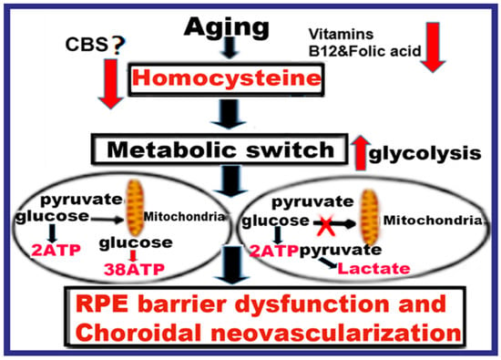
Age-related macular degeneration (AMD) is a major cause of blindness. Recent studies have reported impaired glycolysis in AMD patients with a high lactate/pyruvate ratio. Elevated homocysteine (Hcy) (Hyperhomocysteinemia, HHcy) was observed in several clinical studies, reporting an association between HHcy and AMD. We established the effect of HHcy on barrier function, retinal pigment epithelium (RPE) structure, and induced choroidal neovascularization (CNV) in mice. We hypothesize that HHcy contributes to AMD by inducing a metabolic switch in the mitochondria, in which cells predominantly produce energy by the high rate of glycolysis, or “Warburg”, effect. Increased glycolysis results in an increased production of lactate, cellular acidity, activation of angiogenesis, RPE barrier dysfunction, and CNV. Evaluation of cellular energy production under HHcy was assessed by seahorse analysis, immunofluorescence, and western blot experiments. The seahorse analysis evaluated the extracellular acidification rate (ECAR) as indicative of glycolysis. HHcy showed a significant increase in ECAR both in vivo using (Cystathionine β-synthase) cbs+/− and cbs−/− mice retinas and in vitro (Hcy-treated ARPE-19) compared to wild-type mice and RPE cells. Moreover, HHcy up-regulated glycolytic enzyme (Glucose transporter-1 (GlUT-1), lactate dehydrogenase (LDH), and hexokinase 1 (HK1)) in Hcy-treated ARPE-19 and primary RPE cells isolated from cbs+/+, cbs+/−, and cbs−/− mice retinas. Inhibition of GLUT-1 or blocking of N-methyl-D-aspartate receptors (NMDAR) reduced glycolysis in Hcy-treated RPE and improved albumin leakage and CNV induction in Hcy-injected mice eyes. The current study suggests that HHcy causes a metabolic switch in the RPE cells from mitochondrial respiration to glycolysis during AMD and confirms the involvement of NMDAR in this process. Therefore, targeting Glycolysis or NMDAR could be a novel therapeutic target for AMD. Full article
Show Figures

Figure 1

2022

Jump to: 2024, 2023, 2021, 2020, 2019

17 pages, 1150 KiB  
Article
Novel Arylpiperazine Derivatives of Salicylamide with α1-Adrenolytic Properties Showed Antiarrhythmic and Hypotensive Properties in Rats
by Elżbieta Żmudzka, Klaudia Lustyk, Agata Siwek, Małgorzata Wolak, Adam Gałuszka, Jolanta Jaśkowska, Marcin Kołaczkowski, Jacek Sapa and Karolina Pytka
Int. J. Mol. Sci. 2023, 24(1), 293; https://doi.org/10.3390/ijms24010293 - 24 Dec 2022
Cited by 1 | Viewed by 2017
Abstract
Cardiovascular diseases remain one of the leading causes of death worldwide. Unfortunately, the available pharmacotherapeutic options have limited effectiveness. Therefore, developing new drug candidates remains very important. We selected six novel arylpiperazine alkyl derivatives of salicylamide to investigate their cardiovascular effects. Having in [...] Read more.
Cardiovascular diseases remain one of the leading causes of death worldwide. Unfortunately, the available pharmacotherapeutic options have limited effectiveness. Therefore, developing new drug candidates remains very important. We selected six novel arylpiperazine alkyl derivatives of salicylamide to investigate their cardiovascular effects. Having in mind the beneficial role of α1-adrenergic receptors in restoring sinus rhythm and regulating blood pressure, first, using radioligand binding assays, we evaluated the affinity of the tested compounds for α-adrenergic receptors. Our experiments revealed their high to moderate affinity for α1- but not α2-adrenoceptors. Next, we aimed to determine the antiarrhythmic potential of novel derivatives in rat models of arrhythmia induced by adrenaline, calcium chloride, or aconitine. All compounds showed potent prophylactic antiarrhythmic activity in the adrenaline-induced arrhythmia model and no effects in calcium chloride- or aconitine-induced arrhythmias. Moreover, the tested compounds demonstrated therapeutic antiarrhythmic activity, restoring a normal sinus rhythm immediately after the administration of the arrhythmogen adrenaline. Notably, none of the tested derivatives affected the normal electrocardiogram (ECG) parameters in rodents, which excludes their proarrhythmic potential. Finally, all tested compounds decreased blood pressure in normotensive rats and reversed the pressor response to methoxamine, suggesting that their hypotensive mechanism of action is connected with the blockade of α1-adrenoceptors. Our results confirm the antiarrhythmic and hypotensive activities of novel arylpiperazine derivatives and encourage their further investigation as model structures for potential drugs. Full article
Show Figures

Figure 1

12 pages, 1682 KiB  
Article
A Comprehensive Study of Gemfibrozil Complexation with β-Cyclodextrins in Aqueous Solution Using Different Analytical Techniques
by Arantza Zornoza, Itziar Vélaz, Gustavo González-Gaitano and María Cristina Martínez-Ohárriz
Int. J. Mol. Sci. 2022, 23(24), 16119; https://doi.org/10.3390/ijms232416119 - 17 Dec 2022
Cited by 2 | Viewed by 1518
Abstract
Gemfibrozil (GEM) is a hypolipidemic agent, which is effective in reducing serum cholesterol and triglyceride levels. Complexation of GEM with native β-cyclodextrin (β-CD) and with the derivatives hydroxypropyl-β- and randomly methylated β-CD (HPβ-CD and Meβ-CD) was studied in aqueous solution of pH 2.8 [...] Read more.
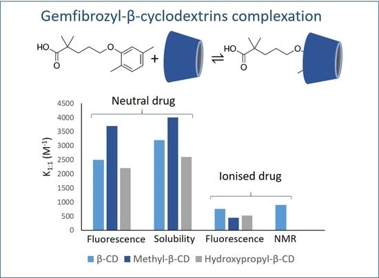
Gemfibrozil (GEM) is a hypolipidemic agent, which is effective in reducing serum cholesterol and triglyceride levels. Complexation of GEM with native β-cyclodextrin (β-CD) and with the derivatives hydroxypropyl-β- and randomly methylated β-CD (HPβ-CD and Meβ-CD) was studied in aqueous solution of pH 2.8 and 7.0. The stability constants were determined by spectrofluorimetry, 1H-NMR spectroscopy and solubility assays. Considering the well-known difficulties to obtain similar stability constants by different techniques, the agreement of the values obtained supports the reliability of the results presented. The advantages and drawbacks of each analytical technique for the study of inclusion complexation were discussed as well. In addition, the thermodynamic parameters of complexation, enthalpy (ΔH) and entropy (ΔS), were determined and related to the type of molecular interactions that take place between GEM and the different cyclodextrins. Finally, solid dispersions were prepared by co-evaporation, kneading, vacuum desiccation, and coprecipitation, and complexation was evaluated by X-ray diffraction. Full article
Show Figures

Graphical abstract

35 pages, 9265 KiB  
Article
Synthesis, Antiproliferative Evaluation and QSAR Analysis of Novel Halogen- and Amidino-Substituted Benzothiazoles and Benzimidazoles
by Valentina Rep Kaulić, Livio Racané, Marijana Leventić, Domagoj Šubarić, Vesna Rastija, Ljubica Glavaš-Obrovac and Silvana Raić-Malić
Int. J. Mol. Sci. 2022, 23(24), 15843; https://doi.org/10.3390/ijms232415843 - 13 Dec 2022
Viewed by 2133
Abstract
Syntheses of 6-halogen-substituted benzothiazoles were performed by condensation of 4-hydroxybenzaldehydes and 2-aminotiophenoles and subsequent O-alkylation with appropriate halides, whereas 6-amidino-substituted benzothiazoles were synthesized by condensation of 5-amidino-2-aminothiophenoles and corresponding benzaldehydes. While most of the compounds from non-substituted and halogen-substituted benzothiazole series showed [...] Read more.
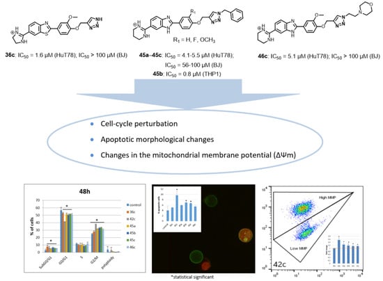
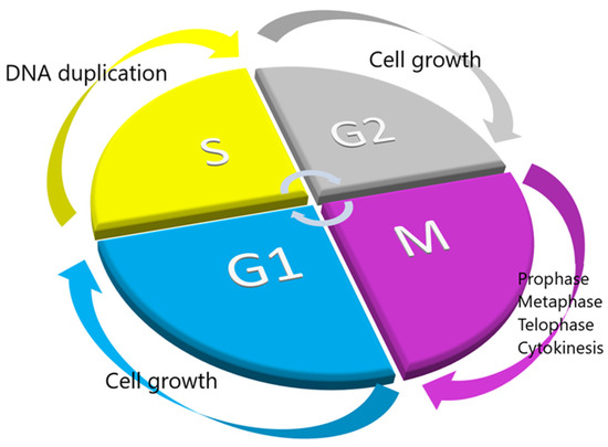
Syntheses of 6-halogen-substituted benzothiazoles were performed by condensation of 4-hydroxybenzaldehydes and 2-aminotiophenoles and subsequent O-alkylation with appropriate halides, whereas 6-amidino-substituted benzothiazoles were synthesized by condensation of 5-amidino-2-aminothiophenoles and corresponding benzaldehydes. While most of the compounds from non-substituted and halogen-substituted benzothiazole series showed marginal antiproliferative activity on tested tumor cell lines, amidino benzazoles exhibited stronger inhibitory activity. Generally, imidazolyl benzothiazoles showed pronounced and nonselective activity, with the exception of 36c which had a strong inhibitory effect on HuT78 cells (IC50 = 1.6 µM) without adverse cytotoxicity on normal BJ cells (IC50 >100 µM). Compared to benzothiazoles, benzimidazole structural analogs 45a45c and 46c containing the 1,2,3-triazole ring exhibited pronounced and selective antiproliferative activity against HuT78 cells with IC50 < 10 µM. Moreover, compounds 45c and 46c containing the methoxy group at the phenoxy unit were not toxic to normal BJ cells. Of all the tested compounds, benzimidazole 45a with the unsubstituted phenoxy central core showed the most pronounced cell growth inhibition on THP1 cells in the nanomolar range (IC50 = 0.8 µM; SI = 70). QSAR models of antiproliferative activity for benzazoles on T-cell lymphoma (HuT78) and non-tumor MDCK-1 cells elucidated the effects of the substituents at position 6 of benzazoles, demonstrating their dependence on the topological and spatial distribution of atomic mass, polarizability, and van der Waals volumes. A notable cell cycle perturbation with higher accumulation of cells in the G2/M phase, and a significant cell increase in subG0/G1 phase were found in HuT78 cells treated with 36c, 42c, 45a45c and 46c. Apoptotic morphological changes, an externalization of phosphatidylserine, and changes in the mitochondrial membrane potential of treated cells were observed as well. Full article
Show Figures

Graphical abstract

13 pages, 3624 KiB  
Article
Molecular Tuning of IR-786 for Improved Brown Adipose Tissue Imaging
by Gayoung Jo, Eun Jeong Kim, Juhyun Song and Hoon Hyun
Int. J. Mol. Sci. 2022, 23(22), 13756; https://doi.org/10.3390/ijms232213756 - 9 Nov 2022
Cited by 4 | Viewed by 1871
Abstract
To overcome the limitations of brown adipose tissue (BAT) imaging with MRI and PET/CT, near-infrared (NIR) fluorescence imaging has been utilized in living animals because it is highly sensitive, noninvasive, nonradioactive, and cost-effective. To date, only a few NIR fluorescent dyes for detecting [...] Read more.
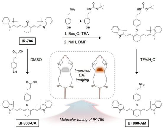
To overcome the limitations of brown adipose tissue (BAT) imaging with MRI and PET/CT, near-infrared (NIR) fluorescence imaging has been utilized in living animals because it is highly sensitive, noninvasive, nonradioactive, and cost-effective. To date, only a few NIR fluorescent dyes for detecting BAT have been reported based on the structure-inherent targeting strategy. Among them, IR-786, a commercial cyanine dye, was used firstly for quantitative NIR imaging of BAT perfusion in 2003. Owing to the high cytotoxicity, poor water solubility, and strong nonspecific background uptake of IR-786, the chemical structure of IR-786 should be redesigned to be more hydrophilic and less toxic so that it can show more BAT-specific accumulation. Here, we developed a BAT-specific NIR dye, BF800-AM, by incorporating the tyramine linker in the original structure of IR-786. After modifying the physicochemical properties of IR-786, in vivo results showed significant uptake of the newly designed BF800-AM in the BAT with improved signal-to-background ratio. Additional in vivo studies using mouse tumor models revealed that BF800-AM targeting to BAT is independent of tumor tissues, as distinct from IR-786 showing uptake in both tissues. Therefore, BF800-AM can be used for improved noninvasive visualization of BAT mass and activity in living animals. Full article
Show Figures

Figure 1

13 pages, 1431 KiB  
Review
Advances in Intraoperative Flow Cytometry
by Marcos V. D’Amato Figueiredo, George A. Alexiou, George Vartholomatos and Roberta Rehder
Int. J. Mol. Sci. 2022, 23(21), 13430; https://doi.org/10.3390/ijms232113430 - 3 Nov 2022
Cited by 5 | Viewed by 3137
Abstract
Flow cytometry is the gold-standard laser-based technique to measure and analyze fluorescence levels of immunostaining and DNA content in individual cells. It provides a valuable tool to assess cells in the G0/G1, S, and G2/M phases, and those with polyploidy, which holds prognostic [...] Read more.
Flow cytometry is the gold-standard laser-based technique to measure and analyze fluorescence levels of immunostaining and DNA content in individual cells. It provides a valuable tool to assess cells in the G0/G1, S, and G2/M phases, and those with polyploidy, which holds prognostic significance. Frozen section analysis is the standard intraoperative assessment for tumor margin evaluation and tumor resection. Here, we present flow cytometry as a promising technique for intraoperative tumor analysis in different pathologies, including brain tumors, leptomeningeal dissemination, breast cancer, head and neck cancer, pancreatic tumor, and hepatic cancer. Flow cytometry is a valuable tool that can provide substantial information on tumor analysis and, consequently, maximize cancer treatment and expedite patients’ survival. Full article
Show Figures

Figure 1

19 pages, 3058 KiB  
Article
Opaganib Protects against Radiation Toxicity: Implications for Homeland Security and Antitumor Radiotherapy
by Lynn W. Maines, Randy S. Schrecengost, Yan Zhuang, Staci N. Keller, Ryan A. Smith, Cecelia L. Green and Charles D. Smith
Int. J. Mol. Sci. 2022, 23(21), 13191; https://doi.org/10.3390/ijms232113191 - 29 Oct 2022
Cited by 4 | Viewed by 6405
Abstract
Exposure to ionizing radiation (IR) is a lingering threat from accidental or terroristic nuclear events, but is also widely used in cancer therapy. In both cases, host inflammatory responses to IR damage normal tissue causing morbidity and possibly mortality to the victim/patient. Opaganib, [...] Read more.
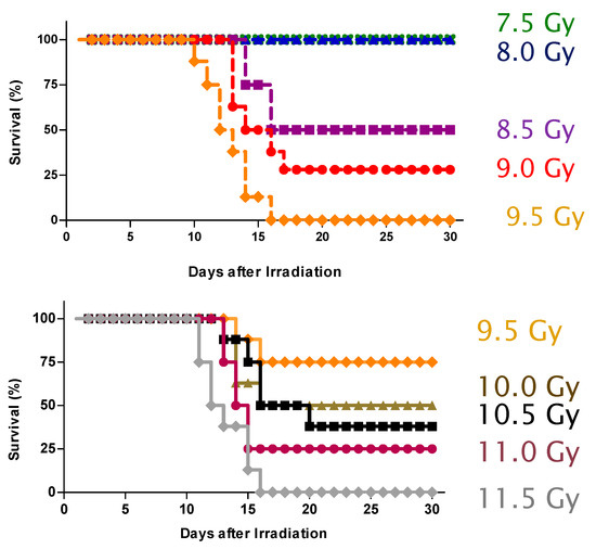
Exposure to ionizing radiation (IR) is a lingering threat from accidental or terroristic nuclear events, but is also widely used in cancer therapy. In both cases, host inflammatory responses to IR damage normal tissue causing morbidity and possibly mortality to the victim/patient. Opaganib, a first-in-class inhibitor of sphingolipid metabolism, has broad anti-inflammatory and anticancer activity. Opaganib elevates ceramide and reduces sphingosine 1-phosphate (S1P) in cells, conditions that increase the antitumor efficacy of radiation while concomitantly suppressing inflammatory damage to normal tissue. Therefore, opaganib may suppress toxicity from unintended IR exposure and improve patient response to chemoradiation. To test these hypotheses, we first examined the effects of opaganib on the toxicity and antitumor activity of radiation in mice exposed to total body irradiation (TBI) or IR with partial bone marrow shielding. Oral treatment with opaganib 2 h before TBI shifted the LD75 from 9.5 Gy to 11.5 Gy, and provided substantial protection against gastrointestinal damage associated with suppression of radiation-induced elevations of S1P and TNFα in the small intestines. In the partially shielded model, opaganib provided dose-dependent survival advantages when administered 4 h before or 24 h after radiation exposure, and was particularly effective when given both prior to and following radiation. Relevant to cancer radiotherapy, opaganib decreased the sensitivity of IEC6 (non-transformed mouse intestinal epithelial) cells to radiation, while sensitizing PAN02 cells to in vitro radiation. Next, the in vivo effects of opaganib in combination with radiation were examined in a syngeneic tumor model consisting of C57BL/6 mice bearing xenografts of PAN02 pancreatic cancer cells and a cross-species xenograft model consisting of nude mice bearing xenografts of human FaDu cells. Mice were treated with opaganib and/or IR (plus cisplatin in the case of FaDu tumors). In both tumor models, the optimal suppression of tumor growth was attained by the combination of opaganib with IR (± cisplatin). Overall, opaganib substantially protects normal tissue from radiation damage that may occur through unintended exposure or cancer radiotherapy. Full article
Show Figures

Figure 1

19 pages, 3836 KiB  
Article
Activation of Piezo1 Increases Na,K-ATPase-Mediated Ion Transport in Mouse Lens
by Mohammad Shahidullah, Joaquin Lopez Rosales and Nicholas Delamere
Int. J. Mol. Sci. 2022, 23(21), 12870; https://doi.org/10.3390/ijms232112870 - 25 Oct 2022
Cited by 8 | Viewed by 2610
Abstract
Lens ion homeostasis depends on Na,K-ATPase and NKCC1. TRPV4 and TRPV1 channels, which are mechanosensitive, play important roles in mechanisms that regulate the activity of these transporters. Here, we examined another mechanosensitive channel, piezo1, which is also expressed in the lens. The purpose [...] Read more.
Lens ion homeostasis depends on Na,K-ATPase and NKCC1. TRPV4 and TRPV1 channels, which are mechanosensitive, play important roles in mechanisms that regulate the activity of these transporters. Here, we examined another mechanosensitive channel, piezo1, which is also expressed in the lens. The purpose of the study was to examine piezo1 function. Recognizing that activation of TRPV4 and TRPV1 causes changes in lens ion transport mechanisms, we carried out studies to determine whether piezo1 activation changes either Na,K-ATPase-mediated or NKCC1-mediated ion transport. We also examined channel function of piezo1 by measuring calcium entry. Rb uptake was measured as an index of inwardly directed potassium transport by intact mouse lenses. Intracellular calcium concentration was measured in Fura-2 loaded cells by a ratiometric imaging technique. Piezo1 immunolocalization was most evident in the lens epithelium. Potassium (Rb) uptake was increased in intact lenses as well as in cultured lens epithelium exposed to Yoda1, a piezo1 agonist. The majority of Rb uptake is Na,K-ATPase-dependent, although there also is a significant NKCC-dependent component. In the presence of ouabain, an Na,K-ATPase inhibitor, Yoda1 did not increase Rb uptake. In contrast, Yoda1 increased Rb uptake to a similar degree in the presence or absence of 1 µM bumetanide, an NKCC inhibitor. The Rb uptake response to Yoda1 was inhibited by the selective piezo1 antagonist GsMTx4, and also by the nonselective antagonists ruthenium red and gadolinium. In parallel studies, Yoda1 was observed to increase cytoplasmic calcium concentration in cells loaded with Fura-2. The calcium response to Yoda1 was abolished by gadolinium or ruthenium red. The calcium and Rb uptake responses to Yoda1 were absent in calcium-free bathing solution, consistent with calcium entry when piezo1 is activated. Taken together, these findings point to stimulation of Na,K-ATPase, but not NKCC, when piezo1 is activated. Na,K-ATPase is the principal mechanism responsible for ion and water homeostasis in the lens. The functional role of lens piezo1 is a topic for further study. Full article
Show Figures

Figure 1

11 pages, 1244 KiB  
Article
miRNA Expression May Have Implications for Immunotherapy in PDGFRA Mutant GISTs
by Gloria Ravegnini, Margherita Nannini, Valentina Indio, Cesar Serrano, Francesca Gorini, Annalisa Astolfi, Aldo Di Vito, Fabiana Morroni, Maria Abbondanza Pantaleo, Patrizia Hrelia and Sabrina Angelini
Int. J. Mol. Sci. 2022, 23(20), 12248; https://doi.org/10.3390/ijms232012248 - 14 Oct 2022
Cited by 2 | Viewed by 1880
Abstract
Gastrointestinal stromal tumors (GISTs) harboring mutations in the PDGFRA gene occur in only about 5–7% of patients. The most common PDGFRA mutation is exon 18 D842V, which is correlated with specific clinico-pathological features compared to the other PDGFRA mutated GISTs. Herein, we present [...] Read more.
Gastrointestinal stromal tumors (GISTs) harboring mutations in the PDGFRA gene occur in only about 5–7% of patients. The most common PDGFRA mutation is exon 18 D842V, which is correlated with specific clinico-pathological features compared to the other PDGFRA mutated GISTs. Herein, we present a miRNA expression profile comparison of PDGFRA D842V mutant GISTs and PDGFRA with mutations other than D842V (non-D842V). miRNA expression profiling was carried out on 10 patients using a TLDA miRNA array. Then, miRNA expression was followed by bioinformatic analysis aimed at evaluating differential expression, pathway enrichment, and miRNA-mRNA networks. We highlighted 24 differentially expressed miRNAs between D842V and non-D842V GIST patients. Pathway enrichment analysis showed that deregulated miRNAs targeted genes that are mainly involved in the immune response pathways. The miRNA-mRNA networks highlighted a signature of miRNAs/mRNA that could explain the indolent behavior of the D842V mutated GIST. The results highlighted a different miRNA fingerprint in PDGFRA D842V GISTs compared to non-D842Vmutated patients, which could explain the different biological behavior of this GIST subset. Full article
Show Figures

Figure 1

25 pages, 4866 KiB  
Article
Selectively Targeting Breast Cancer Stem Cells by 8-Quinolinol and Niclosamide
by Patricia Cámara-Sánchez, Zamira V. Díaz-Riascos, Natalia García-Aranda, Petra Gener, Joaquin Seras-Franzoso, Micaela Giani-Alonso, Miriam Royo, Esther Vázquez, Simó Schwartz, Jr. and Ibane Abasolo
Int. J. Mol. Sci. 2022, 23(19), 11760; https://doi.org/10.3390/ijms231911760 - 4 Oct 2022
Cited by 4 | Viewed by 2972
Abstract
Cancer maintenance, metastatic dissemination and drug resistance are sustained by cancer stem cells (CSCs). Triple negative breast cancer (TNBC) is the breast cancer subtype with the highest number of CSCs and the poorest prognosis. Here, we aimed to identify potential drugs targeting CSCs [...] Read more.
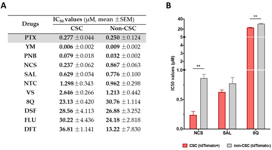
Cancer maintenance, metastatic dissemination and drug resistance are sustained by cancer stem cells (CSCs). Triple negative breast cancer (TNBC) is the breast cancer subtype with the highest number of CSCs and the poorest prognosis. Here, we aimed to identify potential drugs targeting CSCs to be further employed in combination with standard chemotherapy in TNBC treatment. The anti-CSC efficacy of up to 17 small drugs was tested in TNBC cell lines using cell viability assays on differentiated cancer cells and CSCs. Then, the effect of 2 selected drugs (8-quinolinol -8Q- and niclosamide -NCS-) in the cancer stemness features were evaluated using mammosphere growth, cell invasion, migration and anchorage-independent growth assays. Changes in the expression of stemness genes after 8Q or NCS treatment were also evaluated. Moreover, the potential synergism of 8Q and NCS with PTX on CSC proliferation and stemness-related signaling pathways was evaluated using TNBC cell lines, CSC-reporter sublines, and CSC-enriched mammospheres. Finally, the efficacy of NCS in combination with PTX was analyzed in vivo using an orthotopic mouse model of MDA-MB-231 cells. Among all tested drug candidates, 8Q and NCS showed remarkable specific anti-CSC activity in terms of CSC viability, migration, invasion and anchorage independent growth reduction in vitro. Moreover, specific 8Q/PTX and NCS/PTX ratios at which both drugs displayed a synergistic effect in different TNBC cell lines were identified. The sole use of PTX increased the relative presence of CSCs in TNBC cells, whereas the combination of 8Q and NCS counteracted this pro-CSC activity of PTX while significantly reducing cell viability. In vivo, the combination of NCS with PTX reduced tumor growth and limited the dissemination of the disease by reducing circulating tumor cells and the incidence of lung metastasis. The combination of 8Q and NCS with PTX at established ratios inhibits both the proliferation of differentiated cancer cells and the viability of CSCs, paving the way for more efficacious TNBC treatments. Full article
Show Figures

Figure 1

12 pages, 3855 KiB  
Article
Alpha-Lipoic Acid Attenuates Apoptosis and Ferroptosis in Cisplatin-Induced Ototoxicity via the Reduction of Intracellular Lipid Droplets
by Sam Cho, Seok Jin Hong, Sung Hun Kang, YongKeun Park and Sung Kyun Kim
Int. J. Mol. Sci. 2022, 23(18), 10981; https://doi.org/10.3390/ijms231810981 - 19 Sep 2022
Cited by 14 | Viewed by 3433
Abstract
Alpha-lipoic acid (α-LA) is a potent antioxidant that can prevent apoptosis associated with cisplatin-induced ototoxicity through ROS. Ferroptosis is defined as an iron-dependent cell death pathway that has recently been highlighted and is associated with the accumulation of intracellular lipid droplets (LDs) due [...] Read more.
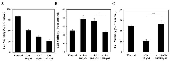
Alpha-lipoic acid (α-LA) is a potent antioxidant that can prevent apoptosis associated with cisplatin-induced ototoxicity through ROS. Ferroptosis is defined as an iron-dependent cell death pathway that has recently been highlighted and is associated with the accumulation of intracellular lipid droplets (LDs) due to an inflammatory process. Herein, we investigated the impact of α-LA on ferroptosis and analyzed the characteristics of LDs in auditory hair cells treated with cisplatin using high-resolution 3D quantitative-phase imaging with reconstruction of the refractive index (RI) distribution. HEI-OC1 cells were treated with 500 μM α-LA for 24 h and then with 15 μM cisplatin for 48 h. With 3D optical diffraction tomography (3D-ODT), the RI values of treated cells were analyzed. Regions with high RI values were considered to be LDs and labelled to measure the count, mass, and volume of LDs. The expression of LC3-B, P62, GPX4, 4-hydroxynonenal (4-HNE), and xCT was evaluated by Western blotting. HEI-OC1 cells damaged by cisplatin showed lipid peroxidation, depletion of xCT, and abnormal accumulation of 4-HNE. Additionally, the count, mass, and volume of LDs increased in the cells. Cells treated with α-LA had inhibited expression of 4-HNE, while the expression of xCT and GPX4 was recovered, which restored LDs to a level that was similar to that in the control group. Our research on LDs with 3D-ODT offers biological evidence of ferroptosis and provides insights on additional approaches for investigating the molecular pathways. Full article
Show Figures

Figure 1

26 pages, 6382 KiB  
Article
Synthesis, Molecular Docking, In Vitro and In Vivo Studies of Novel Dimorpholinoquinazoline-Based Potential Inhibitors of PI3K/Akt/mTOR Pathway
by Maria V. Zapevalova, Ekaterina S. Shchegravina, Irina P. Fonareva, Diana I. Salnikova, Danila V. Sorokin, Alexander M. Scherbakov, Alexander A. Maleev, Stanislav K. Ignatov, Ivan D. Grishin, Alexander N. Kuimov, Maryia V. Konovalova, Elena V. Svirshchevskaya and Alexey Yu. Fedorov
Int. J. Mol. Sci. 2022, 23(18), 10854; https://doi.org/10.3390/ijms231810854 - 17 Sep 2022
Cited by 13 | Viewed by 3498
Abstract
A (series) range of potential dimorpholinoquinazoline-based inhibitors of the PI3K/Akt/mTOR cascade was synthesized. Several compounds exhibited cytotoxicity towards a panel of cancer cell lines in the low and sub-micromolar range. Compound 7c with the highest activity and moderate selectivity towards MCF7 cells which [...] Read more.
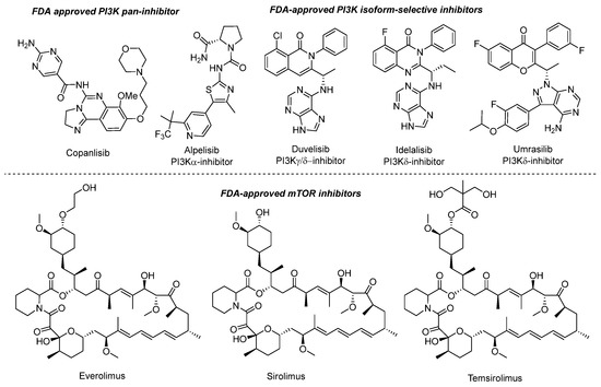
A (series) range of potential dimorpholinoquinazoline-based inhibitors of the PI3K/Akt/mTOR cascade was synthesized. Several compounds exhibited cytotoxicity towards a panel of cancer cell lines in the low and sub-micromolar range. Compound 7c with the highest activity and moderate selectivity towards MCF7 cells which express the mutant type of PI3K was also tested for the ability to inhibit PI3K-(signaling pathway) downstream effectors and associated proteins. Compound 7c inhibited the phosphorylation of Akt, mTOR, and S6K at 125–250 nM. It also triggered PARP1 cleavage, ROS production, and cell death via several mechanisms. Inhibition of PI3Kα was observed at a concentration of 7b 50 µM and of 7c 500 µM and higher, that can indicate minority PI3Kα as a target among other kinases in the titled cascade for 7c. In vivo studies demonstrated an inhibition of tumor growth in the colorectal tumor model. According to the docking studies, the replacement of the triazine core in gedatolisib (8) by a quinazoline fragment, and incorporation of a (hetero)aromatic unit connected with the carbamide group via a flexible spacer, can result in more selective inhibition of the PI3Kα isoform. Full article
Show Figures

Figure 1

20 pages, 3870 KiB  
Article
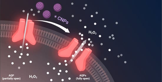
Cerium Oxide Nanoparticles Regulate Oxidative Stress in HeLa Cells by Increasing the Aquaporin-Mediated Hydrogen Peroxide Permeability
by Giorgia Pellavio, Patrizia Sommi, Umberto Anselmi-Tamburini, Maria Paola DeMichelis, Stefania Coniglio and Umberto Laforenza
Int. J. Mol. Sci. 2022, 23(18), 10837; https://doi.org/10.3390/ijms231810837 - 16 Sep 2022
Cited by 7 | Viewed by 2549
Abstract
Some aquaporins (AQPs) allow the diffusion of hydrogen peroxide (H2O2), the most abundant ROS, through the cell membranes. Therefore, the possibility of regulating the AQP-mediated permeability to H2O2, and thus ROS scavenging, appears particularly important [...] Read more.
Some aquaporins (AQPs) allow the diffusion of hydrogen peroxide (H2O2), the most abundant ROS, through the cell membranes. Therefore, the possibility of regulating the AQP-mediated permeability to H2O2, and thus ROS scavenging, appears particularly important for controlling the redox state of cells in physiological and pathophysiological conditions. Several compounds have been screened and characterized for this purpose. This study aimed to analyze the effect of cerium oxide nanoparticles (CNPs) presenting antioxidant activity on AQP functioning. HeLa cells express AQP3, 6, 8, and 11, able to facilitate H2O2. AQP3, 6, and 8 are expressed in the plasma membrane and intracellularly, while AQP11 resides only in intracellular structures. CNPs but not cerium ions treatment significantly increased the water and H2O2 permeability by interacting with AQP3, 6, and especially with AQP8. CNPs increased considerably the AQP-mediated water diffusion in cells with oxidative stress. Functional experiments with silenced HeLa cells revealed that CNPs increased the H2O2 diffusion mainly by modulating the AQP8 permeability but also the AQP3 and AQP6, even if to a lesser extent. Current findings suggest that CNPs represent a promising pharmaceutical agent that might potentially be used in numerous pathologies involving oxidative stress as tumors and neurodegenerative diseases. Full article
Show Figures

Graphical abstract

13 pages, 1998 KiB  
Article
The Antiarrhythmic and Hypotensive Effects of S-61 and S-73, the Pyrrolidin-2-one Derivatives with α1-Adrenolytic Properties
by Klaudia Lustyk, Kinga Sałaciak, Agata Siwek, Jacek Sapa, Paula Zaręba, Adam Gałuszka and Karolina Pytka
Int. J. Mol. Sci. 2022, 23(18), 10381; https://doi.org/10.3390/ijms231810381 - 8 Sep 2022
Cited by 2 | Viewed by 1536
Abstract
Heart rhythm abnormalities are a cause of many deaths worldwide. Unfortunately, the available antiarrhythmic drugs show limited efficacy and proarrhythmic potential. Thus, efforts should be made to search for new, more effective, and safer pharmacotherapies. Several studies suggested that blocking the α1 [...] Read more.
Heart rhythm abnormalities are a cause of many deaths worldwide. Unfortunately, the available antiarrhythmic drugs show limited efficacy and proarrhythmic potential. Thus, efforts should be made to search for new, more effective, and safer pharmacotherapies. Several studies suggested that blocking the α1-adrenoceptors could restore normal heart rhythm in arrhythmia. In this study, we aimed to assess the antiarrhythmic potential of S-61 and S-73, two novel pyrrolidin-2-one derivatives with high affinity for α1-adrenergic receptors. First, using radioligand binding studies, we demonstrated that S-61 and S-73 did not bind with β1-adrenoceptors. Next, we assessed whether S-61 and S-73 could protect rats against arrhythmia in adrenaline-, calcium chloride- and aconitine-induced arrhythmia models. Both compounds showed potent prophylactic antiarrhythmic properties in the adrenaline-induced arrhythmia model, but the effect of S-61 was more pronounced. None of the compounds displayed antiarrhythmic effects in calcium chloride- or aconitine-induced arrhythmia models. Interestingly, both derivatives revealed therapeutic antiarrhythmic activity in the adrenaline-induced arrhythmia, diminishing heart rhythm irregularities. Neither S-61 nor S-73 showed proarrhythmic potential in rats. Finally, the compounds decreased blood pressure in rodents. The hypotensive effects were not observed after coadministration with methoxamine, which suggests the α1-adrenolytic properties of both compounds. Our results confirm that pyrrolidin-2-one derivatives possess potent antiarrhythmic properties. Given the promising results of our experiments, further studies on pyrrolidin-2-one derivatives might result in the development of a new class of antiarrhythmic drugs. Full article
Show Figures

Figure 1

19 pages, 590 KiB  
Article
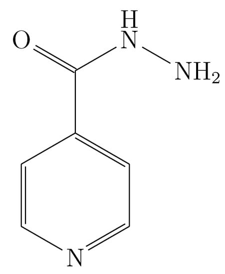
Thermodynamic Analysis of the Solubility of Isoniazid in (PEG 200 + Water) Cosolvent Mixtures from 278.15 K to 318.15 K
by Daniela Baracaldo-Santamaría, Carlos Alberto Calderon-Ospina, Claudia Patricia Ortiz, Rossember Edén Cardenas-Torres, Fleming Martinez and Daniel Ricardo Delgado
Int. J. Mol. Sci. 2022, 23(17), 10190; https://doi.org/10.3390/ijms231710190 - 5 Sep 2022
Cited by 7 | Viewed by 2253
Abstract
The solubility of drugs in cosolvent systems of pharmaceutical interest is of great importance for understanding and optimizing a large number of processes. Here, we report the solubility of isoniazid in nine (PEG 200 + water) cosolvent mixtures at nine temperatures (278.15, 283.15, [...] Read more.
The solubility of drugs in cosolvent systems of pharmaceutical interest is of great importance for understanding and optimizing a large number of processes. Here, we report the solubility of isoniazid in nine (PEG 200 + water) cosolvent mixtures at nine temperatures (278.15, 283.15, 288.15, 293.15, 298.15, 303.15, 308.15, and 318.15 K) determined by UV–vis spectrophotometry. From the solubility data, the thermodynamic solution, mixing, and transfer functions were calculated in addition to performing the enthalpy–entropy compensation analysis. The solubility of isoniazid depends on the concentration of PEG 200 (positive cosolvent effect) and temperature (endothermic process) reaching its maximum solubility in pure PEG 200 at 318.15 K and the lowest solubility in pure water at 278.15 K. The solution process is favored by the solution entropy and according to the enthalpy–entropy compensation analysis it is driven by entropy in mixtures rich in water and by enthalpy in mixtures rich in PEG 200. Full article
Show Figures

Figure 1

28 pages, 4635 KiB  
Article
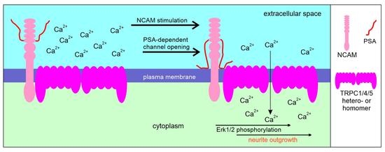
Interactions between the Polysialylated Neural Cell Adhesion Molecule and the Transient Receptor Potential Canonical Channels 1, 4, and 5 Induce Entry of Ca2+ into Neurons
by Laura Amores-Bonet, Ralf Kleene, Thomas Theis and Melitta Schachner
Int. J. Mol. Sci. 2022, 23(17), 10027; https://doi.org/10.3390/ijms231710027 - 2 Sep 2022
Cited by 3 | Viewed by 2373
Abstract
The neural cell adhesion molecule (NCAM) plays important functional roles in the developing and mature nervous systems. Here, we show that the transient receptor potential canonical (TRPC) ion channels TRPC1, −4, and −5 not only interact with the intracellular domains of the transmembrane [...] Read more.
The neural cell adhesion molecule (NCAM) plays important functional roles in the developing and mature nervous systems. Here, we show that the transient receptor potential canonical (TRPC) ion channels TRPC1, −4, and −5 not only interact with the intracellular domains of the transmembrane isoforms NCAM140 and NCAM180, but also with the glycan polysialic acid (PSA) covalently attached to the NCAM protein backbone. NCAM antibody treatment leads to the opening of TRPC1, −4, and −5 hetero- or homomers at the plasma membrane and to the influx of Ca2+ into cultured cortical neurons and CHO cells expressing NCAM, PSA, and TRPC1 and −4 or TRPC1 and −5. NCAM-stimulated Ca2+ entry was blocked by the TRPC inhibitor Pico145 or the bacterial PSA homolog colominic acid. NCAM-stimulated Ca2+ influx was detectable neither in NCAM-deficient cortical neurons nor in TRPC1/4- or TRPC1/5-expressing CHO cells that express NCAM, but not PSA. NCAM-induced neurite outgrowth was reduced by TRPC inhibitors and a function-blocking TRPC1 antibody. A characteristic signaling feature was that extracellular signal-regulated kinase 1/2 phosphorylation was also reduced by TRPC inhibitors. Our findings indicate that the interaction of NCAM with TRPC1, −4, and −5 contributes to the NCAM-stimulated and PSA-dependent Ca2+ entry into neurons thereby influencing essential neural functions. Full article
Show Figures

Graphical abstract

18 pages, 1478 KiB  
Article
Substrates of the Human Brain Proton-Organic Cation Antiporter and Comparison with Organic Cation Transporter 1 Activities
by David A. Doetsch, Salim Ansari, Ole Jensen, Lukas Gebauer, Christof Dücker, Jürgen Brockmöller and Alexandra Sachkova
Int. J. Mol. Sci. 2022, 23(15), 8430; https://doi.org/10.3390/ijms23158430 - 29 Jul 2022
Cited by 8 | Viewed by 2004
Abstract
Many organic cations (OCs) may be transported through membranes by a genetically still uncharacterized proton-organic cation (H + OC) antiporter. Here, we characterized an extended substrate spectrum of this antiporter. We studied the uptake of 72 drugs in hCMEC/D3 cells as a model [...] Read more.
Many organic cations (OCs) may be transported through membranes by a genetically still uncharacterized proton-organic cation (H + OC) antiporter. Here, we characterized an extended substrate spectrum of this antiporter. We studied the uptake of 72 drugs in hCMEC/D3 cells as a model of the human blood–brain barrier. All 72 drugs were tested with exchange transport assays and the transport of 26 of the drugs was studied in more detail concerning concentration-dependent uptake and susceptibility to specific inhibitors. According to exchange transport assays, 37 (51%) drugs were good substrates of the H + OC antiporter. From 26 drugs characterized in more detail, 23 were consistently identified as substrates of the H + OC antiporter in six different assays and transport kinetic constants could be identified with intrinsic clearances between 0.2 (ephedrine) and 201 (imipramine) mL × minute−1 × g protein−1. Excellent substrates of the H + OC antiporter were no substrates of organic cation transporter OCT1 and vice versa. Good substrates of the H + OC antiporter were more hydrophobic and had a lower topological polar surface area than non-substrates or OCT1 substrates. These data and further research on the H + OC antiporter may result in a better understanding of pharmacokinetics, drug–drug interactions and variations in pharmacokinetics. Full article
Show Figures

Figure 1

22 pages, 937 KiB  
Review
Therapeutic Application of Genome Editing Technologies in Viral Diseases
by Tae Hyeong Kim and Seong-Wook Lee
Int. J. Mol. Sci. 2022, 23(10), 5399; https://doi.org/10.3390/ijms23105399 - 12 May 2022
Cited by 7 | Viewed by 3681
Abstract
Viral infections can be fatal and consequently, they are a serious threat to human health. Therefore, the development of vaccines and appropriate antiviral therapeutic agents is essential. Depending on the virus, it can cause an acute or a chronic infection. The characteristics of [...] Read more.
Viral infections can be fatal and consequently, they are a serious threat to human health. Therefore, the development of vaccines and appropriate antiviral therapeutic agents is essential. Depending on the virus, it can cause an acute or a chronic infection. The characteristics of viruses can act as inhibiting factors for the development of appropriate treatment methods. Genome editing technology, including the use of clustered regularly interspaced short palindromic repeats (CRISPR)-CRISPR-associated (Cas) proteins, zinc-finger nucleases (ZFNs), and transcription activator-like effector nucleases (TALENs), is a technology that can directly target and modify genomic sequences in almost all eukaryotic cells. The development of this technology has greatly expanded its applicability in life science research and gene therapy development. Research on the use of this technology to develop therapeutics for viral diseases is being conducted for various purposes, such as eliminating latent infections or providing resistance to new infections. In this review, we will look at the current status of the development of viral therapeutic agents using genome editing technology and discuss how this technology can be used as a new treatment approach for viral diseases. Full article
Show Figures

Figure 1

32 pages, 1153 KiB  
Review
Emerging Antiarrhythmic Drugs for Atrial Fibrillation
by Arnela Saljic, Jordi Heijman and Dobromir Dobrev
Int. J. Mol. Sci. 2022, 23(8), 4096; https://doi.org/10.3390/ijms23084096 - 7 Apr 2022
Cited by 25 | Viewed by 8472
Abstract
Atrial fibrillation (AF), the most common cardiac arrhythmia worldwide, is driven by complex mechanisms that differ between subgroups of patients. This complexity is apparent from the different forms in which AF presents itself (post-operative, paroxysmal and persistent), each with heterogeneous patterns and variable [...] Read more.
Atrial fibrillation (AF), the most common cardiac arrhythmia worldwide, is driven by complex mechanisms that differ between subgroups of patients. This complexity is apparent from the different forms in which AF presents itself (post-operative, paroxysmal and persistent), each with heterogeneous patterns and variable progression. Our current understanding of the mechanisms responsible for initiation, maintenance and progression of the different forms of AF has increased significantly in recent years. Nevertheless, antiarrhythmic drugs for the management of AF have not been developed based on the underlying arrhythmia mechanisms and none of the currently used drugs were specifically developed to target AF. With the increased knowledge on the mechanisms underlying different forms of AF, new opportunities for developing more effective and safer AF therapies are emerging. In this review, we provide an overview of potential novel antiarrhythmic approaches based on the underlying mechanisms of AF, focusing both on the development of novel antiarrhythmic agents and on the possibility of repurposing already marketed drugs. In addition, we discuss the opportunity of targeting some of the key players involved in the underlying AF mechanisms, such as ryanodine receptor type-2 (RyR2) channels and atrial-selective K+-currents (IK2P and ISK) for antiarrhythmic therapy. In addition, we highlight the opportunities for targeting components of inflammatory signaling (e.g., the NLRP3-inflammasome) and upstream mechanisms targeting fibroblast function to prevent structural remodeling and progression of AF. Finally, we critically appraise emerging antiarrhythmic drug principles and future directions for antiarrhythmic drug development, as well as their potential for improving AF management. Full article
Show Figures

Figure 1

37 pages, 4598 KiB  
Review
Small-Molecule RAS Inhibitors as Anticancer Agents: Discovery, Development, and Mechanistic Studies
by Shaila A. Shetu and Debasish Bandyopadhyay
Int. J. Mol. Sci. 2022, 23(7), 3706; https://doi.org/10.3390/ijms23073706 - 28 Mar 2022
Cited by 10 | Viewed by 5265
Abstract
Mutations of RAS oncogenes are responsible for about 30% of all human cancer types, including pancreatic, lung, and colorectal cancers. While KRAS1 is a pseudogene, mutation of KRAS2 (commonly known as KRAS oncogene) is directly or indirectly associated with human cancers. Among the [...] Read more.
Mutations of RAS oncogenes are responsible for about 30% of all human cancer types, including pancreatic, lung, and colorectal cancers. While KRAS1 is a pseudogene, mutation of KRAS2 (commonly known as KRAS oncogene) is directly or indirectly associated with human cancers. Among the RAS family, KRAS is the most abundant oncogene related to uncontrolled cellular proliferation to generate solid tumors in many types of cancer such as pancreatic carcinoma (over 80%), colon carcinoma (40–50%), lung carcinoma (30–50%), and other types of cancer. Once described as ‘undruggable’, RAS proteins have become ‘druggable’, at least to a certain extent, due to the continuous efforts made during the past four decades. In this account, we discuss the chemistry and biology (wherever available) of the small-molecule inhibitors (synthetic, semi-synthetic, and natural) of KRAS proteins that were published in the past decades. Commercial drugs, as well as investigational molecules from preliminary stages to clinical trials, are categorized and discussed in this study. In summary, this study presents an in-depth discussion of RAS proteins, classifies the RAS superfamily, and describes the molecular mechanism of small-molecule RAS inhibitors Full article
Show Figures

Figure 1

19 pages, 655 KiB  
Review
Effects of Antiarrhythmic Drugs on Antiepileptic Drug Action—A Critical Review of Experimental Findings
by Kinga K. Borowicz-Reutt
Int. J. Mol. Sci. 2022, 23(5), 2891; https://doi.org/10.3390/ijms23052891 - 7 Mar 2022
Cited by 5 | Viewed by 5268
Abstract
Severe cardiac arrhythmias developing in the course of seizures increase the risk of SUDEP (sudden unexpected death in epilepsy). Hence, epilepsy patients with pre-existing arrhythmias should receive appropriate pharmacotherapy. Concomitant treatment with antiarrhythmic and antiseizure medications creates, however, the possibility of drug–drug interactions. [...] Read more.
Severe cardiac arrhythmias developing in the course of seizures increase the risk of SUDEP (sudden unexpected death in epilepsy). Hence, epilepsy patients with pre-existing arrhythmias should receive appropriate pharmacotherapy. Concomitant treatment with antiarrhythmic and antiseizure medications creates, however, the possibility of drug–drug interactions. This is due, among other reasons, to a similar mechanism of action. Both groups of drugs inhibit the conduction of electrical impulses in excitable tissues. The aim of this review was the analysis of such interactions in animal seizure models, including the maximal electroshock (MES) test in mice, a widely accepted screening test for antiepileptic drugs. Full article
Show Figures

Figure 1

14 pages, 10116 KiB  
Article
Monitoring TRPC7 Conformational Changes by BRET Following GPCR Activation
by Cécile Pétigny, Audrey-Ann Dumont, Hugo Giguère, Audrey Collette, Brian J. Holleran, Mircea Iftinca, Christophe Altier, Élie Besserer-Offroy, Mannix Auger-Messier and Richard Leduc
Int. J. Mol. Sci. 2022, 23(5), 2502; https://doi.org/10.3390/ijms23052502 - 24 Feb 2022
Cited by 2 | Viewed by 3173
Abstract
Transient receptor potential canonical (TRPC) channels are membrane proteins involved in regulating Ca2+ homeostasis, and whose functions are modulated by G protein-coupled receptors (GPCR). In this study, we developed bioluminescent resonance energy transfer (BRET) biosensors to better study channel conformational changes following [...] Read more.
Transient receptor potential canonical (TRPC) channels are membrane proteins involved in regulating Ca2+ homeostasis, and whose functions are modulated by G protein-coupled receptors (GPCR). In this study, we developed bioluminescent resonance energy transfer (BRET) biosensors to better study channel conformational changes following receptor activation. For this study, two intramolecular biosensors, GFP10-TRPC7-RLucII and RLucII-TRPC7-GFP10, were constructed and were assessed following the activation of various GPCRs. We first transiently expressed receptors and the biosensors in HEK293 cells, and BRET levels were measured following agonist stimulation of GPCRs. The activation of GPCRs that engage Gαq led to a Gαq-dependent BRET response of the functional TRPC7 biosensor. Focusing on the Angiotensin II type-1 receptor (AT1R), GFP10-TRPC7-RLucII was tested in rat neonatal cardiac fibroblasts, expressing endogenous AT1R and TRPC7. We detected similar BRET responses in these cells, thus validating the use of the biosensor in physiological conditions. Taken together, our results suggest that activation of Gαq-coupled receptors induce conformational changes in a novel and functional TRPC7 BRET biosensor. Full article
Show Figures

Figure 1

16 pages, 4020 KiB  
Article
All-Atom Molecular Dynamics Investigations on the Interactions between D2 Subunit Dopamine Receptors and Three 11C-Labeled Radiopharmaceutical Ligands
by Sanda Nastasia Moldovean, Diana-Gabriela Timaru and Vasile Chiş
Int. J. Mol. Sci. 2022, 23(4), 2005; https://doi.org/10.3390/ijms23042005 - 11 Feb 2022
Cited by 3 | Viewed by 2312
Abstract
The D2 subunit dopamine receptor represents a key factor in modulating dopamine release. Moreover, the investigated radiopharmaceutical ligands used in positron emission tomography imaging techniques are known to bind D2 receptors, allowing for dopaminergic pathways quantification in the living human brain. Thus, the [...] Read more.
The D2 subunit dopamine receptor represents a key factor in modulating dopamine release. Moreover, the investigated radiopharmaceutical ligands used in positron emission tomography imaging techniques are known to bind D2 receptors, allowing for dopaminergic pathways quantification in the living human brain. Thus, the biophysical characterization of these radioligands is expected to provide additional insights into the interaction mechanisms between the vehicle molecules and their targets. Using molecular dynamics simulations and QM calculations, the present study aimed to investigate the potential positions in which the D2 dopamine receptor would most likely interact with the three distinctive synthetic 11C-labeled compounds (raclopride (3,5-dichloro-N-[[(2S)-1-ethylpyrrolidin-2-yl]methyl]-2-hydroxy-6-methoxybenzamide)—RACL, FLB457 (5-bromo-N-[[(2S)-1-ethylpyrrolidin-2-yl]methyl]-2,3-dimethoxybenzamide)—FLB457 and SCH23390 (R(+)-7-Chloro-8-hydroxy-3-methyl-1-phenyl-2,3,4,5-tetrahydro-1H-3-benzazepine)—SCH)), as well as to estimate the binding affinities of the ligand-receptor complexes. A docking study was performed prior to multiple 50 ns molecular dynamics productions for the ligands situated at the top and bottom interacting pockets of the receptor. The most prominent motions for the RACL ligand were described by the high fluctuations of the peripheral aliphatic -CH3 groups and by its C-Cl aromatic ring groups. In good agreement with the experimental data, the D2 dopamine receptor-RACL complex showed the highest interacting patterns for ligands docked at the receptor’s top position. Full article
Show Figures

Figure 1

23 pages, 2291 KiB  
Review
Overview of First-Line and Second-Line Pharmacotherapies for Osteoarthritis with Special Focus on Intra-Articular Treatment
by Alicja Nowaczyk, Dawid Szwedowski, Ignacio Dallo and Jacek Nowaczyk
Int. J. Mol. Sci. 2022, 23(3), 1566; https://doi.org/10.3390/ijms23031566 - 29 Jan 2022
Cited by 23 | Viewed by 6192
Abstract
Osteoarthritis (OA) can be defined as the result of pathological processes of various etiologies leading to damage to the articular structures. Although the mechanism of degenerative changes has become better understood due to the plethora of biochemical and genetic studies, the drug that [...] Read more.
Osteoarthritis (OA) can be defined as the result of pathological processes of various etiologies leading to damage to the articular structures. Although the mechanism of degenerative changes has become better understood due to the plethora of biochemical and genetic studies, the drug that could stop the degenerative cascade is still unknown. All available forms of OA therapy are based on symptomatic treatment. According to actual guidelines, comprehensive treatment of OA should always include a combination of various therapeutic options aimed at common goals, which are pain relief in the first place, and then the improvement of function. Local treatment has become more common practice, which takes place between rehabilitation and pharmacological treatment in the hierarchy of procedures. Only in the case of no improvement and the presence of advanced lesions visible in imaging tests, should surgery be considered. Currently, an increasing number of studies are being published suggesting that intra-articular injections may be as effective or even more effective than non-steroidal anti-inflammatory drugs (NSAIDs) and result in fewer systemic adverse events. The most commonly used preparations are hyaluronic acid (HA), glucocorticosteroids (GS), and also platelet-rich plasma (PRP) in recent years. This review aims to present the mechanism of action and clinical effectiveness of different pharmacological options in relieving pain and improving functions in OA as well as the emerging approach in intra-articular treatment with PRP. Full article
Show Figures

Figure 1

13 pages, 636 KiB  
Review
Oral Pathology in COVID-19 and SARS-CoV-2 Infection—Molecular Aspects
by Agnieszka Drozdzik and Marek Drozdzik
Int. J. Mol. Sci. 2022, 23(3), 1431; https://doi.org/10.3390/ijms23031431 - 27 Jan 2022
Cited by 34 | Viewed by 6846
Abstract
This review article was designed to evaluate the existing evidence related to the molecular processes of SARS-CoV-2 infection in the oral cavity. The World Health Organization stated that severe acute respiratory syndrome coronavirus 2 (SARS-CoV-2) infection and transmission is produced by respiratory droplets [...] Read more.
This review article was designed to evaluate the existing evidence related to the molecular processes of SARS-CoV-2 infection in the oral cavity. The World Health Organization stated that severe acute respiratory syndrome coronavirus 2 (SARS-CoV-2) infection and transmission is produced by respiratory droplets and aerosols from the oral cavity of infected patients. The oral cavity structures, keratinized and non-keratinized mucosa, and salivary glands’ epithelia express SARS-CoV-2 entry and transmission factors, especially angiotensin converting enzyme Type 2 (ACE2) and transmembrane serine protease 2 (TMPRSS2). Replication of the virus in cells leads to local and systemic infection spread, and cellular damage is associated with clinical signs and symptoms of the disease in the oral cavity. Saliva, both the cellular and acellular fractions, holds the virus particles and contributes to COVID-19 transmission. The review also presents information about the factors modifying SARS-CoV-2 infection potential and possible local pharmacotherapeutic interventions, which may confine SARS-CoV-2 virus entry and transmission in the oral cavity. The PubMed and Scopus databases were used to search for suitable keywords such as: SARS-CoV-2, COVID-19, oral virus infection, saliva, crevicular fluid, salivary gland, tongue, oral mucosa, periodontium, gingiva, dental pulp, ACE2, TMPRSS2, Furin, diagnosis, topical treatment, vaccine and related words in relevant publications up to 28 December 2021. Data extraction and quality evaluation of the articles were performed by two reviewers, and 63 articles were included in the final review. Full article
Show Figures

Figure 1

2021

Jump to: 2024, 2023, 2022, 2020, 2019

32 pages, 17236 KiB  
Article
Design, Synthesis and Evaluation of Novel Derivatives of Curcuminoids with Cytotoxicity
by Chen-Yin Chen, Jin-Cherng Lien, Chien-Yu Chen, Chin-Chuan Hung and Hui-Chang Lin
Int. J. Mol. Sci. 2021, 22(22), 12171; https://doi.org/10.3390/ijms222212171 - 10 Nov 2021
Cited by 7 | Viewed by 2890
Abstract
Curcumin and curcuminoids have been discussed frequently due to their promising functional groups (such as scaffolds of α,β-unsaturated β-diketone, α,β-unsaturated ketone and β′-hydroxy-α,β-unsaturated ketone connected with aromatic rings on both sides) that play an important role in various bioactivities, including antioxidant, anti-inflammatory, anti-proliferation [...] Read more.
Curcumin and curcuminoids have been discussed frequently due to their promising functional groups (such as scaffolds of α,β-unsaturated β-diketone, α,β-unsaturated ketone and β′-hydroxy-α,β-unsaturated ketone connected with aromatic rings on both sides) that play an important role in various bioactivities, including antioxidant, anti-inflammatory, anti-proliferation and anticancer activity. A series of novel curcuminoid derivatives (a total of 55 new compounds) and three reference compounds were synthesized with good yields using three-step organic synthesis. The anti-proliferative activities of curcumin derivatives were examined for six human cancer cell lines: HeLaS3, KBvin, MCF-7, HepG2, NCI-H460 and NCI-H460/MX20. Compared to the IC50 values of all the synthesized derivatives, most α,β-unsaturated ketones displayed potent anti-proliferative effects against all six human cancer cell lines, whereas β′-hydroxy-α,β-unsaturated ketones and α,β-unsaturated β-diketones presented moderate anti-proliferative effects. Two potent curcuminoid derivatives were found among all the novel derivatives and reference compounds: (E)-5-hydroxy-7-phenyl-1-(3,4,5-trimethoxyphenyl)hept-1-en-3-one (compound 3) and (1E,4E)-1,7-bis(3,4,5-trimethoxyphenyl)hepta-1,4-dien-3-one (compound MD12a). These were selected for further analysis after the evaluation of their anti-proliferative effects against all human cancer cell lines. The results of apoptosis assays revealed that the number of dead cells was increased in early apoptosis and late apoptosis, while cell proliferation was also decreased after applying various concentrations of (E)-5-hydroxy-7-phenyl-1-(3,4,5-trimethoxyphenyl)hept-1-en-3-one (compound 3) and (1E,4E)-1,7-bis(3,4,5-trimethoxyphenyl)hepta-1,4-dien-3-one (compound MD12a) to MCF-7 and HpeG2 cancer cells. Analysis of the gene expression arrays showed that three genes (GADD45B, SESN2 and BBC3) were correlated with the p53 pathway. From the quantitative PCR analysis, it was seen that (1E,4E)-1,7-bis(3,4,5-trimethoxyphenyl)hepta-1,4-dien-3-one (compound MD12a) effectively induced the up-regulated expression of GADD45B, leading to the suppression of MCF-7 cancer cell formation and cell death. Molecular docking analysis was used to predict and sketch the interactions of the GADD45B-α,β-unsaturated ketone complex for help in drug design. Full article
Show Figures

Graphical abstract

28 pages, 25761 KiB  
Review
Experimental Models, Induction Protocols, and Measured Parameters in Dry Eye Disease: Focusing on Practical Implications for Experimental Research
by Md. Mahbubur Rahman, Dong Hyun Kim, Chul-Kyu Park and Yong Ho Kim
Int. J. Mol. Sci. 2021, 22(22), 12102; https://doi.org/10.3390/ijms222212102 - 9 Nov 2021
Cited by 19 | Viewed by 7664
Abstract
Dry eye disease (DED) is one of the major ophthalmological healthcare challenges worldwide. DED is a multifactorial disease characterized by a loss of homeostasis of the tear film, and its main pathogenesis is chronic ocular surface inflammation related with various cellular and molecular [...] Read more.
Dry eye disease (DED) is one of the major ophthalmological healthcare challenges worldwide. DED is a multifactorial disease characterized by a loss of homeostasis of the tear film, and its main pathogenesis is chronic ocular surface inflammation related with various cellular and molecular signaling cascades. The animal model is a reliable and effective tool for understanding the various pathological mechanisms and molecular cascades in DED. Considerable experimental research has focused on developing new strategies for the prevention and treatment of DED. Several experimental models of DED have been developed, and different animal species such as rats, mice, rabbits, dogs, and primates have been used for these models. Although the basic mechanisms of DED in animals are nearly identical to those in humans, proper knowledge about the induction of animal models is necessary to obtain better and more reliable results. Various experimental models (in vitro and in vivo DED models) were briefly discussed in this review, along with pathologic features, analytical approaches, and common measurements, which will help investigators to use the appropriate cell lines, animal, methods, and evaluation parameters depending on their study design. Full article
Show Figures

Figure 1

13 pages, 2782 KiB  
Article
Improvement of an Effective Protocol for Directed Differentiation of Human Adipose Tissue-Derived Adult Mesenchymal Stem Cells to Corneal Endothelial Cells
by Cadenas-Martin Marta, Moratilla Adrian, Fernández-Delgado Jorge, Arnalich-Montiel Francisco and Maria P. De Miguel
Int. J. Mol. Sci. 2021, 22(21), 11982; https://doi.org/10.3390/ijms222111982 - 5 Nov 2021
Cited by 8 | Viewed by 2405
Abstract
Corneal disease affects 12.5 million individuals worldwide, with 2 million new cases each year. The standard treatment consists of a corneal transplantation from a human donor; however, the worldwide demand significantly exceeds the available supply. Lamellar endothelial keratoplasty, the replacement of only the [...] Read more.
Corneal disease affects 12.5 million individuals worldwide, with 2 million new cases each year. The standard treatment consists of a corneal transplantation from a human donor; however, the worldwide demand significantly exceeds the available supply. Lamellar endothelial keratoplasty, the replacement of only the endothelial layer of the cornea, can partially solve the problem. Progressive efforts have succeeded in expanding hCECs; however, the ability to expand hCECs is still limited, and new sources of CECs are being sought. Crucial advances have been achieved by the directed differentiation of embryonic or induced pluripotent stem cells, but these cells have disadvantages, such as the use of oncogenes, and are still difficult to establish. We aimed to transfer such knowledge to obtain hCECs from adipose tissue-derived adult mesenchymal stem cells (ADSC) by modifying four previously published procedures. We present several protocols capable of the directed differentiation of human ADSCs to hCECs. In our hands, the protocol by Ali et al. was the best adapted to such differentiation in terms of efficiency, time, and financial cost; however, the protocol by Wagoner et al. was the best for CEC marker expression. Our results broaden the type of cells of autologous extraocular origin that could be employed in the clinical setting for corneal endothelial deficiency. Full article
Show Figures

Figure 1

17 pages, 6143 KiB  
Article
Tepotinib Inhibits Several Drug Efflux Transporters and Biotransformation Enzymes: The Role in Drug-Drug Interactions and Targeting Cytostatic Resistance In Vitro and Ex Vivo
by Dimitrios Vagiannis, Youssif Budagaga, Anselm Morell, Yu Zhang, Eva Novotná, Adam Skarka, Sarah Kammerer, Jan-Heiner Küpper, Ivo Hanke, Tomáš Rozkoš and Jakub Hofman
Int. J. Mol. Sci. 2021, 22(21), 11936; https://doi.org/10.3390/ijms222111936 - 3 Nov 2021
Cited by 7 | Viewed by 2925
Abstract
Tepotinib is a novel tyrosine kinase inhibitor recently approved for the treatment of non-small cell lung cancer (NSCLC). In this study, we evaluated the tepotinib’s potential to perpetrate pharmacokinetic drug interactions and modulate multidrug resistance (MDR). Accumulation studies showed that tepotinib potently inhibits [...] Read more.
Tepotinib is a novel tyrosine kinase inhibitor recently approved for the treatment of non-small cell lung cancer (NSCLC). In this study, we evaluated the tepotinib’s potential to perpetrate pharmacokinetic drug interactions and modulate multidrug resistance (MDR). Accumulation studies showed that tepotinib potently inhibits ABCB1 and ABCG2 efflux transporters, which was confirmed by molecular docking. In addition, tepotinib inhibited several recombinant cytochrome P450 (CYP) isoforms with varying potency. In subsequent drug combination experiments, tepotinib synergistically reversed daunorubicin and mitoxantrone resistance in cells with ABCB1 and ABCG2 overexpression, respectively. Remarkably, MDR-modulatory properties were confirmed in ex vivo explants derived from NSCLC patients. Furthermore, we demonstrated that anticancer effect of tepotinib is not influenced by the presence of ABC transporters associated with MDR, although monolayer transport assays designated it as ABCB1 substrate. Finally, tested drug was observed to have negligible effect on the expression of clinically relevant drug efflux transporters and CYP enzymes. In conclusion, our findings provide complex overview on the tepotinib’s drug interaction profile and suggest a promising novel therapeutic strategy for future clinical investigations. Full article
Show Figures

Figure 1

17 pages, 3453 KiB  
Article
Structural and Biochemical Analysis of the Dual Inhibition of MG-132 against SARS-CoV-2 Main Protease (Mpro/3CLpro) and Human Cathepsin-L
by Elisa Costanzi, Maria Kuzikov, Francesca Esposito, Simone Albani, Nicola Demitri, Barbara Giabbai, Marianna Camasta, Enzo Tramontano, Giulia Rossetti, Andrea Zaliani and Paola Storici
Int. J. Mol. Sci. 2021, 22(21), 11779; https://doi.org/10.3390/ijms222111779 - 29 Oct 2021
Cited by 55 | Viewed by 5960
Abstract
After almost two years from its first evidence, the COVID-19 pandemic continues to afflict people worldwide, highlighting the need for multiple antiviral strategies. SARS-CoV-2 main protease (Mpro/3CLpro) is a recognized promising target for the development of effective drugs. Because single target inhibition might [...] Read more.
After almost two years from its first evidence, the COVID-19 pandemic continues to afflict people worldwide, highlighting the need for multiple antiviral strategies. SARS-CoV-2 main protease (Mpro/3CLpro) is a recognized promising target for the development of effective drugs. Because single target inhibition might not be sufficient to block SARS-CoV-2 infection and replication, multi enzymatic-based therapies may provide a better strategy. Here we present a structural and biochemical characterization of the binding mode of MG-132 to both the main protease of SARS-CoV-2, and to the human Cathepsin-L, suggesting thus an interesting scaffold for the development of double-inhibitors. X-ray diffraction data show that MG-132 well fits into the Mpro active site, forming a covalent bond with Cys145 independently from reducing agents and crystallization conditions. Docking of MG-132 into Cathepsin-L well-matches with a covalent binding to the catalytic cysteine. Accordingly, MG-132 inhibits Cathepsin-L with nanomolar potency and reversibly inhibits Mpro with micromolar potency, but with a prolonged residency time. We compared the apo and MG-132-inhibited structures of Mpro solved in different space groups and we identified a new apo structure that features several similarities with the inhibited ones, offering interesting perspectives for future drug design and in silico efforts. Full article
Show Figures

Figure 1

21 pages, 1306 KiB  
Review
Pathogenesis and Molecular Mechanisms of Anderson–Fabry Disease and Possible New Molecular Addressed Therapeutic Strategies
by Antonino Tuttolomondo, Irene Simonetta, Renata Riolo, Federica Todaro, Tiziana Di Chiara, Salvatore Miceli and Antonio Pinto
Int. J. Mol. Sci. 2021, 22(18), 10088; https://doi.org/10.3390/ijms221810088 - 18 Sep 2021
Cited by 39 | Viewed by 9230
Abstract
Anderson–Fabry disease (AFD) is a rare disease with an incidenceof approximately 1:117,000 male births. Lysosomal accumulation of globotriaosylceramide (Gb3) is the element characterizing Fabry disease due to a hereditary deficiency α-galactosidase A (GLA) enzyme. The accumulation of Gb3 causes lysosomal dysfunction that compromises [...] Read more.
Anderson–Fabry disease (AFD) is a rare disease with an incidenceof approximately 1:117,000 male births. Lysosomal accumulation of globotriaosylceramide (Gb3) is the element characterizing Fabry disease due to a hereditary deficiency α-galactosidase A (GLA) enzyme. The accumulation of Gb3 causes lysosomal dysfunction that compromises cell signaling pathways. Deposition of sphingolipids occurs in the autonomic nervous system, dorsal root ganglia, kidney epithelial cells, vascular system cells, and myocardial cells, resulting in organ failure. This manuscript will review the molecular pathogenetic pathways involved in Anderson–Fabry disease and in its organ damage. Some studies reported that inhibition of mitochondrial function and energy metabolism plays a significant role in AFD cardiomyopathy and in kidney disease of AFD patients. Furthermore, mitochondrial dysfunction has been reported as linked to the dysregulation of the autophagy–lysosomal pathway which inhibits the mechanistic target of rapamycin kinase (mTOR) mediated control of mitochondrial metabolism in AFD cells. Cerebrovascular complications due to AFD are caused by cerebral micro vessel stenosis. These are caused by wall thickening resulting from the intramural accumulation of glycolipids, luminal occlusion or thrombosis. Other pathogenetic mechanisms involved in organ damage linked to Gb3 accumulation are endocytosis and lysosomal degradation of endothelial calcium-activated intermediate-conductance potassium ion channel 3.1 (KCa3.1) via a clathrin-dependent process. This process represents a crucial event in endothelial dysfunction. Several studies have identified the deacylated form of Gb3, globotriaosylsphingosine (Lyso-Gb3), as the main catabolite that increases in plasma and urine in patients with AFD. The mean concentrations of Gb3 in all organs and plasma of Galactosidase A knockout mice were significantly higher than those of wild-type mice. The distributions of Gb3 isoforms vary from organ to organ. Various Gb3 isoforms were observed mainly in the kidneys, and kidney-specific Gb3 isoforms were hydroxylated. Furthermore, the action of Gb3 on the KCa3.1 channel suggests a possible contribution of this interaction to the Fabry disease process, as this channel is expressed in various cells, including endothelial cells, fibroblasts, smooth muscle cells in proliferation, microglia, and lymphocytes. These molecular pathways could be considered a potential therapeutic target to correct the enzyme in addition to the traditional enzyme replacement therapies (ERT) or drug chaperone therapy. Full article
Show Figures

Figure 1

13 pages, 2394 KiB  
Review
Cystine/Glutamate Antiporter in Schizophrenia: From Molecular Mechanism to Novel Biomarker and Treatment
by Chung-Chieh Hung, Chieh-Hsin Lin and Hsien-Yuan Lane
Int. J. Mol. Sci. 2021, 22(18), 9718; https://doi.org/10.3390/ijms22189718 - 8 Sep 2021
Cited by 18 | Viewed by 6580
Abstract
Glutamate, a crucial excitatory neurotransmitter, plays a major role in the modulation of schizophrenia’s pathogenesis. New drug developments for schizophrenia have been prompted by the hypoglutamatergic hypothesis of schizophrenia. The cystine/glutamate antiporter system xc is related to glutamate-release regulation. Patients with [...] Read more.
Glutamate, a crucial excitatory neurotransmitter, plays a major role in the modulation of schizophrenia’s pathogenesis. New drug developments for schizophrenia have been prompted by the hypoglutamatergic hypothesis of schizophrenia. The cystine/glutamate antiporter system xc is related to glutamate-release regulation. Patients with schizophrenia were recently discovered to exhibit downregulation of xc subunits—the solute carrier (SLC) family 3 member 2 and the SLC family 7 member 11. We searched for relevant studies from 1980, when Bannai and Kitamura first identified the protein subunit system xc in lung fibroblasts, with the aim of compiling the biological, functional, and pharmacological characteristics of antiporter xc, which consists of several subunits. Some of them can significantly stimulate the human brain through the glutamate pathway. Initially, extracellular cysteine activates neuronal xc, causing glutamate efflux. Next, excitatory amino acid transporters enhance the unidirectional transportation of glutamate and sodium. These two biochemical pathways are also crucial to the production of glutathione, a protective agent for neural and glial cells and astrocytes. Investigation of the expression of system xc genes in the peripheral white blood cells of patients with schizophrenia can facilitate better understanding of the mental disorder and future development of novel biomarkers and treatments for schizophrenia. In addition, the findings further support the hypoglutamatergic hypothesis of schizophrenia. Full article
Show Figures

Figure 1

19 pages, 2385 KiB  
Article
Curcumin Derivatives as Potential Mosquito Larvicidal Agents against Two Mosquito Vectors, Culex pipiens and Aedes albopictus
by Dimitris Matiadis, Panagiota G. V. Liggri, Eftichia Kritsi, Niki Tzioumaki, Panagiotis Zoumpoulakis, Dimitrios P. Papachristos, George Balatsos, Marina Sagnou and Antonios Michaelakis
Int. J. Mol. Sci. 2021, 22(16), 8915; https://doi.org/10.3390/ijms22168915 - 18 Aug 2021
Cited by 16 | Viewed by 2814
Abstract
Vector-borne diseases have appeared or re-emerged in many Southern Europe countries making the transmission of infectious diseases by mosquitoes (vectors) one of the greatest worldwide health threats. Larvicides have been used extensively for the control of Aedes (Stegomyia) albopictus (Skuse, 1895) (Diptera: Culicidae) [...] Read more.
Vector-borne diseases have appeared or re-emerged in many Southern Europe countries making the transmission of infectious diseases by mosquitoes (vectors) one of the greatest worldwide health threats. Larvicides have been used extensively for the control of Aedes (Stegomyia) albopictus (Skuse, 1895) (Diptera: Culicidae) and Culex pipiens Linnaeus, 1758 (Diptera: Culicidae) mosquitoes in urban and semi-urban environments, causing the increasing resistance of mosquitoes to commercial insecticides. In this study, 27 curcuminoids and monocarbonyl curcumin derivatives were synthesised and evaluated as potential larvicidal agents against Cx. pipiens and Ae. albopictus. Most of the compounds were more effective against larvae of both mosquito species. Four of the tested compounds, curcumin, demethoxycurcumin, curcumin-BF2 complex and a monocarbonyl tetramethoxy curcumin derivative exhibited high activity against both species. In Cx. pipiens the recorded LC50 values were 6.0, 9.4, 5.0 and 32.5 ppm, respectively, whereas in Ae. albopictus they exhibited LC50 values of 9.2, 36.0, 5.5 and 23.6 ppm, respectively. No conclusive structure activity relationship was evident from the results and the variety of descriptors values generated in silico provided some insight to this end. Full article
Show Figures

Figure 1

14 pages, 1446 KiB  
Article
Influence of Hyperglycemia and Diabetes on Cardioprotection by Humoral Factors Released after Remote Ischemic Preconditioning (RIPC)
by Carolin Torregroza, Lara Gnaegy, Annika Raupach, Martin Stroethoff, Katharina Feige, André Heinen, Markus W. Hollmann and Ragnar Huhn
Int. J. Mol. Sci. 2021, 22(16), 8880; https://doi.org/10.3390/ijms22168880 - 18 Aug 2021
Cited by 10 | Viewed by 2613
Abstract
Remote ischemic preconditioning (RIPC) protects hearts from ischemia–reperfusion (I/R) injury in experimental studies; however, clinical RIPC trials were unsatisfactory. This discrepancy could be caused by a loss of cardioprotection due to comorbidities in patients, including diabetes mellitus (DM) and hyperglycemia (HG). RIPC is [...] Read more.
Remote ischemic preconditioning (RIPC) protects hearts from ischemia–reperfusion (I/R) injury in experimental studies; however, clinical RIPC trials were unsatisfactory. This discrepancy could be caused by a loss of cardioprotection due to comorbidities in patients, including diabetes mellitus (DM) and hyperglycemia (HG). RIPC is discussed to confer protective properties by release of different humoral factors activating cardioprotective signaling cascades. Therefore, we investigated whether DM type 1 and/or HG (1) inhibit the release of humoral factors after RIPC and/or (2) block the cardioprotective effect directly at the myocardium. Experiments were performed on male Wistar rats. Animals in part 1 of the study were either healthy normoglycemic (NG), type 1 diabetic (DM1), or hyperglycemic (HG). RIPC was implemented by four cycles of 5 min bilateral hind-limb ischemia/reperfusion. Control (Con) animals were not treated. Blood plasma taken in vivo was further investigated in isolated rat hearts in vitro. Plasma from diseased animals (DM1 or HG) was administered onto healthy (NG) hearts for 10 min before 33 min of global ischemia and 60 min of reperfusion. Part 2 of the study was performed vice versa—plasma taken in vivo, with or without RIPC, from healthy rats was transferred to DM1 and HG hearts in vitro. Infarct size was determined by TTC staining. Part 1: RIPC plasma from NG (NG Con: 49 ± 8% vs. NG RIPC 29 ± 6%; p < 0.05) and DM1 animals (DM1 Con: 47 ± 7% vs. DM1 RIPC: 38 ± 7%; p < 0.05) reduced infarct size. Interestingly, transfer of HG plasma showed comparable infarct sizes independent of prior treatment (HG Con: 34 ± 9% vs. HG RIPC 35 ± 9%; ns). Part 2: No infarct size reduction was detectable when transferring RIPC plasma from healthy rats to DM1 (DM1 Con: 54 ± 13% vs. DM1 RIPC 53 ± 10%; ns) or HG hearts (HG Con: 60 ± 16% vs. HG RIPC 53 ± 14%; ns). These results suggest that: (1) RIPC under NG and DM1 induces the release of humoral factors with cardioprotective impact, (2) HG plasma might own cardioprotective properties, and (3) RIPC does not confer cardioprotection in DM1 and HG myocardium. Full article
Show Figures

Figure 1

16 pages, 32655 KiB  
Article
Evidence for Protein–Protein Interaction between Dopamine Receptors and the G Protein-Coupled Receptor 143
by Beatriz Bueschbell, Prashiela Manga, Erika Penner and Anke C. Schiedel
Int. J. Mol. Sci. 2021, 22(15), 8328; https://doi.org/10.3390/ijms22158328 - 3 Aug 2021
Cited by 8 | Viewed by 4433
Abstract
Protein-protein interactions between G protein-coupled receptors (GPCRs) can augment their functionality and increase the repertoire of signaling pathways they regulate. New therapeutics designed to modulate such interactions may allow for targeting of a specific GPCR activity, thus reducing potential for side effects. Dopamine [...] Read more.
Protein-protein interactions between G protein-coupled receptors (GPCRs) can augment their functionality and increase the repertoire of signaling pathways they regulate. New therapeutics designed to modulate such interactions may allow for targeting of a specific GPCR activity, thus reducing potential for side effects. Dopamine receptor (DR) heteromers are promising candidates for targeted therapy of neurological conditions such as Parkinson’s disease since current treatments can have severe side effects. To facilitate development of such therapies, it is necessary to identify the various DR binding partners. We report here a new interaction partner for DRD2 and DRD3, the orphan receptor G protein-coupled receptor 143 (GPR143), an atypical GPCR that plays multiple roles in pigment cells and is expressed in several regions of the brain. We previously demonstrated that the DRD2/ DRD3 antagonist pimozide also modulates GPR143 activity. Using confocal microscopy and two FRET methods, we observed that the DRs and GPR143 colocalize and interact at intracellular membranes. Furthermore, co-expression of wildtype GPR143 resulted in a 57% and 67% decrease in DRD2 and DRD3 activity, respectively, as determined by β-Arrestin recruitment assay. GPR143-DR dimerization may negatively modulate DR activity by changing affinity for dopamine or delaying delivery of the DRs to the plasma membrane. Full article
Show Figures

Figure 1

14 pages, 616 KiB  
Review
Epigenetic Changes in Gestational Diabetes Mellitus
by Dominik Franciszek Dłuski, Ewa Wolińska and Maciej Skrzypczak
Int. J. Mol. Sci. 2021, 22(14), 7649; https://doi.org/10.3390/ijms22147649 - 17 Jul 2021
Cited by 27 | Viewed by 4955
Abstract
Gestational diabetes mellitus (GDM) is defined as carbohydrate intolerance that appears or is for the first time diagnosed during pregnancy. It can lead to many complications in the mother and in the offspring, so diagnostics and management of GDM are important to avoid [...] Read more.
Gestational diabetes mellitus (GDM) is defined as carbohydrate intolerance that appears or is for the first time diagnosed during pregnancy. It can lead to many complications in the mother and in the offspring, so diagnostics and management of GDM are important to avoid adverse pregnancy outcomes. Epigenetic studies revealed the different methylation status of genes in pregnancies with GDM compared to pregnancies without GDM. A growing body of evidence shows that the GDM can affect not only the course of the pregnancy, but also the development of the offspring, thus contributing to long-term effects and adverse health outcomes of the progeny. Epigenetic changes occur through histone modification, DNA methylation, and disrupted function of non-coding ribonucleic acid (ncRNA) including microRNAs (miRNAs). In this review, we focus on the recent knowledge about epigenetic changes in GDM. The analysis of this topic may help us to understand pathophysiological mechanisms in GDM and find a solution to prevent their consequences. Full article
Show Figures

Figure 1

15 pages, 2943 KiB  
Review
Derivatives of Natural Chlorophylls as Agents for Antimicrobial Photodynamic Therapy
by Nikita Suvorov, Viktor Pogorilyy, Ekaterina Diachkova, Yuri Vasil’ev, Andrey Mironov and Mikhail Grin
Int. J. Mol. Sci. 2021, 22(12), 6392; https://doi.org/10.3390/ijms22126392 - 15 Jun 2021
Cited by 41 | Viewed by 4531
Abstract
The rapid growth of drug-resistant bacteria all over the world has given rise to a major research challenge, namely a search for alternative treatments to which bacteria will be unable to develop resistance. Photodynamic therapy is an approach of this kind. It involves [...] Read more.
The rapid growth of drug-resistant bacteria all over the world has given rise to a major research challenge, namely a search for alternative treatments to which bacteria will be unable to develop resistance. Photodynamic therapy is an approach of this kind. It involves the use of photosensitizers in combination with visible light at a certain wavelength to excite the former and generate reactive oxygen species. Various synthetic heterocyclic compounds are used as photosensitizers. Of these, derivatives of natural chlorophylls have a special place due to their properties. This review deals with the use of such compounds in antimicrobial PDT. Full article
Show Figures

Figure 1

22 pages, 5062 KiB  
Article
Glycogen Synthase Kinase 3 Beta Regulates the Human Aryl Hydrocarbon Receptor Cellular Content and Activity
by Yujie Yang and William K. Chan
Int. J. Mol. Sci. 2021, 22(11), 6097; https://doi.org/10.3390/ijms22116097 - 5 Jun 2021
Cited by 6 | Viewed by 3072
Abstract
The aryl hydrocarbon receptor (AHR) is a cytosolic receptor which is involved in diverse cellular events in humans. The most well-characterized function of AHR is its ability to upregulate gene transcription after exposure to its ligands, such as environmental toxicants, dietary antioxidants, drugs, [...] Read more.
The aryl hydrocarbon receptor (AHR) is a cytosolic receptor which is involved in diverse cellular events in humans. The most well-characterized function of AHR is its ability to upregulate gene transcription after exposure to its ligands, such as environmental toxicants, dietary antioxidants, drugs, and endogenous ligands. The cellular content of AHR is partly controlled by its degradation via the ubiquitin–proteasome system and the lysosome-dependent autophagy. We used human cervical cancer (HeLa) cells to investigate how AHR undergoes protein degradation and how its activity is modulated. Since the glycogen synthase kinase 3 beta (GSK3β)-mediated phosphorylation can trigger protein degradation and substrates of GSK3β contain stretches of serine/threonine residues which can be found in AHR, we examined whether degradation and activity of AHR can be controlled by GSK3β. We observed that AHR undergoes the GSK3β-dependent, LC3-mediated lysosomal degradation without ligand treatment. The AHR can be phosphorylated in a GSK3β-dependent manner at three putative sites (S436/S440/S444, S689/S693/T697, and S723/S727/T731), which leads to lysosomal degradation of the AHR protein. Inhibition of the GSK3β activity suppresses the ligand-activated transcription of an AHR target gene in HeLa, human liver cancer (Hep3B), and human breast cancer (MCF-7) cells. Collectively, our findings support that phosphorylation of AHR by GSK3β is essential for the optimal activation of its target gene transcription and this phosphorylation may partake as an “off” switch by subjecting the receptor to lysosomal degradation. Full article
Show Figures

Figure 1

39 pages, 845 KiB  
Review
When Good Kinases Go Rogue: GSK3, p38 MAPK and CDKs as Therapeutic Targets for Alzheimer’s and Huntington’s Disease
by Santosh R. D’Mello
Int. J. Mol. Sci. 2021, 22(11), 5911; https://doi.org/10.3390/ijms22115911 - 31 May 2021
Cited by 44 | Viewed by 6845
Abstract
Alzheimer’s disease (AD) is a mostly sporadic brain disorder characterized by cognitive decline resulting from selective neurodegeneration in the hippocampus and cerebral cortex whereas Huntington’s disease (HD) is a monogenic inherited disorder characterized by motor abnormalities and psychiatric disturbances resulting from selective neurodegeneration [...] Read more.
Alzheimer’s disease (AD) is a mostly sporadic brain disorder characterized by cognitive decline resulting from selective neurodegeneration in the hippocampus and cerebral cortex whereas Huntington’s disease (HD) is a monogenic inherited disorder characterized by motor abnormalities and psychiatric disturbances resulting from selective neurodegeneration in the striatum. Although there have been numerous clinical trials for these diseases, they have been unsuccessful. Research conducted over the past three decades by a large number of laboratories has demonstrated that abnormal actions of common kinases play a key role in the pathogenesis of both AD and HD as well as several other neurodegenerative diseases. Prominent among these kinases are glycogen synthase kinase (GSK3), p38 mitogen-activated protein kinase (MAPK) and some of the cyclin-dependent kinases (CDKs). After a brief summary of the molecular and cell biology of AD and HD this review covers what is known about the role of these three groups of kinases in the brain and in the pathogenesis of the two neurodegenerative disorders. The potential of targeting GSK3, p38 MAPK and CDKS as effective therapeutics is also discussed as is a brief discussion on the utilization of recently developed drugs that simultaneously target two or all three of these groups of kinases. Multi-kinase inhibitors either by themselves or in combination with strategies currently being used such as immunotherapy or secretase inhibitors for AD and knockdown for HD could represent a more effective therapeutic approach for these fatal neurodegenerative diseases. Full article
Show Figures

Figure 1

13 pages, 3731 KiB  
Article
Dual Actions of A2A and A3 Adenosine Receptor Ligand Prevents Obstruction-Induced Kidney Fibrosis in Mice
by Eun Seon Pak, Lak Shin Jeong, Xiyan Hou, Sushil K. Tripathi, Jiyoun Lee and Hunjoo Ha
Int. J. Mol. Sci. 2021, 22(11), 5667; https://doi.org/10.3390/ijms22115667 - 26 May 2021
Cited by 10 | Viewed by 3121
Abstract
Kidney fibrosis is the final outcome of chronic kidney disease (CKD). Adenosine plays a significant role in protection against cellular damage by activating four subtypes of adenosine receptors (ARs), A1AR, A2AAR, A2BAR, and A3AR. A [...] Read more.
Kidney fibrosis is the final outcome of chronic kidney disease (CKD). Adenosine plays a significant role in protection against cellular damage by activating four subtypes of adenosine receptors (ARs), A1AR, A2AAR, A2BAR, and A3AR. A2AAR agonists protect against inflammation, and A3AR antagonists effectively inhibit the formation of fibrosis. Here, we showed for the first time that LJ-4459, a newly synthesized dual-acting ligand that is an A2AAR agonist and an A3AR antagonist, prevents the progression of tubulointerstitial fibrosis. Unilateral ureteral obstruction (UUO) surgery was performed on 6-week-old male C57BL/6 mice. LJ-4459 (1 and 10 mg/kg) was orally administered for 7 days, started at 1 day before UUO surgery. Pretreatment with LJ-4459 improved kidney morphology and prevented the progression of tubular injury as shown by decreases in urinary kidney injury molecular-1 (KIM-1) and neutrophil gelatinase-associated lipocalin (NGAL) excretion. Obstruction-induced tubulointerstitial fibrosis was attenuated by LJ-4459, as shown by a decrease in fibrotic protein expression in the kidney. LJ-4459 also inhibited inflammation and oxidative stress in the obstructed kidney, with reduced macrophage infiltration, reduced levels of pro-inflammatory cytokines, as well as reduced levels of reactive oxygen species (ROS). These data demonstrate that LJ-4459 has potential as a therapeutic agent against the progression of tubulointerstitial fibrosis. Full article
Show Figures

Figure 1

16 pages, 5493 KiB  
Article
Neuroprotective Studies of Evodiamine in an Okadaic Acid-Induced Neurotoxicity
by Ching-Hsuan Chou and Chia-Ron Yang
Int. J. Mol. Sci. 2021, 22(10), 5347; https://doi.org/10.3390/ijms22105347 - 19 May 2021
Cited by 28 | Viewed by 4284
Abstract
Background: Alzheimer’s disease (AD) is the most common neurodegenerative disease, and it manifests as progressive memory loss and cognitive decline. However, there are no effective therapies for AD, which is an urgent problem to solve. Evodiamine, one of the main bioactive ingredients of [...] Read more.
Background: Alzheimer’s disease (AD) is the most common neurodegenerative disease, and it manifests as progressive memory loss and cognitive decline. However, there are no effective therapies for AD, which is an urgent problem to solve. Evodiamine, one of the main bioactive ingredients of Evodia rutaecarpa, has been reported to ameliorate blood–brain barrier (BBB) permeability and improve cognitive impairment in ischemia and AD mouse models. However, whether evodiamine alleviates tauopathy remains unclear. This study aimed to examine whether evodiamine ameliorates tau phosphorylation and cognitive deficits in AD models. Methods: A protein phosphatase 2A inhibitor, okadaic acid (OA), was used to induce tau phosphorylation to mimic AD-like models in neuronal cells. Protein expression and cell apoptosis were detected using Western blotting and flow cytometry, respectively. Spatial memory/cognition was assessed using water maze, passive avoidance tests, and magnetic resonance imaging assay in OA-induced mice models, and brain slices were evaluated further by immunohistochemistry. Results: The results showed that evodiamine significantly reduced the expression of phosphor-tau, and further decreased tau aggregation and neuronal cell death in response to OA treatment. This inhibition was found to be via the inhibition of glycogen synthase kinase 3β, cyclin-dependent kinase 5, and mitogen-activated protein kinase pathways. In vivo results indicated that evodiamine treatment ameliorated learning and memory impairments in mice, whereas Western blotting and immunohistochemical analysis of the mouse brain also confirmed the neuroprotective effects of evodiamine. Conclusions: Evodiamine can decrease the neurotoxicity of tau aggregation and exhibit a neuroprotective effect. Our results demonstrate that evodiamine has a therapeutic potential for AD treatment. Full article
Show Figures

Figure 1

22 pages, 3460 KiB  
Article
Chronic Administrations of Guanfacine on Mesocortical Catecholaminergic and Thalamocortical Glutamatergic Transmissions
by Kouji Fukuyama, Tomosuke Nakano, Takashi Shiroyama and Motohiro Okada
Int. J. Mol. Sci. 2021, 22(8), 4122; https://doi.org/10.3390/ijms22084122 - 16 Apr 2021
Cited by 15 | Viewed by 4632
Abstract
It has been established that the selective α2A adrenoceptor agonist guanfacine reduces hyperactivity and improves cognitive impairment in patients with attention-deficit/hyperactivity disorder (ADHD). The major mechanisms of guanfacine are considered to involve the activation of the postsynaptic α2A adrenoceptor of glutamatergic pyramidal neurons [...] Read more.
It has been established that the selective α2A adrenoceptor agonist guanfacine reduces hyperactivity and improves cognitive impairment in patients with attention-deficit/hyperactivity disorder (ADHD). The major mechanisms of guanfacine are considered to involve the activation of the postsynaptic α2A adrenoceptor of glutamatergic pyramidal neurons in the frontal cortex, but the effects of chronic guanfacine administration on catecholaminergic and glutamatergic transmissions associated with the orbitofrontal cortex (OFC) are yet to be clarified. The actions of guanfacine on catecholaminergic transmission, the effects of acutely local and systemically chronic (for 7 days) administrations of guanfacine on catecholamine release in pathways from the locus coeruleus (LC) to OFC, the ventral tegmental area (VTA) and reticular thalamic-nucleus (RTN), from VTA to OFC, from RTN to the mediodorsal thalamic-nucleus (MDTN), and from MDTN to OFC were determined using multi-probe microdialysis with ultra-high performance liquid chromatography. Additionally, the effects of chronic guanfacine administration on the expression of the α2A adrenoceptor in the plasma membrane fraction of OFC, VTA and LC were examined using a capillary immunoblotting system. The acute local administration of therapeutically relevant concentrations of guanfacine into the LC decreased norepinephrine release in the OFC, VTA and RTN without affecting dopamine release in the OFC. Systemically, chronic administration of therapeutically relevant doses of guanfacine for 14 days increased the basal release of norepinephrine in the OFC, VTA, RTN, and dopamine release in the OFC via the downregulation of the α2A adrenoceptor in the LC, OFC and VTA. Furthermore, systemically, chronic guanfacine administration did not affect intrathalamic GABAergic transmission, but it phasically enhanced thalamocortical glutamatergic transmission. The present study demonstrated the dual actions of guanfacine on catecholaminergic transmission—acute attenuation of noradrenergic transmission and chronic enhancement of noradrenergic transmission and thalamocortical glutamatergic transmission. These dual actions of guanfacine probably contribute to the clinical effects of guanfacine against ADHD. Full article
Show Figures

Figure 1

18 pages, 2547 KiB  
Review
Corticosteroids for COVID-19 Therapy: Potential Implications on Tuberculosis
by Radha Gopalaswamy and Selvakumar Subbian
Int. J. Mol. Sci. 2021, 22(7), 3773; https://doi.org/10.3390/ijms22073773 - 6 Apr 2021
Cited by 56 | Viewed by 11816
Abstract
On 11 March 2020, the World Health Organization announced the Corona Virus Disease-2019 (COVID-19) as a global pandemic, which originated in China. At the host level, COVID-19, caused by the Severe Acute Respiratory Syndrome Coronavirus-2 (SARS-CoV-2), affects the respiratory system, with the clinical [...] Read more.
On 11 March 2020, the World Health Organization announced the Corona Virus Disease-2019 (COVID-19) as a global pandemic, which originated in China. At the host level, COVID-19, caused by the Severe Acute Respiratory Syndrome Coronavirus-2 (SARS-CoV-2), affects the respiratory system, with the clinical symptoms ranging from mild to severe or critical illness that often requires hospitalization and oxygen support. There is no specific therapy for COVID-19, as is the case for any common viral disease except drugs to reduce the viral load and alleviate the inflammatory symptoms. Tuberculosis (TB), an infectious disease caused by Mycobacterium tuberculosis (Mtb), also primarily affects the lungs and has clinical signs similar to pulmonary SARS-CoV-2 infection. Active TB is a leading killer among infectious diseases and adds to the burden of the COVID-19 pandemic worldwide. In immunocompetent individuals, primary Mtb infection can also lead to a non-progressive, asymptomatic latency. However, latent Mtb infection (LTBI) can reactivate symptomatic TB disease upon host immune-suppressing conditions. Importantly, the diagnosis and treatment of TB are hampered and admixed with COVID-19 control measures. The US-Center for Disease Control (US-CDC) recommends using antiviral drugs, Remdesivir or corticosteroid (CST), such as dexamethasone either alone or in-combination with specific recommendations for COVID-19 patients requiring hospitalization or oxygen support. However, CSTs can cause immunosuppression, besides their anti-inflammatory properties. The altered host immunity during COVID-19, combined with CST therapy, poses a significant risk for new secondary infections and/or reactivation of existing quiescent infections, such as LTBI. This review highlights CST therapy recommendations for COVID-19, various types and mechanisms of action of CSTs, the deadly combination of two respiratory infectious diseases COVID-19 and TB. It also discusses the importance of screening for LTBI to prevent TB reactivation during corticosteroid therapy for COVID-19. Full article
Show Figures

Figure 1

17 pages, 2243 KiB  
Review
Glucocorticoid Receptor β (GRβ): Beyond Its Dominant-Negative Function
by Patricia Ramos-Ramírez and Omar Tliba
Int. J. Mol. Sci. 2021, 22(7), 3649; https://doi.org/10.3390/ijms22073649 - 31 Mar 2021
Cited by 38 | Viewed by 5724
Abstract
Glucocorticoids (GCs) act via the GC receptor (GR), a receptor ubiquitously expressed in the body where it drives a broad spectrum of responses within distinct cell types and tissues, which vary in strength and specificity. The variability of GR-mediated cell responses is further [...] Read more.
Glucocorticoids (GCs) act via the GC receptor (GR), a receptor ubiquitously expressed in the body where it drives a broad spectrum of responses within distinct cell types and tissues, which vary in strength and specificity. The variability of GR-mediated cell responses is further extended by the existence of GR isoforms, such as GRα and GRβ, generated through alternative splicing mechanisms. While GRα is the classic receptor responsible for GC actions, GRβ has been implicated in the impairment of GRα-mediated activities. Interestingly, in contrast to the popular belief that GRβ actions are restricted to its dominant-negative effects on GRα-mediated responses, GRβ has been shown to have intrinsic activities and “directly” regulates a plethora of genes related to inflammatory process, cell communication, migration, and malignancy, each in a GRα-independent manner. Furthermore, GRβ has been associated with increased cell migration, growth, and reduced sensitivity to GC-induced apoptosis. We will summarize the current knowledge of GRβ-mediated responses, with a focus on the GRα-independent/intrinsic effects of GRβ and the associated non-canonical signaling pathways. Where appropriate, potential links to airway inflammatory diseases will be highlighted. Full article
Show Figures

Figure 1

24 pages, 3159 KiB  
Article
Molecular and Pharmacological Characterization of the Interaction between Human Geranylgeranyltransferase Type I and Ras-Related Protein Rap1B
by Sonja Hinz, Dominik Jung, Dorota Hauert and Hagen S. Bachmann
Int. J. Mol. Sci. 2021, 22(5), 2501; https://doi.org/10.3390/ijms22052501 - 2 Mar 2021
Cited by 8 | Viewed by 2818
Abstract
Geranylgeranyltransferase type-I (GGTase-I) represents an important drug target since it contributes to the function of many proteins that are involved in tumor development and metastasis. This led to the development of GGTase-I inhibitors as anti-cancer drugs blocking the protein function and membrane association [...] Read more.
Geranylgeranyltransferase type-I (GGTase-I) represents an important drug target since it contributes to the function of many proteins that are involved in tumor development and metastasis. This led to the development of GGTase-I inhibitors as anti-cancer drugs blocking the protein function and membrane association of e.g., Rap subfamilies that are involved in cell differentiation and cell growth. In the present study, we developed a new NanoBiT assay to monitor the interaction of human GGTase-I and its substrate Rap1B. Different Rap1B prenylation-deficient mutants (C181G, C181S, and ΔCQLL) were designed and investigated for their interaction with GGTase-I. While the Rap1B mutants C181G and C181S still exhibited interaction with human GGTase-I, mutant ΔCQLL, lacking the entire CAAX motif (defined by a cysteine residue, two aliphatic residues, and the C-terminal residue), showed reduced interaction. Moreover, a specific, peptidomimetic and competitive CAAX inhibitor was able to block the interaction of Rap1B with GGTase-I. Furthermore, activation of both Gαs-coupled human adenosine receptors, A2A (A2AAR) and A2B (A2BAR), increased the interaction between GGTase-I and Rap1B, probably representing a way to modulate prenylation and function of Rap1B. Thus, A2AAR and A2BAR antagonists might be promising candidates for therapeutic intervention for different types of cancer that overexpress Rap1B. Finally, the NanoBiT assay provides a tool to investigate the pharmacology of GGTase-I inhibitors. Full article
Show Figures

Graphical abstract

21 pages, 1389 KiB  
Review
Applications of Biomaterials in 3D Cell Culture and Contributions of 3D Cell Culture to Drug Development and Basic Biomedical Research
by Yujin Park, Kang Moo Huh and Sun-Woong Kang
Int. J. Mol. Sci. 2021, 22(5), 2491; https://doi.org/10.3390/ijms22052491 - 2 Mar 2021
Cited by 78 | Viewed by 12127
Abstract
The process of evaluating the efficacy and toxicity of drugs is important in the production of new drugs to treat diseases. Testing in humans is the most accurate method, but there are technical and ethical limitations. To overcome these limitations, various models have [...] Read more.
The process of evaluating the efficacy and toxicity of drugs is important in the production of new drugs to treat diseases. Testing in humans is the most accurate method, but there are technical and ethical limitations. To overcome these limitations, various models have been developed in which responses to various external stimuli can be observed to help guide future trials. In particular, three-dimensional (3D) cell culture has a great advantage in simulating the physical and biological functions of tissues in the human body. This article reviews the biomaterials currently used to improve cellular functions in 3D culture and the contributions of 3D culture to cancer research, stem cell culture and drug and toxicity screening. Full article
Show Figures

Figure 1

21 pages, 20900 KiB  
Article
Functionalization of Crosslinked Sodium Alginate/Gelatin Wet-Spun Porous Fibers with Nisin Z for the Inhibition of Staphylococcus aureus-Induced Infections
by Natália C. Homem, Tânia D. Tavares, Catarina S. Miranda, Joana C. Antunes, M. Teresa P. Amorim and Helena P. Felgueiras
Int. J. Mol. Sci. 2021, 22(4), 1930; https://doi.org/10.3390/ijms22041930 - 16 Feb 2021
Cited by 24 | Viewed by 3353
Abstract
Nisin Z, an amphipathic peptide, with a significant antibacterial activity against Gram-positive bacteria and low toxicity in humans, has been studied for food preservation applications. Thus far, very little research has been done to explore its potential in biomedicine. Here, we report the [...] Read more.
Nisin Z, an amphipathic peptide, with a significant antibacterial activity against Gram-positive bacteria and low toxicity in humans, has been studied for food preservation applications. Thus far, very little research has been done to explore its potential in biomedicine. Here, we report the modification of sodium alginate (SA) and gelatin (GN) blended microfibers, produced via the wet-spinning technique, with Nisin Z, with the purpose of eradicating Staphylococcus aureus-induced infections. Wet-spun SAGN microfibers were successfully produced at a 70/30% v/v of SA (2 wt%)/GN (1 wt%) polymer ratio by extrusion within a calcium chloride (CaCl2) coagulation bath. Modifications to the biodegradable fibers’ chemical stability and structure were then introduced via crosslinking with CaCl2 and glutaraldehyde (SAGNCL). Regardless of the chemical modification employed, all microfibers were labelled as homogeneous both in size (≈246.79 µm) and shape (cylindrical and defect-free). SA-free microfibers, with an increased surface area for peptide immobilization, originated from the action of phosphate buffer saline solution on SAGN fibers, were also produced (GNCL). Their durability in physiological conditions (simulated body fluid) was, however, compromised very early in the experiment (day 1 and 3, with and without Nisin Z, respectively). Only the crosslinked SAGNCL fibers remained intact for the 28 day-testing period. Their thermal resilience in comparison with the unmodified and SA-free fibers was also demonstrated. Nisin Z was functionalized onto the unmodified and chemically altered fibers at an average concentration of 178 µg/mL. Nisin Z did not impact on the fiber’s morphology nor on their chemical/thermal stability. However, the peptide improved the SA fibers (control) structural integrity, guaranteeing its stability for longer, in physiological conditions. Its main effect was detected on the time-kill kinetics of the bacteria S. aureus. SAGNCL and GNCL loaded with Nisin Z were capable of progressively eliminating the bacteria, reaching an inhibition superior to 99% after 24 h of culture. The peptide-modified SA and SAGN were not as effective, losing their antimicrobial action after 6 h of incubation. Bacteria elimination was consistent with the release kinetics of Nisin Z from the fibers. In general, data revealed the increased potential and durable effect of Nisin Z (significantly superior to its free, unloaded form) against S. aureus-induced infections, while loaded onto prospective biomedical wet-spun scaffolds. Full article
Show Figures

Figure 1

16 pages, 3099 KiB  
Article
Selective Cytotoxicity of Complexes with N,N,N-Donor Dipodal Ligand in Tumor Cells
by Malgorzata Tyszka-Czochara, Anna Adach, Tomasz Grabowski, Paweł Konieczny, Paweł Pasko, Joanna Ortyl, Tomasz Świergosz and Marcin Majka
Int. J. Mol. Sci. 2021, 22(4), 1802; https://doi.org/10.3390/ijms22041802 - 11 Feb 2021
Cited by 14 | Viewed by 2972
Abstract
The present article demonstrates selective cytotoxicity against cancer cells of the complexes [Co(LD)2]I2∙CH3OH (1), [CoLD(NCS)2] (2) and [VOLD(NCS)2]∙C6H5CH3 (3) containing the dipodal [...] Read more.
The present article demonstrates selective cytotoxicity against cancer cells of the complexes [Co(LD)2]I2∙CH3OH (1), [CoLD(NCS)2] (2) and [VOLD(NCS)2]∙C6H5CH3 (3) containing the dipodal tridentate ligand LD = N,N-bis(3,5-dimethylpyrazol-1-ylmethyl)amine), formed in situ. All tested complexes expressed greater anticancer activities and were less toxic towards noncancerous cells than cisplatin. Cobalt complexes (1 and 2) combined high cytotoxicity with selectivity towards cancer cells and caused massive tumour cell death. The vanadium complex (3) induced apoptosis specifically in cancer cells and targeted proteins, controlling their invasive and metastatic properties. The presented experimental data and computational prediction of drug ability of coordination compounds may be helpful for designing novel and less toxic metal-based anticancer species with high specificities towards tumour cells. Full article
Show Figures

Figure 1

12 pages, 569 KiB  
Review
The Effect of Lead Exposure on Autism Development
by Aanya Goel and Michael Aschner
Int. J. Mol. Sci. 2021, 22(4), 1637; https://doi.org/10.3390/ijms22041637 - 6 Feb 2021
Cited by 17 | Viewed by 5430
Abstract
Autism Spectrum Disorder (ASD) remains one of the most detrimental neurodevelopmental conditions in society today. Common symptoms include diminished social and communication ability. Investigations on autism etiology remain largely ambiguous. Previous studies have highlighted exposure to lead (Pb) may play a role in [...] Read more.
Autism Spectrum Disorder (ASD) remains one of the most detrimental neurodevelopmental conditions in society today. Common symptoms include diminished social and communication ability. Investigations on autism etiology remain largely ambiguous. Previous studies have highlighted exposure to lead (Pb) may play a role in ASD. In addition, lead has been shown to be one of the most prevalent metal exposures associated with neurological deficits. A semi-systematic review was conducted using public databases in order to evaluate the extent of lead’s role in the etiology of autism. This review examines the relationship between autistic comorbid symptoms—such as deterioration in intelligence scores, memory, language ability, and social interaction—and lead exposure. Specifically, the mechanisms of action of lead exposure, including changes within the cholinergic, dopaminergic, glutamatergic, gamma aminobutyric acid (GABA)ergic systems, are discussed. The goal of this review is to help illustrate the connections between lead’s mechanistic interference and the possible furthering of the comorbidities of ASD. Considerations of the current data and trends suggest a potential strong role for lead in ASD. Full article
Show Figures

Figure 1

22 pages, 2327 KiB  
Article
Chronic Lorcaserin Treatment Reverses the Nicotine Withdrawal-Induced Disruptions to Behavior and Maturation in Developing Neurons in the Hippocampus of Rats
by Magdalena Zaniewska, Agnieszka Nikiforuk, Urszula Głowacka, Sabina Brygider, Julita Wesołowska, Ewa Litwa and Marzena Maćkowiak
Int. J. Mol. Sci. 2021, 22(2), 868; https://doi.org/10.3390/ijms22020868 - 16 Jan 2021
Cited by 4 | Viewed by 2644
Abstract
Preclinical data have shown that treatment with serotonin (5-HT)2C receptor agonists inhibits the behavioral effects of nicotine, including self-administration, reinstatement, and locomotor responses to nicotine. Since the data on the effects of 5-HT2C receptor agonism on nicotine withdrawal signs are limited, [...] Read more.
Preclinical data have shown that treatment with serotonin (5-HT)2C receptor agonists inhibits the behavioral effects of nicotine, including self-administration, reinstatement, and locomotor responses to nicotine. Since the data on the effects of 5-HT2C receptor agonism on nicotine withdrawal signs are limited, we aimed to investigate whether 5-HT2C receptor agonism alleviated the behavioral and neurobiochemical (hippocampal neurogenesis) consequences of nicotine withdrawal in Sprague-Dawley rats. Our data indicate that withdrawal from nicotine self-administration induced locomotor hyperactivity, lengthened immobility time (the forced swim test), induced ‘drug-seeking’ behavior and deficits in cognition-like behavior (the novel object recognition task). A two-week exposure to the 5-HT2C receptor agonist lorcaserin attenuated locomotor hyperactivity and induced recovery from depression-like behavior. Analyses of brain slices from nicotine-withdrawn animals revealed that lorcaserin treatment recovered the reduced number of doublecortin (DCX)-positive cells, but it did not affect the number of Ki-67- or 5-bromo-2’-deoxyuridine (BrdU)-positive cells or the maturation of proliferating neurons in drug-weaned rats. To summarize, we show that lorcaserin alleviated locomotor responses and depression-like state during nicotine withdrawal. We propose that the modulatory effect of lorcaserin on the ‘affective’ aspects of nicotine cessation may be linked to the positive changes caused by the compound in hippocampal neurogenesis during nicotine withdrawal. Full article
Show Figures

Graphical abstract

2020

Jump to: 2024, 2023, 2022, 2021, 2019

13 pages, 1959 KiB  
Article
G-Quadruplex Modulation of SP1 Functional Binding Sites at the KIT Proximal Promoter
by Silvia Da Ros, Giulia Nicoletto, Riccardo Rigo, Silvia Ceschi, Eleonora Zorzan, Mauro Dacasto, Mery Giantin and Claudia Sissi
Int. J. Mol. Sci. 2021, 22(1), 329; https://doi.org/10.3390/ijms22010329 - 30 Dec 2020
Cited by 26 | Viewed by 3208
Abstract
The regulation of conformational arrangements of gene promoters is a physiological mechanism that has been associated with the fine control of gene expression. Indeed, it can drive the time and the location for the selective recruitment of proteins of the transcriptional machinery. Here, [...] Read more.
The regulation of conformational arrangements of gene promoters is a physiological mechanism that has been associated with the fine control of gene expression. Indeed, it can drive the time and the location for the selective recruitment of proteins of the transcriptional machinery. Here, we address this issue at the KIT proximal promoter where three G-quadruplex forming sites are present (kit1, kit2 and kit*). On this model, we focused on the interplay between G-quadruplex (G4) formation and SP1 recruitment. By site directed mutagenesis, we prepared a library of plasmids containing mutated sequences of the WT KIT promoter that systematically exploited different G4 formation attitudes and SP1 binding properties. Our transfection data showed that the three different G4 sites of the KIT promoter impact on SP1 binding and protein expression at different levels. Notably, kit2 and kit* structural features represent an on-off system for KIT expression through the recruitment of transcription factors. The use of two G4 binders further helps to address kit2-kit* as a reliable target for pharmacological intervention. Full article
Show Figures

Figure 1

20 pages, 6966 KiB  
Article
Interrelated Mechanism by Which the Methide Quinone Celastrol, Obtained from the Roots of Tripterygium wilfordii, Inhibits Main Protease 3CLpro of COVID-19 and Acts as Superoxide Radical Scavenger
by Francesco Caruso, Manrose Singh, Stuart Belli, Molly Berinato and Miriam Rossi
Int. J. Mol. Sci. 2020, 21(23), 9266; https://doi.org/10.3390/ijms21239266 - 4 Dec 2020
Cited by 18 | Viewed by 3070
Abstract
We describe the potential anti coronavirus disease 2019 (COVID-19) action of the methide quinone inhibitor, celastrol. The related methide quinone dexamethasone is, so far, among COVID-19 medications perhaps the most effective drug for patients with severe symptoms. We observe a parallel redox biology [...] Read more.
We describe the potential anti coronavirus disease 2019 (COVID-19) action of the methide quinone inhibitor, celastrol. The related methide quinone dexamethasone is, so far, among COVID-19 medications perhaps the most effective drug for patients with severe symptoms. We observe a parallel redox biology behavior between the antioxidant action of celastrol when scavenging the superoxide radical, and the adduct formation of celastrol with the main COVID-19 protease. The related molecular mechanism is envisioned using molecular mechanics and dynamics calculations. It proposes a covalent bond between the S(Cys145) amino acid thiolate and the celastrol A ring, assisted by proton transfers by His164 and His41 amino acids, and a π interaction from Met49 to the celastrol B ring. Specifically, celastrol possesses two moieties that are able to independently scavenge the superoxide radical: the carboxylic framework located at ring E, and the methide-quinone ring A. The latter captures the superoxide electron, releasing molecular oxygen, and is the feature of interest that correlates with the mechanism of COVID-19 inhibition. This unusual scavenging of the superoxide radical is described using density functional theory (DFT) methods, and is supported experimentally by cyclic voltammetry and X-ray diffraction. Full article
Show Figures

Figure 1

14 pages, 1692 KiB  
Article
New Eugenol Derivatives with Enhanced Insecticidal Activity
by Maria José G. Fernandes, Renato B. Pereira, David M. Pereira, A. Gil Fortes, Elisabete M. S. Castanheira and M. Sameiro T. Gonçalves
Int. J. Mol. Sci. 2020, 21(23), 9257; https://doi.org/10.3390/ijms21239257 - 4 Dec 2020
Cited by 28 | Viewed by 4884
Abstract
Eugenol, the generic name of 4-allyl-2-methoxyphenol, is the major component of clove essential oil, and has demonstrated relevant biological potential with well-known antimicrobial and antioxidant actions. New O-alkylated eugenol derivatives, bearing a propyl chain with terminals like hydrogen, hydroxyl, ester, chlorine, and [...] Read more.
Eugenol, the generic name of 4-allyl-2-methoxyphenol, is the major component of clove essential oil, and has demonstrated relevant biological potential with well-known antimicrobial and antioxidant actions. New O-alkylated eugenol derivatives, bearing a propyl chain with terminals like hydrogen, hydroxyl, ester, chlorine, and carboxylic acid, were synthesized in the present work. These compounds were later subjected to epoxidation conditions to give the corresponding oxiranes. All derivatives were evaluated against their effect upon the viability of insect cell line Sf9 (Spodoptera frugiperda), demonstrating that structural changes elicit marked effects in terms of potency. In addition, the most promising molecules were evaluated for their impact in cell morphology, caspase-like activity, and potential toxicity towards human cells. Some molecules stood out in terms of toxicity towards insect cells, with morphological assessment of treated cells showing chromatin condensation and fragmentation, which are compatible with the occurrence of programmed cell death, later confirmed by evaluation of caspase-like activity. These findings point out the potential use of eugenol derivatives as semisynthetic insecticides from plant natural products. Full article
Show Figures

Figure 1

24 pages, 3534 KiB  
Article
Chiral Cyclobutane-Containing Cell-Penetrating Peptides as Selective Vectors for Anti-Leishmania Drug Delivery Systems
by Ona Illa, José-Antonio Olivares, Nerea Gaztelumendi, Laura Martínez-Castro, Jimena Ospina, María-Ángeles Abengozar, Giuseppe Sciortino, Jean-Didier Maréchal, Carme Nogués, Míriam Royo, Luis Rivas and Rosa M. Ortuño
Int. J. Mol. Sci. 2020, 21(20), 7502; https://doi.org/10.3390/ijms21207502 - 12 Oct 2020
Cited by 6 | Viewed by 4609
Abstract
Two series of new hybrid γ/γ-peptides, γ-CC and γ-CT, formed by (1S,2R)-3-amino-2,2,dimethylcyclobutane-1-carboxylic acid joined in alternation to a Nα-functionalized cis- or trans-γ-amino-l-proline derivative, respectively, have been synthesized and evaluated as cell penetrating peptides [...] Read more.
Two series of new hybrid γ/γ-peptides, γ-CC and γ-CT, formed by (1S,2R)-3-amino-2,2,dimethylcyclobutane-1-carboxylic acid joined in alternation to a Nα-functionalized cis- or trans-γ-amino-l-proline derivative, respectively, have been synthesized and evaluated as cell penetrating peptides (CPP) and as selective vectors for anti-Leishmania drug delivery systems (DDS). They lacked cytotoxicity on the tumoral human cell line HeLa with a moderate cell-uptake on these cells. In contrast, both γ-CC and γ-CT tetradecamers were microbicidal on the protozoan parasite Leishmania beyond 25 μM, with significant intracellular accumulation. They were conjugated to fluorescent doxorubicin (Dox) as a standard drug showing toxicity beyond 1 μM, while free Dox was not toxic. Intracellular accumulation was 2.5 higher than with Dox-TAT conjugate (TAT = transactivator of transcription, taken as a standard CPP). The conformational structure of the conjugates was approached both by circular dichroism spectroscopy and molecular dynamics simulations. Altogether, computational calculations predict that the drug-γ-peptide conjugates adopt conformations that bury the Dox moiety into a cavity of the folded peptide, while the positively charged guanidinium groups face the solvent. The favorable charge/hydrophobicity balance in these CPP improves the solubility of Dox in aqueous media, as well as translocation across cell membranes, making them promising candidates for DDS. Full article
Show Figures

Graphical abstract

16 pages, 3187 KiB  
Article
Minor Ginsenoside Rg2 and Rh1 Attenuates LPS-Induced Acute Liver and Kidney Damages via Downregulating Activation of TLR4-STAT1 and Inflammatory Cytokine Production in Macrophages
by Diem Thi Ngoc Huynh, Naehwan Baek, Sohyun Sim, Chang-Seon Myung and Kyung-Sun Heo
Int. J. Mol. Sci. 2020, 21(18), 6656; https://doi.org/10.3390/ijms21186656 - 11 Sep 2020
Cited by 48 | Viewed by 6367
Abstract
Ginsenosides have been reported to have various biological effects, such as immune regulation and anticancer activity. In this study, we investigated the anti-inflammatory role of a combination of Rg2 and Rh1, which are minor ginsenosides, in lipopolysaccharide (LPS)-stimulated inflammation. In vitro experiments were [...] Read more.
Ginsenosides have been reported to have various biological effects, such as immune regulation and anticancer activity. In this study, we investigated the anti-inflammatory role of a combination of Rg2 and Rh1, which are minor ginsenosides, in lipopolysaccharide (LPS)-stimulated inflammation. In vitro experiments were performed using the RAW264.7 cell line, and an in vivo model of inflammation was established using LPS-treated ICR mice. We employed Griess assay, 3-(4,5-dimethylthiazol-2-yl)-2,5-diphenyltetrazolium bromide (MTT) assay, quantitative reverse transcriptase-polymerase chain reaction, western blotting, immunofluorescence staining, and hematoxylin and eosin staining to evaluate the effect of Rg2 and Rh1. We found that Rg2 and Rh1 significantly decreased LPS-induced major inflammatory mediator production, inducible-nitric oxide synthase expression, and nitric oxide production in macrophages. Moreover, Rg2 and Rh1 combination treatment inhibited the binding of LPS to toll-like receptor 4 (TLR4) on peritoneal macrophages. Therefore, the combination of ginsenoside Rg2 and Rh1 suppressed inflammation by abolishing the binding of LPS to TLR4, thereby inhibiting the TLR4-mediated signaling pathway. The combined ginsenoside synergistically blocked LPS-mediated PKCδ translocation to the plasma membrane, resulting in p38-STAT1 activation and NF-κB translocation. In addition, mRNA levels of pro-inflammatory cytokines, including TNF-α, IL-1β, and IFN-β, were significantly decreased by combined ginsenoside treatment. Notably, the 20 mg/kg ginsenoside treatment significantly reduced LPS-induced acute tissue inflammation levels in vivo, as indicated by the tissue histological damage scores and the levels of biochemical markers for liver and kidney function from mouse serum. These results suggest that the minor ginsenosides Rg2 and Rh1 may play a key role in prevention of LPS-induced acute inflammation and tissue damage. Full article
Show Figures

Graphical abstract

11 pages, 930 KiB  
Communication
“Clicking” an Ionic Liquid to a Potent Antimicrobial Peptide: On the Route towards Improved Stability
by Ana Gomes, Lucinda J. Bessa, Patrícia Correia, Iva Fernandes, Ricardo Ferraz, Paula Gameiro, Cátia Teixeira and Paula Gomes
Int. J. Mol. Sci. 2020, 21(17), 6174; https://doi.org/10.3390/ijms21176174 - 26 Aug 2020
Cited by 16 | Viewed by 3704
Abstract
A covalent conjugate between an antibacterial ionic liquid and an antimicrobial peptide was produced via “click” chemistry, and found to retain the parent peptide’s activity against multidrug-resistant clinical isolates of Gram-negative bacteria, and antibiofilm action on a resistant clinical isolate of Klebsiella pneumoniae [...] Read more.
A covalent conjugate between an antibacterial ionic liquid and an antimicrobial peptide was produced via “click” chemistry, and found to retain the parent peptide’s activity against multidrug-resistant clinical isolates of Gram-negative bacteria, and antibiofilm action on a resistant clinical isolate of Klebsiella pneumoniae, while exhibiting much improved stability towards tyrosinase-mediated modifications. This unprecedented communication is a prelude for the promise held by ionic liquids -based approaches as tools to improve the action of bioactive peptides. Full article
Show Figures

Graphical abstract

31 pages, 1154 KiB  
Review
P2X7 Receptors Amplify CNS Damage in Neurodegenerative Diseases
by Peter Illes
Int. J. Mol. Sci. 2020, 21(17), 5996; https://doi.org/10.3390/ijms21175996 - 20 Aug 2020
Cited by 85 | Viewed by 7699
Abstract
ATP is a (co)transmitter and signaling molecule in the CNS. It acts at a multitude of ligand-gated cationic channels termed P2X to induce rapid depolarization of the cell membrane. Within this receptor-channel family, the P2X7 receptor (R) allows the transmembrane fluxes of Na [...] Read more.
ATP is a (co)transmitter and signaling molecule in the CNS. It acts at a multitude of ligand-gated cationic channels termed P2X to induce rapid depolarization of the cell membrane. Within this receptor-channel family, the P2X7 receptor (R) allows the transmembrane fluxes of Na+, Ca2+, and K+, but also allows the slow permeation of larger organic molecules. This is supposed to cause necrosis by excessive Ca2+ influx, as well as depletion of intracellular ions and metabolites. Cell death may also occur by apoptosis due to the activation of the caspase enzymatic cascade. Because P2X7Rs are localized in the CNS preferentially on microglia, but also at a lower density on neuroglia (astrocytes, oligodendrocytes) the stimulation of this receptor leads to the release of neurodegeneration-inducing bioactive molecules such as pro-inflammatory cytokines, chemokines, proteases, reactive oxygen and nitrogen molecules, and the excitotoxic glutamate/ATP. Various neurodegenerative reactions of the brain/spinal cord following acute harmful events (mechanical CNS damage, ischemia, status epilepticus) or chronic neurodegenerative diseases (neuropathic pain, Alzheimer’s disease, Parkinson’s disease, multiple sclerosis, amyotrophic lateral sclerosis) lead to a massive release of ATP via the leaky plasma membrane of neural tissue. This causes cellular damage superimposed on the original consequences of neurodegeneration. Hence, blood-brain-barrier permeable pharmacological antagonists of P2X7Rs with excellent bioavailability are possible therapeutic agents for these diseases. The aim of this review article is to summarize our present state of knowledge on the involvement of P2X7R-mediated events in neurodegenerative illnesses endangering especially the life quality and duration of the aged human population. Full article
Show Figures

Figure 1

16 pages, 2830 KiB  
Article
Contribution of KCNQ and TREK Channels to the Resting Membrane Potential in Sympathetic Neurons at Physiological Temperature
by Paula Rivas-Ramírez, Antonio Reboreda, Lola Rueda-Ruzafa, Salvador Herrera-Pérez and Jose Antonio Lamas
Int. J. Mol. Sci. 2020, 21(16), 5796; https://doi.org/10.3390/ijms21165796 - 12 Aug 2020
Cited by 6 | Viewed by 3721
Abstract
The ionic mechanisms controlling the resting membrane potential (RMP) in superior cervical ganglion (SCG) neurons have been widely studied and the M-current (IM, KCNQ) is one of the key players. Recently, with the discovery of the presence of functional TREK-2 (TWIK-related [...] Read more.
The ionic mechanisms controlling the resting membrane potential (RMP) in superior cervical ganglion (SCG) neurons have been widely studied and the M-current (IM, KCNQ) is one of the key players. Recently, with the discovery of the presence of functional TREK-2 (TWIK-related K+ channel 2) channels in SCG neurons, another potential main contributor for setting the value of the resting membrane potential has appeared. In the present work, we quantified the contribution of TREK-2 channels to the resting membrane potential at physiological temperature and studied its role in excitability using patch-clamp techniques. In the process we have discovered that TREK-2 channels are sensitive to the classic M-current blockers linopirdine and XE991 (IC50 = 0.310 ± 0.06 µM and 0.044 ± 0.013 µM, respectively). An increase from room temperature (23 °C) to physiological temperature (37 °C) enhanced both IM and TREK-2 currents. Likewise, inhibition of IM by tetraethylammonium (TEA) and TREK-2 current by XE991 depolarized the RMP at room and physiological temperatures. Temperature rise also enhanced adaptation in SCG neurons which was reduced due to TREK-2 and IM inhibition by XE991 application. In summary, TREK-2 and M currents contribute to the resting membrane potential and excitability at room and physiological temperature in the primary culture of mouse SCG neurons. Full article
Show Figures

Figure 1

18 pages, 3796 KiB  
Article
Characterization of Effectiveness in Concerted Ih Inhibition and IK(Ca) Stimulation by Pterostilbene (Trans-3,5-dimethoxy-4′-hydroxystilbene), a Stilbenoid
by Edmund Cheung So, Zi-Han Gao, Shun Yao Ko and Sheng-Nan Wu
Int. J. Mol. Sci. 2020, 21(1), 357; https://doi.org/10.3390/ijms21010357 - 5 Jan 2020
Cited by 5 | Viewed by 3424
Abstract
Pterostilbene (PTER), a natural dimethylated analog of resveratrol, has been demonstrated to produce anti-neoplastic or neuroprotective actions. However, how and whether this compound can entail any perturbations on ionic currents in electrically excitable cells remains unknown. In whole-cell current recordings, addition of PTER [...] Read more.
Pterostilbene (PTER), a natural dimethylated analog of resveratrol, has been demonstrated to produce anti-neoplastic or neuroprotective actions. However, how and whether this compound can entail any perturbations on ionic currents in electrically excitable cells remains unknown. In whole-cell current recordings, addition of PTER decreased the amplitude of macroscopic Ih during long-lasting hyperpolarization in GH3 cells in a concentration-dependent manner, with an effective IC50 value of 0.84 μM. Its presence also shifted the activation curve of Ih along the voltage axis to a more hyperpolarized potential, by 11 mV. PTER at a concentration greater than 10 μM could also suppress l-type Ca2+ and transient outward K+ currents in GH3 cells. With the addition of PTER, IK(Ca) amplitude was increased, with an EC50 value of 2.23 μM. This increase in IK(Ca) amplitude was attenuated by further addition of verruculogen, but not by tolbutamide or TRAM-39. Neither atropine nor nicotine, in the continued presence of PTER, modified the PTER-stimulated IK(Ca). PTER (10 μM) slightly suppressed the amplitude of l-type Ca2+ current and transient outward K+ current. The presence of PTER (3 μM) was also effective at increasing the open-state probability of large-conductance Ca2+-activated K+ (BKCa) channels identified in hippocampal mHippoE-14 neurons; however, its inability to alter single-channel conductance was detected. Our study highlights evidence to show that PTER has the propensity to perturb ionic currents (e.g., Ih and IK(Ca)), thereby influencing the functional activities of neurons, and neuroendocrine or endocrine cells. Full article
Show Figures

Figure 1

2019

Jump to: 2024, 2023, 2022, 2021, 2020

14 pages, 1943 KiB  
Article
Membrane Transporters in Human Parotid Gland-Targeted Proteomics Approach
by Joanna Lapczuk-Romanska, Diana Busch, Ewa Gieruszczak, Agnieszka Drozdzik, Katarzyna Piotrowska, Robert Kowalczyk, Stefan Oswald and Marek Drozdzik
Int. J. Mol. Sci. 2019, 20(19), 4825; https://doi.org/10.3390/ijms20194825 - 28 Sep 2019
Cited by 8 | Viewed by 3109
Abstract
Salivary glands provide secretory functions, including secretion of xenobiotics and among them drugs. However, there is no published information about protein abundance of drug transporters measured using reliable protein quantification methods. Therefore, mRNA expression and absolute protein content of clinically relevant ABC ( [...] Read more.
Salivary glands provide secretory functions, including secretion of xenobiotics and among them drugs. However, there is no published information about protein abundance of drug transporters measured using reliable protein quantification methods. Therefore, mRNA expression and absolute protein content of clinically relevant ABC (n = 6) and SLC (n = 15) family member transporters in the human parotid gland, using the qRT-PCR and liquid chromatography‒tandem mass spectrometry (LC−MS/MS) method, were studied. The abundance of nearly all measured proteins ranged between 0.04 and 0.45 pmol/mg (OCT3 > MRP1 > PEPT2 > MRP4 > MATE1 > BCRP). mRNAs of ABCB1, ABCC2, ABCC3, SLC10A1, SLC10A2, SLC22A1, SLC22A5, SLC22A6, SLC22A7, SLC22A8, SLCO1A2, SLCO1B1, SLCO1B3 and SLCO2B1 were not detected. The present study provides, for the first time, information about the protein abundance of membrane transporters in the human parotid gland, which could further be used to define salivary bidirectional transport (absorption and secretion) mechanisms of endogenous compounds and xenobiotics. Full article
Show Figures

Figure 1

Back to TopTop