Toxicity and Therapeutic Interventions in the Immune System

A topical collection in Toxins (ISSN 2072-6651).

Viewed by 292233

Editor


E-Mail Website
Collection Editor
Department of Clinical Sciences, College of Medicine, University of Sharjah, Sharjah 27272, United Arab Emirates
Interests: cancer; oncology; multiple sclerosis; autoimmune diseases; immunology; chemokines; hematology; drug mechanisms of action (MOA)
Special Issues, Collections and Topics in MDPI journals

Topical Collection Information

Dear Colleagues,

Inflammation and autoimmunity are the major causes of most diseases. They represent one side of the same coin, and are the consequences of deviation the immune system toward detrimental pathways. Diseases such as multiple sclerosis, inflammatory bowel diseases, rheumatoid arthritis, to name few, are manifestations of immune cells attacking normal tissues. On the other side of the coin, low or deficiency of immune cells leads to diseases such as cancer, AIDS, and severe combined immunodeficiency, among many others. Transplantation procedures, particularly those among mismatched individuals require interference with immune cells responsible for tissue rejections and host versus graft diseases. Certain drugs have been developed, whereas others are under evaluation to interfere with immune reactions. This volume of Toxins will deal with all aspects of toxicity that result from over-activity of the immune system. In addition, we will consider papers that deal with therapy of diseases caused by the imbalance of innate and adaptive immune systems. Authors dealing with controlling the toxicity of the immune system as a consequence of therapy, or as a result of genetic or environmental factors are strongly encouraged to submit their papers to this collection of Toxins.

Dr. Azzam A. Maghazachi
Collection Editor

Manuscript Submission Information

Manuscripts should be submitted online at www.mdpi.com by registering and logging in to this website. Once you are registered, click here to go to the submission form. Manuscripts can be submitted until the deadline. All submissions that pass pre-check are peer-reviewed. Accepted papers will be published continuously in the journal (as soon as accepted) and will be listed together on the collection website. Research articles, review articles as well as short communications are invited. For planned papers, a title and short abstract (about 100 words) can be sent to the Editorial Office for announcement on this website.

Submitted manuscripts should not have been published previously, nor be under consideration for publication elsewhere (except conference proceedings papers). All manuscripts are thoroughly refereed through a double-blind peer-review process. A guide for authors and other relevant information for submission of manuscripts is available on the Instructions for Authors page. Toxins is an international peer-reviewed open access monthly journal published by MDPI.

Please visit the Instructions for Authors page before submitting a manuscript. The Article Processing Charge (APC) for publication in this open access journal is 2700 CHF (Swiss Francs). Submitted papers should be well formatted and use good English. Authors may use MDPI's English editing service prior to publication or during author revisions.

Keywords

  • toxicity of the immune system
  • inflammation
  • autoimmune diseases
  • transplantation
  • cancer
  • AIDS
  • drugs

Published Papers (30 papers)

2015

Jump to: 2014, 2013, 2012, 2010, 2009

735 KiB  
Article
Vitamin D3 and Monomethyl Fumarate Enhance Natural Killer Cell Lysis of Dendritic Cells and Ameliorate the Clinical Score in Mice Suffering from Experimental Autoimmune Encephalomyelitis
by Zaidoon Al-Jaderi and Azzam A. Maghazachi
Toxins 2015, 7(11), 4730-4744; https://doi.org/10.3390/toxins7114730 - 13 Nov 2015
Cited by 15 | Viewed by 6341
Abstract
Experimental autoimmune encephalomyelitis (EAE) is a CD4+ T cell mediated inflammatory demyelinating disease that is induced in mice by administration of peptides derived from myelin proteins. We developed EAE in SJL mice by administration of PLP139–151 peptide. The effect of treating [...] Read more.
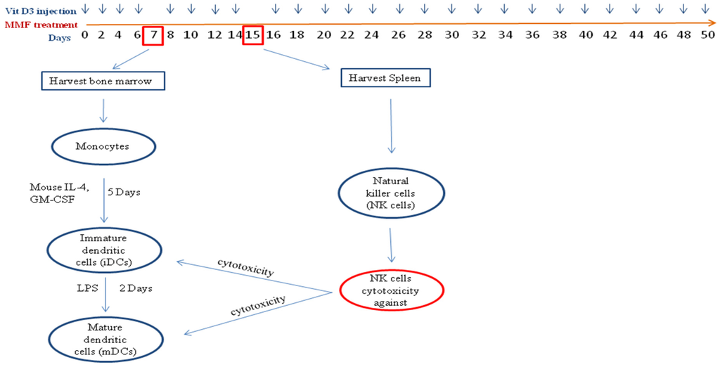
Experimental autoimmune encephalomyelitis (EAE) is a CD4+ T cell mediated inflammatory demyelinating disease that is induced in mice by administration of peptides derived from myelin proteins. We developed EAE in SJL mice by administration of PLP139–151 peptide. The effect of treating these mice with 1α,25-Dihydroxyvitamin D3 (vitamin D3), or with monomethyl fumarate (MMF) was then examined. We observed that both vitamin D3 and MMF inhibited and/or prevented EAE in these mice. These findings were corroborated with isolating natural killer (NK) cells from vitamin D3-treated or MMF-treated EAE mice that lysed immature or mature dendritic cells. The results support and extend other findings indicating that an important mechanism of action for drugs used to treat multiple sclerosis (MS) is to enhance NK cell lysis of dendritic cells. Full article
Show Figures

Figure 1

2030 KiB  
Article
Dendritic Cell Protection from Cisplatin Nephrotoxicity Is Independent of Neutrophils
by Raghu K. Tadagavadi, Guofeng Gao, Wei Wei Wang, Manuel Rovira Gonzalez and W. Brian Reeves
Toxins 2015, 7(8), 3245-3256; https://doi.org/10.3390/toxins7083245 - 19 Aug 2015
Cited by 23 | Viewed by 6421
Abstract
Cisplatin is a very effective chemotherapeutic agent used against a wide range of solid tumors. A major adverse effect of cisplatin therapy is acute kidney injury (AKI). Neutrophils are reported to infiltrate and exacerbate injury in a wide range of sterile inflammatory models [...] Read more.
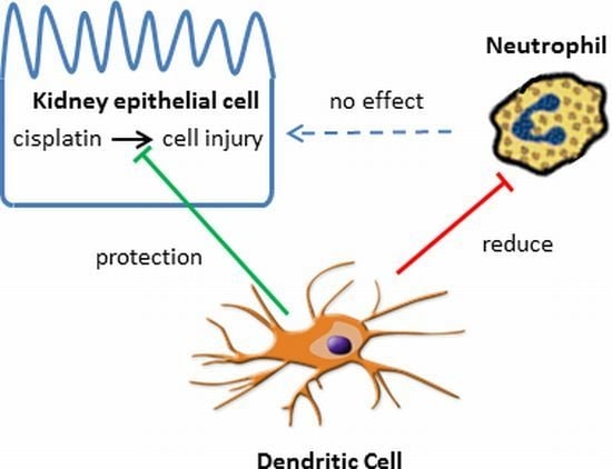
Cisplatin is a very effective chemotherapeutic agent used against a wide range of solid tumors. A major adverse effect of cisplatin therapy is acute kidney injury (AKI). Neutrophils are reported to infiltrate and exacerbate injury in a wide range of sterile inflammatory models of tissue injury. Here, we studied the kinetics of neutrophil infiltration into kidneys and their role in cisplatin-mediated AKI. Mice treated with cisplatin showed an increase in circulating neutrophils 24 and 48 h after cisplatin administration. Cisplatin treatment caused an increase in kidney leukocytes with neutrophils accounting for the majority of the infiltrating leukocytes. The extent of neutrophil infiltration coincided with the severity of kidney injury and renal dysfunction. To examine the functional relevance of infiltrating neutrophils in cisplatin nephrotoxicity, we depleted neutrophils using a neutrophil-specific antibody (anti-Ly-6G). This antibody resulted in greater than 90% depletion of neutrophils in both the blood and kidney. Of note, depletion of neutrophils had no impact on the extent of cisplatin-induced AKI as compared to non-depleted mice. Earlier, we reported that dendritic cell depletion in CD11c-DTRtg mice causes exacerbation of AKI and a dramatic increase in renal neutrophils. Thus, we also examined the role of neutrophils in dendritic cell-depleted mice treated with cisplatin. Dendritic cell depletion exacerbated AKI in spite of neutrophil depletion. These data demonstrate that cisplatin nephrotoxicity is not mediated by neutrophils and that dendritic cells protect kidneys via neutrophil-independent mechanisms. Full article
Show Figures

Graphical abstract

852 KiB  
Review
Monoclonal Antibody Combinations that Present Synergistic Neutralizing Activity: A Platform for Next-Generation Anti-Toxin Drugs
by Eran Diamant, Amram Torgeman, Eyal Ozeri and Ran Zichel
Toxins 2015, 7(6), 1854-1881; https://doi.org/10.3390/toxins7061854 - 29 May 2015
Cited by 29 | Viewed by 8056
Abstract
Monoclonal antibodies (MAbs) are among the fastest-growing therapeutics and are being developed for a broad range of indications, including the neutralization of toxins, bacteria and viruses. Nevertheless, MAbs potency is still relatively low when compared to conventional polyclonal Ab preparations. Moreover, the efficacy [...] Read more.
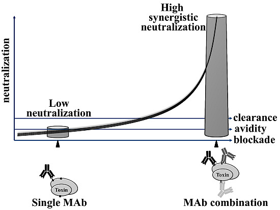
Monoclonal antibodies (MAbs) are among the fastest-growing therapeutics and are being developed for a broad range of indications, including the neutralization of toxins, bacteria and viruses. Nevertheless, MAbs potency is still relatively low when compared to conventional polyclonal Ab preparations. Moreover, the efficacy of an individual neutralizing MAb may significantly be hampered by the potential absence or modification of its target epitope in a mutant or subtype of the infectious agent. These limitations of individual neutralizing MAbs can be overcome by using oligoclonal combinations of several MAbs with different specificities to the target antigen. Studies conducted in our lab and by others show that such combined MAb preparation may present substantial synergy in its potency over the calculated additive potency of its individual MAb components. Moreover, oligoclonal preparation is expected to be better suited to compensating for reduced efficacy due to epitope variation. In this review, the synergistic neutralization properties of combined oligoclonal Ab preparations are described. The effect of Ab affinity, autologous Fc fraction, and targeting a critical number of epitopes, as well as the unexpected contribution of non-neutralizing clones to the synergistic neutralizing effect are presented and discussed. Full article
Show Figures

Graphical abstract

983 KiB  
Article
Toxins Targeting the KV1.3 Channel: Potential Immunomodulators for Autoimmune Diseases
by Yipeng Zhao, Jie Huang, Xiaolu Yuan, Biwen Peng, Wanhong Liu, Song Han and Xiaohua He
Toxins 2015, 7(5), 1749-1764; https://doi.org/10.3390/toxins7051749 - 19 May 2015
Cited by 43 | Viewed by 8803
Abstract
Autoimmune diseases are usually accompanied by tissue injury caused by autoantigen-specific T-cells. KV1.3 channels participate in modulating calcium signaling to induce T-cell proliferation, immune activation and cytokine production. Effector memory T (TEM)-cells, which play major roles in many autoimmune [...] Read more.
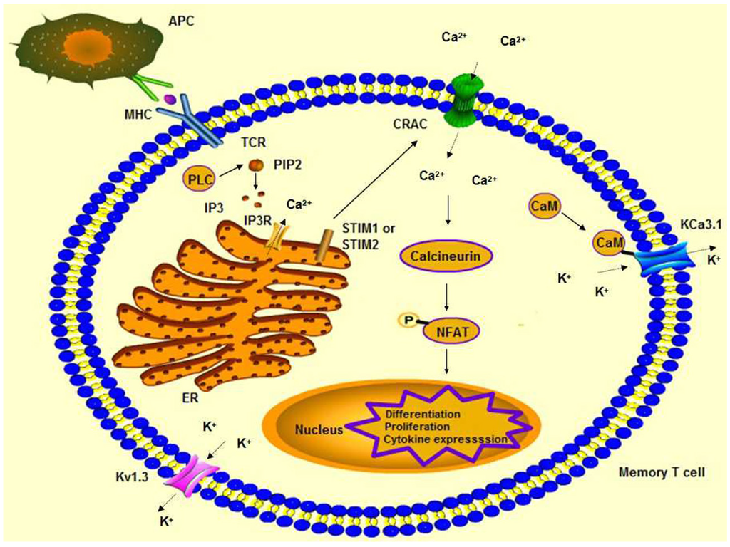
Autoimmune diseases are usually accompanied by tissue injury caused by autoantigen-specific T-cells. KV1.3 channels participate in modulating calcium signaling to induce T-cell proliferation, immune activation and cytokine production. Effector memory T (TEM)-cells, which play major roles in many autoimmune diseases, are controlled by blocking KV1.3 channels on the membrane. Toxins derived from animal venoms have been found to selectively target a variety of ion channels, including KV1.3. By blocking the KV1.3 channel, these toxins are able to suppress the activation and proliferation of TEM cells and may improve TEM cell-mediated autoimmune diseases, such as multiple sclerosis and type I diabetes mellitus. Full article
Show Figures

Figure 1

474 KiB  
Communication
Increased Circulating Levels of Vitamin D Binding Protein in MS Patients
by Arturo Ottavio Rinaldi, Isabella Sanseverino, Cristina Purificato, Antonio Cortese, Rosella Mechelli, Silvia Francisci, Marco Salvetti, Enrico Millefiorini, Sandra Gessani and Maria Cristina Gauzzi
Toxins 2015, 7(1), 129-137; https://doi.org/10.3390/toxins7010129 - 13 Jan 2015
Cited by 31 | Viewed by 9706
Abstract
Vitamin D (vitD) low status is currently considered a main environmental factor in multiple sclerosis (MS) etiology and pathogenesis. VitD and its metabolites are highly hydrophobic and circulate mostly bound to the vitamin D binding protein (DBP) and with lower affinity to albumin, [...] Read more.
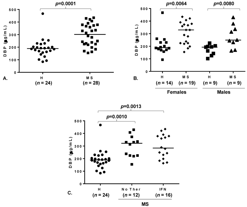
Vitamin D (vitD) low status is currently considered a main environmental factor in multiple sclerosis (MS) etiology and pathogenesis. VitD and its metabolites are highly hydrophobic and circulate mostly bound to the vitamin D binding protein (DBP) and with lower affinity to albumin, while less than 1% are in a free form. The aim of this study was to investigate whether the circulating levels of either of the two vitD plasma carriers and/or their relationship are altered in MS. We measured DBP and albumin plasma levels in 28 MS patients and 24 healthy controls. MS patients were found to have higher DBP levels than healthy subjects. Concomitant interferon beta therapy did not influence DBP concentration, and the difference with the control group was significant in both females and males. No significant correlation between DBP and albumin levels was observed either in healthy controls or in patients. These observations suggest the involvement of DBP in the patho-physiology of MS. Full article
Show Figures

Figure 1

2014

Jump to: 2015, 2013, 2012, 2010, 2009

1088 KiB  
Article
Oxidized Lipids and Lysophosphatidylcholine Induce the Chemotaxis, Up-Regulate the Expression of CCR9 and CXCR4 and Abrogate the Release of IL-6 in Human Monocytes
by Johannes Rolin, Heidi Vego and Azzam A. Maghazachi
Toxins 2014, 6(9), 2840-2856; https://doi.org/10.3390/toxins6092840 - 23 Sep 2014
Cited by 27 | Viewed by 7080
Abstract
Lipids through regulation of chronic inflammation play key roles in the development of various diseases. Here, we report that a mixed population of human primary monocytes migrated towards LPC, as well as oxidized linoleic acid isoforms 9-S-HODE, 9-R-HODE and 13-R-HODE. Incubation with 9-R-HODE, [...] Read more.
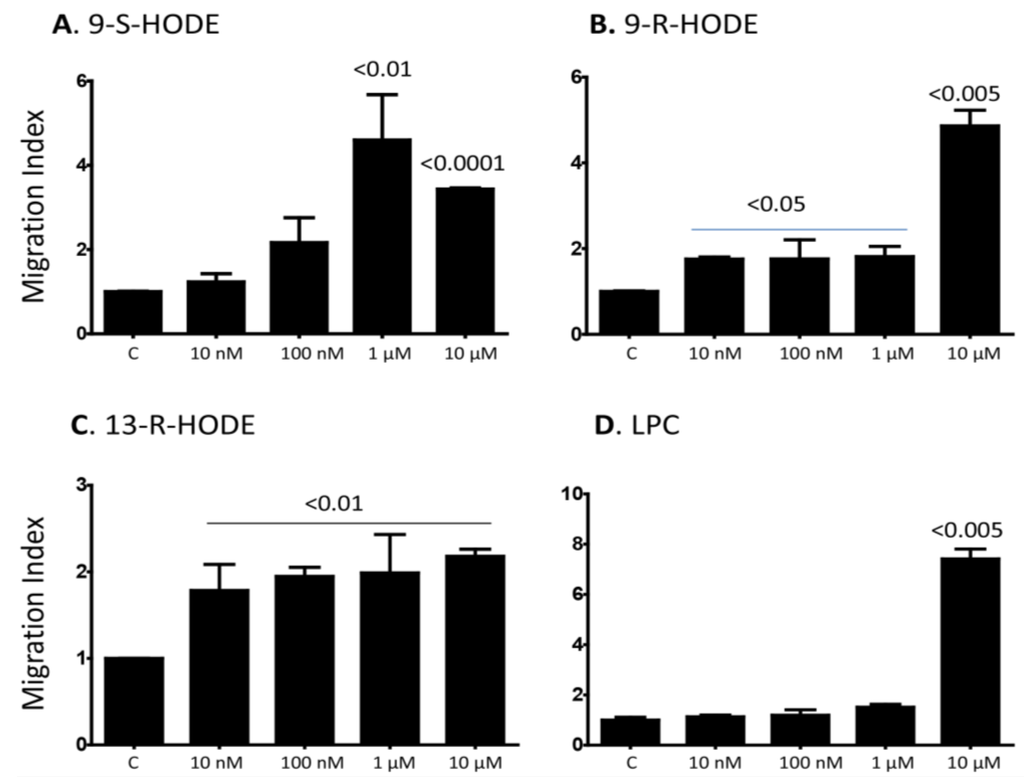
Lipids through regulation of chronic inflammation play key roles in the development of various diseases. Here, we report that a mixed population of human primary monocytes migrated towards LPC, as well as oxidized linoleic acid isoforms 9-S-HODE, 9-R-HODE and 13-R-HODE. Incubation with 9-R-HODE, 13-R-HODE and LPC resulted in increased expression of CXCR4, the receptor for SDF-1α/CXCL12, correlated with increased monocyte migration towards SDF-1α/CXCL12. Further, we report increased expression of CCR9, the receptor for TECK/CCL25, after stimulation with these lipids. Upon examining the migratory response towards TECK/CCL25, it was observed that an increase in CCR9 expression upon pre-treatment with 9-S-HODE, 9-R-HODE, 13-R-HODE and LPC resulted in increased migration of monocytes expressing CCR9. Only LPC but not any other lipid examined increased the influx of intracellular Ca2+ in monocytes. Finally, 9-S-HODE, 9-R-HODE, 13-R-HODE, or LPC inhibited the release of IL-6 from monocytes suggesting that these lipids may play important role in controlling inflammatory responses. Full article
Show Figures

Figure 1

888 KiB  
Review
Type I Interferons as Regulators of Human Antigen Presenting Cell Functions
by Sandra Gessani, Lucia Conti, Manuela Del Cornò and Filippo Belardelli
Toxins 2014, 6(6), 1696-1723; https://doi.org/10.3390/toxins6061696 - 26 May 2014
Cited by 75 | Viewed by 8738
Abstract
Type I interferons (IFNs) are pleiotropic cytokines, initially described for their antiviral activity. These cytokines exhibit a long record of clinical use in patients with some types of cancer, viral infections and chronic inflammatory diseases. It is now well established that IFN action [...] Read more.
Type I interferons (IFNs) are pleiotropic cytokines, initially described for their antiviral activity. These cytokines exhibit a long record of clinical use in patients with some types of cancer, viral infections and chronic inflammatory diseases. It is now well established that IFN action mostly relies on their ability to modulate host innate and adaptive immune responses. Work in recent years has begun to elucidate the mechanisms by which type I IFNs modify the immune response, and this is now recognized to be due to effects on multiple cell types, including monocytes, dendritic cells (DCs), NK cells, T and B lymphocytes. An ensemble of results from both animal models and in vitro studies emphasized the key role of type I IFNs in the development and function of DCs, suggesting the existence of a natural alliance between these cytokines and DCs in linking innate to adaptive immunity. The identification of IFN signatures in DCs and their dysregulation under pathological conditions will therefore be pivotal to decipher the complexity of this DC-IFN interaction and to better exploit the therapeutic potential of these cells. Full article
Show Figures

Graphical abstract

2833 KiB  
Article
Extracellular Hb Enhances Cardiac Toxicity in Endotoxemic Guinea Pigs: Protective Role of Haptoglobin
by Jin Hyen Baek, Xiaoyuan Zhang, Matthew C. Williams, Dominik J. Schaer, Paul W. Buehler and Felice D'Agnillo
Toxins 2014, 6(4), 1244-1259; https://doi.org/10.3390/toxins6041244 - 31 Mar 2014
Cited by 17 | Viewed by 7493
Abstract
Endotoxemia plays a major causative role in the myocardial injury and dysfunction associated with sepsis. Extracellular hemoglobin (Hb) has been shown to enhance the pathophysiology of endotoxemia. In the present study, we examined the myocardial pathophysiology in guinea pigs infused with lipopolysaccharide (LPS), [...] Read more.
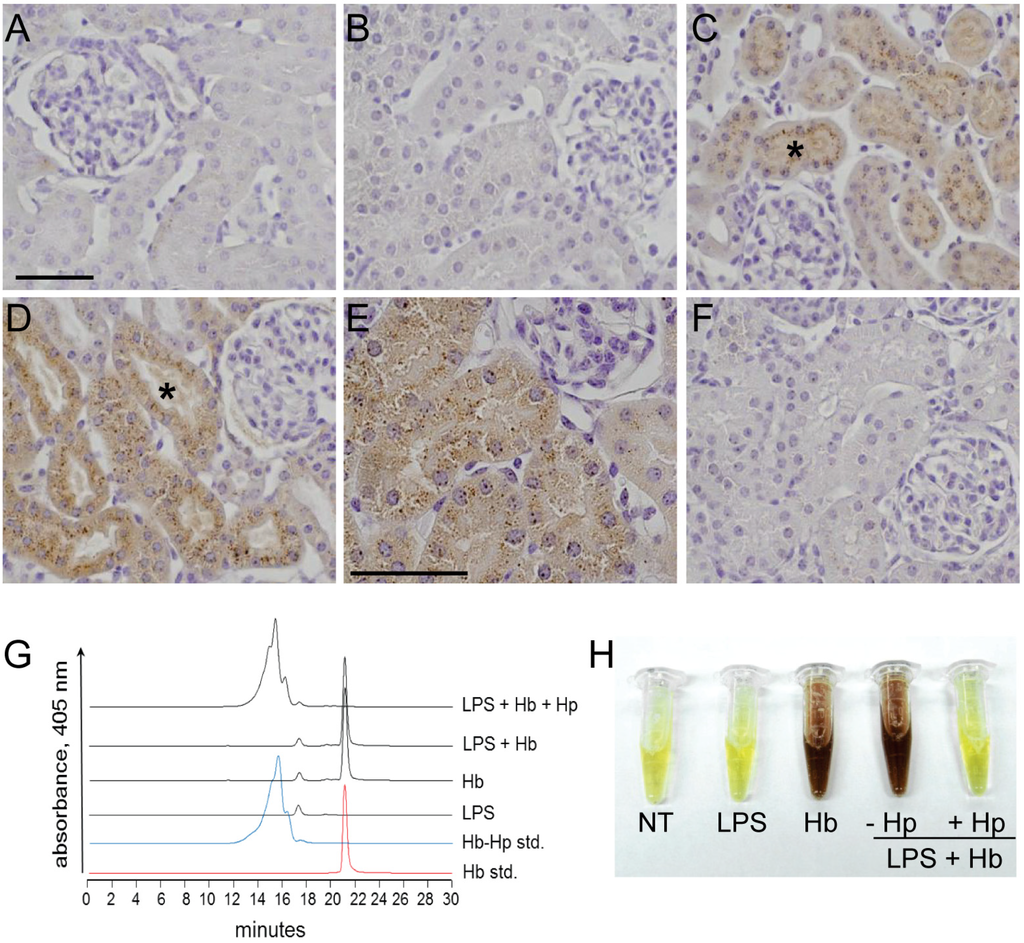
Endotoxemia plays a major causative role in the myocardial injury and dysfunction associated with sepsis. Extracellular hemoglobin (Hb) has been shown to enhance the pathophysiology of endotoxemia. In the present study, we examined the myocardial pathophysiology in guinea pigs infused with lipopolysaccharide (LPS), a Gram-negative bacterial endotoxin, and purified Hb. We also examined whether the administration of the Hb scavenger haptoglobin (Hp) could protect against the effects observed. Here, we show that Hb infusion following LPS administration, but not either insult alone, increased myocardial iron deposition, heme oxygenase-1 expression, phagocyte activation and infiltration, as well as oxidative DNA damage and apoptosis assessed by 8-hydroxy-2'-deoxyguanosine (8-OHdG) and terminal deoxynucleotidyl transferase dUTP nick end labeling (TUNEL) immunostaining, respectively. Co-administration of Hp significantly attenuated the myocardial events induced by the combination of LPS and Hb. These findings may have relevant therapeutic implications for the management of sepsis during concomitant disease or clinical interventions associated with the increased co-exposures to LPS and Hb, such as trauma, surgery or massive blood transfusions. Full article
Show Figures

Figure 1

348 KiB  
Review
Staphylococcal Bicomponent Pore-Forming Toxins: Targets for Prophylaxis and Immunotherapy
by M. Javad Aman and Rajan P. Adhikari
Toxins 2014, 6(3), 950-972; https://doi.org/10.3390/toxins6030950 - 4 Mar 2014
Cited by 37 | Viewed by 8326
Abstract
Staphylococccus aureus represents one of the most challenging human pathogens as well as a common colonizer of human skin and mucosal surfaces. S. aureus causes a wide range of diseases from skin and soft tissue infection (SSTI) to debilitating and life-threatening conditions such [...] Read more.
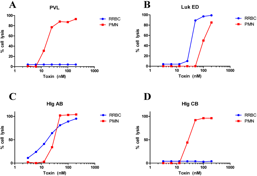
Staphylococccus aureus represents one of the most challenging human pathogens as well as a common colonizer of human skin and mucosal surfaces. S. aureus causes a wide range of diseases from skin and soft tissue infection (SSTI) to debilitating and life-threatening conditions such as osteomyelitis, endocarditis, and necrotizing pneumonia. The range of diseases reflects the remarkable diversity of the virulence factors produced by this pathogen, including surface antigens involved in the establishment of infection and a large number of toxins that mediate a vast array of cellular responses. The staphylococcal toxins are generally believed to have evolved to disarm the innate immune system, the first line of defense against this pathogen. This review focuses on recent advances on elucidating the biological functions of S. aureus bicomponent pore-forming toxins (BCPFTs) and their utility as targets for preventive and therapeutic intervention. These toxins are cytolytic to a variety of immune cells, primarily neutrophils, as well as cells with a critical barrier function. The lytic activity of BCPFTs towards immune cells implies a critical role in immune evasion, and a number of epidemiological studies and animal experiments relate these toxins to clinical disease, particularly SSTI and necrotizing pneumonia. Antibody-mediated neutralization of this lytic activity may provide a strategy for development of toxoid-based vaccines or immunotherapeutics for prevention or mitigation of clinical diseases. However, certain BCPFTs have been proposed to act as danger signals that may alert the immune system through an inflammatory response. The utility of a neutralizing vaccination strategy must be weighed against such immune-activating potential. Full article
Show Figures

Figure 1

3904 KiB  
Article
Immune Checkpoint Blockade in Cancer Treatment: A Double-Edged Sword Cross-Targeting the Host as an “Innocent Bystander”
by Lucia Gelao, Carmen Criscitiello, Angela Esposito, Aron Goldhirsch and Giuseppe Curigliano
Toxins 2014, 6(3), 914-933; https://doi.org/10.3390/toxins6030914 - 3 Mar 2014
Cited by 61 | Viewed by 11346
Abstract
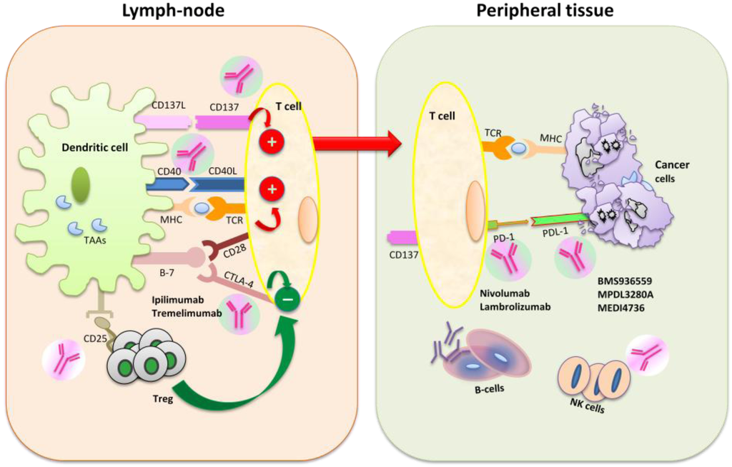
Targeted immune checkpoint blockade augments anti-tumor immunity and induces durable responses in patients with melanoma and other solid tumors. It also induces specific “immune-related adverse events” (irAEs). IrAEs mainly include gastrointestinal, dermatological, hepatic and endocrinological toxicities. Off-target effects that arise appear to account [...] Read more.
Targeted immune checkpoint blockade augments anti-tumor immunity and induces durable responses in patients with melanoma and other solid tumors. It also induces specific “immune-related adverse events” (irAEs). IrAEs mainly include gastrointestinal, dermatological, hepatic and endocrinological toxicities. Off-target effects that arise appear to account for much of the toxicity of the immune checkpoint blockade. These unique “innocent bystander” effects are likely a direct result of breaking immune tolerance upon immune check point blockade and require specific treatment guidelines that include symptomatic therapies or systemic corticosteroids. What do we need going forward to limit immune checkpoint blockade-induced toxicity? Most importantly, we need a better understanding of the roles played by these agents in normal tissues, so that we can begin to predict potentially problematic side effects on the basis of their selectivity profile. Second, we need to focus on the predictive factors of the response and toxicity of the host rather than serially focusing on individual agents. Third, rigorous biomarker-driven clinical trials are needed to further elucidate the mechanisms of both the benefit and toxicity. We will summarize the double-edged sword effect of immunotherapeutics in cancer treatment. Full article
Show Figures

Figure 1

847 KiB  
Review
Monoclonal Antibody Therapy and Renal Transplantation: Focus on Adverse Effects
by Gianluigi Zaza, Paola Tomei, Simona Granata, Luigino Boschiero and Antonio Lupo
Toxins 2014, 6(3), 869-891; https://doi.org/10.3390/toxins6030869 - 28 Feb 2014
Cited by 36 | Viewed by 17706
Abstract
A series of monoclonal antibodies (mAbs) are commonly utilized in renal transplantation as induction therapy (a period of intense immunosuppression immediately before and following the implant of the allograft), to treat steroid-resistant acute rejections, to decrease the incidence and mitigate effects of delayed [...] Read more.
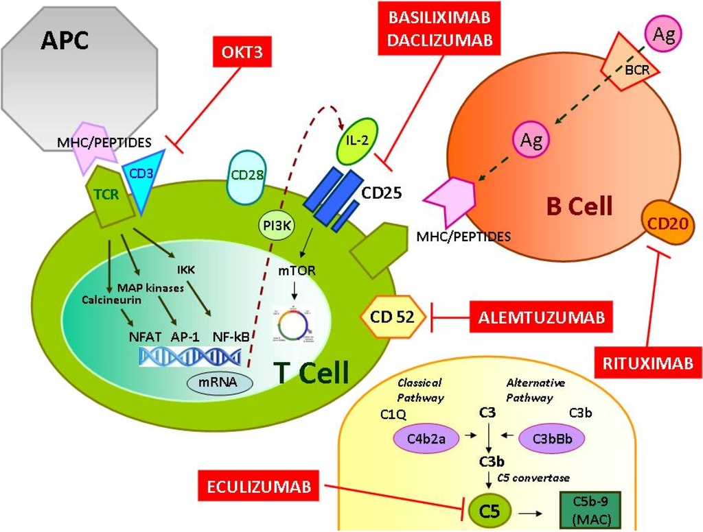
A series of monoclonal antibodies (mAbs) are commonly utilized in renal transplantation as induction therapy (a period of intense immunosuppression immediately before and following the implant of the allograft), to treat steroid-resistant acute rejections, to decrease the incidence and mitigate effects of delayed graft function, and to allow immunosuppressive minimization. Additionally, in the last few years, their use has been proposed for the treatment of chronic antibody-mediated rejection, a major cause of late renal allograft loss. Although the exact mechanism of immunosuppression and allograft tolerance with any of the currently used induction agents is not completely defined, the majority of these medications are targeted against specific CD proteins on the T or B cells surface (e.g., CD3, CD25, CD52). Moreover, some of them have different mechanisms of action. In particular, eculizumab, interrupting the complement pathway, is a new promising treatment tool for acute graft complications and for post-transplant hemolytic uremic syndrome. While it is clear their utility in renal transplantation, it is also unquestionable that by using these highly potent immunosuppressive agents, the body loses much of its innate ability to mount an adequate immune response, thereby increasing the risk of severe adverse effects (e.g., infections, malignancies, haematological complications). Therefore, it is extremely important for clinicians involved in renal transplantation to know the potential side effects of monoclonal antibodies in order to plan a correct therapeutic strategy minimizing/avoiding the onset and development of severe clinical complications. Full article
Show Figures

Figure 1

338 KiB  
Article
Antitumor Cell-Complex Vaccines Employing Genetically Modified Tumor Cells and Fibroblasts
by Antonio Miguel, María José Herrero, Luis Sendra, Rafael Botella, Ana Diaz, Rosa Algás and Salvador F. Aliño
Toxins 2014, 6(2), 636-649; https://doi.org/10.3390/toxins6020636 - 19 Feb 2014
Cited by 2 | Viewed by 5909
Abstract
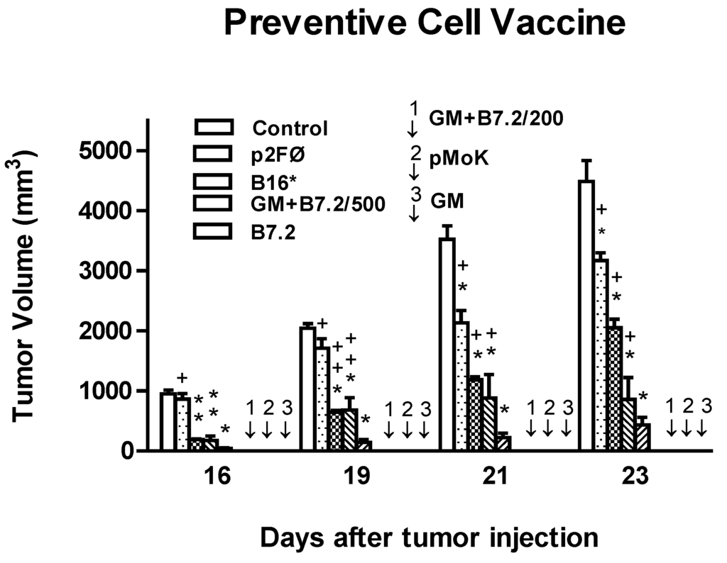
The present study evaluates the immune response mediated by vaccination with cell complexes composed of irradiated B16 tumor cells and mouse fibroblasts genetically modified to produce GM-CSF. The animals were vaccinated with free B16 cells or cell complexes. We employed two gene plasmid [...] Read more.
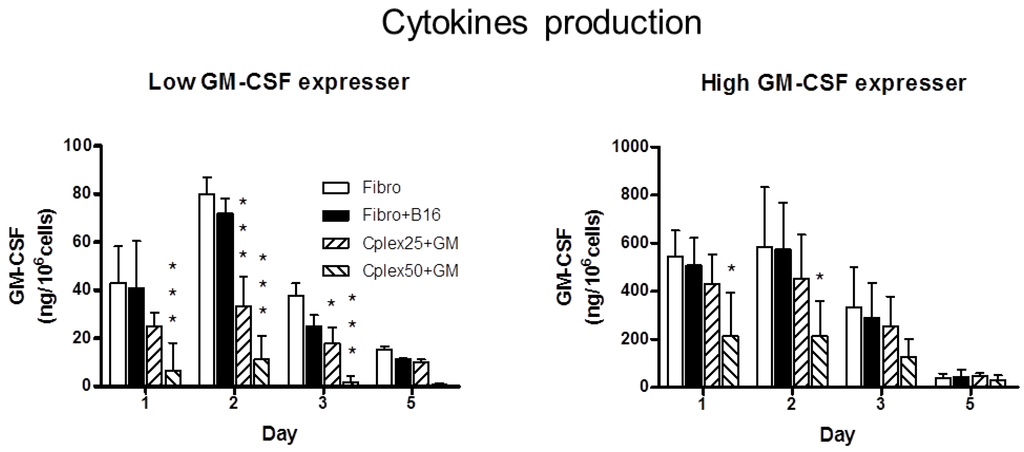
The present study evaluates the immune response mediated by vaccination with cell complexes composed of irradiated B16 tumor cells and mouse fibroblasts genetically modified to produce GM-CSF. The animals were vaccinated with free B16 cells or cell complexes. We employed two gene plasmid constructions: one high producer (pMok) and a low producer (p2F). Tumor transplant was performed by injection of B16 tumor cells. Plasma levels of total IgG and its subtypes were measured by ELISA. Tumor volumes were measured and survival curves were obtained. The study resulted in a cell complex vaccine able to stimulate the immune system to produce specific anti-tumor membrane proteins (TMP) IgG. In the groups vaccinated with cells transfected with the low producer plasmid, IgG production was higher when we used free B16 cell rather than cell complexes. Nonspecific autoimmune response caused by cell complex was not greater than that induced by the tumor cells alone. Groups vaccinated with B16 transfected with low producer plasmid reached a tumor growth delay of 92% (p ≤ 0.01). When vaccinated with cell complex, the best group was that transfected with high producer plasmid, reaching a tumor growth inhibition of 56% (p ≤ 0.05). Significant survival (40%) was only observed in the groups vaccinated with free transfected B16 cells. Full article
Show Figures

Figure 1

2013

Jump to: 2015, 2014, 2012, 2010, 2009

279 KiB  
Article
Impact of Antidepressants on Cytokine Production of Depressed Patients in Vitro
by Alexander Munzer, Ulrich Sack, Roland Mergl, Jeremias Schönherr, Charlotte Petersein, Stefanie Bartsch, Kenneth C. Kirkby, Katrin Bauer and Hubertus Himmerich
Toxins 2013, 5(11), 2227-2240; https://doi.org/10.3390/toxins5112227 - 19 Nov 2013
Cited by 97 | Viewed by 10328
Abstract
The interplay between immune and nervous systems plays a pivotal role in the pathophysiology of depression. In depressive episodes, patients show increased production of pro-inflammatory cytokines such as interleukin (IL)-1β and tumor necrosis factor (TNF)-α. There is limited information on the effect of [...] Read more.
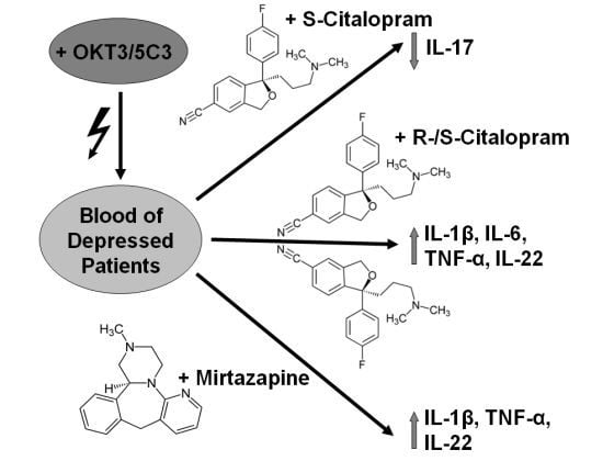
The interplay between immune and nervous systems plays a pivotal role in the pathophysiology of depression. In depressive episodes, patients show increased production of pro-inflammatory cytokines such as interleukin (IL)-1β and tumor necrosis factor (TNF)-α. There is limited information on the effect of antidepressant drugs on cytokines, most studies report on a limited sample of cytokines and none have reported effects on IL-22. We systematically investigated the effect of three antidepressant drugs, citalopram, escitalopram and mirtazapine, on secretion of cytokines IL-1β, IL-2, IL-4, IL-6, IL-17, IL-22 and TNF-α in a whole blood assay in vitro, using murine anti-human CD3 monoclonal antibody OKT3, and 5C3 monoclonal antibody against CD40, to stimulate T and B cells respectively. Citalopram increased production of IL-1β, IL-6, TNF-α and IL-22. Mirtazapine increased IL-1β, TNF-α and IL-22. Escitalopram decreased IL-17 levels. The influence of antidepressants on IL-2 and IL-4 levels was not significant for all three drugs. Compared to escitalopram, citalopram led to higher levels of IL-1β, IL-6, IL-17 and IL-22; and mirtazapine to higher levels of IL-1β, IL-17, IL-22 and TNF-α. Mirtazapine and citalopram increased IL-22 production. The differing profile of cytokine production may relate to differences in therapeutic effects, risk of relapse and side effects. Full article
Show Figures

Graphical abstract

2250 KiB  
Article
Effects of Vitamin D3, Calcipotriol and FTY720 on the Expression of Surface Molecules and Cytolytic Activities of Human Natural Killer Cells and Dendritic Cells
by Zaidoon Al-Jaderi and Azzam A. Maghazachi
Toxins 2013, 5(11), 1932-1947; https://doi.org/10.3390/toxins5111932 - 28 Oct 2013
Cited by 49 | Viewed by 7965
Abstract
We describe here the effects of three drugs that are either approved or have the potential for treating multiple sclerosis (MS) patients through the in vitro activities of human natural killer (NK) cells and dendritic cells (DCs). Our results indicate that 1,25(OH)2 [...] Read more.
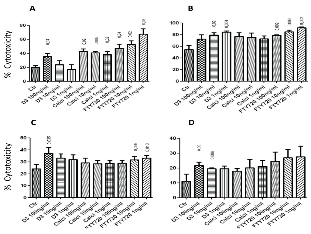
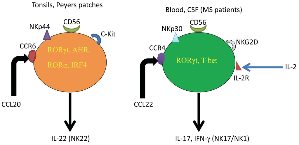
We describe here the effects of three drugs that are either approved or have the potential for treating multiple sclerosis (MS) patients through the in vitro activities of human natural killer (NK) cells and dendritic cells (DCs). Our results indicate that 1,25(OH)2D3, the biologically active metabolite of vitamin D3, calcipotriol and FTY720 augment IL-2-activated NK cell lysis of K562 and RAJI tumor cell lines as well as immature (i) and mature (m) DCs, with variable efficacies. These results are corroborated with the ability of the drugs to up-regulate the expression of NK cytotoxicity receptors NKp30 and NKp44, as well as NKG2D on the surfaces of NK cells. Also, they down-regulate the expression of the killer inhibitory receptor CD158. The three drugs down-regulate the expression of CCR6 on the surface of iDCs, whereas vitamin D3 and calcipotriol tend to up-regulate the expression of CCR7 on mDCs, suggesting that they may influence the migration of DCs into the lymph nodes. Finally, vitamin D3, calcipotriol and FTY720 enhance NK17/NK1 cell lysis of K562 cells, suggesting that a possible mechanism of action for these drugs is via activating these newly described cells. In conclusion, our results show novel mechanisms of action for vitamin D3, calcipotriol and FTY720 on cells of the innate immune system. Full article
Show Figures

Figure 1

192 KiB  
Review
The Snake Venom Rhodocytin from Calloselasma rhodostoma— A Clinically Important Toxin and a Useful Experimental Tool for Studies of C-Type Lectin-like Receptor 2 (CLEC-2)
by Øyvind Bruserud
Toxins 2013, 5(4), 665-674; https://doi.org/10.3390/toxins5040665 - 17 Apr 2013
Cited by 9 | Viewed by 7274
Abstract
The snake venom, rhodocytin, from the Malayan viper, Calloselasma rhodostoma, and the endogenous podoplanin are identified as ligands for the C-type lectin-like receptor 2 (CLEC-2). The snakebites caused by Calloselasma rhodostoma cause a local reaction with swelling, bleeding and eventually necrosis, together [...] Read more.
The snake venom, rhodocytin, from the Malayan viper, Calloselasma rhodostoma, and the endogenous podoplanin are identified as ligands for the C-type lectin-like receptor 2 (CLEC-2). The snakebites caused by Calloselasma rhodostoma cause a local reaction with swelling, bleeding and eventually necrosis, together with a systemic effect on blood coagulation with distant bleedings that can occur in many different organs. This clinical picture suggests that toxins in the venom have effects on endothelial cells and vessel permeability, extravasation and, possibly, activation of immunocompetent cells, as well as effects on platelets and the coagulation cascade. Based on the available biological studies, it seems likely that ligation of CLEC-2 contributes to local extravasation, inflammation and, possibly, local necrosis, due to microthrombi and ischemia, whereas other toxins may be more important for the distant hemorrhagic complications. However, the venom contains several toxins and both local, as well as distant, symptoms are probably complex reactions that cannot be explained by the effects of rhodocytin and CLEC-2 alone. The in vivo reactions to rhodocytin are thus examples of toxin-induced crosstalk between coagulation (platelets), endothelium and inflammation (immunocompetent cells). Very few studies have addressed this crosstalk as a part of the pathogenesis behind local and systemic reactions to Calloselasma rhodostoma bites. The author suggests that detailed biological studies based on an up-to-date methodology of local and systemic reactions to Calloselasma rhodostoma bites should be used as a hypothesis-generating basis for future functional studies of the CLEC-2 receptor. It will not be possible to study the effects of purified toxins in humans, but the development of animal models (e.g., cutaneous injections of rhodocytin to mimic snakebites) would supplement studies in humans. Full article
797 KiB  
Review
On The Role of Natural Killer Cells in Neurodegenerative Diseases
by Azzam A. Maghazachi
Toxins 2013, 5(2), 363-375; https://doi.org/10.3390/toxins5020363 - 19 Feb 2013
Cited by 15 | Viewed by 7183
Abstract
Natural killer (NK) cells exert important immunoregulatory functions by releasing several inflammatory molecules, such as IFN-γ and members of chemokines, which include CCL3/MIP-1α and CCL4/MIP-1β. These cells also express heptahelical receptors, which are coupled to heterotrimeric G proteins that guide them into inflamed [...] Read more.
Natural killer (NK) cells exert important immunoregulatory functions by releasing several inflammatory molecules, such as IFN-γ and members of chemokines, which include CCL3/MIP-1α and CCL4/MIP-1β. These cells also express heptahelical receptors, which are coupled to heterotrimeric G proteins that guide them into inflamed and injured tissues. NK cells have been shown to recognize and destroy transformed cells and virally-infected cells, but their roles in neurodegenerative diseases have not been examined in detail. In this review, I will summarize the effects of NK cells in two neurodegenerative diseases, namely multiple sclerosis and globoid cell leukodystrophy. It is hoped that the knowledge obtained from these diseases may facilitate building rational protocols for treating these and other neurodegenerative or autoimmune diseases using NK cells and drugs that activate them as therapeutic tools. Full article
Show Figures

Figure 1

262 KiB  
Review
The Possible Diagnostic and Prognostic Use of Systemic Chemokine Profiles in Clinical Medicine—The Experience in Acute Myeloid Leukemia from Disease Development and Diagnosis via Conventional Chemotherapy to Allogeneic Stem Cell Transplantation
by Håkon Reikvam, Hanne Fredly, Astrid Olsnes Kittang and Øystein Bruserud
Toxins 2013, 5(2), 336-362; https://doi.org/10.3390/toxins5020336 - 18 Feb 2013
Cited by 28 | Viewed by 6362
Abstract
Chemokines are important regulators of many different biological processes, including (i) inflammation with activation and local recruitment of immunocompetent cells; (ii) angiogenesis as a part of inflammation or carcinogenesis; and (iii) as a bridge between the coagulation system and inflammation/immune activation. The systemic [...] Read more.
Chemokines are important regulators of many different biological processes, including (i) inflammation with activation and local recruitment of immunocompetent cells; (ii) angiogenesis as a part of inflammation or carcinogenesis; and (iii) as a bridge between the coagulation system and inflammation/immune activation. The systemic levels of various chemokines may therefore reflect local disease processes, and such variations may thereby be used in the routine clinical handling of patients. The experience from patients with myeloproliferative diseases, and especially patients with acute myeloid leukemia (AML), suggests that systemic plasma/serum cytokine profiles can be useful, both as a diagnostic tool and for prognostication of patients. However, cytokines/chemokines are released by a wide range of cells and are involved in a wide range of biological processes; the altered levels may therefore mainly reflect the strength and nature of the biological processes, and the optimal clinical use of chemokine/cytokine analyses may therefore require combination with organ-specific biomarkers. Chemokine levels are also altered by clinical procedures, therapeutic interventions and the general status of the patients. A careful standardization of sample collection is therefore important, and the interpretation of the observations will require that the overall clinical context is considered. Despite these limitations, we conclude that analysis of systemic chemokine/cytokine profiles can reflect important clinical characteristics and, therefore, is an important scientific tool that can be used as a part of future clinical studies to identify clinically relevant biomarkers. Full article

2012

Jump to: 2015, 2014, 2013, 2010, 2009

825 KiB  
Article
Interleukin-17 (IL-17) Expression Is Reduced during Acute Myocardial Infarction: Role on Chemokine Receptor Expression in Monocytes and Their in Vitro Chemotaxis towards Chemokines
by Maria Troitskaya, Anton Baysa, Jarle Vaage, Kristin L. Sand, Azzam A. Maghazachi and Guro Valen
Toxins 2012, 4(12), 1427-1439; https://doi.org/10.3390/toxins4121427 - 30 Nov 2012
Cited by 11 | Viewed by 7867
Abstract
The roles of immune cells and their soluble products during myocardial infarction (MI) are not completely understood. Here, we observed that the percentages of IL-17, but not IL-22, producing cells are reduced in mice splenocytes after developing MI. To correlate this finding with [...] Read more.
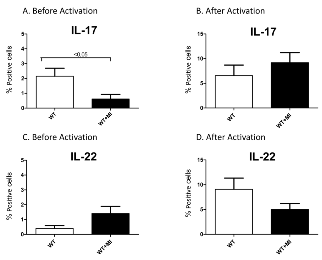
The roles of immune cells and their soluble products during myocardial infarction (MI) are not completely understood. Here, we observed that the percentages of IL-17, but not IL-22, producing cells are reduced in mice splenocytes after developing MI. To correlate this finding with the functional activity of IL-17, we sought to determine its effect on monocytes. In particular, we presumed that this cytokine might affect the chemotaxis of monocytes important for cardiac inflammation and remodeling. We observed that IL-17 tends to reduce the expression of two major chemokine receptors involved in monocyte chemotaxis, namely CCR2 and CXCR4. Further analysis showed that monocytes pretreated with IL-17 have reduced in vitro chemotaxis towards the ligand for CCR2, i.e., MCP-1/CCL2, and the ligand for CXCR4, i.e., SDF-1α/CXCL12. Our results support the possibility that IL-17 may be beneficial in MI, and this could be due to its ability to inhibit the migration of monocytes. Full article
Show Figures

Figure 1

546 KiB  
Review
Glucan–Resveratrol–Vitamin C Combination Offers Protection against Toxic Agents
by Vaclav Vetvicka and Jana Vetvickova
Toxins 2012, 4(11), 1301-1308; https://doi.org/10.3390/toxins4111301 - 9 Nov 2012
Cited by 14 | Viewed by 7786
Abstract
Biological immunomodulators are routinely evaluated as a natural source of molecules with profound effects on the immune system. They belong to a group of physiologically active compounds, collectively termed biological response modifiers. Most of the studies were focused on immune system stimulation. Recently, [...] Read more.
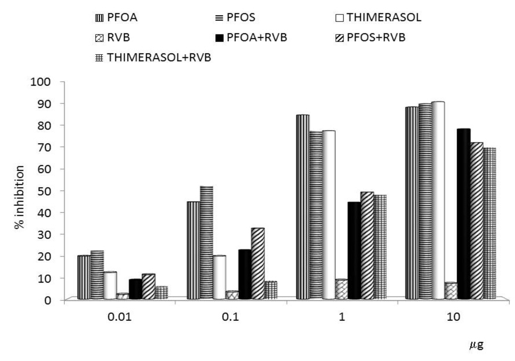
Biological immunomodulators are routinely evaluated as a natural source of molecules with profound effects on the immune system. They belong to a group of physiologically active compounds, collectively termed biological response modifiers. Most of the studies were focused on immune system stimulation. Recently, they have become the focus of studies seeking molecules that are able to overcome negative effects of various immunotoxins. This paper concentrates on the effects of a glucan/resveratrol/vitamin C combination on immunosuppressive effects of mercury and perfluorinated hydrocarbons. Effects described in this review have strong clinical potential, as environmental contaminants have adverse effects on all aspects of the immune system and represent a serious threat to the health of both humans and animals. Full article
Show Figures

Figure 1

1022 KiB  
Article
Comparative Antitumor Effect of Preventive versus Therapeutic Vaccines Employing B16 Melanoma Cells Genetically Modified to Express GM-CSF and B7.2 in a Murine Model
by Antonio Miguel, María José Herrero, Luis Sendra, Rafael Botella, Rosa Algás, Maria Sánchez and Salvador F. Aliño
Toxins 2012, 4(11), 1058-1081; https://doi.org/10.3390/toxins4111058 - 31 Oct 2012
Cited by 6 | Viewed by 7390
Abstract
Cancer vaccines have always been a subject of gene therapy research. One of the most successful approaches has been working with genetically modified tumor cells. In this study, we describe our approach to achieving an immune response against a murine melanoma model, employing [...] Read more.
Cancer vaccines have always been a subject of gene therapy research. One of the most successful approaches has been working with genetically modified tumor cells. In this study, we describe our approach to achieving an immune response against a murine melanoma model, employing B16 tumor cells expressing GM-CSF and B7.2. Wild B16 cells were injected in C57BL6 mice to cause the tumor. Irradiated B16 cells transfected with GM-CSF, B7.2, or both, were processed as a preventive and therapeutic vaccination. Tumor volumes were measured and survival curves were obtained. Blood samples were taken from mice, and IgGs of each treatment group were also measured. The regulatory T cells (Treg) of selected groups were quantified using counts of images taken by confocal microscopy. Results: one hundred percent survival was achieved by preventive vaccination with the group of cells transfected with p2F_GM-CSF. Therapeutic vaccination achieved initial inhibition of tumor growth but did not secure overall survival of the animals. Classical Treg cells did not vary among the different groups in this therapeutic vaccination model. Full article
Show Figures

Figure 1

248 KiB  
Article
Antibody to Heat Shock Protein 70 (HSP70) Inhibits Human T-Cell Lymphoptropic Virus Type I (HTLV-I) Production by Transformed Rabbit T-Cell Lines
by Hanan Fallouh and Wahib Mahana
Toxins 2012, 4(10), 768-777; https://doi.org/10.3390/toxins4100768 - 26 Sep 2012
Cited by 3 | Viewed by 6089
Abstract
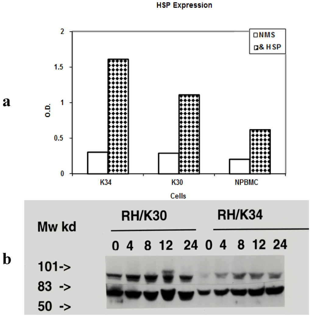
Adult T cell leukemia is a fatal malignant transformation caused by the human T-cell lymphoptropic virus type I (HTLV-I). HTLV-I is only associated with the development of this disease in a small percentage of infected individuals. Using two rabbit transformed T-cell lines; RH/K30 [...] Read more.
Adult T cell leukemia is a fatal malignant transformation caused by the human T-cell lymphoptropic virus type I (HTLV-I). HTLV-I is only associated with the development of this disease in a small percentage of infected individuals. Using two rabbit transformed T-cell lines; RH/K30 (asymptomatic) and RH/K34 (leukemogenic), we have investigated the expression of heat shock proteins (HSP) 90 and 70 and the role of anti-HSPs antibodies on virus production. HSPs surface expression was higher on RH/K34 than RH/K30 cells. Heat treatment of cells increased the expression of HSPs proteins and virus production; HSPs augmentation was stabilized after 12 h and virus production reached a maximum between 8 h–12 h then returned to normal level after 24 h of culture. Incubation of cells only with rabbit anti-HSP 70 antibodies prevented virus production specifically in the leukemogenic cell line. The results indicate a relationship between HSP 70 and virus production. Full article
Show Figures

Figure 1

228 KiB  
Review
The Biological Control of the Malaria Vector
by Layla Kamareddine
Toxins 2012, 4(9), 748-767; https://doi.org/10.3390/toxins4090748 - 19 Sep 2012
Cited by 60 | Viewed by 17903
Abstract
The call for malaria control, over the last century, marked a new epoch in the history of this disease. Many control strategies targeting either the Plasmodium parasite or the Anopheles vector were shown to be effective. Yet, the emergence of drug resistant parasites [...] Read more.
The call for malaria control, over the last century, marked a new epoch in the history of this disease. Many control strategies targeting either the Plasmodium parasite or the Anopheles vector were shown to be effective. Yet, the emergence of drug resistant parasites and insecticide resistant mosquito strains, along with numerous health, environmental, and ecological side effects of many chemical agents, highlighted the need to develop alternative tools that either complement or substitute conventional malaria control approaches. The use of biological means is considered a fundamental part of the recently launched malaria eradication program and has so far shown promising results, although this approach is still in its infancy. This review presents an overview of the most promising biological control tools for malaria eradication, namely fungi, bacteria, larvivorous fish, parasites, viruses and nematodes. Full article
273 KiB  
Brief Report
Intranasal Rapamycin Rescues Mice from Staphylococcal Enterotoxin B-Induced Shock
by Teresa Krakauer and Marilyn Buckley
Toxins 2012, 4(9), 718-728; https://doi.org/10.3390/toxins4090718 - 18 Sep 2012
Cited by 12 | Viewed by 6494
Abstract
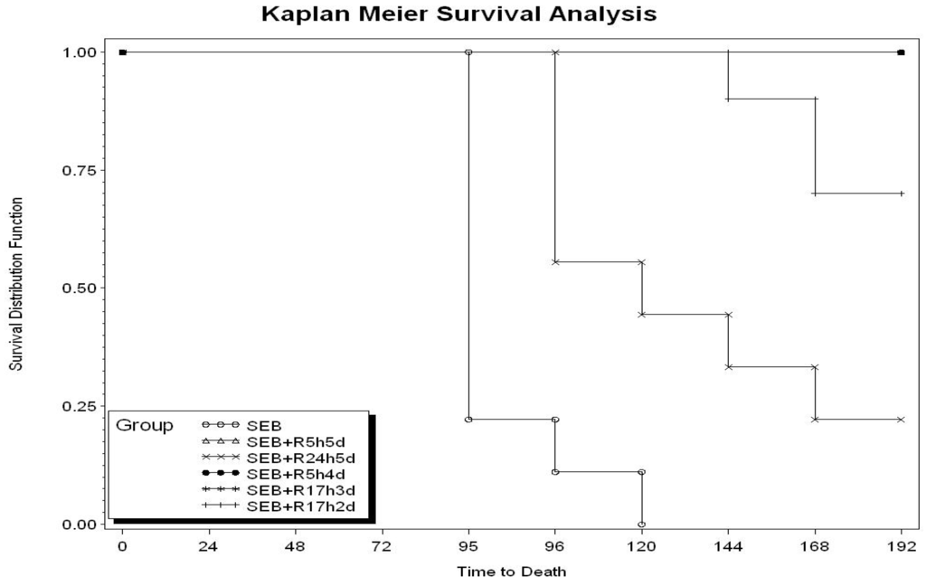
Staphylococcal enterotoxin B (SEB) and related exotoxins produced by Staphylococcus aureus are potent activators of the immune system and cause toxic shock in humans. Currently there is no effective treatment except for the use of intravenous immunoglobulins administered shortly after SEB exposure. Intranasal [...] Read more.
Staphylococcal enterotoxin B (SEB) and related exotoxins produced by Staphylococcus aureus are potent activators of the immune system and cause toxic shock in humans. Currently there is no effective treatment except for the use of intravenous immunoglobulins administered shortly after SEB exposure. Intranasal SEB induces long-lasting lung injury which requires prolonged drug treatment. We investigated the effects of rapamycin, an immunosuppressive drug used to prevent graft rejection, by intranasal administration in a lethal mouse model of SEB-induced shock. The results show that intranasal rapamycin alone delivered as late as 17 h after SEB protected 100% of mice from lethal shock. Additionally, rapamycin diminished the weight loss and temperature fluctuations elicited by SEB. Intranasal rapamycin attenuated lung MCP-1, IL-2, IL-6, and IFNγ by 70%, 30%, 64%, and 68% respectively. Furthermore, short courses (three doses) of rapamycin were sufficient to block SEB-induced shock. Intranasal rapamycin represents a novel use of an immunosuppressant targeting directly to site of toxin exposure, reducing dosages needed and allowing a wider therapeutic window. Full article
Show Figures

Figure 1

506 KiB  
Review
Macrophage-Targeted Therapy: CD64-Based Immunotoxins for Treatment of Chronic Inflammatory Diseases
by Dmitrij Hristodorov, Radoslav Mladenov, Michael Huhn, Stefan Barth and Theo Thepen
Toxins 2012, 4(9), 676-694; https://doi.org/10.3390/toxins4090676 - 14 Sep 2012
Cited by 39 | Viewed by 11771
Abstract
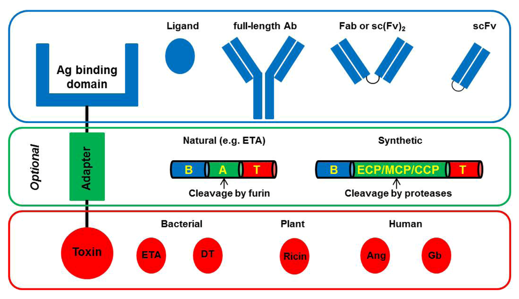
Diseases caused by chronic inflammation (e.g., arthritis, multiple sclerosis and diabetic ulcers) are multicausal, thus making treatment difficult and inefficient. Due to the age-associated nature of most of these disorders and the demographic transition towards an overall older population, efficient therapeutic intervention strategies [...] Read more.
Diseases caused by chronic inflammation (e.g., arthritis, multiple sclerosis and diabetic ulcers) are multicausal, thus making treatment difficult and inefficient. Due to the age-associated nature of most of these disorders and the demographic transition towards an overall older population, efficient therapeutic intervention strategies will need to be developed in the near future. Over the past decades, elimination of activated macrophages using CD64-targeting immunotoxins has proven to be a promising way of resolving inflammation in animal models. More recent data have shown that the M1-polarized population of activated macrophages in particular is critically involved in the chronic phase. We recapitulate the latest progress in the development of IT. These have advanced from full-length antibodies, chemically coupled to bacterial toxins, into single chain variants of antibodies, genetically fused with fully human enzymes. These improvements have increased the range of possible target diseases, which now include chronic inflammatory diseases. At present there are no therapeutic strategies focusing on macrophages to treat chronic disorders. In this review, we focus on the role of different polarized macrophages and the potential of CD64-based IT to intervene in the process of chronic inflammation. Full article
Show Figures

Figure 1

2010

Jump to: 2015, 2014, 2013, 2012, 2009

267 KiB  
Review
The Double-Edged Sword of Autoimmunity: Lessons from Multiple Sclerosis
by Anne Lise K. Hestvik
Toxins 2010, 2(4), 856-877; https://doi.org/10.3390/toxins2040856 - 22 Apr 2010
Cited by 14 | Viewed by 12947
Abstract
The relationship between immune responses to self-antigens and autoimmune disease is unclear. In contrast to its animal model experimental autoimmune encephalomyelitis (EAE), which is driven by T cell responses to myelin antigens, the target antigen of the intrathecal immune response in multiple sclerosis [...] Read more.
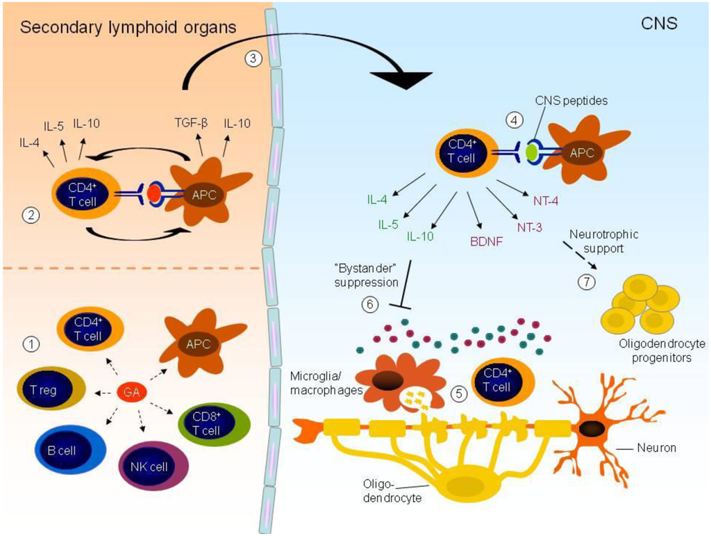
The relationship between immune responses to self-antigens and autoimmune disease is unclear. In contrast to its animal model experimental autoimmune encephalomyelitis (EAE), which is driven by T cell responses to myelin antigens, the target antigen of the intrathecal immune response in multiple sclerosis (MS) has not been identified. Although the immune response in MS contributes significantly to tissue destruction, the action of immunocompetent cells within the central nervous system (CNS) may also hold therapeutic potential. Thus, treatment of MS patients with glatiramer acetate triggers a protective immune response. Here we review the immunopathogenesis of MS and some recent findings on the mechanism of glatiramer acetate (GA). Full article
Show Figures

Figure 1

430 KiB  
Review
Toxins–Useful Biochemical Tools for Leukocyte Research
by Susana Cubillos, Johannes Norgauer and Katja Lehmann
Toxins 2010, 2(4), 428-452; https://doi.org/10.3390/toxins2040428 - 26 Mar 2010
Cited by 9 | Viewed by 11489
Abstract
Leukocytes are a heterogeneous group of cells that display differences in anatomic localization, cell surface phenotype, and function. The different subtypes include e.g., granulocytes, monocytes, dendritic cells, T cells, B cells and NK cells. These different cell types represent the cellular component of [...] Read more.
Leukocytes are a heterogeneous group of cells that display differences in anatomic localization, cell surface phenotype, and function. The different subtypes include e.g., granulocytes, monocytes, dendritic cells, T cells, B cells and NK cells. These different cell types represent the cellular component of innate and adaptive immunity. Using certain toxins such as pertussis toxin, cholera toxin or clostridium difficile toxin, the regulatory functions of Gαi, Gαs and small GTPases of the Rho family in leukocytes have been reported. A summary of these reports is discussed in this review. Full article
Show Figures

Graphical abstract

457 KiB  
Review
The Protein Kinase C Agonist PEP005 (Ingenol 3-Angelate) in the Treatment of Human Cancer: A Balance between Efficacy and Toxicity
by Elisabeth Ersvaer, Astrid Olsnes Kittang, Peter Hampson, Kristoffer Sand, Bjørn Tore Gjertsen, Janet M. Lord and Øystein Bruserud
Toxins 2010, 2(1), 174-194; https://doi.org/10.3390/toxins2010174 - 22 Jan 2010
Cited by 59 | Viewed by 15486
Abstract
The diterpene ester ingenol-3-angelate (referred to as PEP005) is derived from the plant Euphorbia peplus. Crude euphorbia extract causes local toxicity and transient inflammation when applied topically and has been used in the treatment of warts, skin keratoses and skin cancer. PEP005 [...] Read more.
The diterpene ester ingenol-3-angelate (referred to as PEP005) is derived from the plant Euphorbia peplus. Crude euphorbia extract causes local toxicity and transient inflammation when applied topically and has been used in the treatment of warts, skin keratoses and skin cancer. PEP005 is a broad range activator of the classical (α, β, γ) and novel (δ, ε, η, θ) protein kinase C isoenzymes. Direct pro-apoptotic effects of this drug have been demonstrated in several malignant cells, including melanoma cell lines and primary human acute myelogenous leukemia cells. At micromolar concentrations required to kill melanoma cells this agent causes PKC-independent secondary necrosis. In contrast, the killing of leukemic cells occurs in the nanomolar range, requires activation of protein kinase C δ (PKCδ) and is specifically associated with translocation of PKCδ from the cytoplasm to the nuclear membrane. However, in addition to this pro-apoptotic effect the agent seems to have immunostimulatory effects, including: (i) increased chemokine release by malignant cells; (ii) a general increase in proliferation and cytokine release by activated T cells, including T cells derived from patients with chemotherapy-induced lymphopenia; (iii) local infiltration of neutrophils after topical application with increased antibody-dependent cytotoxicity; and (iv) development of specific anti-cancer immune responses by CD8+ T cells in animal models. Published studies mainly describe effects from in vitro investigations or after topical application of the agent, and careful evaluation of the toxicity after systemic administration is required before the possible use of this agent in the treatment of malignancies other than skin cancers. Full article
Show Figures

Figure 1

616 KiB  
Article
Calprotectin (S100A8/S100A9) and Myeloperoxidase: Co-Regulators of Formation of Reactive Oxygen Species
by Arne Bøyum, Knut Kristian Skrede, Oddvar Myhre, Vivi-Ann Tennfjord, Christine Gran Neurauter, Helge Tolleshaug, Eirunn Knudsen, Per Kristian Opstad, Magnar Bjørås and Haakon B. Benestad
Toxins 2010, 2(1), 95-115; https://doi.org/10.3390/toxins2010095 - 20 Jan 2010
Cited by 13 | Viewed by 16217
Abstract
Inflammatory mediators trigger polymorphonuclear neutrophils (PMN) to produce reactive oxygen species (ROS: O2-, H2O2, ∙OH). Mediated by myeloperoxidase in PMN, HOCl is formed, detectable in a chemiluminescence (CL) assay. We have shown that the abundant cytosolic PMN [...] Read more.
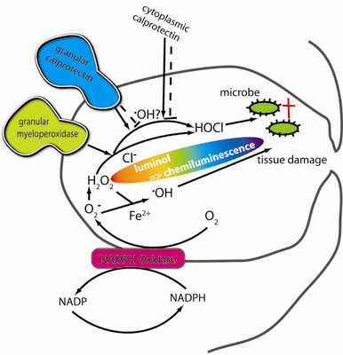
Inflammatory mediators trigger polymorphonuclear neutrophils (PMN) to produce reactive oxygen species (ROS: O2-, H2O2, ∙OH). Mediated by myeloperoxidase in PMN, HOCl is formed, detectable in a chemiluminescence (CL) assay. We have shown that the abundant cytosolic PMN protein calprotectin (S100A8/A9) similarly elicits CL in response to H2O2 in a cell-free system. Myeloperoxidase and calprotectin worked synergistically. Calprotectin-induced CL increased, whereas myeloperoxidase-triggered CL decreased with pH > 7.5. Myeloperoxidase needed NaCl for CL, calprotectin did not. 4-hydroxybenzoic acid, binding ∙OH, almost abrogated calprotectin CL, but moderately increased myeloperoxidase activity. The combination of native calprotectin, or recombinant S100A8/A9 proteins, with NaOCl markedly enhanced CL. NaOCl may be the synergistic link between myeloperoxidase and calprotectin. Surprisingly- and unexplained- at higher concentration of S100A9 the stimulation vanished, suggesting a switch from pro-oxidant to anti-oxidant function. We propose that the ∙OH is predominant in ROS production by calprotectin, a function not described before. Full article
Show Figures

Graphical abstract

640 KiB  
Review
Reciprocal Interactions between Lactoferrin and Bacterial Endotoxins and Their Role in the Regulation of the Immune Response
by Daniela Latorre, Patrizia Puddu, Piera Valenti and Sandra Gessani
Toxins 2010, 2(1), 54-68; https://doi.org/10.3390/toxins2010054 - 8 Jan 2010
Cited by 53 | Viewed by 13276
Abstract
Lactoferrin (Lf), an iron-binding glycoprotein expressed in most biological fluids, represents a major component of the mammalian innate immune system. Lf’s multiple activities rely not only on its capacity to bind iron, but also to interact with molecular and cellular components of both [...] Read more.
Lactoferrin (Lf), an iron-binding glycoprotein expressed in most biological fluids, represents a major component of the mammalian innate immune system. Lf’s multiple activities rely not only on its capacity to bind iron, but also to interact with molecular and cellular components of both host and pathogens. Lf can bind and sequester lipopolysaccharide (LPS), thus preventing pro-inflammatory pathway activation, sepsis and tissue damage. However, Lf-bound LPS may retain the capacity to induce cell activation via Toll-like receptor 4-dependent and -independent mechanisms. This review discusses the complex interplay between Lf and LPS and its relevance in the regulation of the immune response. Full article
Show Figures

Figure 1

2009

Jump to: 2015, 2014, 2013, 2012, 2010

351 KiB  
Article
Immune Response to Chlamydophila abortus POMP91B Protein in the Context of Different Pathogen Associated Molecular Patterns (PAMP); Role of Antigen in the Orientation of Immune Response
by Vincent Le Moigne, Georges Robreau and Wahib Mahana
Toxins 2009, 1(2), 59-73; https://doi.org/10.3390/toxins1020059 - 13 Oct 2009
Viewed by 10125
Abstract
In a previous study, we used bacterial flagellin to deliver antigens such as p27 of Mycobacterium tuberculosis to a host immune system and obtained a potent Th1 responsecompared to those obtained with Freund’s adjuvant and DNA immunization. In the current study, using a [...] Read more.
In a previous study, we used bacterial flagellin to deliver antigens such as p27 of Mycobacterium tuberculosis to a host immune system and obtained a potent Th1 responsecompared to those obtained with Freund’s adjuvant and DNA immunization. In the current study, using a POMP91B antigen of Chlamydophila abortus, a human and animal pathogen, as a model, we found that this antigen is unable to promote Th1 response. However, this antigen, unlike others, was able to induce a good Th2 response and IL-4 production after immunization by recombinant protein in Freund’s adjuvant or in phosphate buffered saline. Our results suggest that immune response is not only dependent on the immunization adjuvant, but also dependent on the nature of antigen used. Full article
Show Figures

Figure 1

Back to TopTop