Next Issue
Volume 22, April-1
Previous Issue
Volume 22, March-1
 
 
ijms-logo

Journal Browser

Journal Browser

Int. J. Mol. Sci., Volume 22, Issue 6 (March-2 2021) – 505 articles

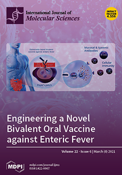
Cover Story (view full-size image): Enteric fever is a major global healthcare issue caused largely by Salmonella enterica serovars Typhi and Paratyphi A. The objective of this study was to develop a novel, bivalent oral vaccine capable of protecting against both serovars. Our approach centred on genetically engineering the attenuated S. Typhi ZH9 strain, to introduce two S. Paratyphi A immunogenic elements: flagellin H:a and lipopolysaccharide (LPS) O:2. The resulting new strain, ZH9PA, incorporated these two genetic changes and exhibited comparable growth kinetics to the parental ZH9 strain. A formulation containing both ZH9 and ZH9PA strains together constitutes a new bivalent vaccine candidate that targets both S. Typhi and S. Paratyphi A antigens to address a major global healthcare gap for enteric fever prophylaxis. View this paper
  • Issues are regarded as officially published after their release is announced to the table of contents alert mailing list.
  • You may sign up for e-mail alerts to receive table of contents of newly released issues.
  • PDF is the official format for papers published in both, html and pdf forms. To view the papers in pdf format, click on the "PDF Full-text" link, and use the free Adobe Reader to open them.
Order results
Result details
Section
Select all
Export citation of selected articles as:
20 pages, 3909 KiB  
Review
Transcriptional Regulation of Postnatal Cardiomyocyte Maturation and Regeneration
by Stephanie L. Padula, Nivedhitha Velayutham and Katherine E. Yutzey
Int. J. Mol. Sci. 2021, 22(6), 3288; https://doi.org/10.3390/ijms22063288 - 23 Mar 2021
Cited by 27 | Viewed by 5672
Abstract
During the postnatal period, mammalian cardiomyocytes undergo numerous maturational changes associated with increased cardiac function and output, including hypertrophic growth, cell cycle exit, sarcomeric protein isoform switching, and mitochondrial maturation. These changes come at the expense of loss of regenerative capacity of the [...] Read more.
During the postnatal period, mammalian cardiomyocytes undergo numerous maturational changes associated with increased cardiac function and output, including hypertrophic growth, cell cycle exit, sarcomeric protein isoform switching, and mitochondrial maturation. These changes come at the expense of loss of regenerative capacity of the heart, contributing to heart failure after cardiac injury in adults. While most studies focus on the transcriptional regulation of embryonic or adult cardiomyocytes, the transcriptional changes that occur during the postnatal period are relatively unknown. In this review, we focus on the transcriptional regulators responsible for these aspects of cardiomyocyte maturation during the postnatal period in mammals. By specifically highlighting this transitional period, we draw attention to critical processes in cardiomyocyte maturation with potential therapeutic implications in cardiovascular disease. Full article
Show Figures

Figure 1

15 pages, 2195 KiB  
Article
Engineering a Novel Bivalent Oral Vaccine against Enteric Fever
by Annelise Soulier, Claudia Prevosto, Mary Chol, Livija Deban and Rocky M. Cranenburgh
Int. J. Mol. Sci. 2021, 22(6), 3287; https://doi.org/10.3390/ijms22063287 - 23 Mar 2021
Cited by 4 | Viewed by 3993
Abstract
Enteric fever is a major global healthcare issue caused largely by Salmonella enterica serovars Typhi and Paratyphi A. The objective of this study was to develop a novel, bivalent oral vaccine capable of protecting against both serovars. Our approach centred on genetically engineering [...] Read more.
Enteric fever is a major global healthcare issue caused largely by Salmonella enterica serovars Typhi and Paratyphi A. The objective of this study was to develop a novel, bivalent oral vaccine capable of protecting against both serovars. Our approach centred on genetically engineering the attenuated S. Typhi ZH9 strain, which has an excellent safety record in clinical trials, to introduce two S. Paratyphi A immunogenic elements: flagellin H:a and lipopolysaccharide (LPS) O:2. We first replaced the native S. Typhi fliC gene encoding flagellin with the highly homologous fliC gene from S. Paratyphi A using Xer-cise technology. Next, we replaced the S. Typhi rfbE gene encoding tyvelose epimerase with a spacer sequence to enable the sustained expression of O:2 LPS and prevent its conversion to O:9 through tyvelose epimerase activity. The resulting new strain, ZH9PA, incorporated these two genetic changes and exhibited comparable growth kinetics to the parental ZH9 strain. A formulation containing both ZH9 and ZH9PA strains together constitutes a new bivalent vaccine candidate that targets both S. Typhi and S. Paratyphi A antigens to address a major global healthcare gap for enteric fever prophylaxis. This vaccine is now being tested in a Phase I clinical trial (NCT04349553). Full article
(This article belongs to the Special Issue Antimicrobial Resistance, Molecular Mechanisms and Fight Strategies)
Show Figures

Graphical abstract

19 pages, 3301 KiB  
Article
Late Health Effects of Partial Body Irradiation Injury in a Minipig Model Are Associated with Changes in Systemic and Cardiac IGF-1 Signaling
by Bernadette Hritzo, Saeed Y. Aghdam, Betre Legesse, Amandeep Kaur, Maohua Cao, Marjan Boerma, Nabarun Chakraborty, George Dimitrov, Aarti Gautam, Rasha Hammamieh, William Wilkins, Alena Tsioplaya, Gregory P. Holmes-Hampton and Maria Moroni
Int. J. Mol. Sci. 2021, 22(6), 3286; https://doi.org/10.3390/ijms22063286 - 23 Mar 2021
Cited by 6 | Viewed by 3434
Abstract
Clinical, epidemiological, and experimental evidence demonstrate non-cancer, cardiovascular, and endocrine effects of ionizing radiation exposure including growth hormone deficiency, obesity, metabolic syndrome, diabetes, and hyperinsulinemia. Insulin-like growth factor-1 (IGF-1) signaling perturbations are implicated in development of cardiovascular disease and metabolic syndrome. The minipig [...] Read more.
Clinical, epidemiological, and experimental evidence demonstrate non-cancer, cardiovascular, and endocrine effects of ionizing radiation exposure including growth hormone deficiency, obesity, metabolic syndrome, diabetes, and hyperinsulinemia. Insulin-like growth factor-1 (IGF-1) signaling perturbations are implicated in development of cardiovascular disease and metabolic syndrome. The minipig is an emerging model for studying radiation effects given its high analogy to human anatomy and physiology. Here we use a minipig model to study late health effects of radiation by exposing male Göttingen minipigs to 1.9–2.0 Gy X-rays (lower limb tibias spared). Animals were monitored for 120 days following irradiation and blood counts, body weight, heart rate, clinical chemistry parameters, and circulating biomarkers were assessed longitudinally. Collagen deposition, histolopathology, IGF-1 signaling, and mRNA sequencing were evaluated in tissues. Our findings indicate a single exposure induced histopathological changes, attenuated circulating IGF-1, and disrupted cardiac IGF-1 signaling. Electrolytes, lipid profiles, liver and kidney markers, and heart rate and rhythm were also affected. In the heart, collagen deposition was significantly increased and transforming growth factor beta-1 (TGF-beta-1) was induced following irradiation; collagen deposition and fibrosis were also observed in the kidney of irradiated animals. Our findings show Göttingen minipigs are a suitable large animal model to study long-term effects of radiation exposure and radiation-induced inhibition of IGF-1 signaling may play a role in development of late organ injuries. Full article
(This article belongs to the Section Molecular Biology)
Show Figures

Figure 1

18 pages, 32313 KiB  
Article
Conformational Heterogeneity and Cooperative Effects of Mammalian ALOX15
by Igor Ivanov, Alejandro Cruz, Alexander Zhuravlev, Almerinda Di Venere, Eleonora Nicolai, Sabine Stehling, José M. Lluch, Àngels González-Lafont and Hartmut Kuhn
Int. J. Mol. Sci. 2021, 22(6), 3285; https://doi.org/10.3390/ijms22063285 - 23 Mar 2021
Cited by 6 | Viewed by 2721
Abstract
Arachidonic acid lipoxygenases (ALOXs) have been suggested to function as monomeric enzymes, but more recent data on rabbit ALOX15 indicated that there is a dynamic monomer-dimer equilibrium in aqueous solution. In the presence of an active site ligand (the ALOX15 inhibitor RS7) rabbit [...] Read more.
Arachidonic acid lipoxygenases (ALOXs) have been suggested to function as monomeric enzymes, but more recent data on rabbit ALOX15 indicated that there is a dynamic monomer-dimer equilibrium in aqueous solution. In the presence of an active site ligand (the ALOX15 inhibitor RS7) rabbit ALOX15 was crystalized as heterodimer and the X-ray coordinates of the two monomers within the dimer exhibit subtle structural differences. Using native polyacrylamide electrophoresis, we here observed that highly purified and predominantly monomeric rabbit ALOX15 and human ALOX15B are present in two conformers with distinct electrophoretic mobilities. In silico docking studies, molecular dynamics simulations, site directed mutagenesis experiments and kinetic measurements suggested that in aqueous solutions the two enzymes exhibit motional flexibility, which may impact the enzymatic properties. Full article
(This article belongs to the Special Issue Structural, Functional and Folding Strategies of Oligomeric Proteins)
Show Figures

Figure 1

14 pages, 2343 KiB  
Article
The GNAQ T96S Mutation Affects Cell Signaling and Enhances the Oncogenic Properties of Hepatocellular Carcinoma
by Eugene Choi, Sung Jean Park, Gunhee Lee, Seung Kew Yoon, Minho Lee and Suk Kyeong Lee
Int. J. Mol. Sci. 2021, 22(6), 3284; https://doi.org/10.3390/ijms22063284 - 23 Mar 2021
Cited by 6 | Viewed by 3489
Abstract
Hepatocellular carcinoma (HCC), the most common malignant tumor in the liver, grows and metastasizes rapidly. Despite advances in treatment modalities, the five-year survival rate of HCC remains less than 30%. We sought genetic mutations that may affect the oncogenic properties of HCC, using [...] Read more.
Hepatocellular carcinoma (HCC), the most common malignant tumor in the liver, grows and metastasizes rapidly. Despite advances in treatment modalities, the five-year survival rate of HCC remains less than 30%. We sought genetic mutations that may affect the oncogenic properties of HCC, using The Cancer Genome Atlas (TCGA) data analysis. We found that the GNAQ T96S mutation (threonine 96 to serine alteration of the Gαq protein) was present in 12 out of 373 HCC patients (3.2%). To examine the effect of the GNAQ T96S mutation on HCC, we transfected the SK-Hep-1 cell line with the wild-type or the mutant GNAQ T96S expression vector. Transfection with the wild-type GNAQ expression vector enhanced anchorage-independent growth, migration, and the MAPK pathways in the SK-Hep-1 cells compared to control vector transfection. Moreover, cell proliferation, anchorage-independent growth, migration, and the MAPK pathways were further enhanced in the SK-Hep-1 cells transfected with the GNAQ T96S expression vector compared to the wild-type GNAQ-transfected cells. In silico structural analysis shows that the substitution of the GNAQ amino acid threonine 96 with a serine may destabilize the interaction between the regulator of G protein signaling (RGS) protein and GNAQ. This may reduce the inhibitory effect of RGS on GNAQ signaling, enhancing the GNAQ signaling pathway. Single nucleotide polymorphism (SNP) genotyping analysis for Korean HCC patients shows that the GNAQ T96S mutation was found in only one of the 456 patients (0.22%). Our data suggest that the GNAQ T96S hotspot mutation may play an oncogenic role in HCC by potentiating the GNAQ signal transduction pathway. Full article
(This article belongs to the Special Issue Molecular Advances in Cancer Genetics)
Show Figures

Figure 1

15 pages, 1125 KiB  
Review
Recent Advances Clarifying the Structure and Function of Plant Apyrases (Nucleoside Triphosphate Diphosphohydrolases)
by Greg Clark, Katherine A. Brown, Manas K. Tripathy and Stanley J. Roux
Int. J. Mol. Sci. 2021, 22(6), 3283; https://doi.org/10.3390/ijms22063283 - 23 Mar 2021
Cited by 16 | Viewed by 3082
Abstract
Studies implicating an important role for apyrase (NTPDase) enzymes in plant growth and development began appearing in the literature more than three decades ago. After early studies primarily in potato, Arabidopsis and legumes, especially important discoveries that advanced an understanding of the biochemistry, [...] Read more.
Studies implicating an important role for apyrase (NTPDase) enzymes in plant growth and development began appearing in the literature more than three decades ago. After early studies primarily in potato, Arabidopsis and legumes, especially important discoveries that advanced an understanding of the biochemistry, structure and function of these enzymes have been published in the last half-dozen years, revealing that they carry out key functions in diverse other plants. These recent discoveries about plant apyrases include, among others, novel findings on its crystal structures, its biochemistry, its roles in plant stress responses and its induction of major changes in gene expression when its expression is suppressed or enhanced. This review will describe and discuss these recent advances and the major questions about plant apyrases that remain unanswered. Full article
(This article belongs to the Special Issue New Horizons in Plant Cell Signaling)
Show Figures

Graphical abstract

13 pages, 2922 KiB  
Article
Anti-Platelet Properties of Phenolic and Nonpolar Fractions Isolated from Various Organs of Elaeagnus rhamnoides (L.) A. Nelson in Whole Blood
by Bartosz Skalski, Joanna Rywaniak, Aleksandra Szustka, Jerzy Żuchowski, Anna Stochmal and Beata Olas
Int. J. Mol. Sci. 2021, 22(6), 3282; https://doi.org/10.3390/ijms22063282 - 23 Mar 2021
Cited by 11 | Viewed by 2480
Abstract
Sea buckthorn (Elaeagnus rhamnoides (L.) A. Nelson) is a shrub growing in coastal areas. Its organs contain a range of bioactive substances including vitamins, fatty acids, various micro and macro elements, as well as phenolic compounds. Numerous studies of sea buckthorn have [...] Read more.
Sea buckthorn (Elaeagnus rhamnoides (L.) A. Nelson) is a shrub growing in coastal areas. Its organs contain a range of bioactive substances including vitamins, fatty acids, various micro and macro elements, as well as phenolic compounds. Numerous studies of sea buckthorn have found it to have anticancer, anti-ulcer, hepatoprotective, antibacterial, and antiviral properties. Some studies suggest that it also affects the hemostasis system. The aim of the study was to determine the effect of six polyphenols rich and triterpenic acids rich fractions (A–F), taken from various organs of sea buckthorn, on the activation of blood platelets using whole blood, and to assess the effect of the tested fractions on platelet proteins: fraction A (polyphenols rich fraction from fruits), fraction B (triterpenic acids rich fraction from fruits), fraction C (polyphenols rich fraction from leaves), fraction D (triterpenic acids rich fraction from leaves), fraction E (polyphenols rich fraction from twigs), and fraction F (triterpenic acids rich fraction from twigs). Hemostasis parameters were determined using flow cytometry and T-TAS (Total Thrombus-formation Analysis System). Additionally, electrophoresis was performed under reducing and non-reducing conditions. Although all tested fractions inhibit platelet activation, the greatest anti-platelet activity was demonstrated by fraction A, which was rich in flavonol glycosides. In addition, none of the tested fractions (A–F) caused any changes in the platelet proteome, and their anti-platelet potential is not dependent on the P2Y12 receptor. Full article
(This article belongs to the Special Issue Flavonoids)
Show Figures

Figure 1

16 pages, 2980 KiB  
Article
Loss of BOK Has a Minor Impact on Acetaminophen Overdose-Induced Liver Damage in Mice
by Samara Naim, Yuniel Fernandez-Marrero, Simone de Brot, Daniel Bachmann and Thomas Kaufmann
Int. J. Mol. Sci. 2021, 22(6), 3281; https://doi.org/10.3390/ijms22063281 - 23 Mar 2021
Cited by 2 | Viewed by 2915
Abstract
Acetaminophen (APAP) is one of the most commonly used analgesic and anti-pyretic drugs, and APAP intoxication is one of the main reasons for liver transplantation following liver failure in the Western world. While APAP poisoning ultimately leads to liver necrosis, various programmed cell [...] Read more.
Acetaminophen (APAP) is one of the most commonly used analgesic and anti-pyretic drugs, and APAP intoxication is one of the main reasons for liver transplantation following liver failure in the Western world. While APAP poisoning ultimately leads to liver necrosis, various programmed cell death modalities have been implicated, including ER stress-triggered apoptosis. The BCL-2 family member BOK (BCL-2-related ovarian killer) has been described to modulate the unfolded protein response and to promote chemical-induced liver injury. We therefore investigated the impact of the loss of BOK following APAP overdosing in mice. Surprisingly, we observed sex-dependent differences in the activation of the unfolded protein response (UPR) in both wildtype (WT) and Bok-/- mice, with increased activation of JNK in females compared with males. Loss of BOK led to a decrease in JNK activation and a reduced percentage of centrilobular necrosis in both sexes after APAP treatment; however, this protection was more pronounced in Bok-/- females. Nevertheless, serum ALT and AST levels of Bok-/- and WT mice were comparable, indicating that there was no major difference in the overall outcome of liver injury. We conclude that after APAP overdosing, loss of BOK affects initiating signaling steps linked to ER stress, but has a more minor impact on the outcome of liver necrosis. Furthermore, we observed sex-dependent differences that might be worthwhile to investigate. Full article
(This article belongs to the Section Molecular Pharmacology)
Show Figures

Figure 1

37 pages, 3229 KiB  
Review
Breast Cancer and the Other Non-Coding RNAs
by Dana Dvorská, Dušan Braný, Marcela Ňachajová, Erika Halašová and Zuzana Danková
Int. J. Mol. Sci. 2021, 22(6), 3280; https://doi.org/10.3390/ijms22063280 - 23 Mar 2021
Cited by 22 | Viewed by 5102
Abstract
Breast cancer is very heterogenous and the most common gynaecological cancer, with various factors affecting its development. While its impact on human lives and national health budgets is still rising in almost all global areas, many molecular mechanisms affecting its onset and development [...] Read more.
Breast cancer is very heterogenous and the most common gynaecological cancer, with various factors affecting its development. While its impact on human lives and national health budgets is still rising in almost all global areas, many molecular mechanisms affecting its onset and development remain unclear. Conventional treatments still prove inadequate in some aspects, and appropriate molecular therapeutic targets are required for improved outcomes. Recent scientific interest has therefore focused on the non-coding RNAs roles in tumour development and their potential as therapeutic targets. These RNAs comprise the majority of the human transcript and their broad action mechanisms range from gene silencing to chromatin remodelling. Many non-coding RNAs also have altered expression in breast cancer cell lines and tissues, and this is often connected with increased proliferation, a degraded extracellular environment, and higher endothelial to mesenchymal transition. Herein, we summarise the known abnormalities in the function and expression of long non-coding RNAs, Piwi interacting RNAs, small nucleolar RNAs and small nuclear RNAs in breast cancer, and how these abnormalities affect the development of this deadly disease. Finally, the use of RNA interference to suppress breast cancer growth is summarised. Full article
(This article belongs to the Special Issue Non-coding RNA (ncRNA) in Cancer)
Show Figures

Figure 1

60 pages, 63296 KiB  
Review
Articular Chondrocyte Phenotype Regulation through the Cytoskeleton and the Signaling Processes That Originate from or Converge on the Cytoskeleton: Towards a Novel Understanding of the Intersection between Actin Dynamics and Chondrogenic Function
by Jasmin C. Lauer, Mischa Selig, Melanie L. Hart, Bodo Kurz and Bernd Rolauffs
Int. J. Mol. Sci. 2021, 22(6), 3279; https://doi.org/10.3390/ijms22063279 - 23 Mar 2021
Cited by 49 | Viewed by 6220
Abstract
Numerous studies have assembled a complex picture, in which extracellular stimuli and intracellular signaling pathways modulate the chondrocyte phenotype. Because many diseases are mechanobiology-related, this review asked to what extent phenotype regulators control chondrocyte function through the cytoskeleton and cytoskeleton-regulating signaling processes. Such [...] Read more.
Numerous studies have assembled a complex picture, in which extracellular stimuli and intracellular signaling pathways modulate the chondrocyte phenotype. Because many diseases are mechanobiology-related, this review asked to what extent phenotype regulators control chondrocyte function through the cytoskeleton and cytoskeleton-regulating signaling processes. Such information would generate leverage for advanced articular cartilage repair. Serial passaging, pro-inflammatory cytokine signaling (TNF-α, IL-1α, IL-1β, IL-6, and IL-8), growth factors (TGF-α), and osteoarthritis not only induce dedifferentiation but also converge on RhoA/ROCK/Rac1/mDia1/mDia2/Cdc42 to promote actin polymerization/crosslinking for stress fiber (SF) formation. SF formation takes center stage in phenotype control, as both SF formation and SOX9 phosphorylation for COL2 expression are ROCK activity-dependent. Explaining how it is molecularly possible that dedifferentiation induces low COL2 expression but high SF formation, this review theorized that, in chondrocyte SOX9, phosphorylation by ROCK might effectively be sidelined in favor of other SF-promoting ROCK substrates, based on a differential ROCK affinity. In turn, actin depolymerization for redifferentiation would “free-up” ROCK to increase COL2 expression. Moreover, the actin cytoskeleton regulates COL1 expression, modulates COL2/aggrecan fragment generation, and mediates a fibrogenic/catabolic expression profile, highlighting that actin dynamics-regulating processes decisively control the chondrocyte phenotype. This suggests modulating the balance between actin polymerization/depolymerization for therapeutically controlling the chondrocyte phenotype. Full article
(This article belongs to the Special Issue The Future of Cartilage Repair in Complex Biological Situations)
Show Figures

Figure 1

28 pages, 7509 KiB  
Article
Utilizing an Animal Model to Identify Brain Neurodegeneration-Related Biomarkers in Aging
by Ming-Hui Yang, Yi-Ming Arthur Chen, Shan-Chen Tu, Pei-Ling Chi, Kuo-Pin Chuang, Chin-Chuan Chang, Chiang-Hsuan Lee, Yi-Ling Chen, Che-Hsin Lee, Cheng-Hui Yuan and Yu-Chang Tyan
Int. J. Mol. Sci. 2021, 22(6), 3278; https://doi.org/10.3390/ijms22063278 - 23 Mar 2021
Cited by 3 | Viewed by 3952
Abstract
Glycine N-methyltransferase (GNMT) regulates S-adenosylmethionine (SAMe), a methyl donor in methylation. Over-expressed SAMe may cause neurogenic capacity reduction and memory impairment. GNMT knockout mice (GNMT-KO) was applied as an experimental model to evaluate its effect on neurons. In this study, proteins from brain [...] Read more.
Glycine N-methyltransferase (GNMT) regulates S-adenosylmethionine (SAMe), a methyl donor in methylation. Over-expressed SAMe may cause neurogenic capacity reduction and memory impairment. GNMT knockout mice (GNMT-KO) was applied as an experimental model to evaluate its effect on neurons. In this study, proteins from brain tissues were studied using proteomic approaches, Haemotoxylin and Eosin staining, immunohistochemistry, Western blotting, and ingenuity pathway analysis. The expression of Receptor-interacting protein 1(RIPK1) and Caspase 3 were up-regulated and activity-dependent neuroprotective protein (ADNP) was down-regulated in GNMT-KO mice regardless of the age. Besides, proteins related to neuropathology, such as excitatory amino acid transporter 2, calcium/calmodulin-dependent protein kinase type II subunit alpha, and Cu-Zn superoxide dismutase were found only in the group of aged wild-type mice; 4-aminobutyrate amino transferase, limbic system-associated membrane protein, sodium- and chloride-dependent GABA transporter 3 and ProSAAS were found only in the group of young GNMT-KO mice and are related to function of neurons; serum albumin and Rho GDP dissociation inhibitor 1 were found only in the group of aged GNMT-KO mice and are connected to neurodegenerative disorders. With proteomic analyses, a pathway involving Gonadotropin-releasing hormone (GnRH) signal was found to be associated with aging. The GnRH pathway could provide additional information on the mechanism of aging and non-aging related neurodegeneration, and these protein markers may be served in developing future therapeutic treatments to ameliorate aging and prevent diseases. Full article
(This article belongs to the Special Issue Peripheral Biomarkers in Neurodegenerative Diseases 2.0)
Show Figures

Figure 1

24 pages, 4409 KiB  
Review
Development of Two-Dimensional Nanomaterials Based Electrochemical Biosensors on Enhancing the Analysis of Food Toxicants
by Iruthayapandi Selestin Raja, Mohan Vedhanayagam, Desingh Raj Preeth, Chuntae Kim, Jong Hun Lee and Dong Wook Han
Int. J. Mol. Sci. 2021, 22(6), 3277; https://doi.org/10.3390/ijms22063277 - 23 Mar 2021
Cited by 21 | Viewed by 4413
Abstract
In recent times, food safety has become a topic of debate as the foodborne diseases triggered by chemical and biological contaminants affect human health and the food industry’s profits. Though conventional analytical instrumentation-based food sensors are available, the consumers did not appreciate them [...] Read more.
In recent times, food safety has become a topic of debate as the foodborne diseases triggered by chemical and biological contaminants affect human health and the food industry’s profits. Though conventional analytical instrumentation-based food sensors are available, the consumers did not appreciate them because of the drawbacks of complexity, greater number of analysis steps, expensive enzymes, and lack of portability. Hence, designing easy-to-use tests for the rapid analysis of food contaminants has become essential in the food industry. Under this context, electrochemical biosensors have received attention among researchers as they bear the advantages of operational simplicity, portability, stability, easy miniaturization, and low cost. Two-dimensional (2D) nanomaterials have a larger surface area to volume compared to other dimensional nanomaterials. Hence, researchers nowadays are inclined to develop 2D nanomaterials-based electrochemical biosensors to significantly improve the sensor’s sensitivity, selectivity, and reproducibility while measuring the food toxicants. In the present review, we compile the contribution of 2D nanomaterials in electrochemical biosensors to test the food toxicants and discuss the future directions in the field. Further, we describe the types of food toxicity, methodologies quantifying food analytes, how the electrochemical food sensor works, and the general biomedical properties of 2D nanomaterials. Full article
Show Figures

Graphical abstract

20 pages, 6581 KiB  
Review
New Insights into the Mammalian Egg Zona Pellucida
by Carla Moros-Nicolás, Pascale Chevret, María Jiménez-Movilla, Blanca Algarra, Paula Cots-Rodríguez, Leopoldo González-Brusi, Manuel Avilés and Mª José Izquierdo-Rico
Int. J. Mol. Sci. 2021, 22(6), 3276; https://doi.org/10.3390/ijms22063276 - 23 Mar 2021
Cited by 26 | Viewed by 8549
Abstract
Mammalian oocytes are surrounded by an extracellular coat called the zona pellucida (ZP), which, from an evolutionary point of view, is the most ancient of the coats that envelope vertebrate oocytes and conceptuses. This matrix separates the oocyte from cumulus cells and is [...] Read more.
Mammalian oocytes are surrounded by an extracellular coat called the zona pellucida (ZP), which, from an evolutionary point of view, is the most ancient of the coats that envelope vertebrate oocytes and conceptuses. This matrix separates the oocyte from cumulus cells and is responsible for species-specific recognition between gametes, preventing polyspermy and protecting the preimplantation embryo. The ZP is a dynamic structure that shows different properties before and after fertilization. Until very recently, mammalian ZP was believed to be composed of only three glycoproteins, ZP1, ZP2 and ZP3, as first described in mouse. However, studies have revealed that this composition is not necessarily applicable to other mammals. Such differences can be explained by an analysis of the molecular evolution of the ZP gene family, during which ZP genes have suffered pseudogenization and duplication events that have resulted in differing models of ZP protein composition. The many discoveries made in recent years related to ZP composition and evolution suggest that a compilation would be useful. Moreover, this review analyses ZP biosynthesis, the role of each ZP protein in different mammalian species and how these proteins may interact among themselves and with other proteins present in the oviductal lumen. Full article
Show Figures

Figure 1

31 pages, 6580 KiB  
Article
Neonatal Mesenchymal Stem Cell Treatment Improves Myelination Impaired by Global Perinatal Asphyxia in Rats
by Andrea Tapia-Bustos, Carolyne Lespay-Rebolledo, Valentina Vío, Ronald Pérez-Lobos, Emmanuel Casanova-Ortiz, Fernando Ezquer, Mario Herrera-Marschitz and Paola Morales
Int. J. Mol. Sci. 2021, 22(6), 3275; https://doi.org/10.3390/ijms22063275 - 23 Mar 2021
Cited by 7 | Viewed by 3685
Abstract
The effect of perinatal asphyxia (PA) on oligodendrocyte (OL), neuroinflammation, and cell viability was evaluated in telencephalon of rats at postnatal day (P)1, 7, and 14, a period characterized by a spur of neuronal networking, evaluating the effect of mesenchymal stem cell (MSCs)-treatment. [...] Read more.
The effect of perinatal asphyxia (PA) on oligodendrocyte (OL), neuroinflammation, and cell viability was evaluated in telencephalon of rats at postnatal day (P)1, 7, and 14, a period characterized by a spur of neuronal networking, evaluating the effect of mesenchymal stem cell (MSCs)-treatment. The issue was investigated with a rat model of global PA, mimicking a clinical risk occurring under labor. PA was induced by immersing fetus-containing uterine horns into a water bath for 21 min (AS), using sibling-caesarean-delivered fetuses (CS) as controls. Two hours after delivery, AS and CS neonates were injected with either 5 μL of vehicle (10% plasma) or 5 × 10⁴ MSCs into the lateral ventricle. Samples were assayed for myelin-basic protein (MBP) levels; Olig-1/Olig-2 transcriptional factors; Gglial phenotype; neuroinflammation, and delayed cell death. The main effects were observed at P7, including: (i) A decrease of MBP-immunoreactivity in external capsule, corpus callosum, cingulum, but not in fimbriae of hippocampus; (ii) an increase of Olig-1-mRNA levels; (iii) an increase of IL-6-mRNA, but not in protein levels; (iv) an increase in cell death, including OLs; and (v) MSCs treatment prevented the effect of PA on myelination, OLs number, and cell death. The present findings show that PA induces regional- and developmental-dependent changes on myelination and OLs maturation. Neonatal MSCs treatment improves survival of mature OLs and myelination in telencephalic white matter. Full article
(This article belongs to the Special Issue Brain Hypoxia: Mechanisms of Resilience and Tolerance)
Show Figures

Figure 1

31 pages, 5795 KiB  
Article
Behavioral Alterations and Decreased Number of Parvalbumin-Positive Interneurons in Wistar Rats after Maternal Immune Activation by Lipopolysaccharide: Sex Matters
by Iveta Vojtechova, Kristyna Maleninska, Viera Kutna, Ondrej Klovrza, Klara Tuckova, Tomas Petrasek and Ales Stuchlik
Int. J. Mol. Sci. 2021, 22(6), 3274; https://doi.org/10.3390/ijms22063274 - 23 Mar 2021
Cited by 24 | Viewed by 4373
Abstract
Maternal immune activation (MIA) during pregnancy represents an important environmental factor in the etiology of schizophrenia and autism spectrum disorders (ASD). Our goal was to investigate the impacts of MIA on the brain and behavior of adolescent and adult offspring, as a rat [...] Read more.
Maternal immune activation (MIA) during pregnancy represents an important environmental factor in the etiology of schizophrenia and autism spectrum disorders (ASD). Our goal was to investigate the impacts of MIA on the brain and behavior of adolescent and adult offspring, as a rat model of these neurodevelopmental disorders. We injected bacterial lipopolysaccharide (LPS, 1 mg/kg) to pregnant Wistar dams from gestational day 7, every other day, up to delivery. Behavior of the offspring was examined in a comprehensive battery of tasks at postnatal days P45 and P90. Several brain parameters were analyzed at P28. The results showed that prenatal immune activation caused social and communication impairments in the adult offspring of both sexes; males were affected already in adolescence. MIA also caused prepulse inhibition deficit in females and increased the startle reaction in males. Anxiety and hypolocomotion were apparent in LPS-affected males and females. In the 28-day-old LPS offspring, we found enlargement of the brain and decreased numbers of parvalbumin-positive interneurons in the frontal cortex in both sexes. To conclude, our data indicate that sex of the offspring plays a crucial role in the development of the MIA-induced behavioral alterations, whereas changes in the brain apparent in young animals are sex-independent. Full article
(This article belongs to the Special Issue Neuroinflammation: The Pathogenic Mechanism of Neurological Disorders)
Show Figures

Figure 1

17 pages, 4049 KiB  
Article
Regulatory Role of Sugars on the Settlement Inducing Activity of a Conspecific Cue in Pacific Oyster Crassostrea gigas
by Mary Grace Sedanza, Hee-Jin Kim, Xerxes Seposo, Asami Yoshida, Kenichi Yamaguchi and Cyril Glenn Satuito
Int. J. Mol. Sci. 2021, 22(6), 3273; https://doi.org/10.3390/ijms22063273 - 23 Mar 2021
Cited by 5 | Viewed by 3373
Abstract
This study evaluated the larval settlement inducing effect of sugars and a conspecific cue from adult shell extract of Crassostrea gigas. To understand how the presence of different chemical cues regulate settlement behavior, oyster larvae were exposed to 12 types of sugars, [...] Read more.
This study evaluated the larval settlement inducing effect of sugars and a conspecific cue from adult shell extract of Crassostrea gigas. To understand how the presence of different chemical cues regulate settlement behavior, oyster larvae were exposed to 12 types of sugars, shell extract-coated and non-coated surfaces, and under varied sugar exposure times. Lectin-glycan interaction effects on settlement and its localization on oyster larval tissues were investigated. The results showed that the conspecific cue elicited a positive concentration dependent settlement inducing trend. Sugars in the absence of a conspecific cue, C. gigas adult shell extract, did not promote settlement. Whereas, in the presence of the cue, showed varied effects, most of which were found inhibitory at different concentrations. Sugar treated larvae exposed for 2 h showed significant settlement inhibition in the presence of a conspecific cue. Neu5Ac, as well as GlcNAc sugars, showed a similar interaction trend with wheat germ agglutinin (WGA) lectin. WGA-FITC conjugate showed positive binding on the foot, velum, and mantle when exposed to GlcNAc sugars. This study suggests that a WGA lectin-like receptor and its endogenous ligand are both found in the larval chemoreceptors and the shell Ethylenediaminetetraacetic acid (EDTA) extract that may complementarily work together to allow the oyster larva greater selectivity during site selection. Full article
(This article belongs to the Special Issue Natural Compounds with Cancer-Selective Toxicity)
Show Figures

Figure 1

41 pages, 2234 KiB  
Review
A Mitocentric View of the Main Bacterial and Parasitic Infectious Diseases in the Pediatric Population
by Sonia Romero-Cordero, Richard Kirwan, Antoni Noguera-Julian, Francesc Cardellach, Clàudia Fortuny and Constanza Morén
Int. J. Mol. Sci. 2021, 22(6), 3272; https://doi.org/10.3390/ijms22063272 - 23 Mar 2021
Cited by 3 | Viewed by 4056
Abstract
Infectious diseases occur worldwide with great frequency in both adults and children. Both infections and their treatments trigger mitochondrial interactions at multiple levels: (i) incorporation of damaged or mutated proteins to the complexes of the electron transport chain, (ii) mitochondrial genome (depletion, deletions, [...] Read more.
Infectious diseases occur worldwide with great frequency in both adults and children. Both infections and their treatments trigger mitochondrial interactions at multiple levels: (i) incorporation of damaged or mutated proteins to the complexes of the electron transport chain, (ii) mitochondrial genome (depletion, deletions, and point mutations) and mitochondrial dynamics (fusion and fission), (iii) membrane potential, (iv) apoptotic regulation, (v) generation of reactive oxygen species, among others. Such alterations may result in serious adverse clinical events with great impact on children’s quality of life, even resulting in death. As such, bacterial agents are frequently associated with loss of mitochondrial membrane potential and cytochrome c release, ultimately leading to mitochondrial apoptosis by activation of caspases-3 and -9. Using Rayyan QCRI software for systematic reviews, we explore the association between mitochondrial alterations and pediatric infections including (i) bacterial: M. tuberculosis, E. cloacae, P. mirabilis, E. coli, S. enterica, S. aureus, S. pneumoniae, N. meningitidis and (ii) parasitic: P. falciparum. We analyze how these pediatric infections and their treatments may lead to mitochondrial deterioration in this especially vulnerable population, with the intention of improving both the understanding of these diseases and their management in clinical practice. Full article
(This article belongs to the Special Issue Mitochondrial Function and Communication)
Show Figures

Figure 1

14 pages, 2327 KiB  
Article
Characterisation of Novel Angiogenic and Potent Anti-Inflammatory Effects of Micro-Fragmented Adipose Tissue
by Baoqiang Guo, Xenia Sawkulycz, Nima Heidari, Ralph Rogers, Donghui Liu and Mark Slevin
Int. J. Mol. Sci. 2021, 22(6), 3271; https://doi.org/10.3390/ijms22063271 - 23 Mar 2021
Cited by 12 | Viewed by 3505
Abstract
Adipose tissue and more specifically micro-fragmented adipose tissue (MFAT) obtained from liposuction has recently been shown to possess interesting medicinal properties whereby its application supports pain reduction and may enhance tissue regeneration particularly in osteoarthritis. Here we have characterised samples of MFAT produced [...] Read more.
Adipose tissue and more specifically micro-fragmented adipose tissue (MFAT) obtained from liposuction has recently been shown to possess interesting medicinal properties whereby its application supports pain reduction and may enhance tissue regeneration particularly in osteoarthritis. Here we have characterised samples of MFAT produced using the Lipogems® International Spa system from eight volunteer individuals in order to understand the critical biological mechanisms through which they act. A variation was found in the MFAT cluster size between individual samples and this translated into a similar variation in the ability of purified mesenchymal stem cells (MSCs) to form colony-forming units. Almost all of the isolated cells were CD105/CD90/CD45+ indicating stemness. An analysis of the secretions of cytokines from MFAT samples in a culture using targeted arrays and an enzyme-linked immunosorbent assay (ELISA) showed a long-term specific and significant expression of proteins associated with anti-inflammation (e.g., interleukin-1 receptor alpha (Il-1Rα) antagonist), pro-regeneration (e.g., hepatocyte growth factor), anti-scarring and pro-angiogenesis (e.g., transforming growth factor beta 1 and 2 (TGFβ1/2) and anti-bacterial (e.g., chemokine C-X-C motif ligand-9 (CXCL-9). Angiogenesis and angiogenic signalling were notably increased in primary bovine aortic endothelial cells (BAEC) to a different extent in each individual sample of the conditioned medium whilst a direct capacity of the conditioned medium to block inflammation induced by lipopolysaccharides was shown. This work characterises the biological mechanisms through which a strong, long-lasting, and potentially beneficial effect can be observed regarding pain reduction, protection and regeneration in osteoarthritic joints treated with MFAT. Full article
(This article belongs to the Special Issue Adipose Stem Cells 3.0)
Show Figures

Figure 1

16 pages, 2859 KiB  
Article
Chemically and Green Synthesized ZnO Nanoparticles Alter Key Immunological Molecules in Common Carp (Cyprinus carpio) Skin Mucus
by Ghasem Rashidian, Carlo C. Lazado, Heba H. Mahboub, Ramin Mohammadi-Aloucheh, Marko D. Prokić, Hend S. Nada and Caterina Faggio
Int. J. Mol. Sci. 2021, 22(6), 3270; https://doi.org/10.3390/ijms22063270 - 23 Mar 2021
Cited by 78 | Viewed by 5049
Abstract
This study was conducted to compare the effects of commercially available (C) and green synthesized (GS) Zinc oxide nanoparticles (ZnO-NPs) on immunological responses of common carp (Cyprinus carpio) skin mucus. GS ZnO-NPs were generated using Thymus pubescent and characterized by UV–vis [...] Read more.
This study was conducted to compare the effects of commercially available (C) and green synthesized (GS) Zinc oxide nanoparticles (ZnO-NPs) on immunological responses of common carp (Cyprinus carpio) skin mucus. GS ZnO-NPs were generated using Thymus pubescent and characterized by UV–vis diffuse reflectance spectroscopy (DRS), Fourier-transform infrared spectroscopy (FTIR), X-ray powder diffraction (XRD), scanning electron microscope (SEM), and energy-dispersive X-ray spectroscopy (EDX). Fish (n = 150) were randomly allocated into five groups in triplicate and received a waterborne concentration of 0% (control), 25%, and 50% of LC50 96 h of commercially available (C1 and C2) and green synthesized ZnO-NPs (GS1 and GS2) for 21 days. Results from XRD displayed ZnO-NPs with 58 nm in size and UV-vis DRS, EDX, and FT-IR analysis showed that some functional groups from plant extract bonded to the surface of NPs. The SEM images showed that ZnO-NPs have conical morphology. Acute toxicity study showed a higher dose of LC5096h for green synthesized ZnO-NPs (78.9 mg.L−1) compared to the commercial source (59.95 mg.L−1). The highest activity of lysozyme and alternative complement activity (ACH50) were found in control and GS1 groups. A significant decrease in alkaline phosphatase activity (ALP) was found in C1 and C2 groups compared to other treatments. Protease activity (P) was significantly decreased in the C2 group compared to the control and GS groups. Total immunoglobulin (total Ig) content was the highest in the control. In addition, total Ig in the GS1 group was higher than GS2. The exposure to ZnO-NPs lowered total protein content in all experimental groups when compared to control. Present findings revealed lower induced immunosuppressive effects by green synthesized ZnO-NPs on key parameters of fish skin mucus. Full article
(This article belongs to the Special Issue Fish Mucosal Physiology and Immunology)
Show Figures

Figure 1

15 pages, 4542 KiB  
Article
Cardiac Protective Effect of Kirenol against Doxorubicin-Induced Cardiac Hypertrophy in H9c2 Cells through Nrf2 Signaling via PI3K/AKT Pathways
by Abdullah M. Alzahrani, Peramaiyan Rajendran, Vishnu Priya Veeraraghavan and Hamza Hanieh
Int. J. Mol. Sci. 2021, 22(6), 3269; https://doi.org/10.3390/ijms22063269 - 23 Mar 2021
Cited by 26 | Viewed by 4144
Abstract
Kirenol (KRL) is a biologically active substance extracted from Herba Siegesbeckiae. This natural type of diterpenoid has been widely adopted for its important anti-inflammatory and anti-rheumatic properties. Despite several studies claiming the benefits of KRL, its cardiac effects have not yet been clarified. [...] Read more.
Kirenol (KRL) is a biologically active substance extracted from Herba Siegesbeckiae. This natural type of diterpenoid has been widely adopted for its important anti-inflammatory and anti-rheumatic properties. Despite several studies claiming the benefits of KRL, its cardiac effects have not yet been clarified. Cardiotoxicity remains a key concern associated with the long-term administration of doxorubicin (DOX). The generation of reactive oxygen species (ROS) causes oxidative stress, significantly contributing to DOX-induced cardiac damage. The purpose of the current study is to investigate the cardio-protective effects of KRL against apoptosis in H9c2 cells induced by DOX. The analysis of cellular apoptosis was performed using the terminal deoxynucleotidyl transferase dUTP nick-end labeling (TUNEL) staining assay and measuring the modulation in the expression levels of proteins involved in apoptosis and Nrf2 signaling, the oxidative stress markers. Furthermore, Western blotting was used to determine cell survival. KRL treatment, with Nrf2 upregulation and activation, accompanied by activation of PI3K/AKT, could prevent the administration of DOX to induce cardiac oxidative stress, remodeling, and other effects. Additionally, the diterpenoid enhanced the activation of Bcl2 and Bcl-xL, while suppressing apoptosis marker proteins. As a result, KRL is considered a potential agent against hypertrophy resulting from cardiac deterioration. The study results show that KRL not only activates the IGF-IR-dependent p-PI3K/p-AKT and Nrf2 signaling pathway, but also suppresses caspase-dependent apoptosis. Full article
(This article belongs to the Special Issue Biomolecular Mediators in Cardiomyopathies)
Show Figures

Figure 1

12 pages, 1941 KiB  
Article
Vaporized Hydrogen Peroxide and Ozone Gas Synergistically Reduce Prion Infectivity on Stainless Steel Wire
by Hideyuki Hara, Junji Chida, Agriani Dini Pasiana, Keiji Uchiyama, Yutaka Kikuchi, Tomoko Naito, Yuichi Takahashi, Junji Yamamura, Hisashi Kuromatsu and Suehiro Sakaguchi
Int. J. Mol. Sci. 2021, 22(6), 3268; https://doi.org/10.3390/ijms22063268 - 23 Mar 2021
Cited by 2 | Viewed by 2841
Abstract
Prions are infectious agents causing prion diseases, which include Creutzfeldt–Jakob disease (CJD) in humans. Several cases have been reported to be transmitted through medical instruments that were used for preclinical CJD patients, raising public health concerns on iatrogenic transmissions of the disease. Since [...] Read more.
Prions are infectious agents causing prion diseases, which include Creutzfeldt–Jakob disease (CJD) in humans. Several cases have been reported to be transmitted through medical instruments that were used for preclinical CJD patients, raising public health concerns on iatrogenic transmissions of the disease. Since preclinical CJD patients are currently difficult to identify, medical instruments need to be adequately sterilized so as not to transmit the disease. In this study, we investigated the sterilizing activity of two oxidizing agents, ozone gas and vaporized hydrogen peroxide, against prions fixed on stainless steel wires using a mouse bioassay. Mice intracerebrally implanted with prion-contaminated stainless steel wires treated with ozone gas or vaporized hydrogen peroxide developed prion disease later than those implanted with control prion-contaminated stainless steel wires, indicating that ozone gas and vaporized hydrogen peroxide could reduce prion infectivity on wires. Incubation times were further elongated in mice implanted with prion-contaminated stainless steel wires treated with ozone gas-mixed vaporized hydrogen peroxide, indicating that ozone gas mixed with vaporized hydrogen peroxide reduces prions on these wires more potently than ozone gas or vaporized hydrogen peroxide. These results suggest that ozone gas mixed with vaporized hydrogen peroxide might be more useful for prion sterilization than ozone gas or vaporized hydrogen peroxide alone. Full article
(This article belongs to the Special Issue Prions and Prion Diseases 2.0)
Show Figures

Figure 1

32 pages, 1731 KiB  
Review
Secreted Extracellular Vesicle Molecular Cargo as a Novel Liquid Biopsy Diagnostics of Central Nervous System Diseases
by Sara Monteiro-Reis, Carina Carvalho-Maia, Genevieve Bart, Seppo J. Vainio, Juliana Pedro, Eunice R. Silva, Goreti Sales, Rui Henrique and Carmen Jerónimo
Int. J. Mol. Sci. 2021, 22(6), 3267; https://doi.org/10.3390/ijms22063267 - 23 Mar 2021
Cited by 15 | Viewed by 4375
Abstract
Secreted extracellular vesicles (EVs) are heterogeneous cell-derived membranous granules which carry a large diversity of molecules and participate in intercellular communication by transferring these molecules to target cells by endocytosis. In the last decade, EVs’ role in several pathological conditions, from etiology to [...] Read more.
Secreted extracellular vesicles (EVs) are heterogeneous cell-derived membranous granules which carry a large diversity of molecules and participate in intercellular communication by transferring these molecules to target cells by endocytosis. In the last decade, EVs’ role in several pathological conditions, from etiology to disease progression or therapy evasion, has been consolidated, including in central nervous system (CNS)-related disorders. For this review, we performed a systematic search of original works published, reporting the presence of molecular components expressed in the CNS via EVs, which have been purified from plasma, serum or cerebrospinal fluid. Our aim is to provide a list of molecular EV components that have been identified from both nonpathological conditions and the most common CNS-related disorders. We discuss the methods used to isolate and enrich EVs from specific CNS-cells and the relevance of its components in each disease context. Full article
Show Figures

Figure 1

19 pages, 4196 KiB  
Article
An Arabidopsis Oxalyl-CoA Decarboxylase, AtOXC, Is Important for Oxalate Catabolism in Plants
by Justin Foster, Ninghui Cheng, Vincent Paris, Lingfei Wang, Jin Wang, Xiaoqiang Wang and Paul A. Nakata
Int. J. Mol. Sci. 2021, 22(6), 3266; https://doi.org/10.3390/ijms22063266 - 23 Mar 2021
Cited by 9 | Viewed by 3387
Abstract
Considering the widespread occurrence of oxalate in nature and its broad impact on a host of organisms, it is surprising that so little is known about the turnover of this important acid. In plants, oxalate oxidase is the most well-studied enzyme capable of [...] Read more.
Considering the widespread occurrence of oxalate in nature and its broad impact on a host of organisms, it is surprising that so little is known about the turnover of this important acid. In plants, oxalate oxidase is the most well-studied enzyme capable of degrading oxalate, but not all plants possess this activity. Recently, acyl-activating enzyme 3 (AAE3), encoding an oxalyl-CoA synthetase, was identified in Arabidopsis. This enzyme has been proposed to catalyze the first step in an alternative pathway of oxalate degradation. Since this initial discovery, this enzyme and proposed pathway have been found to be important to other plants and yeast as well. In this study, we identify, in Arabidopsis, an oxalyl-CoA decarboxylase (AtOXC) that is capable of catalyzing the second step in this proposed pathway of oxalate catabolism. This enzyme breaks down oxalyl-CoA, the product of AtAAE3, into formyl-CoA and CO2. AtOXC:GFP localization suggested that this enzyme functions within the cytosol of the cell. An Atoxc knock-down mutant showed a reduction in the ability to degrade oxalate into CO2. This reduction in AtOXC activity resulted in an increase in the accumulation of oxalate and the enzyme substrate, oxalyl-CoA. Size exclusion studies suggest that the enzyme functions as a dimer. Computer modeling of the AtOXC enzyme structure identified amino acids of predicted importance in co-factor binding and catalysis. Overall, these results suggest that AtOXC catalyzes the second step in this alternative pathway of oxalate catabolism. Full article
(This article belongs to the Section Molecular Plant Sciences)
Show Figures

Figure 1

26 pages, 1199 KiB  
Review
The Role of Innate and Adaptive Immune Cells in Skeletal Muscle Regeneration
by Natalia Ziemkiewicz, Genevieve Hilliard, Nicholas A. Pullen and Koyal Garg
Int. J. Mol. Sci. 2021, 22(6), 3265; https://doi.org/10.3390/ijms22063265 - 23 Mar 2021
Cited by 54 | Viewed by 10101
Abstract
Skeletal muscle regeneration is highly dependent on the inflammatory response. A wide variety of innate and adaptive immune cells orchestrate the complex process of muscle repair. This review provides information about the various types of immune cells and biomolecules that have been shown [...] Read more.
Skeletal muscle regeneration is highly dependent on the inflammatory response. A wide variety of innate and adaptive immune cells orchestrate the complex process of muscle repair. This review provides information about the various types of immune cells and biomolecules that have been shown to mediate muscle regeneration following injury and degenerative diseases. Recently developed cell and drug-based immunomodulatory strategies are highlighted. An improved understanding of the immune response to injured and diseased skeletal muscle will be essential for the development of therapeutic strategies. Full article
(This article belongs to the Section Biochemistry)
Show Figures

Figure 1

33 pages, 1435 KiB  
Review
Genetics of Azoospermia
by Francesca Cioppi, Viktoria Rosta and Csilla Krausz
Int. J. Mol. Sci. 2021, 22(6), 3264; https://doi.org/10.3390/ijms22063264 - 23 Mar 2021
Cited by 81 | Viewed by 12931
Abstract
Azoospermia affects 1% of men, and it can be due to: (i) hypothalamic-pituitary dysfunction, (ii) primary quantitative spermatogenic disturbances, (iii) urogenital duct obstruction. Known genetic factors contribute to all these categories, and genetic testing is part of the routine diagnostic workup of azoospermic [...] Read more.
Azoospermia affects 1% of men, and it can be due to: (i) hypothalamic-pituitary dysfunction, (ii) primary quantitative spermatogenic disturbances, (iii) urogenital duct obstruction. Known genetic factors contribute to all these categories, and genetic testing is part of the routine diagnostic workup of azoospermic men. The diagnostic yield of genetic tests in azoospermia is different in the different etiological categories, with the highest in Congenital Bilateral Absence of Vas Deferens (90%) and the lowest in Non-Obstructive Azoospermia (NOA) due to primary testicular failure (~30%). Whole-Exome Sequencing allowed the discovery of an increasing number of monogenic defects of NOA with a current list of 38 candidate genes. These genes are of potential clinical relevance for future gene panel-based screening. We classified these genes according to the associated-testicular histology underlying the NOA phenotype. The validation and the discovery of novel NOA genes will radically improve patient management. Interestingly, approximately 37% of candidate genes are shared in human male and female gonadal failure, implying that genetic counselling should be extended also to female family members of NOA patients. Full article
Show Figures

Figure 1

26 pages, 9347 KiB  
Review
A Review of Ex Vivo X-ray Microfocus Computed Tomography-Based Characterization of the Cardiovascular System
by Lisa Leyssens, Camille Pestiaux and Greet Kerckhofs
Int. J. Mol. Sci. 2021, 22(6), 3263; https://doi.org/10.3390/ijms22063263 - 23 Mar 2021
Cited by 16 | Viewed by 5114
Abstract
Cardiovascular malformations and diseases are common but complex and often not yet fully understood. To better understand the effects of structural and microstructural changes of the heart and the vasculature on their proper functioning, a detailed characterization of the microstructure is crucial. In [...] Read more.
Cardiovascular malformations and diseases are common but complex and often not yet fully understood. To better understand the effects of structural and microstructural changes of the heart and the vasculature on their proper functioning, a detailed characterization of the microstructure is crucial. In vivo imaging approaches are noninvasive and allow visualizing the heart and the vasculature in 3D. However, their spatial image resolution is often too limited for microstructural analyses, and hence, ex vivo imaging is preferred for this purpose. Ex vivo X-ray microfocus computed tomography (microCT) is a rapidly emerging high-resolution 3D structural imaging technique often used for the assessment of calcified tissues. Contrast-enhanced microCT (CE-CT) or phase-contrast microCT (PC-CT) improve this technique by additionally allowing the distinction of different low X-ray-absorbing soft tissues. In this review, we present the strengths of ex vivo microCT, CE-CT and PC-CT for quantitative 3D imaging of the structure and/or microstructure of the heart, the vasculature and their substructures in healthy and diseased state. We also discuss their current limitations, mainly with regard to the contrasting methods and the tissue preparation. Full article
(This article belongs to the Section Molecular Pathology, Diagnostics, and Therapeutics)
Show Figures

Figure 1

17 pages, 1449 KiB  
Review
CircRNA—Protein Interactions in Muscle Development and Diseases
by Shuailong Zheng, Xujia Zhang, Emmanuel Odame, Xiaoli Xu, Yuan Chen, Jiangfeng Ye, Helin Zhou, Dinghui Dai, Bismark Kyei, Siyuan Zhan, Jiaxue Cao, Jiazhong Guo, Tao Zhong, Linjie Wang, Li Li and Hongping Zhang
Int. J. Mol. Sci. 2021, 22(6), 3262; https://doi.org/10.3390/ijms22063262 - 23 Mar 2021
Cited by 47 | Viewed by 6538
Abstract
Circular RNA (circRNA) is a kind of novel endogenous noncoding RNA formed through back-splicing of mRNA precursor. The biogenesis, degradation, nucleus–cytoplasm transport, location, and even translation of circRNA are controlled by RNA-binding proteins (RBPs). Therefore, circRNAs and the chaperoned RBPs play critical roles [...] Read more.
Circular RNA (circRNA) is a kind of novel endogenous noncoding RNA formed through back-splicing of mRNA precursor. The biogenesis, degradation, nucleus–cytoplasm transport, location, and even translation of circRNA are controlled by RNA-binding proteins (RBPs). Therefore, circRNAs and the chaperoned RBPs play critical roles in biological functions that significantly contribute to normal animal development and disease. In this review, we systematically characterize the possible molecular mechanism of circRNA–protein interactions, summarize the latest research on circRNA–protein interactions in muscle development and myocardial disease, and discuss the future application of circRNA in treating muscle diseases. Finally, we provide several valid prediction methods and experimental verification approaches. Our review reveals the significance of circRNAs and their protein chaperones and provides a reference for further study in this field. Full article
(This article belongs to the Section Molecular Biophysics)
Show Figures

Figure 1

18 pages, 1584 KiB  
Review
Single-Cell Transcriptomics Supports a Role of CHD8 in Autism
by Anke Hoffmann and Dietmar Spengler
Int. J. Mol. Sci. 2021, 22(6), 3261; https://doi.org/10.3390/ijms22063261 - 23 Mar 2021
Cited by 12 | Viewed by 4514
Abstract
Chromodomain helicase domain 8 (CHD8) is one of the most frequently mutated and most penetrant genes in the autism spectrum disorder (ASD). Individuals with CHD8 mutations show leading symptoms of autism, macrocephaly, and facial dysmorphisms. The molecular and cellular mechanisms underpinning [...] Read more.
Chromodomain helicase domain 8 (CHD8) is one of the most frequently mutated and most penetrant genes in the autism spectrum disorder (ASD). Individuals with CHD8 mutations show leading symptoms of autism, macrocephaly, and facial dysmorphisms. The molecular and cellular mechanisms underpinning the early onset and development of these symptoms are still poorly understood and prevent timely and more efficient therapies of patients. Progress in this area will require an understanding of “when, why and how cells deviate from their normal trajectories”. High-throughput single-cell RNA sequencing (sc-RNAseq) directly quantifies information-bearing RNA molecules that enact each cell’s biological identity. Here, we discuss recent insights from sc-RNAseq of CRISPR/Cas9-editing of Chd8/CHD8 during mouse neocorticogenesis and human cerebral organoids. Given that the deregulation of the balance between excitation and inhibition (E/I balance) in cortical and subcortical circuits is thought to represent a major etiopathogenetic mechanism in ASD, we focus on the question of whether, and to what degree, results from current sc-RNAseq studies support this hypothesis. Beyond that, we discuss the pros and cons of these approaches and further steps to be taken to harvest the full potential of these transformative techniques. Full article
Show Figures

Figure 1

19 pages, 1002 KiB  
Review
Maintenance of Cell Wall Integrity under High Salinity
by Jianwei Liu, Wei Zhang, Shujie Long and Chunzhao Zhao
Int. J. Mol. Sci. 2021, 22(6), 3260; https://doi.org/10.3390/ijms22063260 - 23 Mar 2021
Cited by 60 | Viewed by 5649
Abstract
Cell wall biosynthesis is a complex biological process in plants. In the rapidly growing cells or in the plants that encounter a variety of environmental stresses, the compositions and the structure of cell wall can be dynamically changed. To constantly monitor cell wall [...] Read more.
Cell wall biosynthesis is a complex biological process in plants. In the rapidly growing cells or in the plants that encounter a variety of environmental stresses, the compositions and the structure of cell wall can be dynamically changed. To constantly monitor cell wall status, plants have evolved cell wall integrity (CWI) maintenance system, which allows rapid cell growth and improved adaptation of plants to adverse environmental conditions without the perturbation of cell wall organization. Salt stress is one of the abiotic stresses that can severely disrupt CWI, and studies have shown that the ability of plants to sense and maintain CWI is important for salt tolerance. In this review, we highlight the roles of CWI in salt tolerance and the mechanisms underlying the maintenance of CWI under salt stress. The unsolved questions regarding the association between the CWI and salt tolerance are discussed. Full article
(This article belongs to the Special Issue Molecular Aspects of Plant Salinity Stress and Tolerance)
Show Figures

Figure 1

24 pages, 2344 KiB  
Review
Structure and Function of Ion Channels Regulating Sperm Motility—An Overview
by Karolina Nowicka-Bauer and Monika Szymczak-Cendlak
Int. J. Mol. Sci. 2021, 22(6), 3259; https://doi.org/10.3390/ijms22063259 - 23 Mar 2021
Cited by 46 | Viewed by 5593
Abstract
Sperm motility is linked to the activation of signaling pathways that trigger movement. These pathways are mainly dependent on Ca2+, which acts as a secondary messenger. The maintenance of adequate Ca2+ concentrations is possible thanks to proper concentrations of other [...] Read more.
Sperm motility is linked to the activation of signaling pathways that trigger movement. These pathways are mainly dependent on Ca2+, which acts as a secondary messenger. The maintenance of adequate Ca2+ concentrations is possible thanks to proper concentrations of other ions, such as K+ and Na+, among others, that modulate plasma membrane potential and the intracellular pH. Like in every cell, ion homeostasis in spermatozoa is ensured by a vast spectrum of ion channels supported by the work of ion pumps and transporters. To achieve success in fertilization, sperm ion channels have to be sensitive to various external and internal factors. This sensitivity is provided by specific channel structures. In addition, novel sperm-specific channels or isoforms have been found with compositions that increase the chance of fertilization. Notably, the most significant sperm ion channel is the cation channel of sperm (CatSper), which is a sperm-specific Ca2+ channel required for the hyperactivation of sperm motility. The role of other ion channels in the spermatozoa, such as voltage-gated Ca2+ channels (VGCCs), Ca2+-activated Cl-channels (CaCCs), SLO K+ channels or voltage-gated H+ channels (VGHCs), is to ensure the activation and modulation of CatSper. As the activation of sperm motility differs among metazoa, different ion channels may participate; however, knowledge regarding these channels is still scarce. In the present review, the roles and structures of the most important known ion channels are described in regard to regulation of sperm motility in animals. Full article
(This article belongs to the Special Issue Ion Channels in Sperm Physiology 2.0)
Show Figures

Figure 1

Previous Issue
Next Issue
Back to TopTop