Next Issue
Volume 11, November
Previous Issue
Volume 11, September


J. Cardiovasc. Dev. Dis., Volume 11, Issue 10 (October 2024) – 40 articles

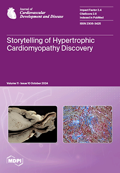
Cover Story (view full-size image): The discovery of hypertrophic cardiomyopathy (HCM) dates back to 1958, when Donald Teare of St. George’s Hospital in London performed autopsies in eight cases with asymmetric hypertrophy of the ventricular septum and bizarre disorganization at histology. In the same year, at the National Institute of Health U.S.A., Eugene Braunwald and Andrew Glenn Morrow put forward the terms “functional” and “hypertrophic idiopathic subaortic stenosis”. In 1980, John F. Goodwin of Westminster Hospital in London postulated the first classification of heart muscle diseases. A familial occurrence was noticed in HCM by Teare and Goodwin, who published their findings in 1960. The same family underwent a molecular genetics investigation by the Seidman team in Boston 30 years later. A missense mutation of the β-cardiac myosin heavy chain was found in chromosome 14. View this paper
  • Issues are regarded as officially published after their release is announced to the table of contents alert mailing list.
  • You may sign up for e-mail alerts to receive table of contents of newly released issues.
  • PDF is the official format for papers published in both, html and pdf forms. To view the papers in pdf format, click on the "PDF Full-text" link, and use the free Adobe Reader to open them.
Order results
Result details
Section
Select all
Export citation of selected articles as:
8 pages, 1135 KiB  
Article
Wearable Activity Trackers and Physical Activity Levels Among Members of the Athens Medical Association in Greece
by Stamatios Lampsas, Georgios Marinos, Dimitrios Lamprinos, Panagiotis Theofilis, George E. Zakynthinos, Ioannis Gialamas, Antonios Lysandrou, Sotirios Pililis, Loukia Pliouta, Georgia Tzioumi, Eleni Anastasopoulou, Vaia Lambadiari, Evangelos Oikonomou and Gerasimos Siasos
J. Cardiovasc. Dev. Dis. 2024, 11(10), 336; https://doi.org/10.3390/jcdd11100336 - 21 Oct 2024
Viewed by 753
Abstract
Introduction: Wearable Activity Trackers (WATs) offer real-time feedback on activity levels. We assessed the impact of WAT usage on physicians’ exercise habits. Methods: Physicians from the Athens Medical Association, Greece (n = 742) responded to a self-administered questionnaire evaluating usage of WAT, demographic [...] Read more.
Introduction: Wearable Activity Trackers (WATs) offer real-time feedback on activity levels. We assessed the impact of WAT usage on physicians’ exercise habits. Methods: Physicians from the Athens Medical Association, Greece (n = 742) responded to a self-administered questionnaire evaluating usage of WAT, demographic characteristics, specialty, and physical exercise habits. WHO guidelines recommend at least 150 min/week of moderate-intensity exercise in all healthy adults. Subjects were divided in Users of WATs (Group A), and Non-Users of WATs (Group B). This is an observational, cross-sectional study. Results: There was no difference in baseline characteristics between the two groups (age, sex, body mass index). WATs were used by 38%. Between Group A and B, there was difference in mean exercise training time (302 ± 304 min vs. 210 ± 268 min, p < 0.001), higher percentage of WHO goal achievement (66.3% vs. 50.7%, p < 0.001), and greater awareness of WHO Guidelines (59.9% vs. 47.4%, p < 0.001). WATs were mostly used by four main specialties, with higher use from Cardiologists: Cardiology (47%), Endocrinology (44%), Surgery (35%) and Internal Medicine (25%), with a p = 0.045. Finally, users of WATs compared to non-users showed higher willingness to reduce body weight (58.5% vs. 48%, p = 0.01), apply dietary restrictions (36.5% vs. 29.6%, p = 0.05), and greater motivation for weekly physical exercise (74.1% vs. 32.4%, p < 0.001); Conclusion: Physicians using WATs demonstrate increased exercise training time, greater awareness of WHO guidelines and a higher propensity to implement dietary restrictions compared to non-users. Variations in WAT usage across medical specialties emphasize the need for targeted interventions to promote physical activity and enhance healthcare professionals’ health. Full article
(This article belongs to the Section Epidemiology, Lifestyle, and Cardiovascular Health)
Show Figures

Figure 1

12 pages, 2960 KiB  
Article
Indirect-Oscillation Sign Suggesting Infective Endocarditis on the Routine Chest CT
by Min Ji Son, Seung Min Yoo, Hwa Yeon Lee and Charles S. White
J. Cardiovasc. Dev. Dis. 2024, 11(10), 335; https://doi.org/10.3390/jcdd11100335 - 21 Oct 2024
Viewed by 524
Abstract
Routine chest CT is not essential for the diagnostic workup for infective endocarditis (IE), but this type of study may be the initial imaging modality in the evaluation of patients ultimately proven to have IE who present to the emergency department with nonspecific [...] Read more.
Routine chest CT is not essential for the diagnostic workup for infective endocarditis (IE), but this type of study may be the initial imaging modality in the evaluation of patients ultimately proven to have IE who present to the emergency department with nonspecific clinical symptoms. Although routine chest CT cannot directly assess valvular oscillating motion due to the lack of cine images, we hypothesized that a combination of elongated nodular valve thickening and abnormal orientation to the normal valve with a blind end on routine CT (indirect-oscillation sign) might suggest movable vegetation indirectly. To evaluate this possibility, we studied 27 patients with IE and 35 controls who underwent both routine chest CT and echocardiography. CT scanning was initiated following a delay of 60–80 s after the administration of the contrast medium. Two cardiothoracic radiologists retrospectively analyzed the CT images to assess the indirect-oscillation sign with consensus. The sensitivity, specificity, positive predictive value, and negative predictive value of the indirect-oscillation sign on routine chest CT were 29.6% (8/27), 100% (35/35), 100% (8/8), and 64.8% (35/54), respectively. Although uncommon, the presence of the indirect-oscillation sign involving the aortic or mitral valve on routine chest CT is a suggestive finding for IE. Full article
Show Figures

Figure 1

12 pages, 2134 KiB  
Article
Left Atrial Low-Voltage Extent Predicts the Recurrence of Supraventricular Arrhythmias
by Yannick Teumer, Luca Gold, Lyuboslav Katov, Carlo Bothner, Wolfgang Rottbauer and Karolina Weinmann-Emhardt
J. Cardiovasc. Dev. Dis. 2024, 11(10), 334; https://doi.org/10.3390/jcdd11100334 - 21 Oct 2024
Viewed by 740
Abstract
The incidence of left atrial (LA) supraventricular arrhythmias is increasing. Even after LA ablation, recurrence of these tachycardias is common. MRI studies show that LA cardiomyopathy is a significant risk factor for recurrence and correlates with low voltage areas detected via 3D electroanatomic [...] Read more.
The incidence of left atrial (LA) supraventricular arrhythmias is increasing. Even after LA ablation, recurrence of these tachycardias is common. MRI studies show that LA cardiomyopathy is a significant risk factor for recurrence and correlates with low voltage areas detected via 3D electroanatomic mapping (EAM). There are limited data on the impact of low voltage extent detected by EAM on recurrence-free survival. Voltage thresholds defining low voltage vary across different studies. This study aims to investigate the impact of the extent of low voltage areas in the LA on recurrence-free survival and to assess whether defining low voltage areas using thresholds of 0.5, 0.4, or 0.3 mV offers better predictive performance. Patients with atrial arrhythmia who underwent LA EAM at Ulm University Heart Center between September 2018 and September 2022 were included from the ATRIUM registry. ROC analysis determined the voltage threshold for predicting recurrence-free survival. Kaplan–Meier and logistic regression models adjusted for patient variables were used to analyze recurrence-free survival. Of 1089 screened patients, 108 met the inclusion criteria. ROC analysis indicated that a 0.4 mV threshold for low voltage provided the best predictive performance. Logistic regression showed a 1.039-fold increase in recurrence risk per percent increase in LA low voltage area (odds ratio = 1.039, 95% CI 1.014–1.064). Low voltage extent in EAM correlates with 1-year recurrence rate after ablation of left atrial supraventricular arrhythmias. The threshold of 0.4 mV is the most suitable for predicting recurrences of those examined. Full article
(This article belongs to the Special Issue Heart Rhythm Disorders: Diagnosis, Treatment, and Management)
Show Figures

Figure 1

15 pages, 3019 KiB  
Systematic Review
The Impact of Frailty and Surgical Risk on Health-Related Quality of Life After TAVI
by Kim E. H. M. van der Velden, Bart P. A. Spaetgens, Wolfgang F. F. A. Buhre, Bart Maesen, Dianne J. D. de Korte-de Boer, Sander M. J. van Kuijk, Arnoud W. J. van ‘t Hof and Jan U. Schreiber
J. Cardiovasc. Dev. Dis. 2024, 11(10), 333; https://doi.org/10.3390/jcdd11100333 - 18 Oct 2024
Viewed by 824
Abstract
Symptomatic aortic stenosis and frailty reduce health-related quality of life (HrQoL). Transcatheter aortic valve implantation (TAVI) in patients at high to extreme risk has been proven to have a beneficial effect on HrQoL. Currently, TAVI is also considered in patients at intermediate risk. [...] Read more.
Symptomatic aortic stenosis and frailty reduce health-related quality of life (HrQoL). Transcatheter aortic valve implantation (TAVI) in patients at high to extreme risk has been proven to have a beneficial effect on HrQoL. Currently, TAVI is also considered in patients at intermediate risk. Our meta-analysis investigates whether benefits to HrQoL after TAVI is more pronounced in frail patients and patients at high to extreme vs. intermediate surgical risk. A systematic search of the literature was performed in November 2021 and updated in November 2023 in PUBMED, EMBASE, and the Cochrane Controlled Trials Register. Statistical analysis was performed according to the inverse variance method and the random effects model. A total of 951 studies were assessed, of which 19 studies were included. Meta-analysis showed a mean increase in the Kansas City Cardiomyopathy Questionnaire (KCCQ) score of 29.6 points (6.0, 33.1) in high to extreme risk patients versus 21.0 (20.9, 21.1) in intermediate risk patients (p < 0.00001) and 24.6 points (21.5, 27.8) in frail patients versus 26.8 (20.2, 33.4) in the general TAVI population (p = 0.55). However, qualitative analyses of non-randomized studies showed the opposite results. In conclusion, TAVI improves HrQoL more in high to extreme than intermediate risk patients. Frailty’s impact on HrQoL post-TAVI is inconclusive due to varying outcomes in RCTs vs. non-RCTs. Full article
(This article belongs to the Special Issue Transcatheter Aortic Valve Implantation (TAVI) II)
Show Figures

Figure 1

12 pages, 953 KiB  
Review
Device-Assisted Left Atrial Appendage Exclusion: From Basic Sciences to Clinical Applications
by Julia Izabela Karpierz, Michał Piotrowski, Krzysztof Bartuś, Radosław Chmiel, Katarzyna Wijatkowska and Artur Słomka
J. Cardiovasc. Dev. Dis. 2024, 11(10), 332; https://doi.org/10.3390/jcdd11100332 - 18 Oct 2024
Viewed by 701
Abstract
Device-assisted left atrial appendage exclusion plays a crucial role in the prevention of fatal ischemic complications in patients with atrial fibrillation and contraindications to anticoagulation treatment. Various devices with different safety profiles and device-related complications are available in daily practice to perform this [...] Read more.
Device-assisted left atrial appendage exclusion plays a crucial role in the prevention of fatal ischemic complications in patients with atrial fibrillation and contraindications to anticoagulation treatment. Various devices with different safety profiles and device-related complications are available in daily practice to perform this procedure. In this review, the anatomy, physiology, and functions of the left atrial appendage were detailed, and all available devices used for epicardial and endocardial exclusion of the left atrial appendage and their clinical outcomes were discussed. Future research should aim to further investigate the long-term effects of left atrial appendage exclusion on body homeostasis, blood coagulation, and cardiac function. Full article
(This article belongs to the Special Issue Feature Review Papers in Stroke and Cerebrovascular Disease)
Show Figures

Figure 1

11 pages, 2397 KiB  
Article
Association of Cardiovascular Risk Factors and Coronary Calcium Burden with Epicardial Adipose Tissue Volume Obtained from PET–CT Imaging in Oncological Patients
by Carmela Nappi, Andrea Ponsiglione, Carlo Vallone, Roberto Lepre, Luigi Basile, Roberta Green, Valeria Cantoni, Ciro Gabriele Mainolfi, Massimo Imbriaco, Mario Petretta and Alberto Cuocolo
J. Cardiovasc. Dev. Dis. 2024, 11(10), 331; https://doi.org/10.3390/jcdd11100331 - 17 Oct 2024
Viewed by 611
Abstract
Whole-body positron emission tomography (PET)–computed tomography (CT) imaging performed for oncological purposes may provide additional parameters such as the coronary artery calcium (CAC) and epicardial adipose tissue (EAT) volume with cost-effective prognostic information in asymptomatic people beyond traditional cardiovascular risk factors. We evaluated [...] Read more.
Whole-body positron emission tomography (PET)–computed tomography (CT) imaging performed for oncological purposes may provide additional parameters such as the coronary artery calcium (CAC) and epicardial adipose tissue (EAT) volume with cost-effective prognostic information in asymptomatic people beyond traditional cardiovascular risk factors. We evaluated the feasibility of measuring the CAC score and EAT volume in cancer patients without known coronary artery disease (CAD) referred to whole-body 18F-FDG PET–CT imaging, regardless of the main clinical problem. We also investigated the potential relationships between traditional cardiovascular risk factors and CAC with EAT volume. A total of 109 oncological patients without overt CAD underwent whole-body PET–CT imaging with 18F-fluorodeoxyglucose (FDG). Unenhanced CT images were retrospectively viewed for CAC and EAT measurements on a dedicated platform. Overall, the mean EAT volume was 99 ± 49 cm3. Patients with a CAC score ≥ 1 were older than those with a CAC = 0 (p < 0.001) and the prevalence of hypertension was higher in patients with detectable CAC as compared to those without (p < 0.005). The EAT volume was higher in patients with CAC than in those without (p < 0.001). For univariable age, body mass index (BMI), hypertension, and CAC were associated with increasing EAT values (all p < 0.005). However, the correlation between the CAC score and EAT volume was weak, and in multivariable analysis only age and BMI were independently associated with increased EAT (both p < 0.001), suggesting that potential prognostic information on CAC and EAT is not redundant. This study demonstrates the feasibility of a cost-effective assessment of CAC scores and EAT volumes in oncological patients undergoing whole-body 18F-FDG PET–CT imaging, enabling staging cancer disease and atherosclerotic burden by a single test already included in the diagnostic work program, with optimization of the radiation dose and without additional costs. Full article
Show Figures

Figure 1

17 pages, 3814 KiB  
Article
A 3D Statistical Shape Model of the Right Ventricular Outflow Tract in Pulmonary Valve Replacement Patients Post-Surgical Repair
by Liam Swanson, Raphaël Sivera, Claudio Capelli, Abdulaziz Alosaimi, Dariusz Mroczek, Christopher Z. Lam, Andrew Cook, Rajiv R. Chaturvedi and Silvia Schievano
J. Cardiovasc. Dev. Dis. 2024, 11(10), 330; https://doi.org/10.3390/jcdd11100330 - 17 Oct 2024
Viewed by 631
Abstract
Assessment of the right ventricular outflow tract and pulmonary arteries (RVOT) for percutaneous pulmonary valve implantation (PPVI) uses discrete measurements (diameters and lengths) from medical images. This multi-centre study identified the 3D RVOT shape features prevalent in patients late after surgical repair of [...] Read more.
Assessment of the right ventricular outflow tract and pulmonary arteries (RVOT) for percutaneous pulmonary valve implantation (PPVI) uses discrete measurements (diameters and lengths) from medical images. This multi-centre study identified the 3D RVOT shape features prevalent in patients late after surgical repair of congenital heart disease (CHD). A 3D RVOT statistical shape model (SSM) was computed from 81 retrospectively selected CHD patients (14.7 ± 6.8 years) who required pulmonary valve replacement late after surgical repair. A principal component analysis identified prevalent shape features (modes) within the population which were compared with standard geometric measurements (diameter, length and surface area) and between sub-groups of diagnosis, RVOT type and dysfunction. Shape mode 1 and 2 represented RVOT size and curvature and tapering and length, respectively. Shape modes 3–5 related to branch pulmonary artery calibre, conical vs. bulbous RVOTs and RVOT curvature, respectively. Tetralogy of Fallot, transannular patch type and regurgitant RVOTs were larger and straighter while conduit and stenotic types were longer and more cylindrical than other subgroups. This SSM analysed the main 3D shape features present in a population of RVOTs, exploiting the wide 3D anatomical information provided by routine imaging. This morphological information may have implications for PPVI patient selection and device design. Full article
(This article belongs to the Section Pediatric Cardiology and Congenital Heart Disease)
Show Figures

Figure 1

13 pages, 2116 KiB  
Article
Gender-Tailored Heart Team Decision Making Equalizes Outcomes for Female Patients after Aortic Valve Replacement through Right Anterior Small Thoracotomy (RAST)
by Isabel Lavanchy, Laina Passos, Thierry Aymard, Jürg Grünenfelder, Maximilian Y. Emmert, Roberto Corti, Oliver Gaemperli, Patric Biaggi and Diana Reser
J. Cardiovasc. Dev. Dis. 2024, 11(10), 329; https://doi.org/10.3390/jcdd11100329 - 16 Oct 2024
Viewed by 928
Abstract
Background: Little is known about gender-dependent outcomes after aortic valve replacement (AVR) through right anterior thoracotomy (RAST). The aim of our study was to analyze the mid-term outcomes of our cohort. Methods: This study is a retrospective analysis of 338 patients (2013–2022). Subgroup [...] Read more.
Background: Little is known about gender-dependent outcomes after aortic valve replacement (AVR) through right anterior thoracotomy (RAST). The aim of our study was to analyze the mid-term outcomes of our cohort. Methods: This study is a retrospective analysis of 338 patients (2013–2022). Subgroup analysis included a gender-dependent comparison of age groups ≤60 and >60 years. Results: Women were older (69.27 ± 7.98 vs. 64.15 ± 11.47, p < 0.001) with higher Euroscore II (1.25 ± 0.73 vs. 0.94 ± 0.45, p < 0.001). Bypass and cross-clamp time were shorter (109.36 ± 30.8 vs. 117.65 ± 33.1 minutes, p = 0.01; 68.26 ± 21.5 vs. 74.36 ± 23.3 minutes, p = 0.01), while ICU, hospital stay and atrial fibrillation were higher (2.48 ± 8.2 vs. 1.35 ± 1.4 days, p = 0.005; 11 ± 7.8 vs. 9.48 ± 2.3 days, p = 0.002; 6.7% vs. 4.4%, p = 0.024). Mortality was 0.9%, while stroke was 0.6%. Age subgroup analysis showed that women were older (p = 0.025) with longer ICU and hospital stays (p < 0.001, p = 0.007). On mid-term follow-up (4.52 ± 2.67 years) of 315 patients (94.3%), there was no significant difference in survival, MACCE and re-intervention comparing gender and age groups. Conclusions: Despite older age, higher Euroscore II, longer ICU and hospital stay in women, mortality, MACCE and reoperation were low and comparable in gender and age groups. We believe that our patient-tailored heart team decision making combined with RAST translates into gender-tailored medicine, which equalizes the widely reported negative outcomes of female patients after cardiac surgery. Full article
(This article belongs to the Special Issue Women and Cardiovascular Disease: The Gender Gap)
Show Figures

Figure 1

17 pages, 647 KiB  
Article
Multivariate Analysis of the Determinants of Total Mortality in the European Union with Focus on Fat Intake, Diabetes, Myocardial Infarction, Life Expectancy, and Preventable Mortality: A Panel Data Fixed-Effects Panel Data Model Approach
by Silviu Marcel Stanciu, Emilia Rusu, Mariana Jinga, Cosmin Gabriel Ursu, Rares Ioan Stanciu, Daniela Miricescu, Valentin Marian Antohi and Elena Barbu
J. Cardiovasc. Dev. Dis. 2024, 11(10), 328; https://doi.org/10.3390/jcdd11100328 - 15 Oct 2024
Viewed by 767
Abstract
Cardiovascular disease is the leading cause of death in the European Union (EU), and while the mortality rates of diabetes, myocardial infarction, and the total fat intake have been extensively studied, we believe that understanding the interaction between such closely correlated determinants is [...] Read more.
Cardiovascular disease is the leading cause of death in the European Union (EU), and while the mortality rates of diabetes, myocardial infarction, and the total fat intake have been extensively studied, we believe that understanding the interaction between such closely correlated determinants is crucial to the development of effective health policies in the EU. Our paper’s novelty is represented by the econometric modelling, and its ability to capture both temporal and unit variations. The research methodology consists of using a panel data model with fixed effects for the 27 EU member states over the period 2010–2021. The results of the study show that the standardized mortality rate for deaths preventable by prevention and treatment and diabetes-related mortality are significant predictors of total mortality in the EU. The standardized mortality rate for deaths preventable by prevention and treatment had a significant positive impact, suggesting that improved preventive and therapeutic interventions can significantly reduce total mortality. Diabetes-associated mortality also showed a strong positive correlation with total mortality, emphasizing the need for effective diabetes management and prevention strategies. These results are useful for the formulation of public health strategies aimed at improving life expectancy and reducing the burden of chronic diseases. Full article
(This article belongs to the Special Issue Clinical Burden of Comorbidities on Cardiovascular System and Beyond)
Show Figures

Figure 1

13 pages, 1154 KiB  
Article
Body Weight’s Role in Infective Endocarditis Surgery
by Ahmed Elderia, Gerold Woll, Anna-Maria Wallau, Walid Bennour, Stephen Gerfer, Ilija Djordjevic, Thorsten Wahlers and Carolyn Weber
J. Cardiovasc. Dev. Dis. 2024, 11(10), 327; https://doi.org/10.3390/jcdd11100327 - 15 Oct 2024
Viewed by 559
Abstract
Objective: to investigate how body mass index (BMI) affects the outcome in patients treated surgically for infective endocarditis (IE). Methods: This is a single-center observational analysis of consecutive patients treated surgically for IE. We divided the cohort into six groups, according to the [...] Read more.
Objective: to investigate how body mass index (BMI) affects the outcome in patients treated surgically for infective endocarditis (IE). Methods: This is a single-center observational analysis of consecutive patients treated surgically for IE. We divided the cohort into six groups, according to the WHO classification of BMI, and performed subsequent outcome analysis. Results: The patient population consisted of 17 (2.6%) underweight, 249 (38.3%) normal weight, 252 (38.8%) overweight, 83 (12.8%) class I obese, 28 (4.3%) class II obese, and 21 (3.8%) class III, or morbidly obese, patients. The median age of the entire cohort was 64.5 [52.5–73.6] years. While only 168 (25.9%) patients were female, women significantly more often exhibited extremes in regards to BMI, including underweight (47.1%) and morbid obesity (52.4%), p = 0.026. Class II and III obese patients displayed more postoperative acute kidney injury (47.9%), p = 0.003, more sternal wound infection (12.9%), p < 0.001, worse 30-day survival (20.4%), p = 0.031, and worse long-term survival, p = 0.026, compared to the results for the other groups. However, the multivariable analysis did not identify obesity as an independent risk factor for 30-day mortality, with an odds ratio of 1.257 [0.613–2.579], p = 0.533. Rather, age > 60, reduced LVEF < 30%, staphylococcal infection, and prosthetic valve endocarditis correlated with mortality. While BMI showed poor discrimination in predicting 30-day mortality on the ROC curve (AUC = 0.609), it showed a fair degree of discrimination in predicting sternal wound infection (AUC = 0.723). Conclusions: Obesity was associated with increased comorbidities, complications, and higher postoperative mortality in IE patients, but it is not an independent mortality risk factor. While BMI is a poor predictor of death, it is a good predictor of sternal wound infections. Full article
Show Figures

Graphical abstract

9 pages, 1120 KiB  
Article
Effect of Aerobic Exercise with Blood Flow Restriction on Postexercise Hypotension in Young Adults: The Role of Histamine Receptors
by Dongnyeuck Seo, Dae Sik Song, William Boyer, Trevor Gillum, Sean Sullivan, Nailiyah Liwanag, Iltark Yoon and Jong-Kyung Kim
J. Cardiovasc. Dev. Dis. 2024, 11(10), 326; https://doi.org/10.3390/jcdd11100326 - 15 Oct 2024
Viewed by 791
Abstract
We tested hypothesis that aerobic exercise with blood flow restriction (BFR) induced postexercise hypotension (PEH), and the reduction in blood pressure (BP) was due to peripheral vasodilation via the histamine receptors. Ten male subjects participated in this study. The subjects were randomly assigned [...] Read more.
We tested hypothesis that aerobic exercise with blood flow restriction (BFR) induced postexercise hypotension (PEH), and the reduction in blood pressure (BP) was due to peripheral vasodilation via the histamine receptors. Ten male subjects participated in this study. The subjects were randomly assigned to walk for 10 min at 6.4 km/h, 0% grade with or without BFR after taking histamine receptor blockade. Following exercise, BP was measured at 10 min interval for 60 min. Heart rate (HR), stroke volume (SV), cardiac output (CO), mean arterial pressure (MAP), and total peripheral resistance (TPR) were evaluated. Our results indicated that MAP was significantly lowered immediately after exercise at 20 min, 30 min, and 40 min before the blockade as opposed to after the blockade. A significant reduction in diastolic BP (DBP) occurred. There were no significant differences in HR, SV, CO, and TPR between before the blockade and after the blockade. MAP was substantially decreased at 20 min, 30 min, and 40 min before the blockade compared to resting (−3.2 ± 2.2, −3.3 ± 2.8, and −2.9 ± 2.5, respectively) while increasing MAP after the blockade. The current study demonstrated that low-intensity aerobic exercise with BFR lowered MAP via histamine receptor-induced peripheral vasodilation. In conclusion, BFR exercise training using short periods and low intensity would be greatly beneficial as a potential treatment to lower BP. Full article
Show Figures

Figure 1

15 pages, 2794 KiB  
Article
Coronary Computed Tomography Angiography (CTA) Findings in COVID-19
by Pietro G. Lacaita, Anna Luger, Fabian Plank, Fabian Barbieri, Christoph Beyer, Theresa Thurner, Yannick Scharll, Johannes Deeg, Gerlig Widmann and Gudrun M. Feuchtner
J. Cardiovasc. Dev. Dis. 2024, 11(10), 325; https://doi.org/10.3390/jcdd11100325 - 14 Oct 2024
Viewed by 1005
Abstract
(1) Background: The novel SARS-CoV-2 virus infects the endothelium. Vasculitis may lead to specific coronary artery wall lesions. Coronary computed tomography angiography (CTA) imaging findings have not been systematically reported. The aim of this study was to describe a case series using CTA. [...] Read more.
(1) Background: The novel SARS-CoV-2 virus infects the endothelium. Vasculitis may lead to specific coronary artery wall lesions. Coronary computed tomography angiography (CTA) imaging findings have not been systematically reported. The aim of this study was to describe a case series using CTA. (2) Methods: Patients with recent RT-PCR confirmed SARS-CoV-2 infection referred for coronary CTA for clinical indications (e.g., chest pain, troponin+, and ECG abnormalities) were included. Coronary CTA findings, such as atypical coronary lesions suggestive of vasculitis, perivascular inflammation measured by using pericoronary fat attenuation (PCAT) index, coronary artery disease, and extracoronary findings were collected. (3) Results: Results for 12 patients (54.8 ± 22 years; four females) with SARS-CoV-2 infection within 60 days (four acute care and eight stable patients) are reported. Time to positive RT-PCR was a mean of 15.1 days (range, 0–51). In four acute patients with signs of myocardial injury, plaque rupture (n = 1), hyperenhancing myocardium/MINOCA (n = 1), MINOCA (n = 1), and pericarditis with acute heart failure (LVEF 20%) (n = 1) were found. All (100%) had pericardial effusion and signs of perivascular inflammation. Among eight stable patients, pericardial effusion or perivascular inflammation were found in only two (25%). Coronary artery disease was ruled out in five (62.5%) (4) Conclusions: Coronary CTA is a useful imaging modality in the diagnostic work up of patients with COVID-19 infection, and is able to describe coronary and other cardiac abnormalities. Full article
Show Figures

Figure 1

15 pages, 5473 KiB  
Review
Electrocardiographic Clues for Early Diagnosis of Ventricular Pre-Excitation and Non-Invasive Risk Stratification in Athletes: A Practical Guide for Sports Cardiologists
by Simone Ungaro, Francesca Graziano, Sergei Bondarev, Matteo Pizzolato, Domenico Corrado and Alessandro Zorzi
J. Cardiovasc. Dev. Dis. 2024, 11(10), 324; https://doi.org/10.3390/jcdd11100324 - 14 Oct 2024
Viewed by 856
Abstract
Ventricular pre-excitation (VP) is a cardiac disorder characterized by the presence of an accessory pathway (AP) that bypasses the atrioventricular node (AVN), which, although often asymptomatic, exposes individuals to an increased risk of re-entrant supraventricular tachycardias and sudden cardiac death (SCD) due to [...] Read more.
Ventricular pre-excitation (VP) is a cardiac disorder characterized by the presence of an accessory pathway (AP) that bypasses the atrioventricular node (AVN), which, although often asymptomatic, exposes individuals to an increased risk of re-entrant supraventricular tachycardias and sudden cardiac death (SCD) due to rapid atrial fibrillation (AF) conduction. This condition is particularly significant in sports cardiology, where preparticipation ECG screening is routinely performed on athletes. Professional athletes, given their elevated risk of developing malignant arrhythmias, require careful assessment. Early identification of VP and proper risk stratification are crucial for determining the most appropriate management strategy and ensuring the safety of these individuals during competitive sports. Non-invasive tools, such as resting electrocardiograms (ECGs), ambulatory ECG monitoring, and exercise stress tests, are commonly employed, although their interpretation can sometimes be challenging. This review aims to provide practical tips and electrocardiographic clues for detecting VP beyond the classical triad (short PR interval, delta wave, and prolonged QRS interval) and offers guidance on non-invasive risk stratification. Although the diagnostic gold standard remains invasive electrophysiological study, appropriate interpretation of the ECG can help limit unnecessary referrals for young, often asymptomatic, athletes. Full article
(This article belongs to the Special Issue The Present and Future of Sports Cardiology and Exercise)
Show Figures

Figure 1

12 pages, 4567 KiB  
Review
Relevance of Anatomical Significance of AV Nodal Structures within Koch’s Triangle and Pyramid
by Andrea Matteucci, Claudio Pandozi, Maurizio Russo, Marco Galeazzi, Giammarco Schiaffini, Marco Valerio Mariani, Carlo Lavalle and Furio Colivicchi
J. Cardiovasc. Dev. Dis. 2024, 11(10), 323; https://doi.org/10.3390/jcdd11100323 - 14 Oct 2024
Viewed by 762
Abstract
The exploration of the cardiac conduction system evolved over a century, marked by groundbreaking discoveries in atrioventricular (AV) nodal physiology. Atrioventricular nodal re-entrant tachycardia (AVNRT), the most prevalent regular tachycardia in humans, remains enigmatic despite extensive research. Detailed examinations of AV nodal anatomy [...] Read more.
The exploration of the cardiac conduction system evolved over a century, marked by groundbreaking discoveries in atrioventricular (AV) nodal physiology. Atrioventricular nodal re-entrant tachycardia (AVNRT), the most prevalent regular tachycardia in humans, remains enigmatic despite extensive research. Detailed examinations of AV nodal anatomy and histology reveal variations in location and shape, influencing electrophysiological properties. Variability in AV nodal extensions and their embryological origins contribute to the complexity of the conduction system. Physiologically, the AV node plays a crucial role in modulating AV conduction, introducing delays for ventricular filling and filtering atrial impulses. Dual-pathway physiology involving fast and slow pathways further complicates AVNRT circuitry. Integrated approaches combining pre-procedural imaging with electroanatomical mapping enhance our understanding of AV nodal structures and high-definition mapping improves precision in identifying ablation targets. Electrophysiological–anatomical correlations may unveil the specific roles of conduction axis components, aiding in the optimization of ablation strategies. This review traces the historical journey from Tawara’s pioneering work to recent integrated approaches aimed at unraveling the intricacies of AV nodal structures while emphasizing the importance of a multidimensional approach, incorporating technological advancements, anatomical understanding, and clinical validation in human mapping studies. Full article
(This article belongs to the Section Electrophysiology and Cardiovascular Physiology)
Show Figures

Figure 1

17 pages, 8240 KiB  
Article
Effect of Spatial Resolution on Accurate Detection and Localization of Arrhythmia Rotors in Human Right Ventricular Tachycardia
by Maria Inês F. Gândara, Igor R. Efimov and Kedar K. Aras
J. Cardiovasc. Dev. Dis. 2024, 11(10), 322; https://doi.org/10.3390/jcdd11100322 - 12 Oct 2024
Viewed by 526
Abstract
The goal of this study was to identify the spatial resolution requirements for accurate rotor detection and localization in human right ventricular tachyarrhythmias. Poor spatial resolution is often cited as a reason for the inaccuracy of cardiac mapping catheters in detecting and localizing [...] Read more.
The goal of this study was to identify the spatial resolution requirements for accurate rotor detection and localization in human right ventricular tachyarrhythmias. Poor spatial resolution is often cited as a reason for the inaccuracy of cardiac mapping catheters in detecting and localizing arrhythmia rotors. High-resolution (0.7 mm) arrhythmia data from optical recordings obtained from human donor hearts (n = 12) were uniformly downsampled to lower resolutions (1.4–7 mm) to approximate the spatial resolution (4 mm) of clinical mapping catheters. Rotors were tracked at various subresolutions and compared to the rotors in the original data by computing F1-scores to create accuracy profiles for both rotor detection and localization. Further comparisons were made according to arrhythmia type, donor sex, anatomical region, and mapped surface: endocardium or epicardium. For a spatial resolution of 4.2 mm, the accuracies of rotor detection and localization were 57% ± 4% and 61% ± 7%, respectively. Arrhythmia type affected the accuracy of rotor detection (monomorphic ventricular tachycardia, 58% ± 4%; ventricular fibrillation, 56% ± 8%) and localization (monomorphic ventricular tachycardia, 70% ± 4%; ventricular fibrillation, 54% ± 13%). However, donor sex, anatomical region (right ventricular outflow tract, mid, and apical), and mapped surface (epicardium and endocardium) did not significantly affect rotor detection or localization accuracy. To achieve rotor detection accuracy of 80%, a spatial resolution of 1.4 mm or better is needed. The accuracy profiles provided here serve as a guideline for future mapping device development. Full article
(This article belongs to the Section Electrophysiology and Cardiovascular Physiology)
Show Figures

Figure 1

14 pages, 1582 KiB  
Article
The Influence of Age and Exercise Training Status on Left Ventricular Systolic Twist Mechanics in Healthy Males—An Exploratory Study
by Alexander J. Beaumont, Amy K. Campbell, Viswanath B. Unnithan, David Oxborough, Fergal Grace, Allan Knox and Nicholas F. Sculthorpe
J. Cardiovasc. Dev. Dis. 2024, 11(10), 321; https://doi.org/10.3390/jcdd11100321 - 12 Oct 2024
Viewed by 530
Abstract
Age-related differences in twist may be mitigated with exercise training, although this remains inconclusive. Moreover, temporal left ventricular (LV) systolic twist mechanics, including early-systolic (twistearly), and beyond peak twist (twistpeak) alone, have not been considered. Therefore, further insights are [...] Read more.
Age-related differences in twist may be mitigated with exercise training, although this remains inconclusive. Moreover, temporal left ventricular (LV) systolic twist mechanics, including early-systolic (twistearly), and beyond peak twist (twistpeak) alone, have not been considered. Therefore, further insights are required to ascertain the influence of age and training status on twist mechanics across systole. Forty males were included and allocated into 1 of 4 groups based on age and training status: young recreationally active (YRA, n = 9; 28 ± 5 years), old recreationally active (ORA, n = 10; 68 ± 6 years), young trained (YT, n = 10; 27 ± 6 years), and old trained (OT, n = 11, 64 ± 4 years) groups. Two-dimensional speckle-tracking echocardiography was performed to determine LV twist mechanics, including twistearly, twistpeak, and total twist (twisttotal), by considering the nadir on the twist time-curve during early systole. Twisttotal was calculated by subtracting twistearly from their peak values. LV twistpeak was higher in older than younger men (p = 0.036), while twistpeak was lower in the trained than recreationally-active (p = 0.004). Twistpeak is underestimated compared with twisttotal (p < 0.001), and when early-systolic mechanics were considered, to calculate twisttotal, the age effect (p = 0.186) was dampened. LV twist was higher in older than younger age, with lower twist in exercise-trained than recreationally-active males. Twistpeak is underestimated when twistearly is not considered, with novel observations demonstrating that the age effect was dampened when considering twistearly. These findings elucidated a smaller age effect when early phases of systole are considered, while lower LV systolic mechanics were observed in older aged trained than recreationally-active males. Full article
(This article belongs to the Section Electrophysiology and Cardiovascular Physiology)
Show Figures

Figure 1

21 pages, 2983 KiB  
Article
FGFR4 Is Required for Concentric Growth of Cardiac Myocytes during Physiologic Cardiac Hypertrophy
by Isaac Campos, Beatrice Richter, Sarah Madison Thomas, Brian Czaya, Christopher Yanucil, Dominik Kentrup, Abul Fajol, Qing Li, Stephen M. Secor and Christian Faul
J. Cardiovasc. Dev. Dis. 2024, 11(10), 320; https://doi.org/10.3390/jcdd11100320 - 12 Oct 2024
Viewed by 692
Abstract
Fibroblast growth factor (FGF) 23 is a bone-derived hormone that promotes renal phosphate excretion. Serum FGF23 is increased in chronic kidney disease (CKD) and contributes to pathologic cardiac hypertrophy by activating FGF receptor (FGFR) 4 on cardiac myocytes, which might lead to the [...] Read more.
Fibroblast growth factor (FGF) 23 is a bone-derived hormone that promotes renal phosphate excretion. Serum FGF23 is increased in chronic kidney disease (CKD) and contributes to pathologic cardiac hypertrophy by activating FGF receptor (FGFR) 4 on cardiac myocytes, which might lead to the high cardiovascular mortality in CKD patients. Increases in serum FGF23 levels have also been observed following endurance exercise and in pregnancy, which are scenarios of physiologic cardiac hypertrophy as an adaptive response of the heart to increased demand. To determine whether FGF23/FGFR4 contributes to physiologic cardiac hypertrophy, we studied FGFR4 knockout mice (FGFR4−/−) during late pregnancy. In comparison to virgin littermates, pregnant wild-type and FGFR4−/− mice showed increases in serum FGF23 levels and heart weight; however, the elevation in myocyte area observed in pregnant wild-type mice was abrogated in pregnant FGFR4−/− mice. This outcome was supported by treatments of cultured cardiac myocytes with serum from fed Burmese pythons, another model of physiologic hypertrophy, where the co-treatment with an FGFR4-specific inhibitor abrogated the serum-induced increase in cell area. Interestingly, we found that in pregnant mice, the heart, and not the bone, shows elevated FGF23 expression, and that increases in serum FGF23 are not accompanied by changes in phosphate metabolism. Our study suggests that in physiologic cardiac hypertrophy, the heart produces FGF23 that contributes to hypertrophic growth of cardiac myocytes in a paracrine and FGFR4-dependent manner, and that the kidney does not respond to heart-derived FGF23. Full article
(This article belongs to the Special Issue Hypertrophic Cardiomyopathy: Pathogenesis, Diagnosis and Management)
Show Figures

Graphical abstract

13 pages, 1797 KiB  
Article
Prognostic Value of Coronary Artery Calcification in Patients with COVID-19 and Interstitial Pneumonia: A Case-Control Study
by Gianni Dall’Ara, Sara Piciucchi, Roberto Carletti, Antonio Vizzuso, Elisa Gardini, Maria De Vita, Chiara Dallaserra, Federica Campacci, Giovanna Di Giannuario, Daniele Grosseto, Giovanni Rinaldi, Sabine Vecchio, Federica Mantero, Lorenzo Mellini, Alessandra Albini, Emanuela Giampalma, Venerino Poletti and Marcello Galvani
J. Cardiovasc. Dev. Dis. 2024, 11(10), 319; https://doi.org/10.3390/jcdd11100319 - 11 Oct 2024
Viewed by 1242
Abstract
Background: Patients suffering from coronavirus disease-19 (COVID-19)-related interstitial pneumonia have variable outcomes, and the risk factors for a more severe course have yet to be comprehensively identified. Cohort studies have suggested that coronary artery calcium (CAC), as estimated at chest computed tomography (CT) [...] Read more.
Background: Patients suffering from coronavirus disease-19 (COVID-19)-related interstitial pneumonia have variable outcomes, and the risk factors for a more severe course have yet to be comprehensively identified. Cohort studies have suggested that coronary artery calcium (CAC), as estimated at chest computed tomography (CT) scan, correlated with patient outcomes. However, given that the prevalence of CAC is gender- and age-dependent, the influence of baseline confounders cannot be completely excluded. Methods: We designed a retrospective, multicenter case-control study including patients with COVID-19, with severe course cases selected based on death within 30 days or requiring invasive ventilation, whereas controls were age- and sex-matched patients surviving up to 30 days without invasive ventilation. The primary outcome was the analysis of moderate-to-severe CAC prevalence between cases and controls. Results: A total of 65 cases and 130 controls were included in the study. Cases had a significantly higher median pulmonary severity score at chest CT scan compared to controls (10 vs. 8, respectively; p = 0.0001), as well as a higher CAC score (5 vs. 2; p = 0.009). The prevalence of moderate-to-severe CAC in cases was significantly greater (41.5% vs. 23.8%; p = 0.013), a difference mainly driven by a higher prevalence in those who died within 30 days (p = 0.000), rather than those requiring invasive ventilation (p = 0.847). White blood cell count, moderate-to-severe CAC, the need for antibiotic therapy, and severe pneumonia at CT scan were independent primary endpoint predictors. Conclusions: This case-control study demonstrated that the CAC burden was higher in COVID-19 patients who did not survive 30 days or who required mechanical ventilation, and CAC played an independent prognostic role. Full article
(This article belongs to the Section Cardiovascular Clinical Research)
Show Figures

Figure 1

11 pages, 1448 KiB  
Review
High-Sensitivity Troponin: Finding a Meaningful Delta
by Catherine X. Wright, Donald S. Wright, Jiun-Ruey Hu and Cesia Gallegos
J. Cardiovasc. Dev. Dis. 2024, 11(10), 318; https://doi.org/10.3390/jcdd11100318 - 11 Oct 2024
Viewed by 1091
Abstract
High-sensitivity cardiac troponin (hs-cTn) assays have significantly refined the resolution of biomarker-level detection and have emerged as the gold standard cardiac biomarker in evaluating myocardial injury. Since its introduction, hs-cTn has been integrated into the Fourth Universal Definition of Myocardial Infarction and various [...] Read more.
High-sensitivity cardiac troponin (hs-cTn) assays have significantly refined the resolution of biomarker-level detection and have emerged as the gold standard cardiac biomarker in evaluating myocardial injury. Since its introduction, hs-cTn has been integrated into the Fourth Universal Definition of Myocardial Infarction and various European Society of Cardiology (ESC) and American College of Cardiology/American Heart Association (ACC/AHA) guidelines for the evaluation and diagnosis of chest pain syndromes. However, despite its integral role in caring for patients with chest pain, there are still substantive gaps in our knowledge of the clinical interpretation of dynamic changes in hs-cTn values. Whether a relative or absolute hs-cTn delta should be used to detect acute myocardial injury remains debatable. There are also emerging considerations of possible sex and racial/ethnic differences in clinically significant troponin deltas. In the emergency department, there is debate about the optimal time frame to recheck hs-cTn after symptom onset for myocardial infarction rule-out and whether hs-cTn deltas should be integrated into clinical risk scores. In this review, we will provide an overview of the history of clinical utilization of cardiac biomarkers, the development of hs-cTn assays, and the ongoing search for a meaningful delta that can be clinically applicable. Full article
(This article belongs to the Section Basic and Translational Cardiovascular Research)
Show Figures

Figure 1

37 pages, 4376 KiB  
Review
Bicuspid Aortic Valve in Children and Young Adults for Cardiologists and Cardiac Surgeons: State-of-the-Art of Literature Review
by Francesco Nappi, Sanjeet Singh Avtaar Singh and Paolo M. de Siena
J. Cardiovasc. Dev. Dis. 2024, 11(10), 317; https://doi.org/10.3390/jcdd11100317 - 11 Oct 2024
Cited by 1 | Viewed by 1403
Abstract
Bicuspid aortic valve disease is the most prevalent congenital heart disease, affecting up to 2% of the general population. The presentation of symptoms may vary based on the patient’s anatomy of fusion, with transthoracic echocardiography being the primary diagnostic tool. Bicuspid aortic valves [...] Read more.
Bicuspid aortic valve disease is the most prevalent congenital heart disease, affecting up to 2% of the general population. The presentation of symptoms may vary based on the patient’s anatomy of fusion, with transthoracic echocardiography being the primary diagnostic tool. Bicuspid aortic valves may also appear with concomitant aortopathy, featuring fundamental structural changes which can lead to valve dysfunction and/or aortic dilatation over time. This article seeks to give a comprehensive overview of the presentation, treatment possibilities and long-term effects of this condition. The databases MEDLINE, Embase, and the Cochrane Library were searched using the terms “endocarditis” or “bicuspid aortic valve” in combination with “epidemiology”, “pathogenesis”, “manifestations”, “imaging”, “treatment”, or “surgery” to retrieve relevant articles. We have identified two types of bicuspid aortic valve disease: aortic stenosis and aortic regurgitation. Valve replacement or repair is often necessary. Patients need to be informed about the benefits and drawbacks of different valve substitutes, particularly with regard to life-long anticoagulation and female patients of childbearing age. Depending on the expertise of the surgeon and institution, the Ross procedure may be a viable alternative. Management of these patients should take into account the likelihood of somatic growth, risk of re-intervention, and anticoagulation risks that are specific to the patient, alongside the expertise of the surgeon or centre. Further research is required on the secondary prevention of patients with bicuspid aortic valve (BAV), such as lifestyle advice and antibiotics to prevent infections, as the guidelines are unclear and lack strong evidence. Full article
Show Figures

Figure 1

10 pages, 1091 KiB  
Article
Lipid Accumulation Product Is Predictive of Cardiovascular Hospitalizations among Patients with Stable Ischemic Heart Disease: Long-Term Follow-Up of the LAERTES Study
by Konstantinos A. Papathanasiou, Christos Eleftherios Roussos, Stylianos Armylagos, Stylianos L. Rallidis and Loukianos S. Rallidis
J. Cardiovasc. Dev. Dis. 2024, 11(10), 316; https://doi.org/10.3390/jcdd11100316 - 10 Oct 2024
Viewed by 642
Abstract
(1) Background: Lipid accumulation product (LAP) is an anthropometric index of abdominal adiposity that has been associated with increased cardiovascular risk. We aimed to explore the association of LAP with cardiovascular hospitalizations and compare its predictive accuracy with other indices such as body [...] Read more.
(1) Background: Lipid accumulation product (LAP) is an anthropometric index of abdominal adiposity that has been associated with increased cardiovascular risk. We aimed to explore the association of LAP with cardiovascular hospitalizations and compare its predictive accuracy with other indices such as body mass index (BMI) and waist circumference. (2) Methods: LAERTES was a prospective, population-based cohort that recruited consecutive patients with stable ischemic heart disease (SIHD) from two Greek hospitals in Athens. Data from 770 participants (13% women, median age 62 years) with a median follow-up of 4.3 years were analyzed in relation to the occurrence of adverse cardiovascular events mandating hospital admission (non-fatal myocardial infarction [MI], non-fatal ischemic stroke and malignant ventricular arrhythmias). (3) Results: A total of 127 (16.5%) of the participants were admitted to cardiology clinics over the follow-up period; 12.4% of them developed MI, 2.6% ventricular arrhythmia and 1.5% ischemic stroke. Patients with cardiovascular hospitalization had higher BMI, larger waist circumference, higher LAP and triglycerides and lower HDL-cholesterol than patients without hospitalization. Upper LAP quartile and hypertension were independent predictors for cardiovascular hospitalization (HR: 2.20, 95% CI: 1.12–4.34, p = 0.02 and HR: 1.57, 95% CI: 1.03–2.39, p = 0.03, respectively). (4) Conclusions: Higher LAP quartiles are predictive of adverse cardiovascular events leading to hospital admission and deserve further evaluation in dedicated studies. Full article
Show Figures

Graphical abstract

13 pages, 1525 KiB  
Review
Atrial Fibrillation in Elite Athletes: A Comprehensive Review of the Literature
by Christos Kourek, Alexandros Briasoulis, Elias Tsougos and Ioannis Paraskevaidis
J. Cardiovasc. Dev. Dis. 2024, 11(10), 315; https://doi.org/10.3390/jcdd11100315 - 9 Oct 2024
Viewed by 1092
Abstract
Although the benefits of exercise training have been shown repeatedly in many studies, its relationship with the occurrence of atrial fibrillation (AF) in competitive athletes still remains controversial. In the present review, we sought to demonstrate a comprehensive report of the incidence, pathophysiology, [...] Read more.
Although the benefits of exercise training have been shown repeatedly in many studies, its relationship with the occurrence of atrial fibrillation (AF) in competitive athletes still remains controversial. In the present review, we sought to demonstrate a comprehensive report of the incidence, pathophysiology, and therapeutic approaches to AF in elite athletes. A 2 to 10 times higher frequency of AF has been shown in many studies in high-intensity endurance athletes compared to individuals who do not exercise. Moreover, a U-shaped relationship between male elite athletes and AF is demonstrated through this finding, while the type and the years of physical activity seem to relate to AF development. A strong correlation seems to exist among the type of exercise (endurance sports), age (>55 years), gender (males), and the time of exercise training, all contributing to an increased risk of AF. The pathophysiology of AF still remains unclear; however, several theories suggest that complex mechanisms are involved, such as bi-atrial dilatation, pulmonary vein stretching, cardiac inflammation, fibrosis, and increased vagal tone. Elite athletes with AF require a comprehensive clinical evaluation and risk factor optimization, similar to the approach taken for nonathletes. Although anticoagulation and rate or rhythm control are cornerstones of AF management, there are still no specific guidelines for elite athletes. Full article
Show Figures

Figure 1

2 pages, 388 KiB  
Correction
Correction: Balleza Alejandri et al. Empagliflozin and Dapagliflozin Improve Endothelial Function in Mexican Patients with Type 2 Diabetes Mellitus: A Double-Blind Clinical Trial. J. Cardiovasc. Dev. Dis. 2024, 11, 182
by Luis Ricardo Balleza Alejandri, Fernando Grover Páez, Erick González Campos, Carlos G. Ramos Becerra, Ernesto Germán Cardona Muñóz, Sara Pascoe González, María Guadalupe Ramos Zavala, Africa Samantha Reynoso Roa, Daniel Osmar Suárez Rico, Alberto Beltrán Ramírez, Jesús Jonathan García Galindo, David Cardona Müller and Claudia Yanette Galán Ruíz
J. Cardiovasc. Dev. Dis. 2024, 11(10), 314; https://doi.org/10.3390/jcdd11100314 - 9 Oct 2024
Viewed by 399
Abstract
In the original publication [...] Full article
16 pages, 264 KiB  
Article
Safety of Magnetic Resonance Imaging in Patients with Cardiac Implantable Electronic Devices
by Hugo Lanz, Katharina Strauß, Julia Höpler, Marie Kraft, Sabine Hoffmann, Leonhard Binzenhöfer, Nils Gade, Daniel Roden, Inas Saleh, Stefan Kääb, Korbinian Lackermair, Sebastian Sadoni, Christian Hagl, Steffen Massberg, Heidi Estner, Stephanie Fichtner and Enzo Lüsebrink
J. Cardiovasc. Dev. Dis. 2024, 11(10), 313; https://doi.org/10.3390/jcdd11100313 - 8 Oct 2024
Viewed by 1198
Abstract
Background: MRI (magnetic resonance imaging) represents the diagnostic image modality of choice in several conditions. With an increasing number of patients requiring MRI for diagnostic purposes, the issue of safety in patients with cardiac implantable electronic devices (CIED) undergoing this imaging modality will [...] Read more.
Background: MRI (magnetic resonance imaging) represents the diagnostic image modality of choice in several conditions. With an increasing number of patients requiring MRI for diagnostic purposes, the issue of safety in patients with cardiac implantable electronic devices (CIED) undergoing this imaging modality will play an ever more important role. The purpose of this study was to assess the safety and device function following MRI in an unrestricted real-world cohort of patients with a wide array of cardiac devices. Methods: We conducted a retrospective single-center study including 1010 MRI studies conducted in adult patients (≥18 years) with an implanted CIED treated in the University Hospital of Munich (LMU) between July 2012 and March 2024. Patients with non-MR conditionally labeled leads, abandoned or epicardial leads, as well as lead fragments, were included for analysis. Results: Across a total of 1010 MRIs (920 total MR-conditional device generators) performed in patients with an implanted CIED, there were no deaths, reports of discomfort, palpitations, heating, or ventricular arrythmias in the 24 h following MRI. Only 2/1010 MRIs were followed by a reported atrial arrhythmia within 24 h, both in patients with an MR-conditional pacemaker (PM) device without an abandoned lead. No significant changes in device function following MRI from baseline were observed across all included CIEDs. Lastly, no instances of severe malfunction, such as generator failure, loss of capture, electrical reset, or inappropriate inhibition of pacing, were found in post-MRI interrogation reports across all MRI studies. Conclusions: Based on the analysis of 1010 MRIs undergone by patients with CIEDs, following standardized device interrogation, manufacturer-advised device programming, monitoring of vital function, and manufacturer-advised reprogramming, MRI can be performed safely and without adverse events or changes in device function. Full article
(This article belongs to the Section Electrophysiology and Cardiovascular Physiology)
Show Figures

Graphical abstract

18 pages, 6875 KiB  
Article
Bicuspid Valve Aortopathy: Is It Reasonable to Define a Different Surgical Cutoff Based on Different Aortic Wall Mechanical Properties Compared to Those of the Tricuspid Valve?
by Pasquale Totaro, Alessandro Caimi, Giulia Formenton, Martina Musto, Martina Schembri, Simone Morganti, Stefano Pelenghi and Ferdinando Auricchio
J. Cardiovasc. Dev. Dis. 2024, 11(10), 312; https://doi.org/10.3390/jcdd11100312 - 8 Oct 2024
Viewed by 624
Abstract
Background: In this study, we examined and compared ex vivo mechanical properties of aortic walls in patients with bicuspid (BAV) and tricuspid (TAV) aortic valve aortopathy to investigate if the anatomical peculiarities in the BAV group are related to an increased frailty of [...] Read more.
Background: In this study, we examined and compared ex vivo mechanical properties of aortic walls in patients with bicuspid (BAV) and tricuspid (TAV) aortic valve aortopathy to investigate if the anatomical peculiarities in the BAV group are related to an increased frailty of the aortic wall and, therefore, if a different surgical cutoff point for ascending aortic replacement could be reasonable in such patients. Methods: Ultimate stress tests were performed on fresh aortic wall specimens harvested during elective aortic surgery in BAV (n. 33) and TAV (n. 77) patients. Three mechanical parameters were evaluated at the failure point, under both longitudinal and circumferential forces: the peak strain (Pstr), peak stress (PS), and maximum elastic modulus (EM). The relationships between the three mechanical parameters and preoperative characteristics were evaluated, with a special focus on evaluating potential risk factors for severely impaired mechanical properties, cumulatively and comparatively (BAV vs. TAV groups). Results: The patient populations were inhomogeneous, as BAV patients reached surgical indication, according to the maximum aortic dilatation, at a younger age (58 ± 15 vs. 64 ± 13; p = 0.0294). The extent of the maximum aortic dilatation was, conversely, similar in the two groups (52 ± 4 vs. 54 ± 7; p = 0.2331), as well as the incidences of different phenotypes of aortic dilatation (with the ascending aorta phenotype being the most frequent in 81% and 66% of the BAV and TAV patients, respectively (p = 0.1134). Cumulatively, the mechanical properties of the aortic wall were influenced mainly by the orientation of the force applied, as both PS and EM were impaired under longitudinal stress. An age of >66 and a maximum dilatation of >52 mm were shown to predict severe Pstr reduction in the overall population. Comparative analysis revealed a trend of increased mechanical properties in the BAV group, regardless of the position, the force orientation, and the phenotype of the aortic dilatation. Conclusions: BAV aortopathy is not correlated with impaired mechanical properties of the aortic wall as such. Different surgical cutoff points for BAV aortopathy, therefore, seem to be unjustified. An age of >66 and a maximum aortic dilatation of >52 mm, however, seem to significantly influence the mechanical properties of the aortic wall in both groups. These findings, therefore, could suggest the need for more accurate monitoring and evaluation in such conditions. Full article
(This article belongs to the Section Acquired Cardiovascular Disease)
Show Figures

Figure 1

9 pages, 803 KiB  
Article
Aortic Aneurysm with and without Dissection and Concomitant Atherosclerosis—Differences in a Retrospective Study
by Andrey V. Suslov, Tatiana V. Kirichenko, Andrey V. Omelchenko, Petr V. Chumachenko, Alexandra Ivanova, Yury Zharikov, Yuliya V. Markina, Alexander M. Markin and Anton Yu. Postnov
J. Cardiovasc. Dev. Dis. 2024, 11(10), 311; https://doi.org/10.3390/jcdd11100311 - 8 Oct 2024
Viewed by 534
Abstract
Background: Thoracic aortic aneurysm is a latent disease with a high risk of death. Today, as data are accumulating, an estimation of the differences in thoracic aneurysm in men and women of different age groups is required. The present study evaluated the type [...] Read more.
Background: Thoracic aortic aneurysm is a latent disease with a high risk of death. Today, as data are accumulating, an estimation of the differences in thoracic aneurysm in men and women of different age groups is required. The present study evaluated the type of atherosclerotic aortic lesions in males and females at different ages regarding the presence or absence of aortic dissection. Methods: A retrospective analysis of clinical and morphological data of 43 patients with thoracic aortic aneurysm was carried out. Patients were divided into groups based on the presence or absence of thoracic aneurysm dissection. Results: Our results of a comparative analysis of the age of study participants showed that patients with aneurysm dissection were younger than patients without dissection. In the subgroup of patients with aortic dissection, the mean age was 50.6 years old, and in patients without aortic dissection, the mean age was 55.0 years old. When conducting a frequency analysis using Fisher’s exact test, it was found that in men and women aneurysm dissection was not associated with atherosclerotic lesions of the aorta. Conclusions: In women and men, aneurysm dissection was not associated with stage of atherosclerotic lesions of the aorta regardless of age; no statistically significant differences were found between the groups with and without aneurysm dissection (p > 0.05). Dissection of the thoracic aneurysm developed in the absence of severe atherosclerosis of the thoracic aorta. Only 18.6% men and women possessed atherosclerotic plaques of types IV and V. Full article
(This article belongs to the Special Issue Current Status and Future Challenges of Aortic Arch Surgery)
Show Figures

Figure 1

11 pages, 1864 KiB  
Article
Association between Left Atrial Function and Survival in Systemic Sclerosis
by Adrian Giucă, Xavier Galloo, Maria Chiara Meucci, Steele C. Butcher, Bogdan Alexandru Popescu, Ruxandra Jurcuț, Adrian Săftoiu, Ciprian Jurcuț, Laura Groșeanu, Anca Emanuela Mușetescu, Saad Ahmed, Jeska De Vries-Bouwstra, Jeroen J. Bax and Nina Ajmone Marsan
J. Cardiovasc. Dev. Dis. 2024, 11(10), 310; https://doi.org/10.3390/jcdd11100310 - 7 Oct 2024
Viewed by 1731
Abstract
Systemic sclerosis (SSc) is a multisystemic autoimmune disorder in which cardiac involvement is frequent and portends negative prognosis. Left ventricular (LV) diastolic dysfunction is one of the most common cardiac alterations in these patients, and left atrial (LA) reservoir strain (ƐR) [...] Read more.
Systemic sclerosis (SSc) is a multisystemic autoimmune disorder in which cardiac involvement is frequent and portends negative prognosis. Left ventricular (LV) diastolic dysfunction is one of the most common cardiac alterations in these patients, and left atrial (LA) reservoir strain (ƐR) measurement using speckle tracking echocardiography has been proposed as a novel parameter for a better assessment of LV diastolic function. Therefore, the aim of this study was to test the prognostic value of ƐR in a large multicenter cohort of SSc patients. In total, 311 SSc patients (54 ± 14 years, 85% female) were included from two different centers. Echocardiography was performed at the time of first visit, including ƐR measurement. Over a median follow-up of 132 (interquartile range: 110 to 157) months, 67 (21.5%) patients experienced the outcome of all-cause mortality. Spline curve analysis identified an optimal cut-off value of 30% for ƐR, and patients with ƐR ≤ 30% showed a 10-year cumulative survival rate of 71% as compared to 88% for patients with ƐR > 30% (log-rank p < 0.001). At the multivariable Cox regression analysis, ƐR was independently associated with the endpoint (HR 1.830; 95% confidence interval (CI) 1.031–3.246; p = 0.039) together with age (HR 1.071, 95% CI 1.043 to 1.099; p < 0.001), sex (female) (HR 0.444, 95% CI 0.229 to 0.861; p = 0.016), and diffusing lung capacity for carbon monoxide (HR 0.969 95% CI 0.956 to 0.982; p < 0.001). ƐR is of independent prognostic value in SSc and might help optimizing risk stratification in these patients. Full article
(This article belongs to the Section Imaging)
Show Figures

Graphical abstract

10 pages, 563 KiB  
Review
Physical Activity and Executive Functioning in Children and Adolescents with Congenital Heart Defects: A Scoping Review
by Amanda Clifton, Neva Kirk-Sanchez, Gerson Cipriano, Jr., James G. Moore and Lawrence P. Cahalin
J. Cardiovasc. Dev. Dis. 2024, 11(10), 309; https://doi.org/10.3390/jcdd11100309 - 5 Oct 2024
Viewed by 665
Abstract
Children and adolescents (C&As) with congenital heart defects (CHDs) have decreased functional capacity and executive functioning (EF) due to brain abnormalities and decreased cerebral perfusion. Exercise may improve EF via increased cognitive demands and cerebral blood supply. The purpose of this review was [...] Read more.
Children and adolescents (C&As) with congenital heart defects (CHDs) have decreased functional capacity and executive functioning (EF) due to brain abnormalities and decreased cerebral perfusion. Exercise may improve EF via increased cognitive demands and cerebral blood supply. The purpose of this review was to identify evidence describing the impact of physical activity (PA) interventions on EF in C&As with CHDs. The following databases were searched from 2000 to 2024: MEDLINE, EMBASE, CINAHL, Scopus, CENTRAL, and PsycInfo. The inclusion criteria consisted of participants aged from birth to 18 years with CHD, interventions related to PA, and EF as an outcome measure. Articles were excluded if adults were included, translation to English was impossible, and full access was unavailable. Of 613 initial articles, 3 were analyzed, with only 1 meeting all inclusion criteria. The included study found significant improvements in self-reported cognitive functioning and parent-reported social functioning after 12 weeks of aerobic exercise in children aged 10–15 years with CHDs. Common themes among the reviewed articles indicated that EF remains impaired throughout the lifespan, children have unique interventional and developmental needs, and research remains limited despite theoretical benefits. Further investigation of the effect of PA on EF in C&As with CHDs is needed. Full article
(This article belongs to the Special Issue Heart Diseases in Children)
Show Figures

Figure 1

15 pages, 2598 KiB  
Article
Effect of Pulmonary Vein Isolation with Left Atrial Wall Isolation Plus Selective CFAE Ablation in Patients with Persistent Atrial Fibrillation
by Yang Pang, Tao Yu, Ye Xu, Qingxing Chen, Yunlong Ling, Guijian Liu, Kuan Cheng, Junbo Ge and Wenqing Zhu
J. Cardiovasc. Dev. Dis. 2024, 11(10), 308; https://doi.org/10.3390/jcdd11100308 - 4 Oct 2024
Viewed by 674
Abstract
Background: Pulmonary vein isolation (PVI) is a foundational treatment for persistent atrial fibrillation (PeAF), but the effectiveness of adding posterior wall isolation (PWI) and selective complex fractionated atrial electrogram (CFAE) ablation in the roof and anterior wall remains debated. The potential of these [...] Read more.
Background: Pulmonary vein isolation (PVI) is a foundational treatment for persistent atrial fibrillation (PeAF), but the effectiveness of adding posterior wall isolation (PWI) and selective complex fractionated atrial electrogram (CFAE) ablation in the roof and anterior wall remains debated. The potential of these additional ablation techniques to improve long-term outcomes for PeAF patients is still uncertain. Methods: This retrospective study included 151 PeAF patients who underwent first-time catheter ablation at our center. The choice of ablation strategy was based on the operator’s clinical judgment, taking into account the patient’s specific condition and anatomical features. Patients were divided into two groups: the PVI group, which received PVI alone, and the modified PWI (MPWI) group, which received PVI along with additional PWI and selective CFAEs ablation in the roof and anterior wall. The primary endpoint was the absence of atrial arrhythmia lasting more than 30 s, without antiarrhythmic drugs, at 12 months. Results: At the 12-month follow-up, 77.3% of the patients in the MPWI group and 52.1% of the patients in the PVI group remained in sinus rhythm without an atrial arrhythmia recurrence (p = 0.001). The BIC-based Cox regression analysis identified the ablation strategy and atrial fibrillation (AF) duration as independent predictors of recurrence across the cohort. It was found that MPWI significantly reduced the risk of recurrence, while a longer AF duration increased it. In the MPWI group, AF duration, left ventricular internal diameter in systole (LVIDs), and moderate or greater tricuspid regurgitation were independent predictors of recurrence. In the PVI group, only the left atrial low voltage area (LVA) index was a significant predictor. Conclusion: The addition of PWI and selective CFAE ablation to PVI significantly improves 12-month arrhythmia-free survival compared to PVI alone, demonstrating the superiority of this combined approach in improving long-term outcomes for patients with persistent AF. Full article
Show Figures

Figure 1

12 pages, 1597 KiB  
Article
Effect of Prior Moderate Aerobic Exercise to Prolonged Sitting on Peripheral and Central Cardiovascular Measures in Young Women
by Abdullah Bandar Alansare, Rawan Tuayes Alotaibi, Ali Mufrih Albarrati, Lee Stoner and Bethany Barone Gibbs
J. Cardiovasc. Dev. Dis. 2024, 11(10), 307; https://doi.org/10.3390/jcdd11100307 - 3 Oct 2024
Viewed by 1016
Abstract
Background: Prolonged sitting is a risk factor for cardiovascular disease (CVD). We examined whether moderate aerobic exercise prior to prolonged sitting (EX + SIT) has protective effects on peripheral and central cardiovascular and autonomic measures. Methods: Young women (n = 26; 23.4 [...] Read more.
Background: Prolonged sitting is a risk factor for cardiovascular disease (CVD). We examined whether moderate aerobic exercise prior to prolonged sitting (EX + SIT) has protective effects on peripheral and central cardiovascular and autonomic measures. Methods: Young women (n = 26; 23.4 ± 4.3 years old; BMI = 23.1 ± 4.3) completed two sessions in random order: (1) EX + SIT, which consisted of 25 min of moderate aerobic exercise followed by a 3 h prolonged sitting bout, and (2) a 3 h prolonged sitting bout only (SIT-only). Seated peripheral and central blood pressure (BP), pulse wave velocity (PWV), and heart rate variability (HRV) were measured at baseline and after 1 h, 2 h, and 3 h of sitting. Generalized linear mixed models with random effects examined the effects of conditions (i.e., EX + SIT vs. SIT) on BP, PWV, and HRV while adjusting for baseline values. Results: Only peripheral and central diastolic BP (β = 2.18; p = 0.016 and β = 1.99; p = 0.034, respectively) were significantly lower in the EX + SIT condition compared to the SIT-only condition. No differences were detected in other BP, PWV, or HRV variables between the two conditions (p > 0.05 for all). Conclusions: Performing moderate aerobic exercise in the morning before engaging in prolonged sitting bouts may reduce some of the prolonged-sitting-induced cardiovascular impairments in young women. Further research is needed to confirm these findings in males and middle-aged/older adults. Full article
(This article belongs to the Special Issue Risk Factors and Prevention of Cardiovascular Diseases)
Show Figures

Figure 1

Previous Issue
Next Issue
Back to TopTop