Feature Papers in 'Cells of the Nervous System' Section
Share This Topical Collection
Editor
Topical Collection Information
Dear Colleagues,
This Topical Collection “Feature Papers in 'Cells of the Nervous System' Section” aims to collect high-quality research articles, review articles, and communications (including opinion and hypothesis papers) in all fields of Cells of the Nervous System, with a focus on molecular and cell biological research. Since the aim of this Topical Collection is to illustrate, through selected works, frontier research in Cells of the Nervous System, we encourage Editorial Board Members of Cells or relevant experts and colleagues to contribute papers reflecting the latest progress in their particular research field.
Topics include, without being limited to:
- Neuron;
- Glia;
- Synapse;
- Neuroglia;
- Oligodendrocyte;
- Dendrites;
- Microglia;
- Axon;
- Neuroimmunology;
- Neurodegeneration;
- neurodegenerative diseases;
- Alzheimer's disease;
- Parkinson's disease;
- Huntington's disease
Prof. Dominique Debanne
Collection Editor
Manuscript Submission Information
Manuscripts should be submitted online at www.mdpi.com by registering and logging in to this website. Once you are registered, click here to go to the submission form. Manuscripts can be submitted until the deadline. All submissions that pass pre-check are peer-reviewed. Accepted papers will be published continuously in the journal (as soon as accepted) and will be listed together on the collection website. Research articles, review articles as well as short communications are invited. For planned papers, a title and short abstract (about 100 words) can be sent to the Editorial Office for announcement on this website.
Submitted manuscripts should not have been published previously, nor be under consideration for publication elsewhere (except conference proceedings papers). All manuscripts are thoroughly refereed through a single-blind peer-review process. A guide for authors and other relevant information for submission of manuscripts is available on the Instructions for Authors page. Cells is an international peer-reviewed open access semimonthly journal published by MDPI.
Please visit the Instructions for Authors page before submitting a manuscript.
The Article Processing Charge (APC) for publication in this open access journal is 2700 CHF (Swiss Francs).
Submitted papers should be well formatted and use good English. Authors may use MDPI's
English editing service prior to publication or during author revisions.
Published Papers (25 papers)
Open AccessArticle
Striatal GDNF Neurons Chemoattract RET-Positive Dopamine Axons at Seven Times Farther Distance Than Medium Spiny Neurons
by
Ana Rosa Montaño-Rodriguez, Tabea Schorling and Jaan-Olle Andressoo
Viewed by 3757
Abstract
Glial cell line-derived neurotrophic factor (GDNF) is among the strongest dopamine neuron function- and survival-promoting factors known. Due to this reason, it has clinical relevance in dopamine disorders such as Parkinson’s disease and schizophrenia. In the striatum, GDNF is exclusively expressed in interneurons,
[...] Read more.
Glial cell line-derived neurotrophic factor (GDNF) is among the strongest dopamine neuron function- and survival-promoting factors known. Due to this reason, it has clinical relevance in dopamine disorders such as Parkinson’s disease and schizophrenia. In the striatum, GDNF is exclusively expressed in interneurons, which make up only about 0.6% of striatal cells. Despite clinical significance, histological analysis of striatal GDNF system arborization and relevance to incoming dopamine axons, which bear its receptor RET, has remained enigmatic. This is mainly due to the lack of antibodies able to visualize GDNF- and RET-positive cellular processes; here, we overcome this problem by using knock-in marker alleles. We find that GDNF neurons chemoattract RET+ axons at least seven times farther in distance than medium spiny neurons (MSNs), which make up 95% of striatal neurons. Furthermore, we provide evidence that tyrosine hydroxylase, the rate-limiting enzyme in dopamine synthesis, is enriched towards GDNF neurons in the dopamine axons. Finally, we find that GDNF neuron arborizations occupy approximately only twelve times less striatal volume than 135 times more abundant MSNs. Collectively, our results improve our understanding of how endogenous GDNF affects striatal dopamine system function.
Full article
►▼
Show Figures
Open AccessArticle
Transcription Factors Sox2 and Sox3 Directly Regulate the Expression of Genes Involved in the Onset of Oligodendrocyte Differentiation
by
Jesse Rupprecht, Simone Reiprich, Tina Baroti, Carmen Christoph, Elisabeth Sock, Franziska Fröb and Michael Wegner
Cited by 1 | Viewed by 854
Abstract
Rapid information processing in the central nervous system requires the myelination of axons by oligodendrocytes. The transcription factor Sox2 and its close relative Sox3 redundantly regulate the development of myelin-forming oligodendrocytes, but little is known about the underlying molecular mechanisms. Here, we characterized
[...] Read more.
Rapid information processing in the central nervous system requires the myelination of axons by oligodendrocytes. The transcription factor Sox2 and its close relative Sox3 redundantly regulate the development of myelin-forming oligodendrocytes, but little is known about the underlying molecular mechanisms. Here, we characterized the expression profile of cultured oligodendroglial cells during early differentiation and identified Bcas1, Enpp6, Zfp488 and Nkx2.2 as major downregulated genes upon Sox2 and Sox3 deletion. An analysis of mice with oligodendrocyte-specific deletion of Sox2 and Sox3 validated all four genes as downstream targets in vivo. Additional functional assays identified regulatory regions in the vicinity of each gene that are responsive to and bind both Sox proteins. Bcas1, Enpp6, Zfp488 and Nkx2.2 therefore likely represent direct target genes and major effectors of Sox2 and Sox3. Considering the preferential expression and role of these genes in premyelinating oligodendrocytes, our findings suggest that Sox2 and Sox3 impact oligodendroglial development at the premyelinating stage with Bcas1, Enpp6, Zfp488 and Nkx2.2 as their major effectors.
Full article
►▼
Show Figures
Open AccessArticle
Systemic LPS Administration Stimulates the Activation of Non-Neuronal Cells in an Experimental Model of Spinal Muscular Atrophy
by
Eleni Karafoulidou, Evangelia Kesidou, Paschalis Theotokis, Chrystalla Konstantinou, Maria-Konstantina Nella, Iliana Michailidou, Olga Touloumi, Eleni Polyzoidou, Ilias Salamotas, Ofira Einstein, Athanasios Chatzisotiriou, Marina-Kleopatra Boziki and Nikolaos Grigoriadis
Viewed by 2022
Abstract
Spinal muscular atrophy (SMA) is a neurodegenerative disease caused by deficiency of the survival motor neuron (SMN) protein. Although SMA is a genetic disease, environmental factors contribute to disease progression. Common pathogen components such as lipopolysaccharides (LPS) are considered significant contributors to inflammation
[...] Read more.
Spinal muscular atrophy (SMA) is a neurodegenerative disease caused by deficiency of the survival motor neuron (SMN) protein. Although SMA is a genetic disease, environmental factors contribute to disease progression. Common pathogen components such as lipopolysaccharides (LPS) are considered significant contributors to inflammation and have been associated with muscle atrophy, which is considered a hallmark of SMA. In this study, we used the SMNΔ7 experimental mouse model of SMA to scrutinize the effect of systemic LPS administration, a strong pro-inflammatory stimulus, on disease outcome. Systemic LPS administration promoted a reduction in SMN expression levels in CNS, peripheral lymphoid organs, and skeletal muscles. Moreover, peripheral tissues were more vulnerable to LPS-induced damage compared to CNS tissues. Furthermore, systemic LPS administration resulted in a profound increase in microglia and astrocytes with reactive phenotypes in the CNS of SMNΔ7 mice. In conclusion, we hereby show for the first time that systemic LPS administration, although it may not precipitate alterations in terms of deficits of motor functions in a mouse model of SMA, it may, however, lead to a reduction in the SMN protein expression levels in the skeletal muscles and the CNS, thus promoting synapse damage and glial cells’ reactive phenotype.
Full article
►▼
Show Figures
Open AccessArticle
Imidacloprid Induces Lysosomal Dysfunction and Cell Death in Human Astrocytes and Fibroblasts—Environmental Implication of a Clinical Case Report
by
Ida Eriksson, Liam J. Ward, Linda Vainikka, Nargis Sultana, Per Leanderson, Ulf Flodin, Wei Li and Xi-Ming Yuan
Cited by 2 | Viewed by 1539
Abstract
Imidacloprid (IMI), a neonicotinoid insecticide, has potential cytotoxic and genotoxic effects on human and experimental models, respectively. While being an emerging environmental contaminant, occupational exposure and related cellular mechanisms are unknown. Herein, we were motivated by a specific patient case where occupational exposure
[...] Read more.
Imidacloprid (IMI), a neonicotinoid insecticide, has potential cytotoxic and genotoxic effects on human and experimental models, respectively. While being an emerging environmental contaminant, occupational exposure and related cellular mechanisms are unknown. Herein, we were motivated by a specific patient case where occupational exposure to an IMI-containing plant protection product was associated with the diagnosis of Bell’s palsy. The aim was to investigate the toxic effects and cellular mechanisms of IMI exposure on glial cells (D384 human astrocytes) and on human fibroblasts (AG01518). IMI-treated astrocytes showed a reduction in cell number and dose-dependent cytotoxicity at 24 h. Lower doses of IMI induced reactive oxygen species (ROS) and lysosomal membrane permeabilisation (LMP), causing apoptosis and autophagic dysfunction, while high doses caused significant necrotic cell death. Using normal fibroblasts, we found that IMI-induced autophagic dysfunction and lysosomal damage, activated lysophagy, and resulted in a compensatory increase in lysosomes. In conclusion, the observed IMI-induced effects on human glial cells and fibroblasts provide a possible link between IMI cytotoxicity and neurological complications observed clinically in the patient exposed to this neonicotinoid insecticide.
Full article
►▼
Show Figures
Open AccessReview
Alzheimer’s Disease and Its Possible Evolutionary Origin: Hypothesis
by
James F. Whitfield, Kerry Rennie and Balu Chakravarthy
Cited by 3 | Viewed by 2913
Abstract
The enormous, 2–3-million-year evolutionary expansion of hominin neocortices to the current enormity enabled humans to take over the planet. However, there appears to have been a glitch, and it occurred without a compensatory expansion of the entorhinal cortical (EC) gateway to the hippocampal
[...] Read more.
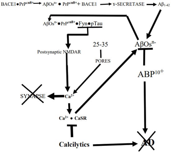
The enormous, 2–3-million-year evolutionary expansion of hominin neocortices to the current enormity enabled humans to take over the planet. However, there appears to have been a glitch, and it occurred without a compensatory expansion of the entorhinal cortical (EC) gateway to the hippocampal memory-encoding system needed to manage the processing of the increasing volume of neocortical data converging on it. The resulting age-dependent connectopathic glitch was unnoticed by the early short-lived populations. It has now surfaced as Alzheimer’s disease (AD) in today’s long-lived populations. With advancing age, processing of the converging neocortical data by the neurons of the relatively small lateral entorhinal cortex (LEC) inflicts persistent strain and high energy costs on these cells. This may result in their hyper-release of harmless Aβ
1–42 monomers into the interstitial fluid, where they seed the formation of toxic amyloid-β oligomers (AβOs) that initiate AD. At the core of connectopathic AD are the postsynaptic cellular prion protein (PrPC). Electrostatic binding of the negatively charged AβOs to the positively charged N-terminus of PrPC induces hyperphosphorylation of tau that destroys synapses. The spread of these accumulating AβOs from ground zero is supported by Aβ’s own production mediated by target cells’ Ca2+-sensing receptors (CaSRs). These data suggest that an early administration of a strongly positively charged, AβOs-interacting peptide or protein, plus an inhibitor of CaSR, might be an effective AD-arresting therapeutic combination.
Full article
►▼
Show Figures
Open AccessArticle
Chikungunya Virus and Its Envelope Protein E2 Induce Hyperalgesia in Mice: Inhibition by Anti-E2 Monoclonal Antibodies and by Targeting TRPV1
by
Carina Z. Segato-Vendrameto, Camila Zanluca, Amanda Z. Zucoloto, Tiago H. Zaninelli, Mariana M. Bertozzi, Telma Saraiva-Santos, Camila R. Ferraz, Larissa Staurengo-Ferrari, Stephanie Badaro-Garcia, Marília F. Manchope, Amanda M. Dionisio, Felipe A. Pinho-Ribeiro, Sergio M. Borghi, Ana Luiza Pamplona Mosimann, Rubia Casagrande, Juliano Bordignon, Victor Fattori, Claudia N. Duarte dos Santos and Waldiceu A. Verri
Cited by 6 | Viewed by 3060
Abstract
Chikungunya virus is an arthropod-borne infectious agent that causes Chikungunya fever disease. About 90% of the infected patients experience intense polyarthralgia, affecting mainly the extremities but also the large joints such as the knees. Chronic disease symptoms persist for months, even after clearance
[...] Read more.
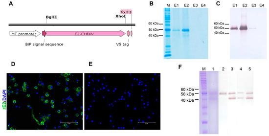
Chikungunya virus is an arthropod-borne infectious agent that causes Chikungunya fever disease. About 90% of the infected patients experience intense polyarthralgia, affecting mainly the extremities but also the large joints such as the knees. Chronic disease symptoms persist for months, even after clearance of the virus from the blood. Envelope proteins stimulate the immune response against the Chikungunya virus, becoming an important therapeutic target. We inactivated the Chikungunya virus (iCHIKV) and produced recombinant E2 (rE2) protein and three different types of anti-rE2 monoclonal antibodies. Using these tools, we observed that iCHIKV and rE2 protein induced mechanical hyperalgesia (electronic aesthesiometer test) and thermal hyperalgesia (Hargreaves test) in mice. These behavioral results were accompanied by the activation of dorsal root ganglia (DRG) neurons in mice, as observed by calcium influx. Treatment with three different types of anti-rE2 monoclonal antibodies and absence or blockade (AMG-9810 treatment) of transient receptor potential vanilloid 1 (TRPV1) channel diminished mechanical and thermal hyperalgesia in mice. iCHIKV and rE2 activated TRPV1+ mouse DRG neurons in vitro, demonstrating their ability to activate nociceptor sensory neurons directly. Therefore, our mouse data demonstrate that targeting E2 CHIKV protein with monoclonal antibodies and inhibiting TRPV1 channels are reasonable strategies to control CHIKV pain.
Full article
►▼
Show Figures
Open AccessReview
Mitochondria Transfer in Brain Injury and Disease
by
Lauren H. Fairley, Amandine Grimm and Anne Eckert
Cited by 22 | Viewed by 5652
Abstract
Intercellular mitochondria transfer is a novel form of cell signalling in which whole mitochondria are transferred between cells in order to enhance cellular functions or aid in the degradation of dysfunctional mitochondria. Recent studies have observed intercellular mitochondria transfer between glia and neurons
[...] Read more.
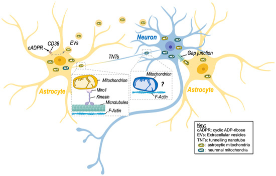
Intercellular mitochondria transfer is a novel form of cell signalling in which whole mitochondria are transferred between cells in order to enhance cellular functions or aid in the degradation of dysfunctional mitochondria. Recent studies have observed intercellular mitochondria transfer between glia and neurons in the brain, and mitochondrial transfer has emerged as a key neuroprotective mechanism in a range of neurological conditions. In particular, artificial mitochondria transfer has sparked widespread interest as a potential therapeutic strategy for brain disorders. In this review, we discuss the mechanisms and effects of intercellular mitochondria transfer in the brain. The role of mitochondrial transfer in neurological conditions, including neurodegenerative disease, brain injury, and neurodevelopmental disorders, is discussed as well as therapeutic strategies targeting mitochondria transfer in the brain.
Full article
►▼
Show Figures
Open AccessArticle
Neuroprotective Effects of a Novel Demeclocycline Derivative Lacking Antibiotic Activity: From a Hit to a Promising Lead Compound
by
Rodrigo Tomas-Grau, Florencia González-Lizárraga, Diego Ploper, César L. Avila, Sergio B. Socías, Pierre Besnault, Aurore Tourville, Rosa M. Mella, Patricia Villacé, Clarisa Salado, Clémence Rose, Blandine Seon-Méniel, Jean-Michel Brunel, Laurent Ferrié, Rita Raisman-Vozari, Patrick P. Michel, Bruno Figadère and Rosana Chehín
Cited by 5 | Viewed by 3460
Abstract
The antibiotic tetracycline demeclocycline (DMC) was recently reported to rescue α-synuclein (α-Syn) fibril-induced pathology. However, the antimicrobial activity of DMC precludes its potential use in long-term neuroprotective treatments. Here, we synthesized a doubly reduced DMC (DDMC) derivative with residual antibiotic activity and improved
[...] Read more.
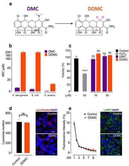
The antibiotic tetracycline demeclocycline (DMC) was recently reported to rescue α-synuclein (α-Syn) fibril-induced pathology. However, the antimicrobial activity of DMC precludes its potential use in long-term neuroprotective treatments. Here, we synthesized a doubly reduced DMC (DDMC) derivative with residual antibiotic activity and improved neuroprotective effects. The molecule was obtained by removal the dimethylamino substituent at position 4 and the reduction of the hydroxyl group at position 12a on ring A of DMC. The modifications strongly diminished its antibiotic activity against Gram-positive and Gram-negative bacteria. Moreover, this compound preserved the low toxicity of DMC in dopaminergic cell lines while improving its ability to interfere with α-Syn amyloid-like aggregation, showing the highest effectiveness of all tetracyclines tested. Likewise, DDMC demonstrated the ability to reduce seeding induced by the exogenous addition of α-Syn preformed fibrils (α-Syn
PFF) in biophysical assays and in a SH-SY5Y-α-Syn-tRFP cell model. In addition, DDMC rendered α-Syn
PFF less inflammogenic. Our results suggest that DDMC may be a promising drug candidate for hit-to-lead development and preclinical studies in Parkinson’s disease and other synucleinopathies.
Full article
►▼
Show Figures
Open AccessArticle
An Epitope-Specific LGI1-Autoantibody Enhances Neuronal Excitability by Modulating Kv1.1 Channel
by
Johanna Extrémet, Oussama El Far, Norbert Ankri, Sarosh R. Irani, Dominique Debanne and Michaël Russier
Cited by 12 | Viewed by 1988
Abstract
Leucine-rich Glioma-Inactivated protein 1 (LGI1) is expressed in the central nervous system and its genetic loss of function is associated with epileptic disorders. Additionally, patients with LGI1-directed autoantibodies have frequent focal seizures as a key feature of their disease. LGI1 is composed of
[...] Read more.
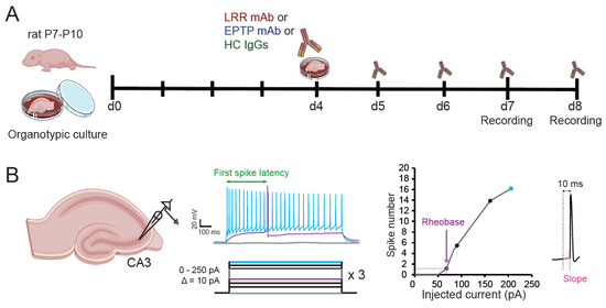
Leucine-rich Glioma-Inactivated protein 1 (LGI1) is expressed in the central nervous system and its genetic loss of function is associated with epileptic disorders. Additionally, patients with LGI1-directed autoantibodies have frequent focal seizures as a key feature of their disease. LGI1 is composed of a Leucine-Rich Repeat (LRR) and an Epitempin (EPTP) domain. These domains are reported to interact with different members of the transsynaptic complex formed by LGI1 at excitatory synapses, including presynaptic Kv1 potassium channels. Patient-derived recombinant monoclonal antibodies (mAbs) are ideal reagents to study whether domain-specific LGI1-autoantibodies induce epileptiform activities in neurons and their downstream mechanisms. We measured the intrinsic excitability of CA3 pyramidal neurons in organotypic cultures from rat hippocampus treated with either an LRR- or an EPTP-reactive patient-derived mAb, or with IgG from control patients. We found an increase in intrinsic excitability correlated with a reduction of the sensitivity to a selective Kv1.1-channel blocker in neurons treated with the LRR mAb, but not in neurons treated with the EPTP mAb. Our findings suggest LRR mAbs are able to modulate neuronal excitability that could account for epileptiform activity observed in patients.
Full article
►▼
Show Figures
Open AccessArticle
Effects of One-Day Application of Levodopa/Carbidopa/Entacapone versus Levodopa/Carbidopa/Opicapone in Parkinson’s Disease Patients
by
Thomas Müller, Eugen Schlegel, Stephanie Zingler and Hans Michael Thiede
Cited by 14 | Viewed by 2579
Abstract
The catechol-O-methyltransferase inhibitors entacapone and opicapone prolong the efficacy of conventional oral levodopa/dopa decarboxylase inhibitor formulations through an increase in levodopa plasma bioavailability. Catechol-O-methyltransferase inhibitors influence the homocysteine metabolism associated with levodopa/dopa decarboxylase application. The objectives of this study were to compare the
[...] Read more.
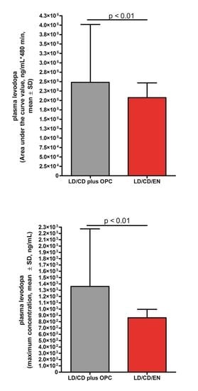
The catechol-O-methyltransferase inhibitors entacapone and opicapone prolong the efficacy of conventional oral levodopa/dopa decarboxylase inhibitor formulations through an increase in levodopa plasma bioavailability. Catechol-O-methyltransferase inhibitors influence the homocysteine metabolism associated with levodopa/dopa decarboxylase application. The objectives of this study were to compare the impact of additional single-day entacapone or opicapone intake on the pharmacokinetic plasma behaviour of levodopa, 3-O-methyldopa and total homocysteine in 15 Parkinson’s disease patients, with concomitant scoring of motor symptoms, under standardized conditions. The patients received opicapone plus two doses of 100 mg levodopa/carbidopa and, one week later, two doses of levodopa/carbidopa/entacapone or vice versa. Levodopa, 3-O-methyldopa and total homocysteine were determined with reversed-phase high-performance liquid chromatography. Levodopa bioavailability and its maximum concentration were higher with opicapone. The computed peak-to-trough difference was lower after the second levodopa administration with entacapone. The fluctuation index of levodopa did not differ between both conditions. 3-O-methyldopa decreased on both days. Homocysteine levels did not significantly vary between both conditions. A significant homocysteine decrease occurred with entacapone, but not with opicapone. Motor behaviour improved with entacapone, but not with opicapone. Opicapone baseline scores were significantly better, and thus the potential for the improvement in motor symptoms was lower compared with the entacapone condition. The higher levodopa bioavailability with opicapone suggests that it is more efficacious than entacapone for the amelioration of “off” phenomena in fluctuating patients when co-administered with a levodopa/dopa decarboxylase inhibitor regimen. Both compounds prevented an increase in homocysteine, which is a metabolic marker for an impaired capacity in the performance of methylation processes.
Full article
►▼
Show Figures
Open AccessReview
The Influence of Gut Dysbiosis in the Pathogenesis and Management of Ischemic Stroke
by
Saravana Babu Chidambaram, Annan Gopinath Rathipriya, Arehally M. Mahalakshmi, Sonali Sharma, Tousif Ahmed Hediyal, Bipul Ray, Tuladhar Sunanda, Wiramon Rungratanawanich, Rajpal Singh Kashyap, M. Walid Qoronfleh, Musthafa Mohamed Essa, Byoung-Joon Song and Tanya M. Monaghan
Cited by 77 | Viewed by 9861
Abstract
Recent research on the gut microbiome has revealed the influence of gut microbiota (GM) on ischemic stroke pathogenesis and treatment outcomes. Alterations in the diversity, abundance, and functions of the gut microbiome, termed gut dysbiosis, results in dysregulated gut–brain signaling, which induces intestinal
[...] Read more.
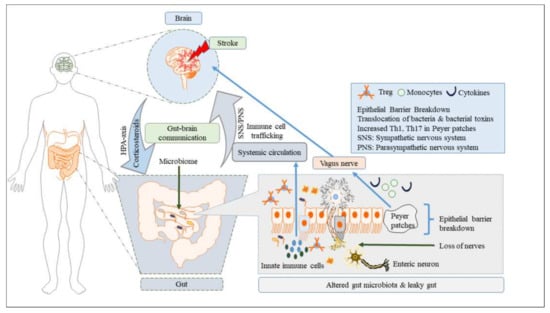
Recent research on the gut microbiome has revealed the influence of gut microbiota (GM) on ischemic stroke pathogenesis and treatment outcomes. Alterations in the diversity, abundance, and functions of the gut microbiome, termed gut dysbiosis, results in dysregulated gut–brain signaling, which induces intestinal barrier changes, endotoxemia, systemic inflammation, and infection, affecting post-stroke outcomes. Gut–brain interactions are bidirectional, and the signals from the gut to the brain are mediated by microbially derived metabolites, such as trimethylamine N-oxide (TMAO) and short-chain fatty acids (SCFAs); bacterial components, such as lipopolysaccharide (LPS); immune cells, such as T helper cells; and bacterial translocation via hormonal, immune, and neural pathways. Ischemic stroke affects gut microbial composition via neural and hypothalamic–pituitary–adrenal (HPA) pathways, which can contribute to post-stroke outcomes. Experimental and clinical studies have demonstrated that the restoration of the gut microbiome usually improves stroke treatment outcomes by regulating metabolic, immune, and inflammatory responses via the gut–brain axis (GBA). Therefore, restoring healthy microbial ecology in the gut may be a key therapeutic target for the effective management and treatment of ischemic stroke.
Full article
►▼
Show Figures
Open AccessArticle
The Effects of Prenatal Exposure to Pregabalin on the Development of Ventral Midbrain Dopaminergic Neurons
by
Walaa F. Alsanie, Majid Alhomrani, Ahmed Gaber, Hamza Habeeballah, Heba A. Alkhatabi, Raed I. Felimban, Sherin Abdelrahman, Charlotte A. E. Hauser, Adeel G. Chaudhary, Abdulhakeem S. Alamri, Bassem M. Raafat, Abdulwahab Alamri, Sirajudheen Anwar, Khaled A. Alswat, Yusuf S. Althobaiti and Yousif A. Asiri
Cited by 6 | Viewed by 3969
Abstract
Pregabalin is widely used as a treatment for multiple neurological disorders; however, it has been reported to have the potential for misuse. Due to a lack of safety studies in pregnancy, pregabalin is considered the last treatment option for various neurological diseases, such
[...] Read more.
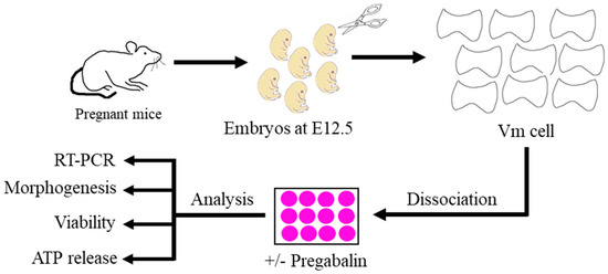
Pregabalin is widely used as a treatment for multiple neurological disorders; however, it has been reported to have the potential for misuse. Due to a lack of safety studies in pregnancy, pregabalin is considered the last treatment option for various neurological diseases, such as neuropathic pain. Therefore, pregabalin abuse in pregnant women, even at therapeutic doses, may impair fetal development. We used primary mouse embryonic neurons to investigate whether exposure to pregabalin can impair the morphogenesis and differentiation of ventral midbrain neurons. This study focused on ventral midbrain dopaminergic neurons, as they are responsible for cognition, movement, and behavior. The results showed that pregabalin exposure during early brain development induced upregulation of the dopaminergic progenitor genes
Lmx1a and
Nurr1 and the mature dopaminergic gene
Pitx3. Interestingly, pregabalin had different effects on the morphogenesis of non-dopaminergic ventral midbrain neurons. Importantly, our findings illustrated that a therapeutic dose of pregabalin (10 μM) did not affect the viability of neurons. However, it caused a decrease in ATP release in ventral midbrain neurons. We demonstrated that exposure to pregabalin during early brain development could interfere with the neurogenesis and morphogenesis of ventral midbrain dopaminergic neurons. These findings are crucial for clinical consideration of the use of pregabalin during pregnancy.
Full article
►▼
Show Figures
Open AccessReview
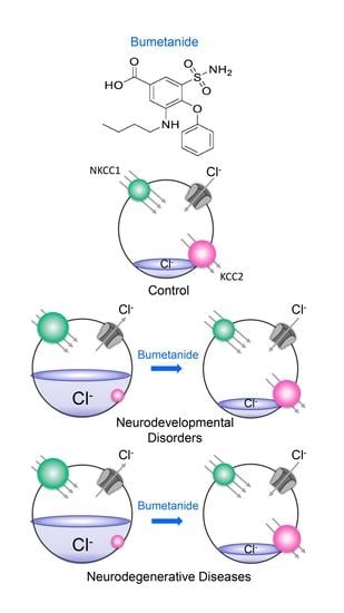
The GABA Polarity Shift and Bumetanide Treatment: Making Sense Requires Unbiased and Undogmatic Analysis
by
Yehezkel Ben-Ari and Enrico Cherubini
Cited by 14 | Viewed by 4315
Abstract
GABA depolarizes and often excites immature neurons in all animal species and brain structures investigated due to a developmentally regulated reduction in intracellular chloride concentration ([Cl
−]
i) levels. The control of [Cl
−]
i levels is mediated by the
[...] Read more.
GABA depolarizes and often excites immature neurons in all animal species and brain structures investigated due to a developmentally regulated reduction in intracellular chloride concentration ([Cl
−]
i) levels. The control of [Cl
−]
i levels is mediated by the chloride cotransporters NKCC1 and KCC2, the former usually importing chloride and the latter exporting it. The GABA polarity shift has been extensively validated in several experimental conditions using often the NKCC1 chloride importer antagonist bumetanide. In spite of an intrinsic heterogeneity, this shift is abolished in many experimental conditions associated with developmental disorders including autism, Rett syndrome, fragile X syndrome, or maternal immune activation. Using bumetanide, an EMA- and FDA-approved agent, many clinical trials have shown promising results with the expected side effects. Kaila et al. have repeatedly challenged these experimental and clinical observations. Here, we reply to the recent reviews by Kaila et al. stressing that the GABA polarity shift is solidly accepted by the scientific community as a major discovery to understand brain development and that bumetanide has shown promising effects in clinical trials.
Full article
►▼
Show Figures
Open AccessReview
Lithium and Erectile Dysfunction: An Overview
by
Mohammad Sheibani, Mehdi Ghasemi and Ahmad Reza Dehpour
Cited by 12 | Viewed by 8775
Abstract
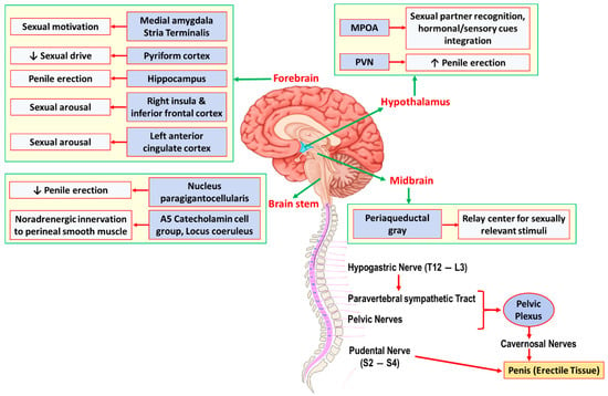
Lithium has been a mainstay of therapy for patients with bipolar disorders for several decades. However, it may exert a variety of adverse effects that can affect patients’ compliance. Sexual and erectile dysfunction has been reported in several studies by patients who take
[...] Read more.
Lithium has been a mainstay of therapy for patients with bipolar disorders for several decades. However, it may exert a variety of adverse effects that can affect patients’ compliance. Sexual and erectile dysfunction has been reported in several studies by patients who take lithium as monotherapy or combined with other psychotherapeutic agents. The exact mechanisms underlying such side effects of lithium are not completely understood. It seems that both central and peripheral mechanisms are involved in the lithium-related sexual dysfunction. Here, we had an overview of the epidemiology of lithium-related sexual and erectile dysfunction in previous clinical studies as well as possible pathologic pathways that could be involved in this adverse effect of lithium based on the previous preclinical studies. Understanding such mechanisms could potentially open a new avenue for therapies that can overcome lithium-related sexual dysfunction and improve patients’ adherence to the medication intake.
Full article
►▼
Show Figures
Open AccessFeature PaperReview
LRRK2 at Striatal Synapses: Cell-Type Specificity and Mechanistic Insights
by
Patrick D. Skelton, Valerie Tokars and Loukia Parisiadou
Cited by 11 | Viewed by 3849
Abstract
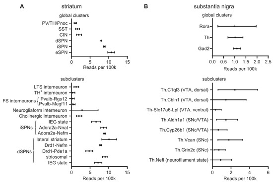
Mutations in leucine-rich repeat kinase 2 (LRRK2) cause Parkinson’s disease with a similar clinical presentation and progression to idiopathic Parkinson’s disease, and common variation is linked to disease risk. Recapitulation of the genotype in rodent models causes abnormal dopamine release and increases the
[...] Read more.
Mutations in leucine-rich repeat kinase 2 (LRRK2) cause Parkinson’s disease with a similar clinical presentation and progression to idiopathic Parkinson’s disease, and common variation is linked to disease risk. Recapitulation of the genotype in rodent models causes abnormal dopamine release and increases the susceptibility of dopaminergic neurons to insults, making LRRK2 a valuable model for understanding the pathobiology of Parkinson’s disease. It is also a promising druggable target with targeted therapies currently in development. LRRK2 mRNA and protein expression in the brain is highly variable across regions and cellular identities. A growing body of work has demonstrated that pathogenic LRRK2 mutations disrupt striatal synapses before the onset of overt neurodegeneration. Several substrates and interactors of LRRK2 have been identified to potentially mediate these pre-neurodegenerative changes in a cell-type-specific manner. This review discusses the effects of pathogenic LRRK2 mutations in striatal neurons, including cell-type-specific and pathway-specific alterations. It also highlights several LRRK2 effectors that could mediate the alterations to striatal function, including Rabs and protein kinase A. The lessons learned from improving our understanding of the pathogenic effects of LRRK2 mutations in striatal neurons will be applicable to both dissecting the cell-type specificity of LRRK2 function in the transcriptionally diverse subtypes of dopaminergic neurons and also increasing our understanding of basal ganglia development and biology. Finally, it will inform the development of therapeutics for Parkinson’s disease.
Full article
►▼
Show Figures
Open AccessReview
Impact of Pharmacological and Non-Pharmacological Modulators on Dendritic Spines Structure and Functions in Brain
by
Arehally M. Mahalakshmi, Bipul Ray, Sunanda Tuladhar, Tousif Ahmed Hediyal, Praveen Raj, Annan Gopinath Rathipriya, M. Walid Qoronfleh, Musthafa Mohamed Essa and Saravana Babu Chidambaram
Cited by 6 | Viewed by 3763
Abstract
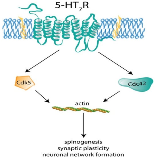
Dendritic spines are small, thin, hair-like protrusions found on the dendritic processes of neurons. They serve as independent compartments providing large amplitudes of Ca
2+ signals to achieve synaptic plasticity, provide sites for newer synapses, facilitate learning and memory. One of the common
[...] Read more.
Dendritic spines are small, thin, hair-like protrusions found on the dendritic processes of neurons. They serve as independent compartments providing large amplitudes of Ca
2+ signals to achieve synaptic plasticity, provide sites for newer synapses, facilitate learning and memory. One of the common and severe complication of neurodegenerative disease is cognitive impairment, which is said to be closely associated with spine pathologies viz., decreased in spine density, spine length, spine volume, spine size etc. Many treatments targeting neurological diseases have shown to improve the spine structure and distribution. However, concise data on the various modulators of dendritic spines are imperative and a need of the hour. Hence, in this review we made an attempt to consolidate the effects of various pharmacological (cholinergic, glutamatergic, GABAergic, serotonergic, adrenergic, and dopaminergic agents) and non-pharmacological modulators (dietary interventions, enriched environment, yoga and meditation) on dendritic spines structure and functions. These data suggest that both the pharmacological and non-pharmacological modulators produced significant improvement in dendritic spine structure and functions and in turn reversing the pathologies underlying neurodegeneration. Intriguingly, the non-pharmacological approaches have shown to improve intellectual performances both in preclinical and clinical platforms, but still more technology-based evidence needs to be studied. Thus, we conclude that a combination of pharmacological and non-pharmacological intervention may restore cognitive performance synergistically via improving dendritic spine number and functions in various neurological disorders.
Full article
►▼
Show Figures
Open AccessArticle
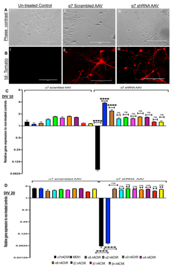
Neuronal Menin Overexpression Rescues Learning and Memory Phenotype in CA1-Specific α7 nAChRs KD Mice
by
Shadab Batool, Basma Akhter, Jawwad Zaidi, Frank Visser, Gavin Petrie, Matthew Hill and Naweed I. Syed
Cited by 6 | Viewed by 2945
Abstract
The perturbation of nicotinic cholinergic receptors is thought to underlie many neurodegenerative and neuropsychiatric disorders, such as Alzheimer’s and schizophrenia. We previously identified that the tumor suppressor gene,
MEN1, regulates both the expression and synaptic targeting of α7 nAChRs in the mouse
[...] Read more.
The perturbation of nicotinic cholinergic receptors is thought to underlie many neurodegenerative and neuropsychiatric disorders, such as Alzheimer’s and schizophrenia. We previously identified that the tumor suppressor gene,
MEN1, regulates both the expression and synaptic targeting of α7 nAChRs in the mouse hippocampal neurons in vitro. Here we sought to determine whether the α7 nAChRs gene expression reciprocally regulates the expression of menin, the protein encoded by the
MEN1 gene, and if this interplay impacts learning and memory. We demonstrate here that α7 nAChRs knockdown (KD) both in in vitro and in vivo, initially upregulated and then subsequently downregulated menin expression. Exogenous expression of menin using an AAV transduction approach rescued α7 nAChRs KD mediated functional and behavioral deficits specifically in hippocampal (CA1) neurons. These effects involved the modulation of the α7 nAChR subunit expression and functional clustering at the synaptic sites. Our data thus demonstrates a novel and important interplay between the
MEN1 gene and the α7 nAChRs in regulating hippocampal-dependent learning and memory.
Full article
►▼
Show Figures
Open AccessReview
Mechanisms of Plasticity in Subcortical Visual Areas
by
Maël Duménieu, Béatrice Marquèze-Pouey, Michaël Russier and Dominique Debanne
Cited by 8 | Viewed by 4952
Abstract
Visual plasticity is classically considered to occur essentially in the primary and secondary cortical areas. Subcortical visual areas such as the dorsal lateral geniculate nucleus (dLGN) or the superior colliculus (SC) have long been held as basic structures responsible for a stable and
[...] Read more.
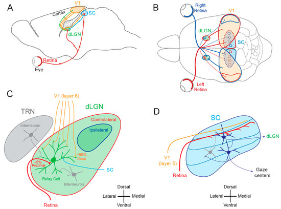
Visual plasticity is classically considered to occur essentially in the primary and secondary cortical areas. Subcortical visual areas such as the dorsal lateral geniculate nucleus (dLGN) or the superior colliculus (SC) have long been held as basic structures responsible for a stable and defined function. In this model, the dLGN was considered as a relay of visual information travelling from the retina to cortical areas and the SC as a sensory integrator orienting body movements towards visual targets. However, recent findings suggest that both dLGN and SC neurons express functional plasticity, adding unexplored layers of complexity to their previously attributed functions. The existence of neuronal plasticity at the level of visual subcortical areas redefines our approach of the visual system. The aim of this paper is therefore to review the cellular and molecular mechanisms for activity-dependent plasticity of both synaptic transmission and cellular properties in subcortical visual areas.
Full article
►▼
Show Figures
Open AccessArticle
Activity of Cerebellar Nuclei Neurons Correlates with ZebrinII Identity of Their Purkinje Cell Afferents
by
Gerrit C. Beekhof, Simona V. Gornati, Cathrin B. Canto, Avraham M. Libster, Martijn Schonewille, Chris I. De Zeeuw and Freek E. Hoebeek
Cited by 10 | Viewed by 3748
Abstract
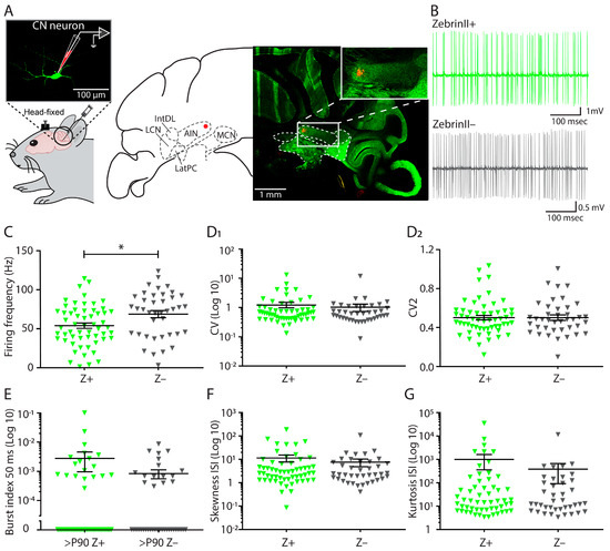
Purkinje cells (PCs) in the cerebellar cortex can be divided into at least two main subpopulations: one subpopulation that prominently expresses ZebrinII (Z+), and shows a relatively low simple spike firing rate, and another that hardly expresses ZebrinII (Z–) and shows higher baseline
[...] Read more.
Purkinje cells (PCs) in the cerebellar cortex can be divided into at least two main subpopulations: one subpopulation that prominently expresses ZebrinII (Z+), and shows a relatively low simple spike firing rate, and another that hardly expresses ZebrinII (Z–) and shows higher baseline firing rates. Likewise, the complex spike responses of PCs, which are evoked by climbing fiber inputs and thus reflect the activity of the inferior olive (IO), show the same dichotomy. However, it is not known whether the target neurons of PCs in the cerebellar nuclei (CN) maintain this bimodal distribution. Electrophysiological recordings in awake adult mice show that the rate of action potential firing of CN neurons that receive input from Z+ PCs was consistently lower than that of CN neurons innervated by Z– PCs. Similar in vivo recordings in juvenile and adolescent mice indicated that the firing frequency of CN neurons correlates to the ZebrinII identity of the PC afferents in adult, but not postnatal stages. Finally, the spontaneous action potential firing pattern of adult CN neurons recorded in vitro revealed no significant differences in intrinsic pacemaking activity between ZebrinII identities. Our findings indicate that all three main components of the olivocerebellar loop, i.e., PCs, IO neurons and CN neurons, operate at a higher rate in the Z– modules.
Full article
►▼
Show Figures
Open AccessFeature PaperArticle
Disproportion in Pericyte/Endothelial Cell Proliferation and Mechanisms of Intussusceptive Angiogenesis Participate in Bizarre Vessel Formation in Glioblastoma
by
Lucio Díaz-Flores, Ricardo Gutiérrez, Miriam González-Gómez, María-del-Pino García, Lucio Díaz-Flores, Jr., Ibrahim González-Marrero, Julio Ávila and Pablo Martín-Vasallo
Cited by 8 | Viewed by 3001
Abstract
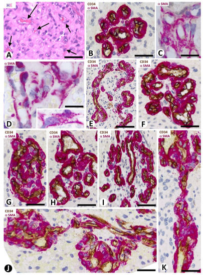
Glioblastoma (GBM) is the most malignant tumor in the brain. In addition to the vascular pattern with thin-walled vessels and findings of sprouting angiogenesis, GBM presents a bizarre microvasculature (BM) formed by vascular clusters, vascular garlands, and glomeruloid bodies. The mechanisms in BM
[...] Read more.
Glioblastoma (GBM) is the most malignant tumor in the brain. In addition to the vascular pattern with thin-walled vessels and findings of sprouting angiogenesis, GBM presents a bizarre microvasculature (BM) formed by vascular clusters, vascular garlands, and glomeruloid bodies. The mechanisms in BM morphogenesis are not well known. Our objective was to assess the role of pericyte/endothelial proliferation and intussusceptive angiogenic mechanisms in the formation of the BM. For this purpose, we studied specimens of 66 GBM cases using immunochemistry and confocal microscopy. In the BM, the results showed (a) transitional forms between the BM patterns, mostly with prominent pericytes covering all the abluminal endothelial cell (EC) surface of the vessels, (b) a proliferation index high in the prominent pericytes and low in ECs (47.85 times higher in pericytes than in ECs), (c) intravascular pillars (hallmark of intussusceptive angiogenesis) formed by transcapillary interendothelial bridges, endothelial contacts of opposite vessel walls, and vessel loops, and (d) the persistence of these findings in complex glomeruloid bodies. In conclusion, disproportion in pericyte/EC proliferation and mechanisms of intussusceptive angiogenesis participate in BM formation. The contributions have morphogenic and clinical interest since pericytes and intussusceptive angiogenesis can condition antiangiogenic therapy in GBM.
Full article
►▼
Show Figures
Open AccessArticle
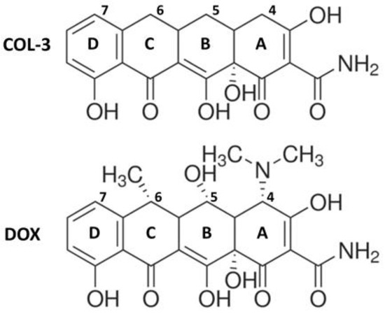
The Chemically-Modified Tetracycline COL-3 and Its Parent Compound Doxycycline Prevent Microglial Inflammatory Responses by Reducing Glucose-Mediated Oxidative Stress
by
Nilson Carlos Ferreira Junior, Maurício dos Santos Pereira, Nour Francis, Paola Ramirez, Paula Martorell, Florencia González-Lizarraga, Bruno Figadère, Rosana Chehin, Elaine Del Bel, Rita Raisman-Vozari and Patrick Pierre Michel
Cited by 12 | Viewed by 3577
Abstract
We used mouse microglial cells in culture activated by lipopolysaccharide (LPS) or α-synuclein amyloid aggregates (αSa) to study the anti-inflammatory effects of COL-3, a tetracycline derivative without antimicrobial activity. Under LPS or αSa stimulation, COL-3 (10, 20 µM) efficiently repressed the induction of
[...] Read more.
We used mouse microglial cells in culture activated by lipopolysaccharide (LPS) or α-synuclein amyloid aggregates (αSa) to study the anti-inflammatory effects of COL-3, a tetracycline derivative without antimicrobial activity. Under LPS or αSa stimulation, COL-3 (10, 20 µM) efficiently repressed the induction of the microglial activation marker protein
Iba-
1 and the stimulated-release of the pro-inflammatory cytokine TNF-α. COL-3′s inhibitory effects on TNF-α were reproduced by the tetracycline antibiotic doxycycline (DOX; 50 µM), the glucocorticoid dexamethasone, and apocynin (APO), an inhibitor of the superoxide-producing enzyme NADPH oxidase. This last observation suggested that COL-3 and DOX might also operate themselves by restraining oxidative stress-mediated signaling events. Quantitative measurement of intracellular reactive oxygen species (ROS) levels revealed that COL-3 and DOX were indeed as effective as APO in reducing oxidative stress and TNF-α release in activated microglia. ROS inhibition with COL-3 or DOX occurred together with a reduction of microglial glucose accumulation and NADPH synthesis. This suggested that COL-3 and DOX might reduce microglial oxidative burst activity by limiting the glucose-dependent synthesis of NADPH, the requisite substrate for NADPH oxidase. Coherent with this possibility, the glycolysis inhibitor 2-deoxy-D-glucose reproduced the immunosuppressive action of COL-3 and DOX in activated microglia. Overall, we propose that COL-3 and its parent compound DOX exert anti-inflammatory effects in microglial cells by inhibiting glucose-dependent ROS production. These effects might be strengthened by the intrinsic antioxidant properties of DOX and COL-3 in a self-reinforcing manner.
Full article
►▼
Show Figures
Open AccessArticle
Alterations in Dendritic Spine Maturation and Neurite Development Mediated by FAM19A1
by
Hyo-Jeong Yong, Jong-Ik Hwang and Jae-Young Seong
Viewed by 3681
Abstract
Neurogenesis and functional brain activity require complex associations of inherently programmed secretory elements that are regulated precisely and temporally. Family with sequence similarity 19 A1 (FAM19A1) is a secreted protein primarily expressed in subsets of terminally differentiated neuronal precursor cells and fully mature
[...] Read more.
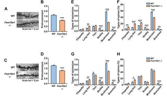
Neurogenesis and functional brain activity require complex associations of inherently programmed secretory elements that are regulated precisely and temporally. Family with sequence similarity 19 A1 (FAM19A1) is a secreted protein primarily expressed in subsets of terminally differentiated neuronal precursor cells and fully mature neurons in specific brain substructures. Several recent studies have demonstrated the importance of FAM19A1 in brain physiology; however, additional information is needed to support its role in neuronal maturation and function. In this study, dendritic spine morphology in
Fam19a1-ablated mice and neurite development during in vitro neurogenesis were examined to understand the putative role of FAM19A1 in neural integrity. Adult
Fam19a1-deficient mice showed low dendritic spine density and maturity with reduced dendrite complexity compared to wild-type (WT) littermates. To further explore the effect of FAM19A1 on neuronal maturation, the neurite outgrowth pattern in primary neurons was analyzed in vitro with and without FAM19A1. In response to FAM19A1, WT primary neurons showed reduced neurite complexity, whereas
Fam19a1-decifient primary neurons exhibited increased neurite arborization, which was reversed by supplementation with recombinant FAM19A1. Together, these findings suggest that FAM19A1 participates in dendritic spine development and neurite arborization.
Full article
►▼
Show Figures
Open AccessArticle
Transcriptomic Profile Reveals Deregulation of Hearing-Loss Related Genes in Vestibular Schwannoma Cells Following Electromagnetic Field Exposure
by
Alessandra Colciago, Matteo Audano, Veronica Bonalume, Valentina Melfi, Tasnim Mohamed, Adam J. Reid, Alessandro Faroni, Peter A. Greer, Nico Mitro and Valerio Magnaghi
Cited by 5 | Viewed by 4330
Abstract
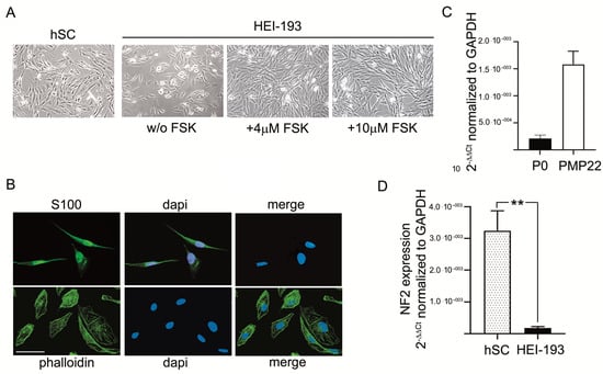
Hearing loss (HL) is the most common sensory disorder in the world population. One common cause of HL is the presence of vestibular schwannoma (VS), a benign tumor of the VIII cranial nerve, arising from Schwann cell (SC) transformation. In the last decade,
[...] Read more.
Hearing loss (HL) is the most common sensory disorder in the world population. One common cause of HL is the presence of vestibular schwannoma (VS), a benign tumor of the VIII cranial nerve, arising from Schwann cell (SC) transformation. In the last decade, the increasing incidence of VS has been correlated to electromagnetic field (EMF) exposure, which might be considered a pathogenic cause of VS development and HL. Here, we explore the molecular mechanisms underlying the biologic changes of human SCs and/or their oncogenic transformation following EMF exposure. Through NGS technology and RNA-Seq transcriptomic analysis, we investigated the genomic profile and the differential display of HL-related genes after chronic EMF. We found that chronic EMF exposure modified the cell proliferation, in parallel with intracellular signaling and metabolic pathways changes, mostly related to translation and mitochondrial activities. Importantly, the expression of HL-related genes such as NEFL, TPRN, OTOGL, GJB2, and REST appeared to be deregulated in chronic EMF exposure. In conclusion, we suggest that, at a preclinical stage, EMF exposure might promote the transformation of VS cells and contribute to HL.
Full article
►▼
Show Figures
Open AccessReview
A Review on Lactoferrin and Central Nervous System Diseases
by
Yu-Qi Li and Chuang Guo
Cited by 49 | Viewed by 6223
Abstract
Central nervous system (CNS) diseases are currently one of the major health issues around the world. Most CNS disorders are characterized by high oxidative stress levels and intense inflammatory responses in affected tissues. Lactoferrin (Lf), a multifunctional iron-binding glycoprotein, plays a significant role
[...] Read more.
Central nervous system (CNS) diseases are currently one of the major health issues around the world. Most CNS disorders are characterized by high oxidative stress levels and intense inflammatory responses in affected tissues. Lactoferrin (Lf), a multifunctional iron-binding glycoprotein, plays a significant role in anti-inflammatory, antibacterial, antiviral, reactive oxygen species (ROS) modulator, antitumor immunity, and anti-apoptotic processes. Previous studies have shown that Lf is abnormally expressed in a variety of neurological diseases, especially neurodegenerative diseases. Recently, the promotion of neurodevelopment and neuroprotection by Lf has attracted widespread attention, and Lf could be exploited both as an active therapeutic agent and drug nanocarrier. However, our understanding of the roles of Lf proteins in the initiation or progression of CNS diseases is limited, especially the roles of Lf in regulating neurogenesis. This review highlights recent advances in the understanding of the major pharmacological effects of Lf in CNS diseases, including neurodegenerative diseases, cerebrovascular disease, developmental delays in children, and brain tumors.
Full article
Open AccessReview
Role of SNAREs in Neurodegenerative Diseases
by
Azzurra Margiotta
Cited by 33 | Viewed by 5610
Abstract
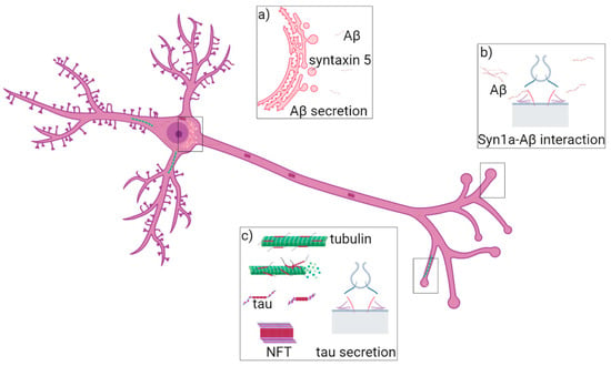
Neurodegenerative diseases are pathologies of the central and peripheral nervous systems characterized by loss of brain functions and problems in movement which occur due to the slow and progressive degeneration of cellular elements. Several neurodegenerative diseases are known such as Alzheimer’s disease, Parkinson’s
[...] Read more.
Neurodegenerative diseases are pathologies of the central and peripheral nervous systems characterized by loss of brain functions and problems in movement which occur due to the slow and progressive degeneration of cellular elements. Several neurodegenerative diseases are known such as Alzheimer’s disease, Parkinson’s disease and amyotrophic lateral sclerosis and many studies on the molecular mechanisms underlying these pathologies have been conducted. Altered functions of some key proteins and the presence of intraneuronal aggregates have been identified as responsible for the development of the diseases. Interestingly, the formation of the SNARE complex has been discovered to be fundamental for vesicle fusion, vesicle recycling and neurotransmitter release. Indeed, inhibition of the formation of the SNARE complex, defects in the SNARE-dependent exocytosis and altered regulation of SNARE-mediated vesicle fusion have been associated with neurodegeneration. In this review, the biological aspects of neurodegenerative diseases and the role of SNARE proteins in relation to the onset of these pathologies are described.
Full article
►▼
Show Figures
Planned Papers
The below list represents only planned manuscripts. Some of these
manuscripts have not been received by the Editorial Office yet. Papers
submitted to MDPI journals are subject to peer-review.
Title: Role of Complement as a Presynaptic Modulator of the Synaptic Transmission in the CNS
Authors: Anna Pittaluga
Affiliation: Dipartimento di Farmacia (DIFAR), Università degli Studi di Genova, 16132 Genoa, Italy


