ijms-logo

Journal Browser

Journal Browser

Molecular Mechanisms and Pathophysiology of Cardiovascular Disease

A special issue of International Journal of Molecular Sciences (ISSN 1422-0067). This special issue belongs to the section "Molecular Biology".

Deadline for manuscript submissions: closed (31 July 2022) | Viewed by 48899

Special Issue Editors


E-Mail Website1 Website2
Guest Editor
Vascular Pathophysiology Lab, Hospital Nacional de Parapléjicos, Finca la peraleda s/n, 45071 Toledo, Spain
Interests: proteomics; precision medicine; biomarkers; cardiology and aging

E-Mail Website
Guest Editor
Department of Pharmacology and Therapeutics, School of Medicine, Universidad Autónoma de Madrid, Madrid, Spain
Interests: atherosclerosis; cardiovascular; extracellular vesicles; omics; cell senescence

Special Issue Information

Dear Colleagues,

A Special Issue on the topic of “Molecular Mechanisms and Pathophysiology of Cardiovascular Disease” is being prepared for the journal IJMS (Impact Factor 4.556 2020 Journal Citation Reports®).

Even in the 21st century, cardiovascular diseases (CVD) remain a significant health issue worldwide. As early as in 2001, these diseases were recognized as the largest single cause of mortality on the planet. Today, despite the huge growth of pathophysiology knowledge and advances in molecular mechanisms, prognosis, diagnosis, and treatment, CVD remain at the world’s top rank of all human death causes. In fact, the impact of CVD is growing and has reached pandemic proportions, mainly due to the great incidence of obesity in the global population. This Special Issue focuses on molecular mechanisms (vascular calcification, endothelial dysfunction, inflammation, oxidative stress, etc.) (endothelial cells, macrophages, vSMCs, platelets, cardiac myocytes, and extracellular vesicles) and pathophysiology of CVD contributing to better understanding their development, providing insights to guide designing future, more personalized, medicine.

We warmly welcome submissions, including original papers and reviews, on this widely discussed topic.

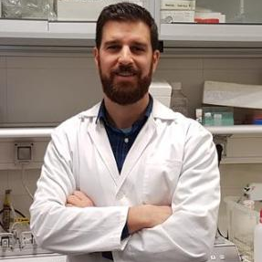
Dr. Maria Gonzalez Barderas
Dr. Fernando de la Cuesta
Guest Editors

Manuscript Submission Information

Manuscripts should be submitted online at www.mdpi.com by registering and logging in to this website. Once you are registered, click here to go to the submission form. Manuscripts can be submitted until the deadline. All submissions that pass pre-check are peer-reviewed. Accepted papers will be published continuously in the journal (as soon as accepted) and will be listed together on the special issue website. Research articles, review articles as well as short communications are invited. For planned papers, a title and short abstract (about 100 words) can be sent to the Editorial Office for announcement on this website.

Submitted manuscripts should not have been published previously, nor be under consideration for publication elsewhere (except conference proceedings papers). All manuscripts are thoroughly refereed through a single-blind peer-review process. A guide for authors and other relevant information for submission of manuscripts is available on the Instructions for Authors page. International Journal of Molecular Sciences is an international peer-reviewed open access semimonthly journal published by MDPI.

Please visit the Instructions for Authors page before submitting a manuscript. There is an Article Processing Charge (APC) for publication in this open access journal. For details about the APC please see here. Submitted papers should be well formatted and use good English. Authors may use MDPI's English editing service prior to publication or during author revisions.

Keywords

  • cardiovascular disease
  • molecular mechanisms
  • cardiovascular pathophysiology
  • inflammation
  • endothelial dysfunction
  • molecular markers

Benefits of Publishing in a Special Issue

  • Ease of navigation: Grouping papers by topic helps scholars navigate broad scope journals more efficiently.
  • Greater discoverability: Special Issues support the reach and impact of scientific research. Articles in Special Issues are more discoverable and cited more frequently.
  • Expansion of research network: Special Issues facilitate connections among authors, fostering scientific collaborations.
  • External promotion: Articles in Special Issues are often promoted through the journal's social media, increasing their visibility.
  • e-Book format: Special Issues with more than 10 articles can be published as dedicated e-books, ensuring wide and rapid dissemination.

Further information on MDPI's Special Issue polices can be found here.

Published Papers (15 papers)

Order results
Result details
Select all
Export citation of selected articles as:

Editorial

Jump to: Research, Review

3 pages, 194 KiB  
Editorial
A Molecular (Not Very Becoming) Picture of Stressed Arteries and Heart, with Some Therapeutic Hope
by Maria G. Barderas and Fernando de la Cuesta
Int. J. Mol. Sci. 2023, 24(4), 3870; https://doi.org/10.3390/ijms24043870 - 15 Feb 2023
Viewed by 1195
Abstract
This Special Issue has focused on molecular mechanisms (vascular calcification, endothelial dysfunction, cardiac remodelling, inflammation, oxidative stress, etc [...] Full article
(This article belongs to the Special Issue Molecular Mechanisms and Pathophysiology of Cardiovascular Disease)

Research

Jump to: Editorial, Review

13 pages, 1697 KiB  
Article
1,3,8-Triazaspiro[4.5]decane Derivatives Inhibit Permeability Transition Pores through a FO-ATP Synthase c Subunit Glu119-Independent Mechanism That Prevents Oligomycin A-Related Side Effects
by Gaia Pedriali, Daniela Ramaccini, Esmaa Bouhamida, Alessio Branchini, Giulia Turrin, Elisabetta Tonet, Antonella Scala, Simone Patergnani, Mirko Pinotti, Claudio Trapella, Carlotta Giorgi, Elena Tremoli, Gianluca Campo, Giampaolo Morciano and Paolo Pinton
Int. J. Mol. Sci. 2023, 24(7), 6191; https://doi.org/10.3390/ijms24076191 - 24 Mar 2023
Cited by 3 | Viewed by 1530
Abstract
Permeability transition pore (PTP) molecular composition and activity modulation have been a matter of research for several years, especially due to their importance in ischemia reperfusion injury (IRI). Notably, c subunit of ATP synthase (Csub) has been identified as one of the PTP-forming [...] Read more.
Permeability transition pore (PTP) molecular composition and activity modulation have been a matter of research for several years, especially due to their importance in ischemia reperfusion injury (IRI). Notably, c subunit of ATP synthase (Csub) has been identified as one of the PTP-forming proteins and as a target for cardioprotection. Oligomycin A is a well-known Csub interactor that has been chemically modified in-depth for proposed new pharmacological approaches against cardiac reperfusion injury. Indeed, by taking advantage of its scaffold and through focused chemical improvements, innovative Csub-dependent PTP inhibitors (1,3,8-Triazaspiro[4.5]decane) have been synthetized in the past. Interestingly, four critical amino acids have been found to be involved in Oligomycin A-Csub binding in yeast. However, their position on the human sequence is unknown, as is their function in PTP inhibition. The aims of this study are to (i) identify for the first time the topologically equivalent residues in the human Csub sequence; (ii) provide their in vitro validation in Oligomycin A-mediated PTP inhibition and (iii) understand their relevance in the binding of 1,3,8-Triazaspiro[4.5]decane small molecules, as Oligomycin A derivatives, in order to provide insights into Csub interactions. Notably, in this study we demonstrated that 1,3,8-Triazaspiro[4.5]decane derivatives inhibit permeability transition pores through a FO-ATP synthase c subunit Glu119-independent mechanism that prevents Oligomycin A-related side effects. Full article
(This article belongs to the Special Issue Molecular Mechanisms and Pathophysiology of Cardiovascular Disease)
Show Figures

Figure 1

16 pages, 2217 KiB  
Article
The Proteome of Circulating Large Extracellular Vesicles in Diabetes and Hypertension
by Akram Abolbaghaei, Maddison Turner, Jean-François Thibodeau, Chet E. Holterman, Christopher R. J. Kennedy and Dylan Burger
Int. J. Mol. Sci. 2023, 24(5), 4930; https://doi.org/10.3390/ijms24054930 - 3 Mar 2023
Cited by 5 | Viewed by 2410
Abstract
Hypertension and diabetes induce vascular injury through processes that are not fully understood. Changes in extracellular vesicle (EV) composition could provide novel insights. Here, we examined the protein composition of circulating EVs from hypertensive, diabetic and healthy mice. EVs were isolated from transgenic [...] Read more.
Hypertension and diabetes induce vascular injury through processes that are not fully understood. Changes in extracellular vesicle (EV) composition could provide novel insights. Here, we examined the protein composition of circulating EVs from hypertensive, diabetic and healthy mice. EVs were isolated from transgenic mice overexpressing human renin in the liver (TtRhRen, hypertensive), OVE26 type 1 diabetic mice and wild-type (WT) mice. Protein content was analyzed using liquid chromatography–mass spectrometry. We identified 544 independent proteins, of which 408 were found in all groups, 34 were exclusive to WT, 16 were exclusive to OVE26 and 5 were exclusive to TTRhRen mice. Amongst the differentially expressed proteins, haptoglobin (HPT) was upregulated and ankyrin-1 (ANK1) was downregulated in OVE26 and TtRhRen mice compared with WT controls. Conversely, TSP4 and Co3A1 were upregulated and SAA4 was downregulated exclusively in diabetic mice; and PPN was upregulated and SPTB1 and SPTA1 were downregulated in hypertensive mice, compared to WT mice. Ingenuity pathway analysis identified enrichment in proteins associated with SNARE signaling, the complement system and NAD homeostasis in EVs from diabetic mice. Conversely, in EVs from hypertensive mice, there was enrichment in semaphroin and Rho signaling. Further analysis of these changes may improve understanding of vascular injury in hypertension and diabetes. Full article
(This article belongs to the Special Issue Molecular Mechanisms and Pathophysiology of Cardiovascular Disease)
Show Figures

Figure 1

11 pages, 1194 KiB  
Article
Sustained Increase in Serum Glial Fibrillary Acidic Protein after First ST-Elevation Myocardial Infarction
by Jan Traub, Katja Grondey, Tobias Gassenmaier, Dominik Schmitt, Georg Fette, Stefan Frantz, Valérie Boivin-Jahns, Roland Jahns, Stefan Störk, Guido Stoll, Theresa Reiter, Ulrich Hofmann, Martin S. Weber and Anna Frey
Int. J. Mol. Sci. 2022, 23(18), 10304; https://doi.org/10.3390/ijms231810304 - 7 Sep 2022
Cited by 2 | Viewed by 1998
Abstract
Acute ischemic cardiac injury predisposes one to cognitive impairment, dementia, and depression. Pathophysiologically, recent positron emission tomography data suggest astroglial activation after experimental myocardial infarction (MI). We analyzed peripheral surrogate markers of glial (and neuronal) damage serially within 12 months after the first [...] Read more.
Acute ischemic cardiac injury predisposes one to cognitive impairment, dementia, and depression. Pathophysiologically, recent positron emission tomography data suggest astroglial activation after experimental myocardial infarction (MI). We analyzed peripheral surrogate markers of glial (and neuronal) damage serially within 12 months after the first ST-elevation MI (STEMI). Serum levels of glial fibrillary acidic protein (GFAP) and neurofilament light chain (NfL) were quantified using ultra-sensitive molecular immunoassays. Sufficient biomaterial was available from 45 STEMI patients (aged 28 to 78 years, median 56 years, 11% female). The median (quartiles) of GFAP was 63.8 (47.0, 89.9) pg/mL and of NfL 10.6 (7.2, 14.8) pg/mL at study entry 0–4 days after STEMI. GFAP after STEMI increased in the first 3 months, with a median change of +7.8 (0.4, 19.4) pg/mL (p = 0.007). It remained elevated without further relevant increases after 6 months (+11.7 (0.6, 23.5) pg/mL; p = 0.015), and 12 months (+10.3 (1.5, 22.7) pg/mL; p = 0.010) compared to the baseline. Larger relative infarction size was associated with a higher increase in GFAP (ρ = 0.41; p = 0.009). In contrast, NfL remained unaltered in the course of one year. Our findings support the idea of central nervous system involvement after MI, with GFAP as a potential peripheral biomarker of chronic glial damage as one pathophysiologic pathway. Full article
(This article belongs to the Special Issue Molecular Mechanisms and Pathophysiology of Cardiovascular Disease)
Show Figures

Figure 1

15 pages, 4096 KiB  
Article
Dock10 Regulates Cardiac Function under Neurohormonal Stress
by Liad Segal, Sharon Etzion, Sigal Elyagon, Moran Shahar, Hadar Klapper-Goldstein, Aviva Levitas, Michael S. Kapiloff, Ruti Parvari and Yoram Etzion
Int. J. Mol. Sci. 2022, 23(17), 9616; https://doi.org/10.3390/ijms23179616 - 25 Aug 2022
Cited by 3 | Viewed by 2259
Abstract
Dedicator of cytokinesis 10 (Dock10) is a guanine nucleotide exchange factor for Cdc42 and Rac1 that regulates the JNK (c-Jun N-terminal kinase) and p38 MAPK (mitogen-activated protein kinase) signaling cascades. In this study, we characterized the roles of Dock10 in the myocardium. In [...] Read more.
Dedicator of cytokinesis 10 (Dock10) is a guanine nucleotide exchange factor for Cdc42 and Rac1 that regulates the JNK (c-Jun N-terminal kinase) and p38 MAPK (mitogen-activated protein kinase) signaling cascades. In this study, we characterized the roles of Dock10 in the myocardium. In vitro: we ablated Dock10 in neonatal mouse floxed Dock10 cardiomyocytes (NMCMs) and cardiofibroblasts (NMCFs) by transduction with an adenovirus expressing Cre-recombinase. In vivo, we studied mice in which the Dock10 gene was constitutively and globally deleted (Dock10 KO) and mice with cardiac myocyte-specific Dock10 KO (Dock10 CKO) at baseline and in response to two weeks of Angiotensin II (Ang II) infusion. In vitro, Dock10 ablation differentially inhibited the α-adrenergic stimulation of p38 and JNK in NMCM and NMCF, respectively. In vivo, the stimulation of both signaling pathways was markedly attenuated in the heart. The Dock10 KO mice had normal body weight and cardiac size. However, echocardiography revealed mildly reduced systolic function, and IonOptix recordings demonstrated reduced contractility and elevated diastolic calcium levels in isolated cardiomyocytes. Remarkably, Dock10 KO, but not Dock10 CKO, exaggerated the pathological response to Ang II infusion. These data suggest that Dock10 regulates cardiac stress-related signaling. Although Dock10 can regulate MAPK signaling in both cardiomyocytes and cardiofibroblasts, the inhibition of pathological cardiac remodeling is not apparently due to the Dock10 signaling in the cardiomyocyte. Full article
(This article belongs to the Special Issue Molecular Mechanisms and Pathophysiology of Cardiovascular Disease)
Show Figures

Graphical abstract

15 pages, 1752 KiB  
Article
Unilateral Acute Renal Ischemia-Reperfusion Injury Induces Cardiac Dysfunction through Intracellular Calcium Mishandling
by Carolina Victoria Cruz Junho, Laura González-Lafuente, José Alberto Navarro-García, Elena Rodríguez-Sánchez, Marcela Sorelli Carneiro-Ramos and Gema Ruiz-Hurtado
Int. J. Mol. Sci. 2022, 23(4), 2266; https://doi.org/10.3390/ijms23042266 - 18 Feb 2022
Cited by 11 | Viewed by 2398
Abstract
Background: Acute renal failure (ARF) following renal ischemia-reperfusion (I/R) injury is considered a relevant risk factor for cardiac damage, but the underlying mechanisms, particularly those triggered at cardiomyocyte level, are unknown. Methods: We examined intracellular Ca2+ dynamics in adult ventricular cardiomyocytes isolated [...] Read more.
Background: Acute renal failure (ARF) following renal ischemia-reperfusion (I/R) injury is considered a relevant risk factor for cardiac damage, but the underlying mechanisms, particularly those triggered at cardiomyocyte level, are unknown. Methods: We examined intracellular Ca2+ dynamics in adult ventricular cardiomyocytes isolated from C57BL/6 mice 7 or 15 days following unilateral renal I/R. Results: After 7 days of I/R, the cell contraction was significantly lower in cardiomyocytes compared to sham-treated mice. It was accompanied by a significant decrease in both systolic Ca2+ transients and sarco/endoplasmic reticulum Ca2+-ATPase (SERCA2a) activity measured as Ca2+ transients decay. Moreover, the incidence of pro-arrhythmic events, measured as the number of Ca2+ sparks, waves or automatic Ca2+ transients, was greater in cardiomyocytes from mice 7 days after I/R than from sham-treated mice. Ca2+ mishandling related to systolic Ca2+ transients and contraction were recovered to sham values 15 days after I/R, but Ca2+ sparks frequency and arrhythmic events remained elevated. Conclusions: Renal I/R injury causes a cardiomyocyte Ca2+ cycle dysfunction at medium (contraction-relaxation dysfunction) and long term (Ca2+ leak), after 7 and 15 days of renal reperfusion, respectively. Full article
(This article belongs to the Special Issue Molecular Mechanisms and Pathophysiology of Cardiovascular Disease)
Show Figures

Figure 1

22 pages, 5863 KiB  
Article
Type 1 Diabetes Impairs Cardiomyocyte Contractility in the Left and Right Ventricular Free Walls but Preserves It in the Interventricular Septum
by Anastasia Khokhlova, Tatiana Myachina, Denis Volzhaninov, Xenia Butova, Anastasia Kochurova, Valentina Berg, Irina Gette, Gleb Moroz, Svetlana Klinova, Ilzira Minigalieva, Olga Solovyova, Irina Danilova, Ksenia Sokolova, Galina Kopylova and Daniil Shchepkin
Int. J. Mol. Sci. 2022, 23(3), 1719; https://doi.org/10.3390/ijms23031719 - 2 Feb 2022
Cited by 7 | Viewed by 3091
Abstract
Type 1 diabetes (T1D) leads to ischemic heart disease and diabetic cardiomyopathy. We tested the hypothesis that T1D differently affects the contractile function of the left and right ventricular free walls (LV, RV) and the interventricular septum (IS) using a rat model of [...] Read more.
Type 1 diabetes (T1D) leads to ischemic heart disease and diabetic cardiomyopathy. We tested the hypothesis that T1D differently affects the contractile function of the left and right ventricular free walls (LV, RV) and the interventricular septum (IS) using a rat model of alloxan-induced T1D. Single-myocyte mechanics and cytosolic Ca2+ concentration transients were studied on cardiomyocytes (CM) from LV, RV, and IS in the absence and presence of mechanical load. In addition, we analyzed the phosphorylation level of sarcomeric proteins and the characteristics of the actin-myosin interaction. T1D similarly affected the characteristics of actin-myosin interaction in all studied regions, decreasing the sliding velocity of native thin filaments over myosin in an in vitro motility assay and its Ca2+ sensitivity. A decrease in the thin-filament velocity was associated with increased expression of β-myosin heavy-chain isoform. However, changes in the mechanical function of single ventricular CM induced by T1D were different. T1D depressed the contractility of CM from LV and RV; it decreased the auxotonic tension amplitude and the slope of the active tension–length relationship. Nevertheless, the contractile function of CM from IS was principally preserved. Full article
(This article belongs to the Special Issue Molecular Mechanisms and Pathophysiology of Cardiovascular Disease)
Show Figures

Figure 1

14 pages, 3642 KiB  
Article
Attenuation of Adverse Postinfarction Left Ventricular Remodeling with Empagliflozin Enhances Mitochondria-Linked Cellular Energetics and Mitochondrial Biogenesis
by Yang Song, Chengqun Huang, Jon Sin, Juliana de F. Germano, David J. R. Taylor, Reetu Thakur, Roberta A. Gottlieb, Robert M. Mentzer, Jr. and Allen M. Andres
Int. J. Mol. Sci. 2022, 23(1), 437; https://doi.org/10.3390/ijms23010437 - 31 Dec 2021
Cited by 25 | Viewed by 2893
Abstract
Sodium–glucose cotransporter 2 (SGLT2) inhibitors such as empagliflozin are known to reduce the risk of hospitalizations related to heart failure irrespective of diabetic state. Meanwhile, adverse cardiac remodeling remains the leading cause of heart failure and death in the USA. Thus, understanding the [...] Read more.
Sodium–glucose cotransporter 2 (SGLT2) inhibitors such as empagliflozin are known to reduce the risk of hospitalizations related to heart failure irrespective of diabetic state. Meanwhile, adverse cardiac remodeling remains the leading cause of heart failure and death in the USA. Thus, understanding the mechanisms that are responsible for the beneficial effects of SGLT2 inhibitors is of the utmost relevance and importance. Our previous work illustrated a connection between adverse cardiac remodeling and the regulation of mitochondrial turnover and cellular energetics using a short-acting glucagon-like peptide-1 receptor agonist (GLP1Ra). Here, we sought to determine if the mechanism of the SGLT2 inhibitor empagliflozin (EMPA) in ameliorating adverse remodeling was similar and/or to identify what differences exist, if any. To this end, we administered permanent coronary artery ligation to induce adverse remodeling in wild-type and Parkin knockout mice and examined the progression of adverse cardiac remodeling with or without EMPA treatment over time. Like GLP1Ra, we found that EMPA affords a robust attenuation of PCAL-induced adverse remodeling. Interestingly, unlike the GLP1Ra, EMPA does not require Parkin to improve/maintain mitochondria-related cellular energetics and afford its benefits against developing adverse remodeling. These findings suggests that further investigation of EMPA is warranted as a potential path for developing therapy against adverse cardiac remodeling for patients that may have Parkin and/or mitophagy-related deficiencies. Full article
(This article belongs to the Special Issue Molecular Mechanisms and Pathophysiology of Cardiovascular Disease)
Show Figures

Figure 1

16 pages, 3435 KiB  
Article
Quantitative Analysis of the Cardiac Phosphoproteome in Response to Acute β-Adrenergic Receptor Stimulation In Vivo
by Alican Güran, Yanlong Ji, Pan Fang, Kuan-Ting Pan, Henning Urlaub, Metin Avkiran and Christof Lenz
Int. J. Mol. Sci. 2021, 22(22), 12584; https://doi.org/10.3390/ijms222212584 - 22 Nov 2021
Cited by 4 | Viewed by 2768
Abstract
β-adrenergic receptor (β-AR) stimulation represents a major mechanism of modulating cardiac output. In spite of its fundamental importance, its molecular basis on the level of cell signalling has not been characterised in detail yet. We employed mass spectrometry-based proteome and phosphoproteome analysis using [...] Read more.
β-adrenergic receptor (β-AR) stimulation represents a major mechanism of modulating cardiac output. In spite of its fundamental importance, its molecular basis on the level of cell signalling has not been characterised in detail yet. We employed mass spectrometry-based proteome and phosphoproteome analysis using SuperSILAC (spike-in stable isotope labelling by amino acids in cell culture) standardization to generate a comprehensive map of acute phosphoproteome changes in mice upon administration of isoprenaline (ISO), a synthetic β-AR agonist that targets both β1-AR and β2-AR subtypes. Our data describe 8597 quantitated phosphopeptides corresponding to 10,164 known and novel phospho-events from 2975 proteins. In total, 197 of these phospho-events showed significantly altered phosphorylation, indicating an intricate signalling network activated in response to β-AR stimulation. In addition, we unexpectedly detected significant cardiac expression and ISO-induced fragmentation of junctophilin-1, a junctophilin isoform hitherto only thought to be expressed in skeletal muscle. Data are available via ProteomeXchange with identifier PXD025569. Full article
(This article belongs to the Special Issue Molecular Mechanisms and Pathophysiology of Cardiovascular Disease)
Show Figures

Figure 1

18 pages, 3880 KiB  
Article
Endothelial Contribution to Warfarin-Induced Arterial Media Calcification in Mice
by Geoffrey Van den Bergh, Sofie De Moudt, Astrid Van den Branden, Ellen Neven, Hanne Leysen, Stuart Maudsley, Guido R. Y. De Meyer, Patrick D’Haese and Anja Verhulst
Int. J. Mol. Sci. 2021, 22(21), 11615; https://doi.org/10.3390/ijms222111615 - 27 Oct 2021
Cited by 10 | Viewed by 2860
Abstract
Arterial media calcification (AMC) is predominantly regulated by vascular smooth muscle cells (VSMCs), which transdifferentiate into pro-calcifying cells. In contrast, there is little evidence for endothelial cells playing a role in the disease. The current study investigates cellular functioning and molecular pathways underlying [...] Read more.
Arterial media calcification (AMC) is predominantly regulated by vascular smooth muscle cells (VSMCs), which transdifferentiate into pro-calcifying cells. In contrast, there is little evidence for endothelial cells playing a role in the disease. The current study investigates cellular functioning and molecular pathways underlying AMC, respectively by, an ex vivo isometric organ bath set-up to explore the interaction between VSMCs and ECs and quantitative proteomics followed by functional pathway interpretation. AMC development, which was induced in mice by dietary warfarin administration, was proved by positive Von Kossa staining and a significantly increased calcium content in the aorta compared to that of control mice. The ex vivo organ bath set-up showed calcified aortic segments to be significantly more sensitive to phenylephrine induced contraction, compared to control segments. This, together with the fact that calcified segments as compared to control segments, showed a significantly smaller contraction in the absence of extracellular calcium, argues for a reduced basal NO production in the calcified segments. Moreover, proteomic data revealed a reduced eNOS activation to be part of the vascular calcification process. In summary, this study identifies a poor endothelial function, next to classic pro-calcifying stimuli, as a possible initiator of arterial calcification. Full article
(This article belongs to the Special Issue Molecular Mechanisms and Pathophysiology of Cardiovascular Disease)
Show Figures

Graphical abstract

21 pages, 6787 KiB  
Article
Proteomics of Mouse Heart Ventricles Reveals Mitochondria and Metabolism as Major Targets of a Post-Infarction Short-Acting GLP1Ra-Therapy
by Juliana de Freitas Germano, Ankush Sharma, Miroslava Stastna, Chengqun Huang, Marianne Aniag, Angie Aceves, Jennifer E. Van Eyk, Robert M. Mentzer, Jr., Honit Piplani, Allen M. Andres and Roberta A. Gottlieb
Int. J. Mol. Sci. 2021, 22(16), 8711; https://doi.org/10.3390/ijms22168711 - 13 Aug 2021
Cited by 7 | Viewed by 3754
Abstract
Cardiovascular disease is the main cause of death worldwide, making it crucial to search for new therapies to mitigate major adverse cardiac events (MACEs) after a cardiac ischemic episode. Drugs in the class of the glucagon-like peptide-1 receptor agonists (GLP1Ra) have demonstrated benefits [...] Read more.
Cardiovascular disease is the main cause of death worldwide, making it crucial to search for new therapies to mitigate major adverse cardiac events (MACEs) after a cardiac ischemic episode. Drugs in the class of the glucagon-like peptide-1 receptor agonists (GLP1Ra) have demonstrated benefits for heart function and reduced the incidence of MACE in patients with diabetes. Previously, we demonstrated that a short-acting GLP1Ra known as DMB (2-quinoxalinamine, 6,7-dichloro-N-[1,1-dimethylethyl]-3-[methylsulfonyl]-,6,7-dichloro-2-methylsulfonyl-3-N-tert-butylaminoquinoxaline or compound 2, Sigma) also mitigates adverse postinfarction left ventricular remodeling and cardiac dysfunction in lean mice through activation of parkin-mediated mitophagy following infarction. Here, we combined proteomics with in silico analysis to characterize the range of effects of DMB in vivo throughout the course of early postinfarction remodeling. We demonstrate that the mitochondrion is a key target of DMB and mitochondrial respiration, oxidative phosphorylation and metabolic processes such as glycolysis and fatty acid beta-oxidation are the main biological processes being regulated by this compound in the heart. Moreover, the overexpression of proteins with hub properties identified by protein–protein interaction networks, such as Atp2a2, may also be important to the mechanism of action of DMB. Data are available via ProteomeXchange with identifier PXD027867. Full article
(This article belongs to the Special Issue Molecular Mechanisms and Pathophysiology of Cardiovascular Disease)
Show Figures

Figure 1

Review

Jump to: Editorial, Research

20 pages, 1886 KiB  
Review
Role of Extracellular Matrix and Inflammation in Abdominal Aortic Aneurysm
by Karolina L. Stepien, Karolina Bajdak-Rusinek, Agnieszka Fus-Kujawa, Wacław Kuczmik and Katarzyna Gawron
Int. J. Mol. Sci. 2022, 23(19), 11078; https://doi.org/10.3390/ijms231911078 - 21 Sep 2022
Cited by 27 | Viewed by 3445
Abstract
Abdominal aortic aneurysm (AAA) is one of the most dangerous cardiovascular diseases, occurring mainly in men over the age of 55 years. As it is asymptomatic, patients are diagnosed very late, usually when they suffer pain in the abdominal cavity. The late detection [...] Read more.
Abdominal aortic aneurysm (AAA) is one of the most dangerous cardiovascular diseases, occurring mainly in men over the age of 55 years. As it is asymptomatic, patients are diagnosed very late, usually when they suffer pain in the abdominal cavity. The late detection of AAA contributes to the high mortality rate. Many environmental, genetic, and molecular factors contribute to the development and subsequent rupture of AAA. Inflammation, apoptosis of smooth muscle cells, and degradation of the extracellular matrix in the AAA wall are believed to be the major molecular processes underlying AAA formation. Until now, no pharmacological treatment has been implemented to prevent the formation of AAA or to cure the disease. Therefore, it is important that patients are diagnosed at a very early stage of the disease. Biomarkers contribute to the assessment of the concentration level, which will help to determine the level and rate of AAA development. The potential biomarkers today include homocysteine, cathepsins, osteopontin, and osteoprotegerin. In this review, we describe the major aspects of molecular processes that take place in the aortic wall during AAA formation. In addition, biomarkers, the monitoring of which will contribute to the prompt diagnosis of AAA patients over the age of 55 years, are described. Full article
(This article belongs to the Special Issue Molecular Mechanisms and Pathophysiology of Cardiovascular Disease)
Show Figures

Figure 1

32 pages, 873 KiB  
Review
From Classic to Modern Prognostic Biomarkers in Patients with Acute Myocardial Infarction
by Cristian Stătescu, Larisa Anghel, Bogdan-Sorin Tudurachi, Andreea Leonte, Laura-Cătălina Benchea and Radu-Andy Sascău
Int. J. Mol. Sci. 2022, 23(16), 9168; https://doi.org/10.3390/ijms23169168 - 15 Aug 2022
Cited by 15 | Viewed by 6708
Abstract
Despite all the important advances in its diagnosis and treatment, acute myocardial infarction (AMI) is still one of the most prominent causes of morbidity and mortality worldwide. Early identification of patients at high risk of poor outcomes through the measurement of various biomarker [...] Read more.
Despite all the important advances in its diagnosis and treatment, acute myocardial infarction (AMI) is still one of the most prominent causes of morbidity and mortality worldwide. Early identification of patients at high risk of poor outcomes through the measurement of various biomarker concentrations might contribute to more accurate risk stratification and help to guide more individualized therapeutic strategies, thus improving prognoses. The aim of this article is to provide an overview of the role and applications of cardiac biomarkers in risk stratification and prognostic assessment for patients with myocardial infarction. Although there is no ideal biomarker that can provide prognostic information for risk assessment in patients with AMI, the results obtained in recent years are promising. Several novel biomarkers related to the pathophysiological processes found in patients with myocardial infarction, such as inflammation, neurohormonal activation, myocardial stress, myocardial necrosis, cardiac remodeling and vasoactive processes, have been identified; they may bring additional value for AMI prognosis when included in multi-biomarker strategies. Furthermore, the use of artificial intelligence algorithms for risk stratification and prognostic assessment in these patients may have an extremely important role in improving outcomes. Full article
(This article belongs to the Special Issue Molecular Mechanisms and Pathophysiology of Cardiovascular Disease)
Show Figures

Figure 1

21 pages, 3695 KiB  
Review
Impact of Uremic Toxins on Endothelial Dysfunction in Chronic Kidney Disease: A Systematic Review
by Eva Harlacher, Julia Wollenhaupt, Constance C. F. M. J. Baaten and Heidi Noels
Int. J. Mol. Sci. 2022, 23(1), 531; https://doi.org/10.3390/ijms23010531 - 4 Jan 2022
Cited by 52 | Viewed by 4704
Abstract
Patients with chronic kidney disease (CKD) are at a highly increased risk of cardiovascular complications, with increased vascular inflammation, accelerated atherogenesis and enhanced thrombotic risk. Considering the central role of the endothelium in protecting from atherogenesis and thrombosis, as well as its cardioprotective [...] Read more.
Patients with chronic kidney disease (CKD) are at a highly increased risk of cardiovascular complications, with increased vascular inflammation, accelerated atherogenesis and enhanced thrombotic risk. Considering the central role of the endothelium in protecting from atherogenesis and thrombosis, as well as its cardioprotective role in regulating vasorelaxation, this study aimed to systematically integrate literature on CKD-associated endothelial dysfunction, including the underlying molecular mechanisms, into a comprehensive overview. Therefore, we conducted a systematic review of literature describing uremic serum or uremic toxin-induced vascular dysfunction with a special focus on the endothelium. This revealed 39 studies analyzing the effects of uremic serum or the uremic toxins indoxyl sulfate, cyanate, modified LDL, the advanced glycation end products N-carboxymethyl-lysine and N-carboxyethyl-lysine, p-cresol and p-cresyl sulfate, phosphate, uric acid and asymmetric dimethylarginine. Most studies described an increase in inflammation, oxidative stress, leukocyte migration and adhesion, cell death and a thrombotic phenotype upon uremic conditions or uremic toxin treatment of endothelial cells. Cellular signaling pathways that were frequently activated included the ROS, MAPK/NF-κB, the Aryl-Hydrocarbon-Receptor and RAGE pathways. Overall, this review provides detailed insights into pathophysiological and molecular mechanisms underlying endothelial dysfunction in CKD. Targeting these pathways may provide new therapeutic strategies reducing increased the cardiovascular risk in CKD. Full article
(This article belongs to the Special Issue Molecular Mechanisms and Pathophysiology of Cardiovascular Disease)
Show Figures

Figure 1

16 pages, 1423 KiB  
Review
Diabetes Mellitus and Its Implications in Aortic Stenosis Patients
by Laura Mourino-Alvarez, Nerea Corbacho-Alonso, Tamara Sastre-Oliva, Cecilia Corros-Vicente, Jorge Solis, Teresa Tejerina, Luis R. Padial and Maria G. Barderas
Int. J. Mol. Sci. 2021, 22(12), 6212; https://doi.org/10.3390/ijms22126212 - 9 Jun 2021
Cited by 14 | Viewed by 4612
Abstract
Aortic stenosis (AS) and diabetes mellitus (DM) are both progressive diseases that if left untreated, result in significant morbidity and mortality. Several studies revealed that the prevalence of DM is substantially higher in patients with AS and, thus, the progression from mild to [...] Read more.
Aortic stenosis (AS) and diabetes mellitus (DM) are both progressive diseases that if left untreated, result in significant morbidity and mortality. Several studies revealed that the prevalence of DM is substantially higher in patients with AS and, thus, the progression from mild to severe AS is greater in those patients with DM. DM and common comorbidities associated with both diseases, DM and AS, increase patient management complexity and make aortic valve replacement the only effective treatment. For that reason, a better understanding of the pathogenesis underlying both these diseases and the relationships between them is necessary to design more appropriate preventive and therapeutic approaches. In this review, we provided an overview of the main aspects of the relationship between AS and DM, including common comorbidities and risk factors. We also discuss the established treatments/therapies in patients with AS and DM. Full article
(This article belongs to the Special Issue Molecular Mechanisms and Pathophysiology of Cardiovascular Disease)
Show Figures

Figure 1

Back to TopTop