ijms-logo

Journal Browser

Journal Browser

Radiation Toxicity in Cells (Closed)

A topical collection in International Journal of Molecular Sciences (ISSN 1422-0067). This collection belongs to the section "Molecular Toxicology".

Viewed by 699431

Editor

Topical Collection Information

Dear Colleagues,

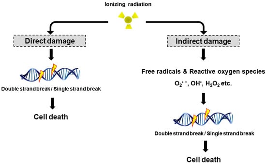
Life forms on the planet are exposed to different forms of radiation. Such radiation may be emitted from the sun, such as X-rays, radio waves, and visible, ultraviolet or infrared light. Radiation may also come from other natural sources, such as α-, β- or γ-radiation.  Concern has also been raised about the effects of exposure to microwaves, naturally occurring radioactivity, and heat (thermal radiation).  The mechanisms by which these forms of radiation affect cell function differ considerably, but high levels of exposure can result in cell death, and long-term exposure is known to cause a range of cancers.

This issue will look at the cytotoxic effects of radiation on living systems; studies will range from those concerning cultured cells to those discussing population and epidemiology.   While emphasis will be placed on radiation-induced cytotoxicity, studies discussing radiation’s connections with DNA damage, intracellular signaling pathways, caspase activation, membrane damage, ROS/RNS production, gene activation, organelle dysfunction, and animal and epidemiological studies are also encouraged.  Reviews of the mechanisms involved in the cytotoxic effects of electromagnetic radiation are welcomed, as well as studies discussing the effects of long-term human exposure to these forms of radiation.  I encourage you to submit a manuscript to this issue so as to help shed further “light” on the lethal effects that radiation has on cells and individuals.

Prof. Dr. Terrence Piva
Collection Editor

Manuscript Submission Information

Manuscripts for the topical collection can be submitted online at www.mdpi.com by registering and logging in to this website. Once you are registered, click here to go to the submission form. All papers will be peer-reviewed. Accepted papers will be published continuously in the journal (as soon as accepted) and will be listed together on this website. The topical collection considers regular research articles, short communications and review articles. A guide for authors and other relevant information for submission of manuscripts is available on the Instructions for Authors page.

Please visit the Instructions for Authors page before submitting a manuscript. The article processing charge (APC) for publication in this open access journal is 2900 CHF (Swiss Francs).

Keywords

  • radiation
  • ultraviolet light
  • X-rays
  • γ radiation
  • microwaves
  • cell death pathways
  • DNA damage
  • cell membrane
  • ROS
  • signaling pathways
  • epidemiology studies
  • animal studies
Published Papers (73 papers)

2022

Jump to: 2021, 2020, 2019, 2018, 2017, 2016, 2015, 2014, 2013

21 pages, 7852 KiB  
Article
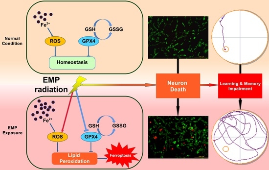
Lipid Peroxides Mediated Ferroptosis in Electromagnetic Pulse-Induced Hippocampal Neuronal Damage via Inhibition of GSH/GPX4 Axis
by Yunfei Lai, Ji Dong, You Wu, Li Zhao, Hui Wang, Jing Zhang, Binwei Yao, Xinping Xu, Yong Zou, Haixia Zhao, Hanlin Yue, Yiwei Song, Haoyu Wang and Ruiyun Peng
Int. J. Mol. Sci. 2022, 23(16), 9277; https://doi.org/10.3390/ijms23169277 - 17 Aug 2022
Cited by 13 | Viewed by 2674
Abstract
Electromagnetic pulse (EMP) radiation was reported to be harmful to hippocampal neurons. However, the mechanism underlying EMP-induced neuronal damage remains unclear. In this paper, for the first time, we attempted to investigate the involvement of ferroptosis in EMP-induced neuronal damage and its underlying [...] Read more.
Electromagnetic pulse (EMP) radiation was reported to be harmful to hippocampal neurons. However, the mechanism underlying EMP-induced neuronal damage remains unclear. In this paper, for the first time, we attempted to investigate the involvement of ferroptosis in EMP-induced neuronal damage and its underlying mechanism. In vivo studies were conducted with a rat model to examine the association of ferroptosis and EMP-induced hippocampal neuronal damage. Moreover, in vitro studies were conducted with HT22 neurons to investigate the underlying mechanism of EMP-induced neuronal ferroptosis. In vivo results showed that EMP could induce learning and memory impairment of rats, ferroptotic morphological damages to mitochondria, accumulation of malonaldehyde (MDA) and iron, overexpression of prostaglandin-endoperoxide synthase 2 (PTGS2) mRNA, and downregulation of GPX4 protein in rat hippocampus. In vitro results showed that EMP could induce neuronal death, MDA accumulation, iron overload, PTGS2 overexpression, and GPX4 downregulation in HT22 neurons. These adverse effects could be reversed by either lipid peroxides scavenger ferrostatin-1 or overexpression of GPX4. These results suggest that EMP radiation can induce ferroptosis in hippocampal neurons via a vicious cycle of lipid peroxides accumulation and GSH/GPX4 axis downregulation. Lipid peroxides and the GSH/GPX4 axis provide potential effective intervention targets to EMP-induced hippocampal neuronal damage. Full article
Show Figures

Graphical abstract

2021

Jump to: 2022, 2020, 2019, 2018, 2017, 2016, 2015, 2014, 2013

14 pages, 3271 KiB  
Article
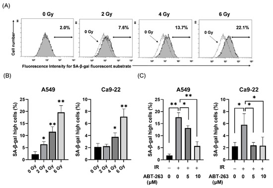
Effects and Related Mechanisms of the Senolytic Agent ABT-263 on the Survival of Irradiated A549 and Ca9-22 Cancer Cells
by Kota Sato, Soichiro Iwasaki and Hironori Yoshino
Int. J. Mol. Sci. 2021, 22(24), 13233; https://doi.org/10.3390/ijms222413233 - 8 Dec 2021
Cited by 6 | Viewed by 4743
Abstract
Senolytic agents eliminate senescent cells and are expected to reduce senescent cell-mediated adverse effects in cancer therapy. However, the effects of senolytic agents on the survival of irradiated cancer cells remain unknown. Here, the effects of the senolytic agent ABT-263 on the survival [...] Read more.
Senolytic agents eliminate senescent cells and are expected to reduce senescent cell-mediated adverse effects in cancer therapy. However, the effects of senolytic agents on the survival of irradiated cancer cells remain unknown. Here, the effects of the senolytic agent ABT-263 on the survival of irradiated A549 and Ca9-22 cancer cells were investigated. ABT-263 was added to the culture medium after irradiation. SA-β-gal activity and cell size, which are hallmarks of cell senescence, were evaluated using a flow cytometer. The colony-forming assay and annexin V staining were performed to test cell survival. We first confirmed that radiation increased the proportion of cells with high SA-β-gal activity and that ABT-263 decreased it. Of note, ABT-263 decreased the survival of irradiated cancer cells and increased the proportion of radiation-induced annexin V+ cells. Furthermore, the caspase inhibitor suppressed the ABT-263-induced decrease in the survival of irradiated cells. Intriguingly, ABT-263 decreased the proportion of SA-β-gal low-activity/large cells in the irradiated A549 cells, which was recovered by the caspase inhibitor. Together, these findings suggest that populations maintaining the ability to proliferate existed among the irradiated cancer cells showing senescence-related features and that ABT-263 eliminated the population, which led to decreased survival of irradiated cancer cells. Full article
Show Figures

Figure 1

15 pages, 29622 KiB  
Article
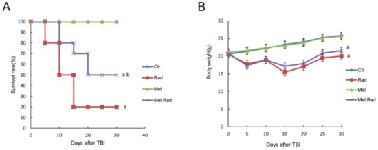
The Effects of Melatonin Administration on Intestinal Injury Caused by Abdominal Irradiation from Mice
by Qin Wang, Yan Wang, Liqing Du, Chang Xu, Qiang Liu and Saijun Fan
Int. J. Mol. Sci. 2021, 22(18), 9715; https://doi.org/10.3390/ijms22189715 - 8 Sep 2021
Cited by 6 | Viewed by 2710
Abstract
Intestinal injury caused by ionizing radiation (IR) is a main clinical issue for patients with cancer receiving abdominal or pelvic radiotherapy. Melatonin (N-acetyl-5-methoxytryptamine) is a neurohormone that the pineal gland in the brain normally secretes. The study aimed to disclose the [...] Read more.
Intestinal injury caused by ionizing radiation (IR) is a main clinical issue for patients with cancer receiving abdominal or pelvic radiotherapy. Melatonin (N-acetyl-5-methoxytryptamine) is a neurohormone that the pineal gland in the brain normally secretes. The study aimed to disclose the potential function of melatonin in intestinal injury induced by IR and its mechanism. Pretreatment with melatonin enhanced the 30-day survival rate of the irradiated mice and promoted the recovery of the intestinal epithelium and hematopoietic function following abdominal irradiation (ABI). Melatonin altered the gene profile of the small intestines from mice following ABI. The enriched biological process terms for melatonin treatment prior to radiation were mainly involved in the immune process. LPS/IL-1-mediated inhibition of RXR Function, TWEAK signaling, and Toll-like receptor signaling were the most activated canonical pathways targeted by melatonin. An upstream analysis network showed that Tripartite motif-containing 24 (TRIM24) was the most significantly inhibited and S100 calcium binding protein A9 (S100A9) activated. TRIM24 activated atherogenesis and cell viability in breast cancer cell lines and S100A9 inhibited the metabolism of amino acids. Melatonin has radioprotective effects on ABI-caused intestinal injury. The mechanisms behind the beneficial effects of melatonin were involved in activation of the immunity. It is necessary to conduct further experiments to explore the underlying mechanisms. Full article
Show Figures

Figure 1

13 pages, 2613 KiB  
Article
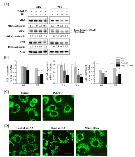
DAP3 Is Involved in Modulation of Cellular Radiation Response by RIG-I-Like Receptor Agonist in Human Lung Adenocarcinoma Cells
by Yoshiaki Sato, Hironori Yoshino, Ikuo Kashiwakura and Eichi Tsuruga
Int. J. Mol. Sci. 2021, 22(1), 420; https://doi.org/10.3390/ijms22010420 - 3 Jan 2021
Cited by 11 | Viewed by 3999
Abstract
Retinoic acid-inducible gene-I (RIG-I)-like receptors (RLRs) mediate anti-viral response through mitochondria. In addition, RLR activation induces anti-tumor effects on various cancers. We previously reported that the RLR agonist Poly(I:C)-HMW/LyoVec™ (Poly(I:C)) enhanced radiosensitivity and that cotreatment with Poly(I:C) and ionizing radiation (IR) more than [...] Read more.
Retinoic acid-inducible gene-I (RIG-I)-like receptors (RLRs) mediate anti-viral response through mitochondria. In addition, RLR activation induces anti-tumor effects on various cancers. We previously reported that the RLR agonist Poly(I:C)-HMW/LyoVec™ (Poly(I:C)) enhanced radiosensitivity and that cotreatment with Poly(I:C) and ionizing radiation (IR) more than additively increased cell death in lung adenocarcinoma cells, indicating that Poly(I:C) modulates the cellular radiation response. However, it remains unclear how mitochondria are involved in the modulation of this response. Here, we investigated the involvement of mitochondrial dynamics and mitochondrial ribosome protein death-associated protein 3 (DAP3) in the modulation of cellular radiation response by Poly(I:C) in A549 and H1299 human lung adenocarcinoma cell lines. Western blotting revealed that Poly(I:C) decreased the expression of mitochondrial dynamics-related proteins and DAP3. In addition, siRNA experiments showed that DAP3, and not mitochondrial dynamics, is involved in the resistance of lung adenocarcinoma cells to IR-induced cell death. Finally, we revealed that a more-than-additive effect of cotreatment with Poly(I:C) and IR on increasing cell death was diluted by DAP3-knockdown because of an increase in cell death induced by IR alone. Together, our findings suggest that RLR agonist Poly(I:C) modulates the cellular radiation response of lung adenocarcinoma cells by downregulating DAP3 expression. Full article
Show Figures

Figure 1

2020

Jump to: 2022, 2021, 2019, 2018, 2017, 2016, 2015, 2014, 2013

12 pages, 1532 KiB  
Brief Report
Geomagnetic Shielding Enhances Radiation Resistance by Promoting DNA Repair Process in Human Bronchial Epithelial Cells
by Xunwen Xue, Yasser F. Ali, Caorui Liu, Zhiqiang Hong, Wanrong Luo, Jing Nie, Bingyan Li, Yang Jiao and Ning-Ang Liu
Int. J. Mol. Sci. 2020, 21(23), 9304; https://doi.org/10.3390/ijms21239304 - 6 Dec 2020
Cited by 12 | Viewed by 2841
Abstract
With the advent of long-duration space explorations, ionizing radiation (IR) may pose a constant threat to astronauts without the protection of Earth’s magnetic field, or hypomagnetic field (HMF). However, the potential biological effects of a HMF on the cellular response to IR have [...] Read more.
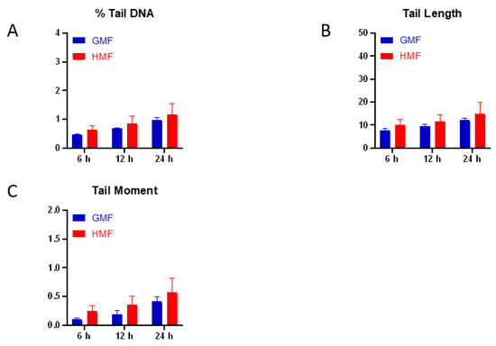
With the advent of long-duration space explorations, ionizing radiation (IR) may pose a constant threat to astronauts without the protection of Earth’s magnetic field, or hypomagnetic field (HMF). However, the potential biological effects of a HMF on the cellular response to IR have not been well characterized so far. In this study, immortalized human bronchial epithelial cells were exposed to X-rays under either a geomagnetic field (GMF, ~50 uT) or HMF (<50 nT) culture condition. A significant increase of the cell survival rate in HMF after radiation was observed by colony formation analysis. The kinetics of DNA double-strand breaks (DSBs), determined by γH2AX foci formation and disappearance, presented a faster decrease of foci-positive cells and a significantly lower mean number of γH2AX foci per nucleus in HMF-cultured cells than in GMF-cultured cells after radiation. In addition, a γH2AX/53BP1 colocalization assay showed an upregulated DSB recovery rate in HMF cultured cells. These findings provided the first evidence that HMF exposure may enhance the cellular DSB repair efficiency upon radiation, and consequently modulate the genotoxic effects of IR. Full article
Show Figures

Figure 1

21 pages, 4060 KiB  
Article
The p53–53BP1-Related Survival of A549 and H1299 Human Lung Cancer Cells after Multifractionated Radiotherapy Demonstrated Different Response to Additional Acute X-ray Exposure
by Margarita Pustovalova, Lina Alhaddad, Nadezhda Smetanina, Anna Chigasova, Taisia Blokhina, Roman Chuprov-Netochin, Andreyan N. Osipov and Sergey Leonov
Int. J. Mol. Sci. 2020, 21(9), 3342; https://doi.org/10.3390/ijms21093342 - 8 May 2020
Cited by 23 | Viewed by 5369
Abstract
Radiation therapy is one of the main methods of treating patients with non-small cell lung cancer (NSCLC). However, the resistance of tumor cells to exposure remains the main factor that limits successful therapeutic outcome. To study the molecular/cellular mechanisms of increased resistance of [...] Read more.
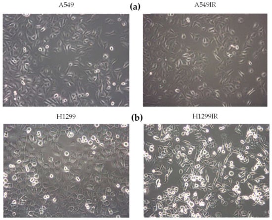
Radiation therapy is one of the main methods of treating patients with non-small cell lung cancer (NSCLC). However, the resistance of tumor cells to exposure remains the main factor that limits successful therapeutic outcome. To study the molecular/cellular mechanisms of increased resistance of NSCLC to ionizing radiation (IR) exposure, we compared A549 (p53 wild-type) and H1299 (p53-deficient) cells, the two NSCLC cell lines. Using fractionated X-ray irradiation of these cells at a total dose of 60 Gy, we obtained the survived populations and named them A549IR and H1299IR, respectively. Further characterization of these cells showed multiple alterations compared to parental NSCLC cells. The additional 2 Gy exposure led to significant changes in the kinetics of γH2AX and phosphorylated ataxia telangiectasia mutated (pATM) foci numbers in A549IR and H1299IR compared to parental NSCLC cells. Whereas A549, A549IR, and H1299 cells demonstrated clear two-component kinetics of DNA double-strand break (DSB) repair, H1299IR showed slower kinetics of γH2AX foci disappearance with the presence of around 50% of the foci 8 h post-IR. The character of H2AX phosphorylation in these cells was pATM-independent. A decrease of residual γH2AX/53BP1 foci number was observed in both A549IR and H1299IR compared to parental cells post-IR at extra doses of 2, 4, and 6 Gy. This process was accompanied with the changes in the proliferation, cell cycle, apoptosis, and the expression of ATP-binding cassette sub-family G member 2 (ABCG2, also designated as CDw338 and the breast cancer resistance protein (BCRP)) protein. Our study provides strong evidence that different DNA repair mechanisms are activated by multifraction radiotherapy (MFR), as well as single-dose IR, and that the enhanced cellular survival after MFR is reliant on both p53 and 53BP1 signaling along with non-homologous end-joining (NHEJ). Our results are of clinical significance as they can guide the choice of the most effective IR regimen by analyzing the expression status of the p53–53BP1 pathway in tumors and thereby maximize therapeutic benefits for the patients while minimizing collateral damage to normal tissue. Full article
Show Figures

Figure 1

2019

Jump to: 2022, 2021, 2020, 2018, 2017, 2016, 2015, 2014, 2013

10 pages, 1185 KiB  
Article
Laser-Driven Ultrashort Pulsed Electron Beam Radiation at Doses of 0.5 and 1.0 Gy Induces Apoptosis in Human Fibroblasts
by Nelly Babayan, Bagrat Grigoryan, Lusine Khondkaryan, Gohar Tadevosyan, Natalya Sarkisyan, Ruzanna Grigoryan, Lilit Apresyan, Rouben Aroutiounian, Natalia Vorobyeva, Margarita Pustovalova, Anna Grekhova and Andreyan N. Osipov
Int. J. Mol. Sci. 2019, 20(20), 5140; https://doi.org/10.3390/ijms20205140 - 17 Oct 2019
Cited by 20 | Viewed by 3539
Abstract
Rapidly evolving laser technologies have led to the development of laser-generated particle accelerators as an alternative to conventional facilities. However, the radiobiological characteristics need to be determined to enhance their applications in biology and medicine. In this study, the radiobiological effects of ultrashort [...] Read more.
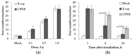
Rapidly evolving laser technologies have led to the development of laser-generated particle accelerators as an alternative to conventional facilities. However, the radiobiological characteristics need to be determined to enhance their applications in biology and medicine. In this study, the radiobiological effects of ultrashort pulsed electron beam (UPEB) and X-ray radiation in human lung fibroblasts (MRC-5 cell line) exposed to doses of 0.1, 0.5, and 1 Gy are compared. The changes of γH2AX foci number as a marker of DNA double-strand breaks (DSBs) were analyzed. In addition, the micronuclei induction and cell death via apoptosis were studied. We found that the biological action of UPEB-radiation compared to X-rays was characterized by significantly slower γH2AX foci elimination (with a dose of 1 Gy) and strong apoptosis induction (with doses of 0.5 and 1.0 Gy), accompanied by a slight increase in micronuclei formation (dose of 1 Gy). Our data suggest that UPEB radiation produces more complex DNA damage than X-ray radiation, leading to cell death rather than cytogenetic disturbance. Full article
Show Figures

Figure 1

12 pages, 3103 KiB  
Article
Effect of Ionizing Radiation from Computed Tomography on Differentiation of Human Embryonic Stem Cells into Neural Precursors
by Christine Hanu, Burk W. Loeliger, Irina V. Panyutin, Roberto Maass-Moreno, Paul Wakim, William F. Pritchard, Ronald D. Neumann and Igor G. Panyutin
Int. J. Mol. Sci. 2019, 20(16), 3900; https://doi.org/10.3390/ijms20163900 - 10 Aug 2019
Cited by 5 | Viewed by 3945
Abstract
We studied the effect of radiation from computed tomography (CT) scans on differentiation of human embryonic stem cells (hESCs) into neuronal lineage. hESCs were divided into three radiation exposure groups: 0-dose, low-dose, or high-dose exposure. Low dose was accomplished with a single 15 [...] Read more.
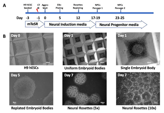
We studied the effect of radiation from computed tomography (CT) scans on differentiation of human embryonic stem cells (hESCs) into neuronal lineage. hESCs were divided into three radiation exposure groups: 0-dose, low-dose, or high-dose exposure. Low dose was accomplished with a single 15 mGy CT dose index (CTDI) CT scan that approximated the dose for abdominal/pelvic CT examinations in adults while the high dose was achieved with several consecutive CT scans yielding a cumulative dose of 500 mGy CTDI. The neural induction was characterized by immunocytochemistry. Quantitative polymerase chain reaction (qPCR) and Western blots were used to measure expression of the neuronal markers PAX6 and NES and pluripotency marker OCT4. We did not find any visible morphological differences between neural precursors from irradiated and non-irradiated cells. However, quantitative analyses of neuronal markers showed that PAX6 expression was reduced following exposure to the high dose compared to 0-dose controls, while no such decrease in PAX6 expression was observed following exposure to the low dose. Similarly, a statistically significant reduction in expression of NES was observed following high-dose exposure, while after low-dose exposure, a modest but statistically significant reduction in NES expression was only observed on Day 8 of differentiation. Further studies are warranted to elucidate how lower or delayed expression of PAX6 and NES can impact human fetal brain development. Full article
Show Figures

Figure 1

13 pages, 1177 KiB  
Communication
Formation of γH2AX and pATM Foci in Human Mesenchymal Stem Cells Exposed to Low Dose-Rate Gamma-Radiation
by Stepan Ulyanenko, Margarita Pustovalova, Sergey Koryakin, Evgenii Beketov, Anatolii Lychagin, Liliya Ulyanenko, Andrey Kaprin, Anna Grekhova, Alexandra M. Ozerova, Ivan V. Ozerov, Natalia Vorobyeva, Peter Shegay, Sergey Ivanov, Sergey Leonov, Dmitry Klokov and Andreyan N. Osipov
Int. J. Mol. Sci. 2019, 20(11), 2645; https://doi.org/10.3390/ijms20112645 - 29 May 2019
Cited by 31 | Viewed by 5061
Abstract
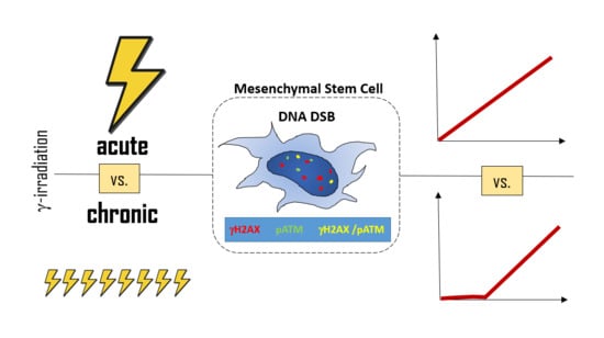
DNA double-strand breaks (DSB) are among the most harmful DNA lesions induced by ionizing radiation (IR). Although the induction and repair of radiation-induced DSB is well studied for acute irradiation, responses to DSB produced by chronic IR exposures are poorly understood, especially in [...] Read more.
DNA double-strand breaks (DSB) are among the most harmful DNA lesions induced by ionizing radiation (IR). Although the induction and repair of radiation-induced DSB is well studied for acute irradiation, responses to DSB produced by chronic IR exposures are poorly understood, especially in human stem cells. The aim of this study was to examine the formation of DSB markers (γH2AX and phosphorylated kinase ATM, pATM, foci) in human mesenchymal stem cells (MSCs) exposed to chronic gamma-radiation (0.1 mGy/min) in comparison with acute irradiation (30 mGy/min) at cumulative doses of 30, 100, 160, 240 and 300 mGy. A linear dose-dependent increase in the number of both γH2AX and pATM foci, as well as co-localized γH2AX/pATM foci (“true” DSB), were observed after an acute radiation exposure. In contrast, the response of MSCs to a chronic low dose-rate IR exposure deviated from linearity towards a threshold model, for γH2AX, pATM foci and γH2AX/pATM foci, with an indication of a “plateau”. The state of equilibrium between newly formed DSB at a low rate during the protracted exposure time and the elimination of a fraction of DSB is proposed as a mechanistic explanation of the non-linear DSB responses following a low dose-rate irradiation. This notion is supported by the observation of the elimination of a substantial fraction of DSB 6 h after the cessation of the exposures. Our results demonstrate non-linear dose responses for γH2AX and pATM foci in human MSCs exposed to low dose-rate IR and showed the existence of a threshold, which may have implications for radiation protection in humans. Full article
Show Figures

Graphical abstract

20 pages, 5813 KiB  
Article
Bone Marrow Endothelial Cells Influence Function and Phenotype of Hematopoietic Stem and Progenitor Cells after Mixed Neutron/Gamma Radiation
by Lynnette Cary, Daniel Noutai, Rudolph Salber, Opeyemi Fadiyimu, Arthur Gross, Graca Almeida-Porada, Yared Kidane and Mark Whitnall
Int. J. Mol. Sci. 2019, 20(7), 1795; https://doi.org/10.3390/ijms20071795 - 11 Apr 2019
Cited by 10 | Viewed by 4725
Abstract
The bone marrow (BM) microenvironment plays a crucial role in the maintenance and regeneration of hematopoietic stem (HSC) and progenitor cells (HSPC). In particular, the vascular niche is responsible for regulating HSC maintenance, differentiation, and migration of cells in and out of the [...] Read more.
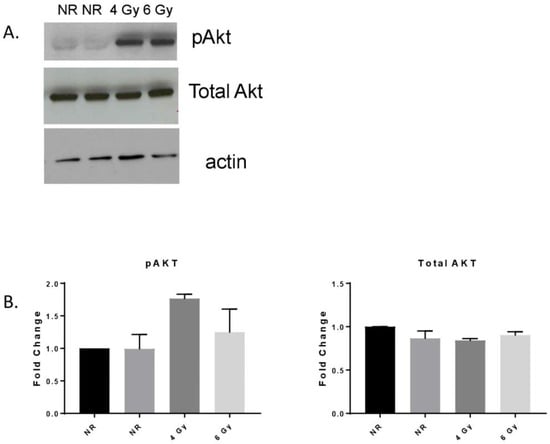
The bone marrow (BM) microenvironment plays a crucial role in the maintenance and regeneration of hematopoietic stem (HSC) and progenitor cells (HSPC). In particular, the vascular niche is responsible for regulating HSC maintenance, differentiation, and migration of cells in and out of the BM. Damage to this niche upon exposure to ionizing radiation, whether accidental or as a result of therapy, can contribute to delays in HSC recovery and/or function. The ability of BM derived-endothelial cells (BMEC) to alter and/or protect HSPC after exposure to ionizing radiation was investigated. Our data show that exposure of BMEC to ionizing radiation resulted in alterations in Akt signaling, increased expression of PARP-1, IL6, and MCP-1, and decreased expression of MMP1 and MMP9. In addition, global analysis of gene expression of HSC and BMEC in response to mixed neutron/gamma field (MF) radiation identified 60 genes whose expression was altered after radiation in both cell types, suggesting that a subset of genes is commonly affected by this type of radiation. Focused gene analysis by RT-PCR revealed two categories of BMEC alterations: (a) a subset of genes whose expression was altered in response to radiation, with no additional effect observed during coculture with HSPC, and (b) a subset of genes upregulated in response to radiation, and altered when cocultured with HSPC. Coculture of BMEC with CD34+ HSPC induced HSPC proliferation, and improved BM function after MF radiation. Nonirradiated HSPC exhibited reduced CD34 expression over time, but when irradiated, they maintained higher CD34 expression. Nonirradiated HSPC cocultured with nonirradiated BMEC expressed lower levels of CD34 expression compared to nonirradiated alone. These data characterize the role of each cell type in response to MF radiation and demonstrate the interdependence of each cell’s response to ionizing radiation. The identified genes modulated by radiation and coculture provide guidance for future experiments to test hypotheses concerning specific factors mediating the beneficial effects of BMEC on HSPC. This information will prove useful in the search for medical countermeasures to radiation-induced hematopoietic injury. Full article
Show Figures

Figure 1

2018

Jump to: 2022, 2021, 2020, 2019, 2017, 2016, 2015, 2014, 2013

14 pages, 2245 KiB  
Article
Melatonin Sensitizes Human Colorectal Cancer Cells to γ-ray Ionizing Radiation In Vitro and In Vivo
by Qin Wang, Zhijuan Sun, Liqing Du, Chang Xu, Yan Wang, Bing Yang, Ningning He, Jinhan Wang, Kaihua Ji, Yang Liu and Qiang Liu
Int. J. Mol. Sci. 2018, 19(12), 3974; https://doi.org/10.3390/ijms19123974 - 10 Dec 2018
Cited by 31 | Viewed by 4660
Abstract
Colorectal cancer is the most commonly reported gastrointestinal malignancy, with a recent, rapid increase of the annual incidence all over the world. Enhancing the radiosensitivity of cancer cells while preserving the health of normal cells is one of the most important tasks in [...] Read more.
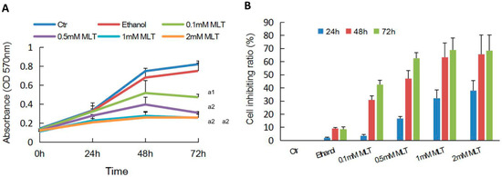
Colorectal cancer is the most commonly reported gastrointestinal malignancy, with a recent, rapid increase of the annual incidence all over the world. Enhancing the radiosensitivity of cancer cells while preserving the health of normal cells is one of the most important tasks in clinical radiobiology. However, resistance to radiotherapy for colorectal cancer greatly decreases the therapeutic outcome. Melatonin (N-acetyl-5-methoxytryptamine), a natural secretory product that the pineal gland in the brain normally produces, has been reported to have anticancer properties. In the study, we investigated the combination of melatonin with radiotherapy as a treatment for colorectal cancer. We firstly explored the anti-tumor activity of melatonin combined with ionizing radiation (IR) against colorectal carcinoma in vitro. It was found that melatonin effectively inhibited human colorectal carcinoma cell line HCT 116 cellular proliferation, colony formation rate and cell migration counts following IR. Increasing the radiosensitivity of colorectal cancer cells by melatonin treatment was found to be associated with cell cycle arrest in the G2/M phase, downregulation of proteins involved in DNA double-strand break repair and activation of the caspase-dependent apoptotic pathway. Moreover, we also investigated the combined effect of IR and melatonin on colorectal tumor in vivo. Results from a tumor xenograft showed that melatonin plus IR treatment significantly suppressed tumor cell growth compared with melatonin or IR alone, resulting in a much higher tumor inhibition rate for the combined treatment. The data suggested that melatonin combined with IR could improve the radiosensitivity of colorectal cancer and thus enhance the therapeutic effect of the patients, implying melatonin could function as a potential sensitizer in tumor radiotherapy. Full article
Show Figures

Figure 1

17 pages, 3415 KiB  
Article
Relationship between the Regulation of Caspase-8-Mediated Apoptosis and Radioresistance in Human THP-1-Derived Macrophages
by Hironori Yoshino, Haruka Konno, Koya Ogura, Yoshiaki Sato and Ikuo Kashiwakura
Int. J. Mol. Sci. 2018, 19(10), 3154; https://doi.org/10.3390/ijms19103154 - 13 Oct 2018
Cited by 10 | Viewed by 5228
Abstract
Radiosensitivity varies depending on the cell type; highly differentiated cells typically exhibit greater radioresistance. We recently demonstrated that human macrophages derived from THP-1 monocytic cells, which lack TP53, are highly resistant to radiation-induced apoptosis compared with undifferentiated THP-1 cells. However, the mechanisms [...] Read more.
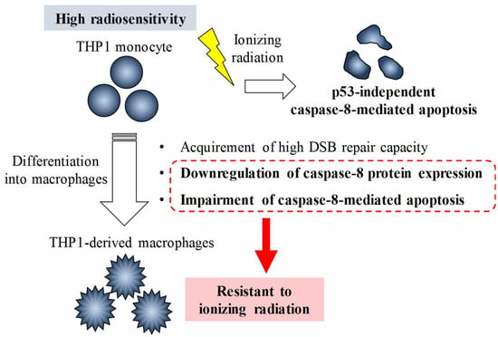
Radiosensitivity varies depending on the cell type; highly differentiated cells typically exhibit greater radioresistance. We recently demonstrated that human macrophages derived from THP-1 monocytic cells, which lack TP53, are highly resistant to radiation-induced apoptosis compared with undifferentiated THP-1 cells. However, the mechanisms by which THP-1 cells acquire radioresistance during differentiation remain unknown. Herein, we investigated the mechanisms by which THP-1-derived macrophages develop p53-independent radioresistance by analyzing DNA damage responses and apoptotic pathways. Analysis of γ-H2AX foci, which indicates the formation of DNA double-strand breaks (DSB), suggested that a capacity to repair DSB of macrophages is comparable to that of radiosensitive THP-1 cells. Furthermore, treatment with inhibitors against DSB repair-related proteins failed to enhance radiation-induced apoptosis in THP-1-derrived macrophages. Analysis of the apoptotic pathways showed that radiosensitive THP-1 cells undergo apoptosis through the caspase-8/caspase-3 cascade after irradiation, whereas this was not observed in the macrophages. Caspase-8 protein expression was lower in macrophages than in THP-1 cells, whereas mRNA expressions were comparable between both cell types. Co-treatment with a proteasome inhibitor and ionizing radiation effectively induced apoptosis in macrophages in a caspase-8-dependent manner. Results suggest that the regulation of caspase-8-mediated apoptosis during differentiation plays a role in the p53-independent radioresistance of THP-1-derived macrophages. Full article
Show Figures

Graphical abstract

13 pages, 2832 KiB  
Article
Oridonin Enhances Radiation-Induced Cell Death by Promoting DNA Damage in Non-Small Cell Lung Cancer Cells
by Hyejin Park, Ye Ji Jeong, Na-Kyung Han, Joong Sun Kim and Hae-June Lee
Int. J. Mol. Sci. 2018, 19(8), 2378; https://doi.org/10.3390/ijms19082378 - 13 Aug 2018
Cited by 29 | Viewed by 5068
Abstract
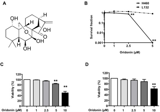
Although many attempts have been made to improve the efficacy of radiotherapy to treat cancer, radiation resistance is still an obstacle in lung cancer treatment. Oridonin is a natural compound with promising antitumor efficacy that can trigger cancer cell death; however, its direct [...] Read more.
Although many attempts have been made to improve the efficacy of radiotherapy to treat cancer, radiation resistance is still an obstacle in lung cancer treatment. Oridonin is a natural compound with promising antitumor efficacy that can trigger cancer cell death; however, its direct cellular targets, efficacy as a radiosensitizer, and underlying mechanisms of activity remain unclear. Herein, we report that oridonin exhibits additive cytotoxic and antitumor activity with radiation using the H460 non-small cell lung cancer cell lines. We assessed the effect of oridonin by proliferation, clonogenic, reactive oxygen species (ROS) production, DNA damage, and apoptosis assays. In vitro, oridonin enhanced the radiation-induced inhibition of cell growth and clonogenic survival. Oridonin also facilitated radiation-induced ROS production and DNA damage and enhanced apoptotic cell death. In vivo, the combination of oridonin and radiation effectively inhibited H460 xenograft tumor growth, with higher caspase-3 activation and H2A histone family member X (H2AX) phosphorylation compared with that of radiation alone. Our findings suggest that oridonin possesses a novel mechanism to enhance radiation therapeutic responses by increasing DNA damage and apoptosis. In conclusion, oridonin may be a novel small molecule to improve radiotherapy in non-small cell lung cancer. Full article
Show Figures

Figure 1

2017

Jump to: 2022, 2021, 2020, 2019, 2018, 2016, 2015, 2014, 2013

897 KiB  
Review
Molecular Pathogenesis of Radiation-Induced Cell Toxicity in Stem Cells
by Wonhee Hur and Seung Kew Yoon
Int. J. Mol. Sci. 2017, 18(12), 2749; https://doi.org/10.3390/ijms18122749 - 18 Dec 2017
Cited by 33 | Viewed by 16189
Abstract
Radiation therapy is an effective cancer therapy, but damage to normal tissues surrounding the tumor due to radiotherapy causes severe complications. The importance of the therapeutic area between tumor suppression and normal tissue injury has long been highlighted in radiation therapy. Recent advances [...] Read more.
Radiation therapy is an effective cancer therapy, but damage to normal tissues surrounding the tumor due to radiotherapy causes severe complications. The importance of the therapeutic area between tumor suppression and normal tissue injury has long been highlighted in radiation therapy. Recent advances in stem cell biology have shown that stem cell (SC) responses to genotoxic stresses of ionizing radiation can improve the therapeutic effect of radiation by repairing damaged cells. In contrast, cancer stem cells (CSCs), a small subpopulation of cells within tumors, are generally resistant to chemotherapy and radiotherapy and cause tumor recurrence. Although the underlying mechanisms are not clearly understood in detail, efforts are still underway to identify SC treatment or CSC resistant pathogenesis of DNA damage agents such as radiation therapy. In response to radiation, CSCs differ from normal SCs in their biological properties due to severe deregulation of the self-renewal ability in CSCs. Differences of cleavage mode, cell cycle characteristics, replication potential, and activation/inactivation of DNA damage treatment and cancer-specific molecular pathways between normal SCs and CSCs confer a malignant phenotype upon CSCs. However, further studies are needed to identify normal SC and CSC-specific targets. In this review, we summarize the current advances in research regarding how normal SCs and CSCs respond to ionizing radiation, with a special emphasis on cell toxicity, radiosensitivity, signaling networks, DNA damage response (DDR) and DNA repair. In addition, we discuss strategies to develop new diagnostic and therapeutic techniques for predicting responses to cancer treatment and overcoming radiation-related toxicity. Full article
Show Figures

Figure 1

3627 KiB  
Article
Impact of UVR Exposure Pattern on Squamous Cell Carcinoma-A Dose–Delivery and Dose–Response Study in Pigmented Hairless Mice
by Catharina M. Lerche, Katrine Togsverd-Bo, Peter A. Philipsen and Hans Christian Wulf
Int. J. Mol. Sci. 2017, 18(12), 2738; https://doi.org/10.3390/ijms18122738 - 16 Dec 2017
Cited by 10 | Viewed by 4575
Abstract
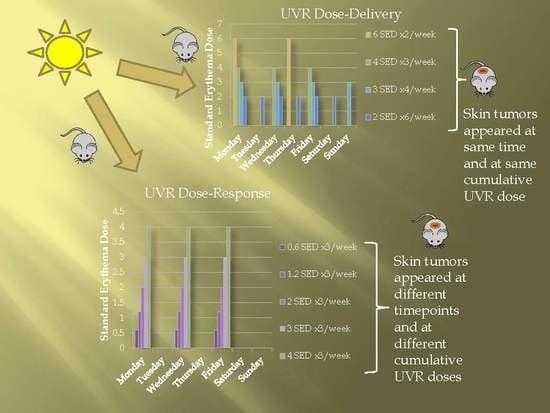
Cumulative lifetime ultraviolet radiation (UVR) is an important factor in the development of squamous cell carcinoma. This study examines the impact of UVR exposure pattern on tumor development. Hairless C3.Cg/TifBomTac immunocompetent pigmented mice (n = 351) were irradiated with 12 standard erythema [...] Read more.
Cumulative lifetime ultraviolet radiation (UVR) is an important factor in the development of squamous cell carcinoma. This study examines the impact of UVR exposure pattern on tumor development. Hairless C3.Cg/TifBomTac immunocompetent pigmented mice (n = 351) were irradiated with 12 standard erythema doses (SED)/week, given as 2 SED ×6, 3 SED ×4, 4 SED ×3, or 6 SED ×2 (dose–delivery study) or 0, 0.6, 1.2, 2, 3 or 4 SED ×3/week (dose–response study). All mice were irradiated until development of 3 tumors of 4 mm each. Pigmentation was measured once monthly. In the dose–delivery study, the median time until tumor development was independent of dose fractions. In the dose–response study, higher UVR doses resulted in faster tumor appearance. When the weekly UVR dose was decreased from 12 to 6 SED, the cumulative UVR dose needed for tumor development was reduced by 40%. In conclusion, delivery schedules of a fixed weekly UVR dose did not affect tumor development. When using different weekly UVR doses, longer time to tumor development was observed using lower UVR doses. Lower weekly UVR doses however resulted in lower cumulative UVR doses to induce tumors in hairless pigmented mice. Full article
Show Figures

Graphical abstract

299 KiB  
Review
Pharmacological Modulation of Radiation Damage. Does It Exist a Chance for Other Substances than Hematopoietic Growth Factors and Cytokines?
by Michal Hofer, Zuzana Hoferová and Martin Falk
Int. J. Mol. Sci. 2017, 18(7), 1385; https://doi.org/10.3390/ijms18071385 - 28 Jun 2017
Cited by 27 | Viewed by 5012
Abstract
In recent times, cytokines and hematopoietic growth factors have been at the center of attention for many researchers trying to establish pharmacological therapeutic procedures for the treatment of radiation accident victims. Two granulocyte colony-stimulating factor-based radiation countermeasures have been approved for the treatment [...] Read more.
In recent times, cytokines and hematopoietic growth factors have been at the center of attention for many researchers trying to establish pharmacological therapeutic procedures for the treatment of radiation accident victims. Two granulocyte colony-stimulating factor-based radiation countermeasures have been approved for the treatment of the hematopoietic acute radiation syndrome. However, at the same time, many different substances with varying effects have been tested in animal studies as potential radioprotectors and mitigators of radiation damage. A wide spectrum of these substances has been studied, comprising various immunomodulators, prostaglandins, inhibitors of prostaglandin synthesis, agonists of adenosine cell receptors, herbal extracts, flavonoids, vitamins, and others. These agents are often effective, relatively non-toxic, and cheap. This review summarizes the results of animal experiments, which show the potential for some of these untraditional or new radiation countermeasures to become a part of therapeutic procedures applicable in patients with the acute radiation syndrome. The authors consider β-glucan, 5-AED (5-androstenediol), meloxicam, γ-tocotrienol, genistein, IB-MECA (N6-(3-iodobezyl)adenosine-5’-N-methyluronamide), Ex-RAD (4-carboxystyryl-4-chlorobenzylsulfone), and entolimod the most promising agents, with regards to their contingent use in clinical practice. Full article
1247 KiB  
Article
Pre-Exposure Gene Expression in Baboons with and without Pancytopenia after Radiation Exposure
by Matthias Port, Francis Hérodin, Marco Valente, Michel Drouet, Reinhard Ullmann, Matthäus Majewski and Michael Abend
Int. J. Mol. Sci. 2017, 18(3), 541; https://doi.org/10.3390/ijms18030541 - 2 Mar 2017
Cited by 19 | Viewed by 5064
Abstract
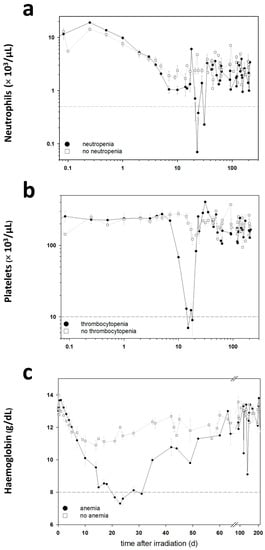
Radiosensitivity differs in humans and likely among primates. The reasons are not well known. We examined pre-exposure gene expression in baboons (n = 17) who developed haematologic acute radiation syndrome (HARS) without pancytopenia or a more aggravated HARS with pancytopenia after irradiation. [...] Read more.
Radiosensitivity differs in humans and likely among primates. The reasons are not well known. We examined pre-exposure gene expression in baboons (n = 17) who developed haematologic acute radiation syndrome (HARS) without pancytopenia or a more aggravated HARS with pancytopenia after irradiation. We evaluated gene expression in a two stage study design where stage I comprised a whole genome screen for messenger RNAs (mRNA) (microarray) and detection of 667 microRNAs (miRNA) (real-time quantitative polymerase chain reaction (qRT-PCR) platform). Twenty candidate mRNAs and nine miRNAs were selected for validation in stage II (qRT-PCR). None of the mRNA species could be confirmed during the validation step, but six of the nine selected candidate miRNA remained significantly different during validation. In particular, miR-425-5p (receiver operating characteristic = 0.98; p = 0.0003) showed nearly complete discrimination between HARS groups with and without pancytopenia. Target gene searches of miR-425-5p identified new potential mRNAs and associated biological processes linked with radiosensitivity. We found that one miRNA species examined in pre-exposure blood samples was associated with HARS characterized by pancytopenia and identified new target mRNAs that might reflect differences in radiosensitivity of irradiated normal tissue. Full article
Show Figures

Figure 1

2016

Jump to: 2022, 2021, 2020, 2019, 2018, 2017, 2015, 2014, 2013

5869 KiB  
Article
Development of Orally Administered γ-Tocotrienol (GT3) Nanoemulsion for Radioprotection
by Grace A. Ledet, Shukla Biswas, Vidya P. Kumar, Richard A. Graves, Demaurian M. Mitchner, Taylor M. Parker, Levon A. Bostanian, Sanchita P. Ghosh and Tarun K. Mandal
Int. J. Mol. Sci. 2017, 18(1), 28; https://doi.org/10.3390/ijms18010028 - 24 Dec 2016
Cited by 15 | Viewed by 5619
Abstract
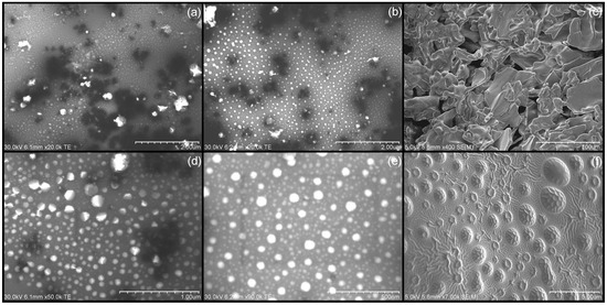
The purpose of this study was two-fold: (1) to formulate γ-tocotrienol (GT3) in a nanoemulsion formulation as a prophylactic orally administered radioprotective agent; and (2) to optimize the storage conditions to preserve the structural integrity of both the formulation and the compound. γ-tocotrienol [...] Read more.
The purpose of this study was two-fold: (1) to formulate γ-tocotrienol (GT3) in a nanoemulsion formulation as a prophylactic orally administered radioprotective agent; and (2) to optimize the storage conditions to preserve the structural integrity of both the formulation and the compound. γ-tocotrienol was incorporated into a nanoemulsion and lyophilized with lactose. Ultra performance liquid chromatography–mass spectroscopy (UPLC–MS) was used to monitor the chemical stability of GT3 over time, the particle size and ζ potential, and scanning electron microscopy (SEM) were used to study the physical stability of the nanoemulsion. Radioprotective and toxicity studies were performed in mice. The liquid formulation exhibited GT3 degradation at all storage temperatures. Lyophilization, in the presence of lactose, significantly reduced GT3 degradation. Both the liquid and lyophilized nanoemulsions had stable particle size and ζ potential when stored at 4 °C. Toxicity studies of the nanoemulsion resulted in no observable toxicity in mice at an oral dose of 600 mg/kg GT3. The nano-formulated GT3 (300 mg/kg) demonstrated enhanced survival efficacy compared to GT3 alone (200 and 400 mg/kg) in CD2F1 mice exposed to total body gamma radiation. The optimal long-term storage of formulated GT3 is as a powder at −20 °C to preserve drug and formulation integrity. Formulation of GT3 as a nanoemulsion for oral delivery as a prophylactic radioprotectant shows promise and warrants further investigation. Full article
Show Figures

Figure 1

1684 KiB  
Article
Low Concentration of Exogenous Carbon Monoxide Modulates Radiation-Induced Bystander Effect in Mammalian Cell Cluster Model
by Wenqing Wu, Lili Nie, K. N. Yu, Lijun Wu, Peizhong Kong, Lingzhi Bao, Guodong Chen, Haoran Yang and Wei Han
Int. J. Mol. Sci. 2016, 17(12), 2051; https://doi.org/10.3390/ijms17122051 - 8 Dec 2016
Cited by 4 | Viewed by 6533
Abstract
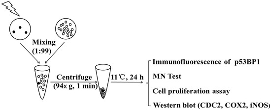
During radiotherapy procedures, radiation-induced bystander effect (RIBE) can potentially lead to genetic hazards to normal tissues surrounding the targeted regions. Previous studies showed that RIBE intensities in cell cluster models were much higher than those in monolayer cultured cell models. On the other [...] Read more.
During radiotherapy procedures, radiation-induced bystander effect (RIBE) can potentially lead to genetic hazards to normal tissues surrounding the targeted regions. Previous studies showed that RIBE intensities in cell cluster models were much higher than those in monolayer cultured cell models. On the other hand, low-concentration carbon monoxide (CO) was previously shown to exert biological functions via binding to the heme domain of proteins and then modulating various signaling pathways. In relation, our previous studies showed that exogenous CO generated by the CO releasing molecule, tricarbonyldichlororuthenium (CORM-2), at a relatively low concentration (20 µM), effectively attenuated the formation of RIBE-induced DNA double-strand breaks (DSB) and micronucleus (MN). In the present work, we further investigated the capability of a low concentration of exogenous CO (CORM-2) of attenuating or inhibiting RIBE in a mixed-cell cluster model. Our results showed that CO (CORM-2) with a low concentration of 30 µM could effectively suppress RIBE-induced DSB (p53 binding protein 1, p53BP1), MN formation and cell proliferation in bystander cells but not irradiated cells via modulating the inducible nitric oxide synthase (iNOS) andcyclooxygenase-2 (COX-2). The results can help mitigate RIBE-induced hazards during radiotherapy procedures. Full article
Show Figures

Figure 1

2257 KiB  
Article
NOD Scid Gamma Mice Are Permissive to Allogeneic HSC Transplantation without Prior Conditioning
by Tom Verbiest, Rosemary Finnon, Natalie Brown, Paul Finnon, Simon Bouffler and Christophe Badie
Int. J. Mol. Sci. 2016, 17(11), 1850; https://doi.org/10.3390/ijms17111850 - 7 Nov 2016
Cited by 8 | Viewed by 6446
Abstract
Scid hematopoietic stem cells (HSCs) have an intrinsic defect in their maintenance within the bone marrow (BM) niche which facilitates HSC transplantation without the absolute requirement of prior conditioning. Nevertheless, NOD scid mice have a significantly altered life span due to early development [...] Read more.
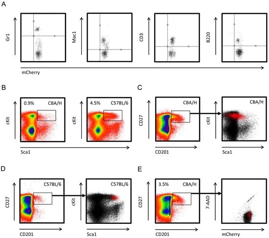
Scid hematopoietic stem cells (HSCs) have an intrinsic defect in their maintenance within the bone marrow (BM) niche which facilitates HSC transplantation without the absolute requirement of prior conditioning. Nevertheless, NOD scid mice have a significantly altered life span due to early development of thymic lymphomas, which compromises the ability to study the long-term fate of exogenous HSCs and their progeny. Here, we present data on the transplantation of HSCs into NOD scid gamma (NSG) mice to achieve long-term engraftment without prior conditioning. We transplanted allogeneic HSCs constitutively expressing the mCherry fluorescent marker into age-matched NSG mice and assessed donor chimerism 6 months post-transplantation. All transplanted NSG mice showed long-term myeloid and lymphoid cell chimerism. Also, in vivo irradiated HSCs showed long-term engraftment, although overall white blood cell (WBC) donor chimerism was lower compared with non-irradiated HSCs. Using this novel NSG transplantation model, we will be able to study the effects of low dose in vivo X-ray exposure on the long-term fate of HSCs, without the requirement of prior radio-ablation of the recipient, and thus leaving the recipient’s BM microenvironment uncompromised. In conclusion, we demonstrated for the first time that allogeneic HSCs from a different inbred strain can compete for niches in the BM compartment of NSG mice. Full article
Show Figures

Figure 1

1026 KiB  
Review
Transcriptional and Posttranslational Regulation of Nucleotide Excision Repair: The Guardian of the Genome against Ultraviolet Radiation
by Jeong-Min Park and Tae-Hong Kang
Int. J. Mol. Sci. 2016, 17(11), 1840; https://doi.org/10.3390/ijms17111840 - 4 Nov 2016
Cited by 30 | Viewed by 8047
Abstract
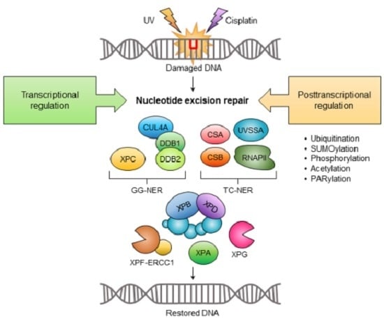
Ultraviolet (UV) radiation from sunlight represents a constant threat to genome stability by generating modified DNA bases such as cyclobutane pyrimidine dimers (CPD) and pyrimidine-pyrimidone (6-4) photoproducts (6-4PP). If unrepaired, these lesions can have deleterious effects, including skin cancer. Mammalian cells are able [...] Read more.
Ultraviolet (UV) radiation from sunlight represents a constant threat to genome stability by generating modified DNA bases such as cyclobutane pyrimidine dimers (CPD) and pyrimidine-pyrimidone (6-4) photoproducts (6-4PP). If unrepaired, these lesions can have deleterious effects, including skin cancer. Mammalian cells are able to neutralize UV-induced photolesions through nucleotide excision repair (NER). The NER pathway has multiple components including seven xeroderma pigmentosum (XP) proteins (XPA to XPG) and numerous auxiliary factors, including ataxia telangiectasia and Rad3-related (ATR) protein kinase and RCC1 like domain (RLD) and homologous to the E6-AP carboxyl terminus (HECT) domain containing E3 ubiquitin protein ligase 2 (HERC2). In this review we highlight recent data on the transcriptional and posttranslational regulation of NER activity. Full article
Show Figures

Graphical abstract

1510 KiB  
Article
Dose and Radioadaptive Response Analysis of Micronucleus Induction in Mouse Bone Marrow
by Laura A. Bannister, Rebecca R. Mantha, Yvonne Devantier, Eugenia S. Petoukhov, Chantal L. A. Brideau, Mandy L. Serran and Dmitry Y. Klokov
Int. J. Mol. Sci. 2016, 17(9), 1548; https://doi.org/10.3390/ijms17091548 - 13 Sep 2016
Cited by 16 | Viewed by 7123
Abstract
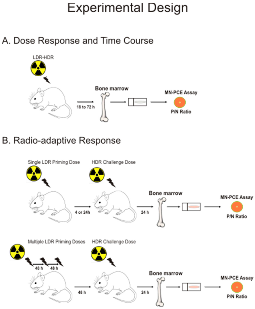
Enhanced cellular DNA repair efficiency and suppression of genomic instability have been proposed as mechanisms underlying radio-adaptive responses following low-dose radiation exposures. We previously showed that low-dose γ irradiation does not generate radio-adaptation by lowering radiation-induced cytogenetic damage in mouse spleen. Since radiation [...] Read more.
Enhanced cellular DNA repair efficiency and suppression of genomic instability have been proposed as mechanisms underlying radio-adaptive responses following low-dose radiation exposures. We previously showed that low-dose γ irradiation does not generate radio-adaptation by lowering radiation-induced cytogenetic damage in mouse spleen. Since radiation may exert tissue-specific effects, we extended these results here by examining the effects of γ radiation on cytogenetic damage and proliferative index in bone marrow erythrocytes of C57BL/6 and BALB/c mice. In C57BL/6 mice, the induction of micronuclei in polychromatic erythrocytes (MN-PCE) was observed at radiation doses of 100 mGy and greater, and suppression of erythroblast maturation occurred at doses of >500 mGy. A linear dose–response relationship for MN-PCE frequencies in C57BL/6 mice was established for radiation doses between 100 mGy and 1 Gy, with departure from linearity at doses of >1 Gy. BALB/c mice exhibited increased MN-PCE frequencies above baseline following a 20 mGy radiation exposure but did not exhibit radio-sensitivity relative to C57BL/6 mice following 2 Gy exposure. Radio-adaptation of bone marrow erythrocytes was not observed in either strain of mice exposed to low-dose priming γ irradiation (single doses of 20 mGy or 100 mGy or multiple 20 mGy doses) administered at various times prior to acute 2 Gy irradiation, confirming the lack of radio-adaptive response for induction of cytogenetic damage or suppression or erythrocyte proliferation/maturation in bone marrow of these mouse strains. Full article
Show Figures

Figure 1

2952 KiB  
Article
Brusatol Enhances the Radiosensitivity of A549 Cells by Promoting ROS Production and Enhancing DNA Damage
by Xiaohui Sun, Qin Wang, Yan Wang, Liqing Du, Chang Xu and Qiang Liu
Int. J. Mol. Sci. 2016, 17(7), 997; https://doi.org/10.3390/ijms17070997 - 24 Jun 2016
Cited by 87 | Viewed by 8617
Abstract
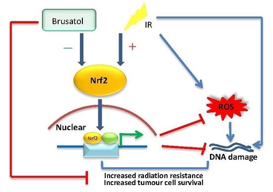
NF-E2-related factor 2 (Nrf2) has been identified as a master regulatory factor in the protection of cells from oxidative and electrophilic stress. However, overexpression of Nrf2 in lung cancer may cause chemoresistance, as well as radioresistance. In this study, we examined the relationship [...] Read more.
NF-E2-related factor 2 (Nrf2) has been identified as a master regulatory factor in the protection of cells from oxidative and electrophilic stress. However, overexpression of Nrf2 in lung cancer may cause chemoresistance, as well as radioresistance. In this study, we examined the relationship between radioresistance and Nrf2 protein levels in H1299, A549, and H460 cells, and finally chose the A549 cell line to continue with due to its strong radioresistance and high Nrf2 protein levels. We found that the Nrf2 inhibitor, brusatol, could prevent the increase and accumulation of Nrf2 after exposure to irradiation. Additionally, following treatment with 80 nM brusatol, A549 cells became sensitive to irradiation, suffering severe DNA damage. Combination treatment with brusatol and ionizing radiation (IR) can distinctly increase the level of reactive oxygen species in A549 cells, causing a 1.8-fold increase compared with the control, and a 1.4-fold increase compared with IR alone. In fact, in the treatment with both brusatol and IR, lung cancer cell proliferation is halted, gradually leading to cell death. Because Nrf2 is closely linked to DNA damage repair, inhibiting the function of Nrf2, as in brusatol treatment, may increase the DNA damage caused by radiotherapy or chemotherapy, possibly enhancing the efficacy of chemotherapeutic drugs. Our study is the first to demonstrate brusatol’s ability to enhance the responsiveness of lung cancer cells to irradiation, and its potential application as a natural sensitizer in radiotherapy. Full article
Show Figures

Graphical abstract

5189 KiB  
Article
The Protective Effects of 5-Methoxytryptamine-α-lipoic Acid on Ionizing Radiation-Induced Hematopoietic Injury
by Deguan Li, Zhenyuan Tian, Weisheng Tang, Junling Zhang, Lu Lu, Zhaojin Sun, Zewei Zhou and Feiyue Fan
Int. J. Mol. Sci. 2016, 17(6), 935; https://doi.org/10.3390/ijms17060935 - 14 Jun 2016
Cited by 37 | Viewed by 6041
Abstract
Antioxidants are prospective radioprotectors because of their ability to scavenge radiation-induced reactive oxygen species (ROS). The hematopoietic system is widely studied in radiation research because of its high radiosensitivity. In the present study, we describe the beneficial effects of 5-methoxytryptamine-α-lipoic acid (MLA), which [...] Read more.
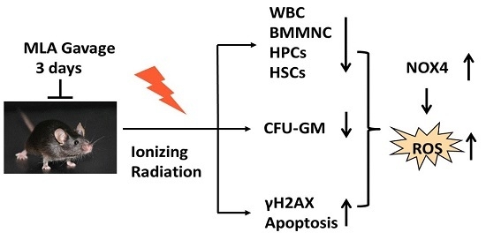
Antioxidants are prospective radioprotectors because of their ability to scavenge radiation-induced reactive oxygen species (ROS). The hematopoietic system is widely studied in radiation research because of its high radiosensitivity. In the present study, we describe the beneficial effects of 5-methoxytryptamine-α-lipoic acid (MLA), which was synthesized from melatonin and α-lipoic acid, against radiation-induced hematopoietic injury. MLA administration significantly enhanced the survival rate of mice after 7.2 Gy total body irradiation. The results showed that MLA not only markedly increased the numbers and clonogenic potential of hematopoietic cells but also decreased DNA damage, as determined by flow cytometric analysis of histone H2AX phosphorylation. In addition, MLA decreased the levels of ROS in hematopoietic cells by inhibiting NOX4 expression. These data demonstrate that MLA prevents radiation-induced hematopoietic syndrome by increasing the number and function of and by inhibiting DNA damage and ROS production in hematopoietic cells. These data suggest MLA is beneficial for the protection of radiation injuries. Full article
Show Figures

Graphical abstract

12356 KiB  
Article
Induction of Lipocalin2 in a Rat Model of Lung Irradiation
by Sadaf Sultan, Shakil Ahmad, Margret Rave-Fränk, Ihtzaz Ahmed Malik, Clemens F. Hess, Hans Christiansen and Silke Cameron
Int. J. Mol. Sci. 2016, 17(5), 637; https://doi.org/10.3390/ijms17050637 - 28 Apr 2016
Cited by 3 | Viewed by 6130
Abstract
Previously, we showed that lipocalin2 (LCN2) serum levels increased after liver irradiation and during acute-phase conditions. Here, we evaluate LCN2 expression and serum levels after single-dose lung irradiation with 25 Gy, percutaneously administered to the lung of randomly-paired male Wistar rats. Due to [...] Read more.
Previously, we showed that lipocalin2 (LCN2) serum levels increased after liver irradiation and during acute-phase conditions. Here, we evaluate LCN2 expression and serum levels after single-dose lung irradiation with 25 Gy, percutaneously administered to the lung of randomly-paired male Wistar rats. Due to the concave anatomy of the lung recesses, the irradiation field included the upper part of the liver. No rat died due to irradiation. In control tissue, lung immunohistochemistry showed a high constitutive expression of LCN2+ granulocytes. LCN2 mRNA levels in lung tissue increased up to 24 h (9 ± 2.3-fold) after irradiation. However, serum LCN2 levels remained undetectable after lung irradiation. LCN2 expression in the upper part of the liver increased up to 4.2-fold after lung irradiation, but the lower liver showed an early decrease. Acute-phase cytokines (IL-1β and TNF-α) showed a significant increase on transcript level in both lung and upper liver, whilst the lower liver did not show any considerable increase. In conclusion, constitutive expression of LCN2 in local immune cells demonstrates its local role during stress conditions in the lung. The absence of LCN2 in the serum strengthens our previous findings that the liver is the key player in secreting LCN2 during stress conditions with liver involvement. Full article
Show Figures

Figure 1

8407 KiB  
Article
Starvation after Cobalt-60 γ-Ray Radiation Enhances Metastasis in U251 Glioma Cells by Regulating the Transcription Factor SP1
by Tuo Zhao, Hailong Wang, Hong Ma, Hao Wang, Bo Chen and Yulin Deng
Int. J. Mol. Sci. 2016, 17(4), 386; https://doi.org/10.3390/ijms17040386 - 5 Apr 2016
Cited by 9 | Viewed by 5779
Abstract
Radiation is of clinical importance during glioma therapy; however, vasculature damage is observed over the treatment course. This type of tissue damage might lead to starvation conditions, affecting tumor metastasis. To test this possibility, we compared starvation conditions in conjunction with radiation treatment [...] Read more.
Radiation is of clinical importance during glioma therapy; however, vasculature damage is observed over the treatment course. This type of tissue damage might lead to starvation conditions, affecting tumor metastasis. To test this possibility, we compared starvation conditions in conjunction with radiation treatment to monitor metastatic ability in the U251 glioma cell line. Transcriptome, western blot, and immunofluorescence analyses were used to measure the RNA and protein expression changes of the U251 cells after various treatments. We found that starvation combined with radiation treatment yielded the most significant expression changes in metastasis-related factors compared to that in the control groups. In addition, a metastasis assay was used to directly measure the metastatic ability of the treated cells, which confirmed that the U251 cells treated with starvation combined with radiation possessed the highest metastatic ability. Furthermore, bioinformatics analysis demonstrated that SP1 represented a common transcription factor associated with changes in metastasis-related factors. Blocking SP1 activity by an inhibitor suppressed the starvation-plus-radiation treatment-mediated enhancement of U251 cell metastasis. Our study provides the first evidence that starvation caused by radiation might play a significant role in enhancing the ability of the glioma cell line U251 to metastasize via regulation of the transcription factor SP1. Full article
Show Figures

Graphical abstract

1754 KiB  
Article
Assessment of Radiation Induced Therapeutic Effect and Cytotoxicity in Cancer Patients Based on Transcriptomic Profiling
by Sajjad Karim, Zeenat Mirza, Adeel G. Chaudhary, Adel M. Abuzenadah, Mamdooh Gari and Mohammed H. Al-Qahtani
Int. J. Mol. Sci. 2016, 17(2), 250; https://doi.org/10.3390/ijms17020250 - 19 Feb 2016
Cited by 17 | Viewed by 7420
Abstract
Toxicity induced by radiation therapy is a curse for cancer patients undergoing treatment. It is imperative to understand and define an ideal condition where the positive effects notably outweigh the negative. We used a microarray meta-analysis approach to measure global gene-expression before and [...] Read more.
Toxicity induced by radiation therapy is a curse for cancer patients undergoing treatment. It is imperative to understand and define an ideal condition where the positive effects notably outweigh the negative. We used a microarray meta-analysis approach to measure global gene-expression before and after radiation exposure. Bioinformatic tools were used for pathways, network, gene ontology and toxicity related studies. We found 429 differentially expressed genes at fold change >2 and p-value <0.05. The most significantly upregulated genes were synuclein alpha (SNCA), carbonic anhydrase I (CA1), X-linked Kx blood group (XK), glycophorin A and B (GYPA and GYPB), and hemogen (HEMGN), while downregulated ones were membrane-spanning 4-domains, subfamily A member 1 (MS4A1), immunoglobulin heavy constant mu (IGHM), chemokine (C-C motif) receptor 7 (CCR7), BTB and CNC homology 1 transcription factor 2 (BACH2), and B-cell CLL/lymphoma 11B (BCL11B). Pathway analysis revealed calcium-induced T lymphocyte apoptosis and the role of nuclear factor of activated T-cells (NFAT) in regulation of the immune response as the most inhibited pathways, while apoptosis signaling was significantly activated. Most of the normal biofunctions were significantly decreased while cell death and survival process were activated. Gene ontology enrichment analysis revealed the immune system process as the most overrepresented group under the biological process category. Toxicity function analysis identified liver, kidney and heart to be the most affected organs during and after radiation therapy. The identified biomarkers and alterations in molecular pathways induced by radiation therapy should be further investigated to reduce the cytotoxicity and development of fatigue. Full article
Show Figures

Graphical abstract

1468 KiB  
Review
Cellular Pathways in Response to Ionizing Radiation and Their Targetability for Tumor Radiosensitization
by Patrick Maier, Linda Hartmann, Frederik Wenz and Carsten Herskind
Int. J. Mol. Sci. 2016, 17(1), 102; https://doi.org/10.3390/ijms17010102 - 14 Jan 2016
Cited by 315 | Viewed by 17515
Abstract
During the last few decades, improvements in the planning and application of radiotherapy in combination with surgery and chemotherapy resulted in increased survival rates of tumor patients. However, the success of radiotherapy is impaired by two reasons: firstly, the radioresistance of tumor cells [...] Read more.
During the last few decades, improvements in the planning and application of radiotherapy in combination with surgery and chemotherapy resulted in increased survival rates of tumor patients. However, the success of radiotherapy is impaired by two reasons: firstly, the radioresistance of tumor cells and, secondly, the radiation-induced damage of normal tissue cells located in the field of ionizing radiation. These limitations demand the development of drugs for either radiosensitization of tumor cells or radioprotection of normal tissue cells. In order to identify potential targets, a detailed understanding of the cellular pathways involved in radiation response is an absolute requirement. This review describes the most important pathways of radioresponse and several key target proteins for radiosensitization. Full article
Show Figures

Graphical abstract

10419 KiB  
Article
Effect of Chromatin Structure on the Extent and Distribution of DNA Double Strand Breaks Produced by Ionizing Radiation; Comparative Study of hESC and Differentiated Cells Lines
by Priyanka Venkatesh, Irina V. Panyutin, Evgenia Remeeva, Ronald D. Neumann and Igor G. Panyutin
Int. J. Mol. Sci. 2016, 17(1), 58; https://doi.org/10.3390/ijms17010058 - 2 Jan 2016
Cited by 22 | Viewed by 8032
Abstract
Chromatin structure affects the extent of DNA damage and repair. Thus, it has been shown that heterochromatin is more protective against DNA double strand breaks (DSB) formation by ionizing radiation (IR); and that DNA DSB repair may proceed differently in hetero- and euchromatin [...] Read more.
Chromatin structure affects the extent of DNA damage and repair. Thus, it has been shown that heterochromatin is more protective against DNA double strand breaks (DSB) formation by ionizing radiation (IR); and that DNA DSB repair may proceed differently in hetero- and euchromatin regions. Human embryonic stem cells (hESC) have a more open chromatin structure than differentiated cells. Here, we study the effect of chromatin structure in hESC on initial DSB formation and subsequent DSB repair. DSB were scored by comet assay; and DSB repair was assessed by repair foci formation via 53BP1 antibody staining. We found that in hESC, heterochromatin is confined to distinct regions, while in differentiated cells it is distributed more evenly within the nuclei. The same dose of ionizing radiation produced considerably more DSB in hESC than in differentiated derivatives, normal human fibroblasts; and one cancer cell line. At the same time, the number of DNA repair foci were not statistically different among these cells. We showed that in hESC, DNA repair foci localized almost exclusively outside the heterochromatin regions. We also noticed that exposure to ionizing radiation resulted in an increase in heterochromatin marker H3K9me3 in cancer HT1080 cells, and to a lesser extent in IMR90 normal fibroblasts, but not in hESCs. These results demonstrate the importance of chromatin conformation for DNA protection and DNA damage repair; and indicate the difference of these processes in hESC. Full article
Show Figures

Graphical abstract

2015

Jump to: 2022, 2021, 2020, 2019, 2018, 2017, 2016, 2014, 2013

231 KiB  
Review
Global Gene Expression Alterations as a Crucial Constituent of Human Cell Response to Low Doses of Ionizing Radiation Exposure
by Mykyta Sokolov and Ronald Neumann
Int. J. Mol. Sci. 2016, 17(1), 55; https://doi.org/10.3390/ijms17010055 - 31 Dec 2015
Cited by 50 | Viewed by 6134
Abstract
Exposure to ionizing radiation (IR) is inevitable to humans in real-life scenarios; the hazards of IR primarily stem from its mutagenic, carcinogenic, and cell killing ability. For many decades, extensive research has been conducted on the human cell responses to IR delivered at [...] Read more.
Exposure to ionizing radiation (IR) is inevitable to humans in real-life scenarios; the hazards of IR primarily stem from its mutagenic, carcinogenic, and cell killing ability. For many decades, extensive research has been conducted on the human cell responses to IR delivered at a low dose/low dose (LD) rate. These studies have shown that the molecular-, cellular-, and tissue-level responses are different after low doses of IR (LDIR) compared to those observed after a short-term high-dose IR exposure (HDIR). With the advent of high-throughput technologies in the late 1990s, such as DNA microarrays, changes in gene expression have also been found to be ubiquitous after LDIR. Very limited subset of genes has been shown to be consistently up-regulated by LDIR, including CDKN1A. Further research on the biological effects and mechanisms induced by IR in human cells demonstrated that the molecular and cellular processes, including transcriptional alterations, activated by LDIR are often related to protective responses and, sometimes, hormesis. Following LDIR, some distinct responses were observed, these included bystander effects, and adaptive responses. Changes in gene expression, not only at the level of mRNA, but also miRNA, have been found to crucially underlie these effects having implications for radiation protection purposes. Full article
2943 KiB  
Article
Combination Treatment with Sublethal Ionizing Radiation and the Proteasome Inhibitor, Bortezomib, Enhances Death-Receptor Mediated Apoptosis and Anti-Tumor Immune Attack
by Ercan Cacan, Alexander M. Spring, Anita Kumari, Susanna F. Greer and Charlie Garnett-Benson
Int. J. Mol. Sci. 2015, 16(12), 30405-30421; https://doi.org/10.3390/ijms161226238 - 21 Dec 2015
Cited by 19 | Viewed by 5842
Abstract
Sub-lethal doses of radiation can modulate gene expression, making tumor cells more susceptible to T-cell-mediated immune attack. Proteasome inhibitors demonstrate broad anti-tumor activity in clinical and pre-clinical cancer models. Here, we use a combination treatment of proteasome inhibition and irradiation to further induce [...] Read more.
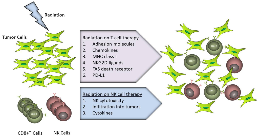
Sub-lethal doses of radiation can modulate gene expression, making tumor cells more susceptible to T-cell-mediated immune attack. Proteasome inhibitors demonstrate broad anti-tumor activity in clinical and pre-clinical cancer models. Here, we use a combination treatment of proteasome inhibition and irradiation to further induce immunomodulation of tumor cells that could enhance tumor-specific immune responses. We investigate the effects of the 26S proteasome inhibitor, bortezomib, alone or in combination with radiotherapy, on the expression of immunogenic genes in normal colon and colorectal cancer cell lines. We examined cells for changes in the expression of several death receptors (DR4, DR5 and Fas) commonly used by T cells for killing of target cells. Our results indicate that the combination treatment resulted in increased cell surface expression of death receptors by increasing their transcript levels. The combination treatment further increases the sensitivity of carcinoma cells to apoptosis through FAS and TRAIL receptors but does not change the sensitivity of normal non-malignant epithelial cells. Furthermore, the combination treatment significantly enhances tumor cell killing by tumor specific CD8+ T cells. This study suggests that combining radiotherapy and proteasome inhibition may simultaneously enhance tumor immunogenicity and the induction of antitumor immunity by enhancing tumor-specific T-cell activity. Full article
Show Figures

Graphical abstract

1395 KiB  
Review
Human Anti-Oxidation Protein A1M—A Potential Kidney Protection Agent in Peptide Receptor Radionuclide Therapy
by Jonas Ahlstedt, Thuy A. Tran, Sven-Erik Strand, Magnus Gram and Bo Åkerström
Int. J. Mol. Sci. 2015, 16(12), 30309-30320; https://doi.org/10.3390/ijms161226234 - 18 Dec 2015
Cited by 11 | Viewed by 7247
Abstract
Peptide receptor radionuclide therapy (PRRT) has been in clinical use for 15 years to treat metastatic neuroendocrine tumors. PRRT is limited by reabsorption and retention of the administered radiolabeled somatostatin analogues in the proximal tubule. Consequently, it is essential to develop and employ [...] Read more.
Peptide receptor radionuclide therapy (PRRT) has been in clinical use for 15 years to treat metastatic neuroendocrine tumors. PRRT is limited by reabsorption and retention of the administered radiolabeled somatostatin analogues in the proximal tubule. Consequently, it is essential to develop and employ methods to protect the kidneys during PRRT. Today, infusion of positively charged amino acids is the standard method of kidney protection. Other methods, such as administration of amifostine, are still under evaluation and show promising results. α1-microglobulin (A1M) is a reductase and radical scavenging protein ubiquitously present in plasma and extravascular tissue. Human A1M has antioxidation properties and has been shown to prevent radiation-induced in vitro cell damage and protect non-irradiated surrounding cells. It has recently been shown in mice that exogenously infused A1M and the somatostatin analogue octreotide are co-localized in proximal tubules of the kidney after intravenous infusion. In this review we describe the current situation of kidney protection during PRRT, discuss the necessity and implications of more precise dosimetry and present A1M as a new, potential candidate for renal protection during PRRT and related targeted radionuclide therapies. Full article
Show Figures

Figure 1

8520 KiB  
Article
Histone H2AX Is Involved in FoxO3a-Mediated Transcriptional Responses to Ionizing Radiation to Maintain Genome Stability
by Stephane Tarrade, Tanya Bhardwaj, Matthew Flegal, Lindsey Bertrand, Ilya Velegzhaninov, Alexey Moskalev and Dmitry Klokov
Int. J. Mol. Sci. 2015, 16(12), 29996-30014; https://doi.org/10.3390/ijms161226216 - 16 Dec 2015
Cited by 13 | Viewed by 8443
Abstract
Histone H2AX plays a crucial role in molecular and cellular responses to DNA damage and in the maintenance of genome stability. It is downstream of ataxia telangiectasia mutated (ATM) damage signaling pathway and there is an emerging role of the transcription factor FoxO3a, [...] Read more.
Histone H2AX plays a crucial role in molecular and cellular responses to DNA damage and in the maintenance of genome stability. It is downstream of ataxia telangiectasia mutated (ATM) damage signaling pathway and there is an emerging role of the transcription factor FoxO3a, a regulator of a variety of other pathways, in activating this signaling. We asked whether H2AX may feedback to FoxO3a to affect respective FoxO3a-dependent pathways. We used a genetically matched pair of mouse embryonic fibroblast H2AX+/+ and H2AX/ cell lines to carry out comprehensive time-course and dose-response experiments and to show that the expression of several FoxO3a-regulated genes was altered in H2AX/ compared to H2AX+/+ cells at both basal and irradiated conditions. Hspa1b and Gadd45a were down-regulated four- to five-fold and Ddit3, Cdkn1a and Sod2 were up-regulated 2–3-fold in H2AX/ cells. Using the luciferase reporter assay, we directly demonstrated that transcriptional activity of FoxoO3a was reduced in H2AX/ cells. FoxO3a localization within the nuclear phospho-ATM (Ser1981) foci in irradiated cells was affected by the H2AX status, as well as its posttranslational modification (phospho-Thr32). These differences were associated with genomic instability and radiosensitivity in H2AX/ cells. Finally, knockdown of H2AX in H2AX+/+ cells resulted in FoxO3a-dependent gene expression patterns and increased radiosensitivity that partially mimicked those found in H2AX/ cells. Taken together, our data suggest a role for FoxO3a in the maintenance of genome integrity in response to DNA damage that is mediated by H2AX via yet unknown mechanisms. Full article
Show Figures

Graphical abstract

5864 KiB  
Article
Carbon Ion Irradiated Neural Injury Induced the Peripheral Immune Effects in Vitro or in Vivo
by Runhong Lei, Tuo Zhao, Qiang Li, Xiao Wang, Hong Ma and Yulin Deng
Int. J. Mol. Sci. 2015, 16(12), 28334-28346; https://doi.org/10.3390/ijms161226109 - 30 Nov 2015
Cited by 21 | Viewed by 6344
Abstract
Carbon ion radiation is a promising treatment for brain cancer; however, the immune system involved long-term systemic effects evoke a concern of complementary and alternative therapies in clinical treatment. To clarify radiotherapy caused fundamental changes in peripheral immune system, examinations were performed based [...] Read more.
Carbon ion radiation is a promising treatment for brain cancer; however, the immune system involved long-term systemic effects evoke a concern of complementary and alternative therapies in clinical treatment. To clarify radiotherapy caused fundamental changes in peripheral immune system, examinations were performed based on established models in vitro and in vivo. We found that brain-localized carbon ion radiation of neural cells induced complex changes in the peripheral blood, thymus, and spleen at one, two, and three months after its application. Atrophy, apoptosis, and abnormal T-cell distributions were observed in rats receiving a single high dose of radiation. Radiation downregulated the expression of proteins involved in T-cell development at the transcriptional level and increased the proportion of CD3+CD4CD8+ T-cells in the thymus and the proportion of CD3+CD4+CD8 T-cells in the spleen. These data show that brain irradiation severely affects the peripheral immune system, even at relatively long times after irradiation. In addition, they provide valuable information that will implement the design of biological-based strategies that will aid brain cancer patients suffering from the long-term side effects of radiation. Full article
Show Figures

Figure 1

659 KiB  
Review
Molecular, Cellular and Functional Effects of Radiation-Induced Brain Injury: A Review
by Sona Balentova and Marian Adamkov
Int. J. Mol. Sci. 2015, 16(11), 27796-27815; https://doi.org/10.3390/ijms161126068 - 24 Nov 2015
Cited by 128 | Viewed by 8965
Abstract
Radiation therapy is the most effective non-surgical treatment of primary brain tumors and metastases. Preclinical studies have provided valuable insights into pathogenesis of radiation-induced injury to the central nervous system. Radiation-induced brain injury can damage neuronal, glial and vascular compartments of the brain [...] Read more.
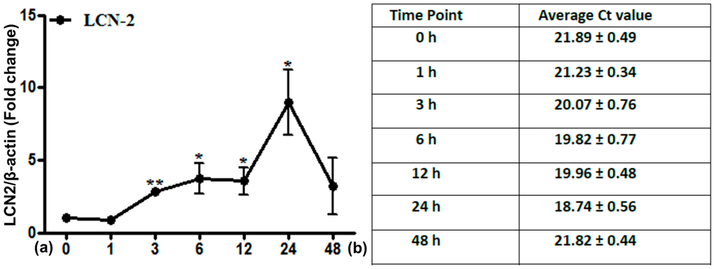
Radiation therapy is the most effective non-surgical treatment of primary brain tumors and metastases. Preclinical studies have provided valuable insights into pathogenesis of radiation-induced injury to the central nervous system. Radiation-induced brain injury can damage neuronal, glial and vascular compartments of the brain and may lead to molecular, cellular and functional changes. Given its central role in memory and adult neurogenesis, the majority of studies have focused on the hippocampus. These findings suggested that hippocampal avoidance in cranial radiotherapy prevents radiation-induced cognitive impairment of patients. However, multiple rodent studies have shown that this problem is more complex. As the radiation-induced cognitive impairment reflects hippocampal and non-hippocampal compartments, it is of critical importance to investigate molecular, cellular and functional modifications in various brain regions as well as their integration at clinically relevant doses and schedules. We here provide a literature overview, including our previously published results, in order to support the translation of preclinical findings to clinical practice, and improve the physical and mental status of patients with brain tumors. Full article
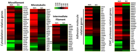
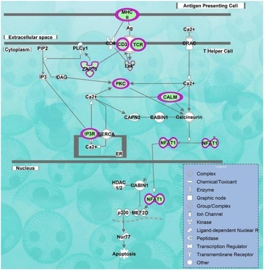
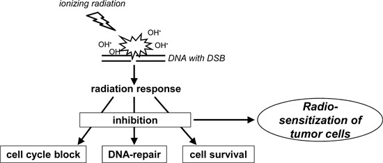
Show Figures

Figure 1

1517 KiB  
Article
Betulinyl Sulfamates as Anticancer Agents and Radiosensitizers in Human Breast Cancer Cells
by Matthias Bache, Christin Münch, Antje Güttler, Henri Wichmann, Katharina Theuerkorn, Daniel Emmerich, Reinhard Paschke and Dirk Vordermark
Int. J. Mol. Sci. 2015, 16(11), 26249-26262; https://doi.org/10.3390/ijms161125953 - 3 Nov 2015
Cited by 25 | Viewed by 7009
Abstract
Betulinic acid (BA), a natural compound of birch bark, is cytotoxic for many tumors. Recently, a betulinyl sulfamate was described that inhibits carbonic anhydrases (CA), such as CAIX, an attractive target for tumor-selective therapy strategies in hypoxic cancer cells. Data on combined CAIX [...] Read more.
Betulinic acid (BA), a natural compound of birch bark, is cytotoxic for many tumors. Recently, a betulinyl sulfamate was described that inhibits carbonic anhydrases (CA), such as CAIX, an attractive target for tumor-selective therapy strategies in hypoxic cancer cells. Data on combined CAIX inhibition with radiotherapy are rare. In the human breast cancer cell lines MDA-MB231 and MCF7, the effects of BA and betulinyl sulfamates on cellular and radiobiological behavior under normoxia and hypoxia were evaluated. The two most effective betulinyl sulfamates CAI 1 and CAI 3 demonstrated a 1.8–2.8-fold higher cytotoxicity than BA under normoxia in breast cancer cells, with IC50 values between 11.1 and 18.1 µM. BA exhibits its strongest cytotoxicity with IC50 values of 8.2 and 16.4 µM under hypoxia. All three substances show a dose-dependent increase in apoptosis, inhibition of migration, and inhibition of hypoxia-induced gene expression. In combination with irradiation, betulinyl sulfamates act as radiosensitizers, with DMF10 values of 1.47 (CAI 1) and 1.75 (CAI 3) under hypoxia in MDA-MB231 cells. BA showed additive effects in combination with irradiation. Taken together; our results suggest that BA and betulinyl sulfamates seem to be attractive substances to combine with radiotherapy; particularly for hypoxic breast cancer. Full article
Show Figures

Figure 1

1382 KiB  
Article
A Co-Culture Model of Fibroblasts and Adipose Tissue-Derived Stem Cells Reveals New Insights into Impaired Wound Healing After Radiotherapy
by Frank Haubner, Dominique Muschter, Fabian Pohl, Stephan Schreml, Lukas Prantl and Holger G. Gassner
Int. J. Mol. Sci. 2015, 16(11), 25947-25958; https://doi.org/10.3390/ijms161125935 - 29 Oct 2015
Cited by 38 | Viewed by 7656
Abstract
External radiation seems to be associated with increased amounts of cytokines and other cellular modulators. Impaired microcirculation and fibrosis are examples of typical long term damage caused by radiotherapy. Adipose tissue-derived stem cells (ASC) are discussed to enhance wound healing, but their role [...] Read more.
External radiation seems to be associated with increased amounts of cytokines and other cellular modulators. Impaired microcirculation and fibrosis are examples of typical long term damage caused by radiotherapy. Adipose tissue-derived stem cells (ASC) are discussed to enhance wound healing, but their role in wounds due to radiotherapy is poorly understood. Normal human fibroblasts (NHF) and ASCs were co-cultured and external radiation with doses from 2–12 Gray (Gy) was delivered. Cell proliferation and mRNA levels of matrix metalloproteinases (MMP1, MMP2 and MMP13) were determined 48 h after irradiation of the co-cultures by qPCR. Additionally, tissue inhibitors of matrix metalloproteinases (TIMP1, TIMP2) were determined by enzyme-linked immunosorbent assay (ELISA). There was a reduction of cell proliferation after external radiation in mono-cultures of NHFs and ASCs compared to controls without irradiation. The co-culture of ASCs and NHFs showed reduced impairment of cell proliferation after external radiation. Gene expression of MMP1 and MMP13 was reduced after external irradiation in NHF. MMP2 expression of irradiated NHFs was increased. In the co-culture setting, MMP1 and MMP2 gene expression levels were upregulated. TIMP1 and TIMP2 protein expression was increased after irradiation in NHFs and their co-cultures with ASCs. ASCs seem to stimulate cell proliferation of NHFs and modulate relevant soluble mediators as well as proteinases after external radiation. Full article
Show Figures

Figure 1

2310 KiB  
Article
TP53inp1 Gene Is Implicated in Early Radiation Response in Human Fibroblast Cells
by Nikolett Sándor, Boglárka Schilling-Tóth, Enikő Kis, Lili Fodor, Fruzsina Mucsányi, Géza Sáfrány and Hargita Hegyesi
Int. J. Mol. Sci. 2015, 16(10), 25450-25465; https://doi.org/10.3390/ijms161025450 - 23 Oct 2015
Cited by 11 | Viewed by 7697
Abstract
Tumor protein 53-induced nuclear protein-1 (TP53inp1) is expressed by activation via p53 and p73. The purpose of our study was to investigate the role of TP53inp1 in response of fibroblasts to ionizing radiation. γ-Ray radiation dose-dependently induces the expression of TP53inp1 [...] Read more.
Tumor protein 53-induced nuclear protein-1 (TP53inp1) is expressed by activation via p53 and p73. The purpose of our study was to investigate the role of TP53inp1 in response of fibroblasts to ionizing radiation. γ-Ray radiation dose-dependently induces the expression of TP53inp1 in human immortalized fibroblast (F11hT) cells. Stable silencing of TP53inp1 was done via lentiviral transfection of shRNA in F11hT cells. After irradiation the clonogenic survival of TP53inp1 knockdown (F11hT-shTP) cells was compared to cells transfected with non-targeting (NT) shRNA. Radiation-induced senescence was measured by SA-β-Gal staining and autophagy was detected by Acridine Orange dye and microtubule-associated protein-1 light chain 3 (LC3B) immunostaining. The expression of TP53inp1, GDF-15, and CDKN1A and alterations in radiation induced mitochondrial DNA deletions were evaluated by qPCR. TP53inp1 was required for radiation (IR) induced maximal elevation of CDKN1A and GDF-15 expressions. Mitochondrial DNA deletions were increased and autophagy was deregulated following irradiation in the absence of TP53inp1. Finally, we showed that silencing of TP53inp1 enhances the radiation sensitivity of fibroblast cells. These data suggest functional roles for TP53inp1 in radiation-induced autophagy and survival. Taken together, we suppose that silencing of TP53inp1 leads radiation induced autophagy impairment and induces accumulation of damaged mitochondria in primary human fibroblasts. Full article
Show Figures

Graphical abstract

1086 KiB  
Article
Comparative Analysis of Whole-Genome Gene Expression Changes in Cultured Human Embryonic Stem Cells in Response to Low, Clinical Diagnostic Relevant, and High Doses of Ionizing Radiation Exposure
by Mykyta Sokolov, Van Nguyen and Ronald Neumann
Int. J. Mol. Sci. 2015, 16(7), 14737-14748; https://doi.org/10.3390/ijms160714737 - 30 Jun 2015
Cited by 10 | Viewed by 5518
Abstract
The biological effects of low-dose ionizing radiation (LDIR) exposure in humans are not comprehensively understood, generating a high degree of controversy in published literature. The earliest stages of human development are known to be among the most sensitive to stress exposures, especially genotoxic [...] Read more.
The biological effects of low-dose ionizing radiation (LDIR) exposure in humans are not comprehensively understood, generating a high degree of controversy in published literature. The earliest stages of human development are known to be among the most sensitive to stress exposures, especially genotoxic stresses. However, the risks stemming from exposure to LDIR, particularly within the clinical diagnostic relevant dose range, have not been directly evaluated in human embryonic stem cells (hESCs). Here, we describe the dynamics of the whole genome transcriptional responses of different hESC lines to both LDIR and, as a reference, high-dose IR (HDIR). We found that even doses as low as 0.05 Gy could trigger statistically significant transient changes in a rather limited subset of genes in all hESCs lines examined. Gene expression signatures of hESCs exposed to IR appear to be highly dose-, time-, and cell line-dependent. We identified 50 genes constituting consensus gene expression signature as an early response to HDIR across all lines of hESC examined. We observed substantial differences in biological pathways affected by either LDIR or HDIR in hESCs, suggesting that the molecular mechanisms underpinning the responses of hESC may fundamentally differ depending on radiation doses. Full article
Show Figures

Figure 1

1542 KiB  
Article
The Effect of Diagnostic Absorbed Doses from 131I on Human Thyrocytes in Vitro
by Zbigniew Adamczewski, Mariusz Stasiołek, Bolesław Karwowski, Marek Dedecjus, Daria Orszulak-Michalak, Anna Merecz, Przemysław W. Śliwka, Bartosz Puła and Andrzej Lewiński
Int. J. Mol. Sci. 2015, 16(7), 14608-14622; https://doi.org/10.3390/ijms160714608 - 29 Jun 2015
Cited by 2 | Viewed by 5294
Abstract
Background: Administration of diagnostic activities of 131I, performed in order to detect thyroid remnants after surgery and/or thyroid cancer recurrence/metastases, may lead to reduction of iodine uptake. This phenomenon is called “thyroid stunning”. We estimated radiation absorbed dose-dependent changes in genetic material, [...] Read more.
Background: Administration of diagnostic activities of 131I, performed in order to detect thyroid remnants after surgery and/or thyroid cancer recurrence/metastases, may lead to reduction of iodine uptake. This phenomenon is called “thyroid stunning”. We estimated radiation absorbed dose-dependent changes in genetic material, in particular in sodium iodide symporter (NIS) gene promoter, and NIS protein level in human thyrocytes (HT). Materials and Methods: We used unmodified HT isolated from patients subjected to thyroidectomy exposed to 131I in culture. The different 131I activities applied were calculated to result in absorbed doses of 5, 10, and 20 Gy. Results: According to flow cytometry analysis and comet assay, 131I did not influence the HT viability in culture. Temporary increase of 8-oxo-dG concentration in HT directly after 24 h (p < 0.05) and increase in the number of AP-sites 72 h after termination of exposition to 20 Gy dose (p < 0.0001) were observed. The signs of dose-dependent DNA damage were not associated with essential changes in the NIS expression on mRNA and protein levels. Conclusions: Our observation constitutes a first attempt to evaluate the effect of the absorbed dose of 131I on HT. The results have not confirmed the theory that the “thyroid stunning” reduces the NIS protein synthesis. Full article
Show Figures

Figure 1

4008 KiB  
Review
Biological Mechanisms Underlying the Ultraviolet Radiation-Induced Formation of Skin Wrinkling and Sagging II: Over-Expression of Neprilysin Plays an Essential Role
by Genji Imokawa, Hiroaki Nakajima and Koichi Ishida
Int. J. Mol. Sci. 2015, 16(4), 7776-7795; https://doi.org/10.3390/ijms16047776 - 8 Apr 2015
Cited by 55 | Viewed by 12128
Abstract
Our previous studies strongly indicated that the up-regulated activity of skin fibroblast-derived elastase plays a pivotal role in wrinkling and/or sagging of the skin via the impairment of elastic fiber configuration and the subsequent loss of skin elasticity. Fortunately, we succeeded in identifying [...] Read more.
Our previous studies strongly indicated that the up-regulated activity of skin fibroblast-derived elastase plays a pivotal role in wrinkling and/or sagging of the skin via the impairment of elastic fiber configuration and the subsequent loss of skin elasticity. Fortunately, we succeeded in identifying human skin fibroblast-derived elastase as a previously known enzyme, neprilysin or neutral endopeptidase (NEP). We have also characterized epithelial-mesenchymal paracrine cytokine interactions between UVB-exposed-keratinocytes and dermal fibroblasts and found that interleukin-1α and granulocyte macrophage colony stimulatory factor (GM-CSF) are intrinsic cytokines secreted by UVB-exposed keratinocytes that stimulate the expression of neprilysin by fibroblasts. On the other hand, direct UVA exposure of human fibroblasts significantly stimulates the secretion of IL-6 and also elicits a significant increase in the gene expression of matrix metallo-protease(MMP)-1 as well as neprilysin (to a lesser extent), which is followed by distinct increases in their protein and enzymatic activity levels. Direct UVA exposure of human keratinocytes also stimulates the secretion of IL-6, IL-8 and GM-CSF but not of IL-1 and endothelin-1. These findings suggest that GM-CSF secreted by UVA-exposed keratinocytes as well as IL-6 secreted by UVA-exposed dermal fibroblasts play important and additional roles in UVA-induced sagging and wrinkling by up-regulation of neprilysin and MMP-1, respectively, in dermal fibroblasts. Full article
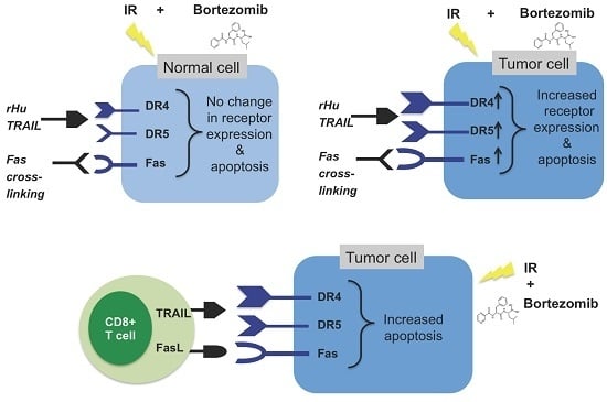
Show Figures

Figure 1

3839 KiB  
Review
Biological Mechanisms Underlying the Ultraviolet Radiation-Induced Formation of Skin Wrinkling and Sagging I: Reduced Skin Elasticity, Highly Associated with Enhanced Dermal Elastase Activity, Triggers Wrinkling and Sagging
by Genji Imokawa and Koichi Ishida
Int. J. Mol. Sci. 2015, 16(4), 7753-7775; https://doi.org/10.3390/ijms16047753 - 8 Apr 2015
Cited by 136 | Viewed by 16976
Abstract
The repetitive exposure of skin to ultraviolet B (UVB) preferentially elicits wrinkling while ultraviolet A (UVA) predominantly elicits sagging. In chronically UVB or UVA-exposed rat skin there is a similar tortuous deformation of elastic fibers together with decreased skin elasticity, whose magnitudes are [...] Read more.
The repetitive exposure of skin to ultraviolet B (UVB) preferentially elicits wrinkling while ultraviolet A (UVA) predominantly elicits sagging. In chronically UVB or UVA-exposed rat skin there is a similar tortuous deformation of elastic fibers together with decreased skin elasticity, whose magnitudes are greater in UVB-exposed skin than in UVA-exposed skin. Comparison of skin elasticity with the activity of matrix metalloproteinases (MMPs) in the dermis of ovariectomized rats after UVB or UVA irradiation demonstrates that skin elasticity is more significantly decreased in ovariectomized rats than in sham-operated rats, which is accompanied by a reciprocal increase in elastase activity but not in the activities of collagenases I or IV. Clinical studies using animal skin and human facial skin demonstrated that topical treatment with a specific inhibitor or an inhibitory extract of skin fibroblast-derived elastase distinctly attenuates UVB and sunlight-induced formation of wrinkling. Our results strongly indicated that the upregulated activity of skin fibroblast-derived elastase plays a pivotal role in wrinkling and/or sagging of the skin via the impairment of elastic fiber configuration and the subsequent loss of skin elasticity. Full article
Show Figures

Figure 1

1299 KiB  
Article
Protective Effects of Resveratrol against UVA-Induced Damage in ARPE19 Cells
by Chi-Ming Chan, Cheng-Hua Huang, Hsin-Ju Li, Chien-Yu Hsiao, Ching-Chieh Su, Pei-Lan Lee and Chi-Feng Hung
Int. J. Mol. Sci. 2015, 16(3), 5789-5802; https://doi.org/10.3390/ijms16035789 - 12 Mar 2015
Cited by 56 | Viewed by 8773
Abstract
Ultraviolet radiation, especially UVA, can penetrate the lens, reach the retina, and induce oxidative stress to retinal pigment epithelial (RPE) cells. Even though it is weakly absorbed by protein and DNA, it may trigger the production of reactive oxygen species (ROS) and generate [...] Read more.
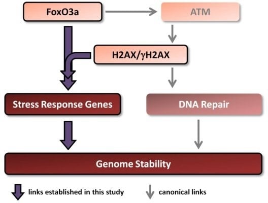
Ultraviolet radiation, especially UVA, can penetrate the lens, reach the retina, and induce oxidative stress to retinal pigment epithelial (RPE) cells. Even though it is weakly absorbed by protein and DNA, it may trigger the production of reactive oxygen species (ROS) and generate oxidative injury; oxidative injury to the retinal pigment epithelium has been implicated to play a contributory role in age-related macular degeneration (AMD). Studies showed that resveratrol, an abundant and active component of red grapes, can protect several cell types from oxidative stress. In this study, adult RPE cells being treated with different concentrations of resveratrol were used to evaluate the protective effect of resveratrol on RPE cells against UVA-induced damage. Cell viability assay showed that resveratrol reduced the UVA-induced decrease in RPE cell viability. Through flow cytometry analysis, we found that the generation of intracellular H2O2 induced by UVA irradiation in RPE cells could be suppressed by resveratrol in a concentration-dependent manner. Results of Western blot analysis demonstrated that resveratrol lowered the activation of UVA-induced extracellular signal-regulated kinase, c-jun-NH2 terminal kinase and p38 kinase in RPE cells. In addition, there was also a reduction in UVA-induced cyclooxygenase-2 (COX-2) expression in RPE cells pretreated with resveratrol. Our observations suggest that resveratrol is effective in preventing RPE cells from being damaged by UVA radiation, and is worth considering for further development as a chemoprotective agent for the prevention of early AMD. Full article
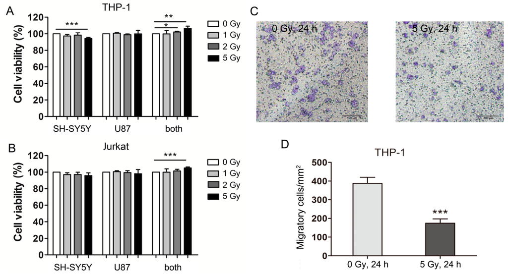
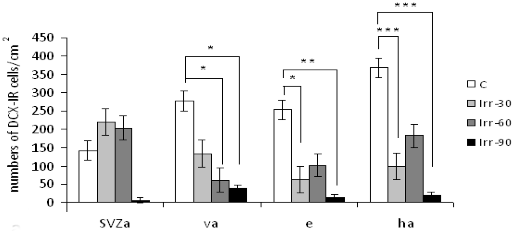
Show Figures

Graphical abstract

2014

Jump to: 2022, 2021, 2020, 2019, 2018, 2017, 2016, 2015, 2013

2272 KiB  
Review
Exposure to Non-Extreme Solar UV Daylight: Spectral Characterization, Effects on Skin and Photoprotection
by Claire Marionnet, Caroline Tricaud and Françoise Bernerd
Int. J. Mol. Sci. 2015, 16(1), 68-90; https://doi.org/10.3390/ijms16010068 - 23 Dec 2014
Cited by 127 | Viewed by 13549
Abstract
The link between chronic sun exposure of human skin and harmful clinical consequences such as photo-aging and skin cancers is now indisputable. These effects are mostly due to ultraviolet (UV) rays (UVA, 320–400 nm and UVB, 280–320 nm). The UVA/UVB ratio can vary [...] Read more.
The link between chronic sun exposure of human skin and harmful clinical consequences such as photo-aging and skin cancers is now indisputable. These effects are mostly due to ultraviolet (UV) rays (UVA, 320–400 nm and UVB, 280–320 nm). The UVA/UVB ratio can vary with latitude, season, hour, meteorology and ozone layer, leading to different exposure conditions. Zenithal sun exposure (for example on a beach around noon under a clear sky) can rapidly induce visible and well-characterized clinical consequences such as sunburn, predominantly induced by UVB. However, a limited part of the global population is exposed daily to such intense irradiance and until recently little attention has been paid to solar exposure that does not induce any short term clinical impact. This paper will review different studies on non-extreme daily UV exposures with: (1) the characterization and the definition of the standard UV daylight and its simulation in the laboratory; (2) description of the biological and clinical effects of such UV exposure in an in vitro reconstructed human skin model and in human skin in vivo, emphasizing the contribution of UVA rays and (3) analysis of photoprotection approaches dedicated to prevent the harmful impact of such UV exposure. Full article
Show Figures

Figure 1

1212 KiB  
Article
The Daidzein Metabolite, 6,7,4'-Trihydroxyisoflavone, Is a Novel Inhibitor of PKCα in Suppressing Solar UV-Induced Matrix Metalloproteinase 1
by Tae-Gyu Lim, Jong-Eun Kim, Sung-Young Lee, Jun Seong Park, Myung Hun Yeom, Hanyong Chen, Ann M. Bode, Zigang Dong and Ki Won Lee
Int. J. Mol. Sci. 2014, 15(11), 21419-21432; https://doi.org/10.3390/ijms151121419 - 19 Nov 2014
Cited by 22 | Viewed by 8825
Abstract
Soy isoflavone is an attractive source of functional cosmetic materials with anti-wrinkle, whitening and skin hydration effects. After consumption, the majority of soy isoflavones are converted to their metabolites in the human gastrointestinal tract. To understand the physiological impact of soy isoflavone on [...] Read more.
Soy isoflavone is an attractive source of functional cosmetic materials with anti-wrinkle, whitening and skin hydration effects. After consumption, the majority of soy isoflavones are converted to their metabolites in the human gastrointestinal tract. To understand the physiological impact of soy isoflavone on the human body, it is necessary to evaluate and address the biological function of its metabolites. In this study, we investigated the effect of 6,7,4'-trihydroxyisoflavone (6,7,4'-THIF), a major metabolite of daidzein, against solar UV (sUV)-induced matrix metalloproteinases (MMPs) in normal human dermal fibroblasts. MMPs play a critical role in the degradation of collagen in skin, thereby accelerating the aging process of skin. The mitogen-activated protein/extracellular signal-regulated kinase (MEK)/extracellular signal-regulated kinase (ERK), mitogen-activated protein kinase (MKK)3/6/p38 and MKK4/c-Jun N-terminal kinases (JNK) signaling pathways are known to modulate MMP-1 function, and their activation by sUV was significantly reduced by 6,7,4'-THIF pretreatment. Our results also indicated that the enzyme activity of protein kinase C (PKC)α, an upstream regulator of MKKs signaling, is suppressed by 6,7,4'-THIF using the in vitro kinase assay. Furthermore, the direct interaction between 6,7,4'-THIF and endogenous PKCα was confirmed using the pull-down assay. Not only sUV-induced MMP-1 expression, but also sUV-induced signaling pathway activation were decreased in PKCα knockdown cells. Overall, we elucidated the inhibitory effect of 6,7,4'-THIF on sUV-induced MMPs and suggest PKCα as its direct molecular target. Full article
Show Figures

Figure 1

1829 KiB  
Article
Betulinic Acid Derivatives NVX-207 and B10 for Treatment of Glioblastoma—An in Vitro Study of Cytotoxicity and Radiosensitization
by Matthias Bache, Stephan Bernhardt, Sarina Passin, Henri Wichmann, Anja Hein, Martin P Zschornak, Matthias Kappler, Helge Taubert, Reinhard Paschke and Dirk Vordermark
Int. J. Mol. Sci. 2014, 15(11), 19777-19790; https://doi.org/10.3390/ijms151119777 - 30 Oct 2014
Cited by 31 | Viewed by 8492
Abstract
Betulinic acid (BA), a pentacyclic triterpene, represents a new therapeutic substance that has potential benefits for treating glioblastoma. Recently, new strategies for producing BA derivatives with improved properties have evolved. However, few studies have examined the combination of BA or BA derivatives using [...] Read more.
Betulinic acid (BA), a pentacyclic triterpene, represents a new therapeutic substance that has potential benefits for treating glioblastoma. Recently, new strategies for producing BA derivatives with improved properties have evolved. However, few studies have examined the combination of BA or BA derivatives using radiotherapy. The effects of two BA derivatives, NVX-207 and B10, on cellular and radiobiological behavior were analyzed using glioblastoma cell lines (U251MG, U343MG and LN229). Based on IC50 values under normoxic conditions, we detected a 1.3–2.9-fold higher cytotoxicity of the BA derivatives B10 and NVX-207, respectively, compared to BA. Incubation using both BA derivatives led to decreased cell migration, cleavage of PARP and decreased protein expression levels of Survivin. Weak radiation sensitivity enhancement was observed in U251MG cells after treatment with both BA derivatives. The enhancement factors at an irradiation dose of 6 Gy after treatment with 5 µM NVX-207 and 5 µM B10 were 1.32 (p = 0.029) and 1.55 (p = 0.002), respectively. In contrast to BA, neither NVX-207 nor B10 had additional effects under hypoxic conditions. Our results suggest that the BA derivatives NVX-207 and B10 improve the effects of radiotherapy on human malignant glioma cells, particularly under normoxic conditions. Full article
Show Figures

Figure 1

3377 KiB  
Article
Protective Effects of a New Phloretin Derivative against UVB-Induced Damage in Skin Cell Model and Human Volunteers
by Seoungwoo Shin, Hyunwoo Kum, Dehun Ryu, Minkyung Kim, Eunsun Jung and Deokhoon Park
Int. J. Mol. Sci. 2014, 15(10), 18919-18940; https://doi.org/10.3390/ijms151018919 - 20 Oct 2014
Cited by 39 | Viewed by 9083
Abstract
The phenolic compound phloretin is a prominent member of the chemical class of dihydrochalcones. Phloretin is specifically found in apple and apple juice and known for its biological properties. We were particularly interested in its potential dermo-cosmetic applications. However, practical limitations of phloretin [...] Read more.
The phenolic compound phloretin is a prominent member of the chemical class of dihydrochalcones. Phloretin is specifically found in apple and apple juice and known for its biological properties. We were particularly interested in its potential dermo-cosmetic applications. However, practical limitations of phloretin do exist due to its poor water-solubility. Phloretin was sulfonated with sulfuric acid (98%, wt) and mixed with saturated salt water to produce phloretin 3',3-disulfonate in order to increase its water-solubility. Here we reported the photoprotective effect of phloretin 3',3-disulfonate (PS), a new semi-synthetic derivative of phloretin. Results showed that PS attenuated cyclobutane pyrimidine dimer (CPDs) formation, glutathione (GSH) depletion and apoptosis induced by ultraviolet B (UVB). The photoprotective effect of PS is tightly correlated to the enhancement of nucleotide excision repair (NER) gene expression. Furthemore, PS had inhibitory effects on UVB-induced release of the inflammatory mediators, such as IL-6 and prostaglandin-E2. We also confirmed the safety and clinical efficacy of PS on human skin. Overall, the results demonstrated significant benefits of PS on the protection of keratinocytes against UVB-induced injuries and suggested its potential use in skin photoprotection. Full article
Show Figures

Figure 1

1085 KiB  
Article
Critical Role for the Protons in FRTL-5 Thyroid Cells: Nuclear Sphingomyelinase Induced-Damage
by Elisabetta Albi, Giuseppina Perrella, Andrea Lazzarini, Samuela Cataldi, Remo Lazzarini, Alessandro Floridi, Francesco Saverio Ambesi-Impiombato and Francesco Curcio
Int. J. Mol. Sci. 2014, 15(7), 11555-11565; https://doi.org/10.3390/ijms150711555 - 27 Jun 2014
Cited by 5 | Viewed by 5341
Abstract
Proliferating thyroid cells are more sensitive to UV-C radiations than quiescent cells. The effect is mediated by nuclear phosphatidylcholine and sphingomyelin metabolism. It was demonstrated that proton beams arrest cell growth and stimulate apoptosis but until now there have been no indications in [...] Read more.
Proliferating thyroid cells are more sensitive to UV-C radiations than quiescent cells. The effect is mediated by nuclear phosphatidylcholine and sphingomyelin metabolism. It was demonstrated that proton beams arrest cell growth and stimulate apoptosis but until now there have been no indications in the literature about their possible mechanism of action. Here we studied the effect of protons on FRTL-5 cells in culture. We showed that proton beams stimulate slightly nuclear neutral sphingomyelinase activity and inhibit nuclear sphingomyelin-synthase activity in quiescent cells whereas stimulate strongly nuclear neutral sphingomyelinase activity and do not change nuclear sphingomyelin-synthase activity in proliferating cells. The study of neutral sphingomyelinase/sphingomyelin-synthase ratio, a marker of functional state of the cells, indicated that proton beams induce FRTL-5 cells in a proapoptotic state if the cells are quiescent and in an initial apoptotic state if the cells are proliferating. The changes of cell life are accompanied by a decrease of nuclear sphingomyelin and increase of bax protein. Full article
Show Figures

Figure 1

503 KiB  
Article
shRNA-Mediated XRCC2 Gene Knockdown Efficiently Sensitizes Colon Tumor Cells to X-ray Irradiation in Vitro and in Vivo
by Qin Wang, Yan Wang, Liqing Du, Chang Xu, Yuanming Sun, Bing Yang, Zhijuan Sun, Yue Fu, Lu Cai, Saijun Fan, Feiyue Fan and Qiang Liu
Int. J. Mol. Sci. 2014, 15(2), 2157-2171; https://doi.org/10.3390/ijms15022157 - 29 Jan 2014
Cited by 10 | Viewed by 9202
Abstract
Colon cancer is one of the most common tumors of the digestive tract. Resistance to ionizing radiation (IR) decreased therapeutic efficiency in these patients’ radiotherapy. XRCC2 is the key protein of DNA homologous recombination repair, and its high expression is associated with enhanced [...] Read more.
Colon cancer is one of the most common tumors of the digestive tract. Resistance to ionizing radiation (IR) decreased therapeutic efficiency in these patients’ radiotherapy. XRCC2 is the key protein of DNA homologous recombination repair, and its high expression is associated with enhanced resistance to DNA damage induced by IR. Here, we investigated the effect of XRCC2 silencing on colon tumor cells’ growth and sensitivity to X-radiation in vitro and in vivo. Colon tumor cells (T84 cell line) were cultivated in vitro and tumors originated from the cell line were propagated as xenografts in nude mice. The suppression of XRCC2 expression was achieved by using vector-based short hairpin RNA (shRNA) in T84 cells. We found that the knockdown of XRCC2 expression effectively decreased T84 cellular proliferation and colony formation, and led to cell apoptosis and cell cycle arrested in G2/M phase induced by X-radiation in vitro. In addition, tumor xenograft studies suggested that XRCC2 silencing inhibited tumorigenicity after radiation treatment in vivo. Our data suggest that the suppression of XRCC2 expression rendered colon tumor cells more sensitive to radiation therapy in vitro and in vivo, implying XRCC2 as a promising therapeutic target for the treatment of radioresistant human colon cancer. Full article
Show Figures

Graphical abstract

465 KiB  
Review
The Effect of Radiation on the Immune Response to Cancers
by Bonggoo Park, Cassian Yee and Kyung-Mi Lee
Int. J. Mol. Sci. 2014, 15(1), 927-943; https://doi.org/10.3390/ijms15010927 - 10 Jan 2014
Cited by 221 | Viewed by 15673
Abstract
In cancer patients undergoing radiation therapy, the beneficial effects of radiation can extend beyond direct cytotoxicity to tumor cells. Delivery of localized radiation to tumors often leads to systemic responses at distant sites, a phenomenon known as the abscopal effect which has been [...] Read more.
In cancer patients undergoing radiation therapy, the beneficial effects of radiation can extend beyond direct cytotoxicity to tumor cells. Delivery of localized radiation to tumors often leads to systemic responses at distant sites, a phenomenon known as the abscopal effect which has been attributed to the induction and enhancement of the endogenous anti-tumor innate and adaptive immune response. The mechanisms surrounding the abscopal effect are diverse and include trafficking of lymphocytes into the tumor microenvironment, enhanced tumor recognition and killing via up-regulation of tumor antigens and antigen presenting machinery and, induction of positive immunomodulatory pathways. Here, we discuss potential mechanisms of radiation-induced enhancement of the anti-tumor response through its effect on the host immune system and explore potential combinational immune-based strategies such as adoptive cellular therapy using ex vivo expanded NK and T cells as a means of delivering a potent effector population in the context of radiation-enhanced anti-tumor immune environment. Full article
Show Figures

2013

Jump to: 2022, 2021, 2020, 2019, 2018, 2017, 2016, 2015, 2014

401 KiB  
Article
The Lack of Cytotoxic Effect and Radioadaptive Response in Splenocytes of Mice Exposed to Low Level Internal β-Particle Irradiation through Tritiated Drinking Water in Vivo
by Matthew Flegal, Melinda Blimkie, Sandrine Roch-Lefevre, Eric Gregoire and Dmitry Klokov
Int. J. Mol. Sci. 2013, 14(12), 23791-23800; https://doi.org/10.3390/ijms141223791 - 5 Dec 2013
Cited by 11 | Viewed by 6229
Abstract
Health effects of tritium, a β-emitter and a by-product of the nuclear industry, is a subject of significant controversy. This mouse in vivo study was undertaken to monitor biological effects of low level tritium exposure. Mice were exposed to tritiated drinking water (HTO) [...] Read more.
Health effects of tritium, a β-emitter and a by-product of the nuclear industry, is a subject of significant controversy. This mouse in vivo study was undertaken to monitor biological effects of low level tritium exposure. Mice were exposed to tritiated drinking water (HTO) at 10 KBq/L, 1 MBq/L and 20 MBq/L concentrations for one month. The treatment did not result in a significant increase of apoptosis in splenocytes. To examine if this low level tritium exposure alters radiosensitivity, the extracted splenocytes were challenged in vitro with 2 Gy γ-radiation, and apoptotic responses at 1 and 24 h were measured. No alterations in the radiosensitivity were detected in cells from mice exposed to tritium compared to sham-treated mice. In contrast, low dose γ-irradiation at 20 or 100 mGy, resulted in a significant increase in resistance to apoptotic cell death after 2 Gy irradiation; an indication of the radioadaptive response. Overall, our data suggest that low concentrations of tritium given to mice as HTO in drinking water do not exert cytotoxic effect in splenocytes, nor do they change cellular sensitivity to additional high dose γ-radiation. The latter may be considered as the lack of a radioadaptive response, typically observed after low dose γ-irradiation. Full article
Show Figures

408 KiB  
Review
Membrane Signaling Induced by High Doses of Ionizing Radiation in the Endothelial Compartment. Relevance in Radiation Toxicity
by Isabelle Corre, Maëva Guillonneau and François Paris
Int. J. Mol. Sci. 2013, 14(11), 22678-22696; https://doi.org/10.3390/ijms141122678 - 18 Nov 2013
Cited by 58 | Viewed by 8911
Abstract
Tumor areas can now be very precisely delimited thanks to technical progress in imaging and ballistics. This has also led to the development of novel radiotherapy protocols, delivering higher doses of ionizing radiation directly to cancer cells. Despite this, radiation toxicity in healthy [...] Read more.
Tumor areas can now be very precisely delimited thanks to technical progress in imaging and ballistics. This has also led to the development of novel radiotherapy protocols, delivering higher doses of ionizing radiation directly to cancer cells. Despite this, radiation toxicity in healthy tissue remains a major issue, particularly with dose-escalation in these new protocols. Acute and late tissue damage following irradiation have both been linked to the endothelium irrigating normal tissues. The molecular mechanisms involved in the endothelial response to high doses of radiation are associated with signaling from the plasma membrane, mainly via the acid sphingomyelinase/ceramide pathway. This review describes this signaling pathway and discusses the relevance of targeting endothelial signaling to protect healthy tissues from the deleterious effects of high doses of radiation. Full article
Show Figures

801 KiB  
Article
Evaluation of the Comet Assay for Assessing the Dose-Response Relationship of DNA Damage Induced by Ionizing Radiation
by Yan Wang, Chang Xu, Li Qing Du, Jia Cao, Jian Xiang Liu, Xu Su, Hui Zhao, Fei-Yue Fan, Bing Wang, Takanori Katsube, Sai Jun Fan and Qiang Liu
Int. J. Mol. Sci. 2013, 14(11), 22449-22461; https://doi.org/10.3390/ijms141122449 - 14 Nov 2013
Cited by 51 | Viewed by 14720
Abstract
Dose- and time-response curves were combined to assess the potential of the comet assay in radiation biodosimetry. The neutral comet assay was used to detect DNA double-strand breaks in lymphocytes caused by γ-ray irradiation. A clear dose-response relationship with DNA double-strand breaks using [...] Read more.
Dose- and time-response curves were combined to assess the potential of the comet assay in radiation biodosimetry. The neutral comet assay was used to detect DNA double-strand breaks in lymphocytes caused by γ-ray irradiation. A clear dose-response relationship with DNA double-strand breaks using the comet assay was found at different times after irradiation (p < 0.001). A time-response relationship was also found within 72 h after irradiation (p < 0.001). The curves for DNA double-strand breaks and DNA repair in vitro of human lymphocytes presented a nice model, and a smooth, three-dimensional plane model was obtained when the two curves were combined. Full article
Show Figures

2141 KiB  
Article
A Combination of Pre- and Post-Exposure Ascorbic Acid Rescues Mice from Radiation-Induced Lethal Gastrointestinal Damage
by Yasutoshi Ito, Manabu Kinoshita, Tetsuo Yamamoto, Tomohito Sato, Takeyuki Obara, Daizoh Saitoh, Shuhji Seki and Yukihiro Takahashi
Int. J. Mol. Sci. 2013, 14(10), 19618-19635; https://doi.org/10.3390/ijms141019618 - 27 Sep 2013
Cited by 26 | Viewed by 9404
Abstract
The development of an effective therapy for radiation-induced gastrointestinal damage is important, because it is currently a major complication of treatment and there are few effective therapies available. Although we have recently demonstrated that pretreatment with ascorbic acid attenuates lethal gastrointestinal damage in [...] Read more.
The development of an effective therapy for radiation-induced gastrointestinal damage is important, because it is currently a major complication of treatment and there are few effective therapies available. Although we have recently demonstrated that pretreatment with ascorbic acid attenuates lethal gastrointestinal damage in irradiated mice, more than half of mice eventually died, thus indicating that better approach was needed. We then investigated a more effective therapy for radiation-induced gastrointestinal damage. Mice receiving abdominal radiation at 13 Gy were orally administered ascorbic acid (250 mg/kg/day) for three days before radiation (pretreatment), one shot of engulfment (250 mg/kg) at 8 h before radiation, or were administered the agent for seven days after radiation (post-treatment). None of the control mice survived the abdominal radiation at 13 Gy due to severe gastrointestinal damage (without bone marrow damage). Neither pretreatment with ascorbic acid (20% survival), engulfment (20%), nor post-treatment (0%) was effective in irradiated mice. However, combination therapy using ascorbic acid, including pretreatment, engulfment and post-treatment, rescued all of the mice from lethal abdominal radiation, and was accompanied by remarkable improvements in the gastrointestinal damage (100% survival). Omitting post-treatment from the combination therapy with ascorbic acid markedly reduced the mouse survival (20% survival), suggesting the importance of post-treatment with ascorbic acid. Combination therapy with ascorbic acid may be a potent therapeutic tool for radiation-induced gastrointestinal damage. Full article
Show Figures

537 KiB  
Article
X-Ray Induced DNA Damage and Repair in Germ Cells of PARP1−/− Male Mice
by Paola Villani, Anna Maria Fresegna, Roberto Ranaldi, Patrizia Eleuteri, Lorena Paris, Francesca Pacchierotti and Eugenia Cordelli
Int. J. Mol. Sci. 2013, 14(9), 18078-18092; https://doi.org/10.3390/ijms140918078 - 4 Sep 2013
Cited by 13 | Viewed by 7668
Abstract
Poly(ADP-ribose)polymerase-1 (PARP1) is a nuclear protein implicated in DNA repair, recombination, replication, and chromatin remodeling. The aim of this study was to evaluate possible differences between PARP1/ and wild-type mice regarding induction and repair of DNA lesions in irradiated male [...] Read more.
Poly(ADP-ribose)polymerase-1 (PARP1) is a nuclear protein implicated in DNA repair, recombination, replication, and chromatin remodeling. The aim of this study was to evaluate possible differences between PARP1/ and wild-type mice regarding induction and repair of DNA lesions in irradiated male germ cells. Comet assay was applied to detect DNA damage in testicular cells immediately, and two hours after 4 Gy X-ray irradiation. A similar level of spontaneous and radiation-induced DNA damage was observed in PARP1/ and wild-type mice. Conversely, two hours after irradiation, a significant level of residual damage was observed in PARP1/ cells only. This finding was particularly evident in round spermatids. To evaluate if PARP1 had also a role in the dynamics of H2AX phosphorylation in round spermatids, in which γ-H2AX foci had been shown to persist after completion of DNA repair, we carried out a parallel analysis of γ-H2AX foci at 0.5, 2, and 48 h after irradiation in wild-type and PARP1/ mice. No evidence was obtained of an effect of PARP1 depletion on H2AX phosphorylation induction and removal. Our results suggest that, in round spermatids, under the tested experimental conditions, PARP1 has a role in radiation-induced DNA damage repair rather than in long-term chromatin modifications signaled by phosphorylated H2AX. Full article
Show Figures

755 KiB  
Article
The DNA-Damage Response to γ-Radiation Is Affected by miR-27a in A549 Cells
by Andrea Di Francesco, Cristiano De Pittà, Francesca Moret, Vito Barbieri, Lucia Celotti and Maddalena Mognato
Int. J. Mol. Sci. 2013, 14(9), 17881-17896; https://doi.org/10.3390/ijms140917881 - 2 Sep 2013
Cited by 33 | Viewed by 8323
Abstract
Perturbations during the cell DNA-Damage Response (DDR) can originate from alteration in the functionality of the microRNA-mediated gene regulation, being microRNAs (miRNAs), small non-coding RNAs that act as post-transcriptional regulators of gene expression. The oncogenic miR-27a is over-expressed in several tumors and, in [...] Read more.
Perturbations during the cell DNA-Damage Response (DDR) can originate from alteration in the functionality of the microRNA-mediated gene regulation, being microRNAs (miRNAs), small non-coding RNAs that act as post-transcriptional regulators of gene expression. The oncogenic miR-27a is over-expressed in several tumors and, in the present study, we investigated its interaction with ATM, the gene coding for the main kinase of DDR pathway. Experimental validation to confirm miR-27a as a direct regulator of ATM was performed by site-direct mutagenesis of the luciferase reporter vector containing the 3'UTR of ATM gene, and by miRNA oligonucleotide mimics. We then explored the functional miR-27a/ATM interaction under biological conditions, i.e., during the response of A549 cells to ionizing radiation (IR) exposure. To evaluate if miR-27a over-expression affects IR-induced DDR activation in A549 cells we determined cell survival, cell cycle progression and DNA double-strand break (DSB) repair. Our results show that up-regulation of miR-27a promotes cell proliferation of non-irradiated and irradiated cells. Moreover, increased expression of endogenous mature miR-27a in A549 cells affects DBS rejoining kinetics early after irradiation. Full article
Show Figures

266 KiB  
Article
Cytogenetic Abnormalities in Lymphocytes from Victims Exposed to Cobalt-60 Radiation
by Jia Cao, Jing Zhang, Yan Wang, Li Qing Du, Chang Xu, Qin Wang, Jian Xiang Liu, Xu Su, Fei Yue Fan, Qiang Liu and Sai Jun Fan
Int. J. Mol. Sci. 2013, 14(9), 17525-17535; https://doi.org/10.3390/ijms140917525 - 27 Aug 2013
Cited by 9 | Viewed by 11369
Abstract
The present study investigates cytogenetic damage in lymphocytes, derived from three victims who were unfortunately exposed to cobalt-60 (60Co) radiation (the 1999 accident occurred in a village in China’s Henan province). Case A of the three victims was exposed to a [...] Read more.
The present study investigates cytogenetic damage in lymphocytes, derived from three victims who were unfortunately exposed to cobalt-60 (60Co) radiation (the 1999 accident occurred in a village in China’s Henan province). Case A of the three victims was exposed to a higher dose of 60Co radiation than Cases B and C. The chromosomal aberrations, cytokinesis-block micronucleus (CBMN, the CBMN assay), and DNA double-strand breaks (DSBs, the comet assay) examined in this study are biomarkers for cytogenetic abnormalities. After the lymphocytes collected from the victims were cultured, the frequencies of dicentric chromosomes and rings (dic + r) and CBMN in the first mitotic division detected in the lymphocytes of Case A were found to be substantially higher than in Cases B and C. Similarly, the DNA-DSB level found in the peripheral blood collected from Case A was much higher than those of Cases B and C. These results suggest that an acutely enhanced induction of the 60Co-induced cytogenetic abnormality frequency in humans depends on the dose of 60Co radiation. This finding is supported by the data obtained using practical techniques to evaluate early lymphoid-tissue abnormalities induced after exposure to acute radiation. Full article
Show Figures

Graphical abstract

1853 KiB  
Article
UVB-Stimulated TNFα Release from Human Melanocyte and Melanoma Cells Is Mediated by p38 MAPK
by Visalini Muthusamy and Terrence J. Piva
Int. J. Mol. Sci. 2013, 14(8), 17029-17054; https://doi.org/10.3390/ijms140817029 - 19 Aug 2013
Cited by 9 | Viewed by 7854
Abstract
Ultraviolet (UV) radiation activates cell signaling pathways in melanocytes. As a result of altered signaling pathways and UV-induced cellular damage, melanocytes can undergo oncogenesis and develop into melanomas. In this study, we investigated the effect of UV-radiation on p38 MAPK (mitogen-activated protein kinase), [...] Read more.
Ultraviolet (UV) radiation activates cell signaling pathways in melanocytes. As a result of altered signaling pathways and UV-induced cellular damage, melanocytes can undergo oncogenesis and develop into melanomas. In this study, we investigated the effect of UV-radiation on p38 MAPK (mitogen-activated protein kinase), JNK and NFκB pathways to determine which plays a major role in stimulating TNFα secretion in human HEM (melanocytes) and MM96L (melanoma) cells. MM96L cells exhibited 3.5-fold higher p38 activity than HEM cells at 5 min following UVA + B radiation and 1.6-fold higher JNK activity at 15–30 min following UVB+A radiation, while NFκB was minimally activated in both cells. Irradiated HEM cells had the greatest fold of TNFα secretion (UVB: 109-fold, UVA + B: 103-fold & UVB+A: 130-fold) when co-exposed to IL1α. The p38 inhibitor, SB202190, inhibited TNFα release by 93% from UVB-irradiated HEM cells. In the UVB-irradiated MM96L cells, both SB202190 and sulfasalazine (NFκB inhibitor) inhibited TNFα release by 52%. Although, anisomycin was a p38 MAPK activator, it inhibited TNFα release in UV-irradiated cells. This suggests that UV-mediated TNFα release may occur via different p38 pathway intermediates compared to those stimulated by anisomycin. As such, further studies into the functional role p38 MAPK plays in regulating TNFα release in UV-irradiated melanocyte-derived cells are warranted. Full article
Show Figures

353 KiB  
Article
The Association of IFI27 Expression and Fatigue Intensification during Localized Radiation Therapy: Implication of a Para-Inflammatory Bystander Response
by Chao-Pin Hsiao, Maria Araneta, Xiao Min Wang and Leorey N. Saligan
Int. J. Mol. Sci. 2013, 14(8), 16943-16957; https://doi.org/10.3390/ijms140816943 - 16 Aug 2013
Cited by 25 | Viewed by 8509
Abstract
The mechanisms behind fatigue intensification during cancer therapy remain elusive. The interferon alpha-inducible protein 27 (IFI27) was the most up-regulated gene based on our previous microarray data in fatigued men with non-metastatic prostate cancer receiving localized external beam radiation therapy (EBRT). [...] Read more.
The mechanisms behind fatigue intensification during cancer therapy remain elusive. The interferon alpha-inducible protein 27 (IFI27) was the most up-regulated gene based on our previous microarray data in fatigued men with non-metastatic prostate cancer receiving localized external beam radiation therapy (EBRT). The purpose of this study was to confirm the IFI27 up-regulation and determine its association with fatigue intensification during EBRT. Peripheral blood samples and fatigue scores were collected at three time points—prior to EBRT, at midpoint, and at completion of EBRT. Confirmatory quantitative real time polymerase chain reaction (qPCR) and enzyme-linked immunosorbent assay (ELISA) were utilized to verify the microarray results. Subjects were a total of 40 Caucasian men with prostate cancer; 20 scheduled for EBRT (65.6 ± 7.5 years old), and 20 on active surveillance as controls (62.8 ± 6.1 years old). Significant IFI27 expression overtime during EBRT was confirmed by qPCR (p < 0.5), which correlated with fatigue scores during EBRT (R = −0.90, p = 0.006). Alterations in mechanisms associated with immune response and mitochondrial function that explain the up-regulation of IFI27 may provide an understanding of the pathways related to the intensification of fatigue during localized radiation therapy. Full article
Show Figures

Graphical abstract

446 KiB  
Review
Mechanisms of Radiation Toxicity in Transformed and Non-Transformed Cells
by Ronald-Allan M. Panganiban, Andrew L. Snow and Regina M. Day
Int. J. Mol. Sci. 2013, 14(8), 15931-15958; https://doi.org/10.3390/ijms140815931 - 31 Jul 2013
Cited by 90 | Viewed by 14159
Abstract
Radiation damage to biological systems is determined by the type of radiation, the total dosage of exposure, the dose rate, and the region of the body exposed. Three modes of cell death—necrosis, apoptosis, and autophagy—as well as accelerated senescence have been demonstrated to [...] Read more.
Radiation damage to biological systems is determined by the type of radiation, the total dosage of exposure, the dose rate, and the region of the body exposed. Three modes of cell death—necrosis, apoptosis, and autophagy—as well as accelerated senescence have been demonstrated to occur in vitro and in vivo in response to radiation in cancer cells as well as in normal cells. The basis for cellular selection for each mode depends on various factors including the specific cell type involved, the dose of radiation absorbed by the cell, and whether it is proliferating and/or transformed. Here we review the signaling mechanisms activated by radiation for the induction of toxicity in transformed and normal cells. Understanding the molecular mechanisms of radiation toxicity is critical for the development of radiation countermeasures as well as for the improvement of clinical radiation in cancer treatment. Full article
Show Figures

1841 KiB  
Article
Highly Sensitive Automated Method for DNA Damage Assessment: Gamma-H2AX Foci Counting and Cell Cycle Sorting
by Laia Hernández, Mariona Terradas, Marta Martín, Laura Tusell and Anna Genescà
Int. J. Mol. Sci. 2013, 14(8), 15810-15826; https://doi.org/10.3390/ijms140815810 - 30 Jul 2013
Cited by 34 | Viewed by 13573
Abstract
Phosphorylation of the H2AX protein is an early step in the double strand break (DSB) repair pathway; therefore, phosphorylated histone (γH2AX) foci scoring is widely used as a measure for DSBs. Foci scoring is performed either manually or semi-automatically using hand-operated capturing and [...] Read more.
Phosphorylation of the H2AX protein is an early step in the double strand break (DSB) repair pathway; therefore, phosphorylated histone (γH2AX) foci scoring is widely used as a measure for DSBs. Foci scoring is performed either manually or semi-automatically using hand-operated capturing and image analysis software. In general, both techniques are laborious and prone to artifacts associated with manual scoring. While a few fully automated methods have been described in the literature, none of them have been used to quantify γH2AX foci in combination with a cell cycle phase analysis. Adding this feature to a rapid automated γH2AX foci quantification method would reduce the scoring uncertainty that arises from the variations in the background level of the γH2AX signal throughout the cell cycle. The method was set up to measure DNA damage induced in human mammary epithelial cells by irradiation under a mammogram device. We adapted a FISH (fluorescent in situ hybridization) Spot-counting system, which has a slide loader with automatic scanning and cell capture system throughout the thickness of each cell (z-stack), to meet our assay requirements. While scanning the sample, the system classifies the selected nuclei according to the signal patterns previously described by the user. For our purposes, a double staining immunofluorescence was carried out with antibodies to detect γH2AX and pericentrin, an integral component of the centrosome. We could thus distinguish both the number of γH2AX foci per cell and the cell cycle phase. Furthermore, restrictive settings of the program classifier reduced the “touching nuclei” problem described in other image analysis software. The automated scoring was faster than and as sensitive as its manually performed counterpart. This system is a reliable tool for γH2AX radio-induced foci counting and provides essential information about the cell cycle stage. It thus offers a more complete and rapid assessment of DNA damage. Full article
Show Figures

Graphical abstract

372 KiB  
Review
Lessons Learned about Human Stem Cell Responses to Ionizing Radiation Exposures: A Long Road Still Ahead of Us
by Mykyta Sokolov and Ronald Neumann
Int. J. Mol. Sci. 2013, 14(8), 15695-15723; https://doi.org/10.3390/ijms140815695 - 29 Jul 2013
Cited by 23 | Viewed by 8885
Abstract
Human stem cells (hSC) possess several distinct characteristics that set them apart from other cell types. First, hSC are self-renewing, capable of undergoing both asymmetric and symmetric cell divisions. Second, these cells can be coaxed to differentiate into various specialized cell types and, [...] Read more.
Human stem cells (hSC) possess several distinct characteristics that set them apart from other cell types. First, hSC are self-renewing, capable of undergoing both asymmetric and symmetric cell divisions. Second, these cells can be coaxed to differentiate into various specialized cell types and, as such, hold great promise for regenerative medicine. Recent progresses in hSC biology fostered the characterization of the responses of hSC to genotoxic stresses, including ionizing radiation (IR). Here, we examine how different types of hSC respond to IR, with a special emphasis on their radiosensitivity, cell cycle, signaling networks, DNA damage response (DDR) and DNA repair. We show that human embryonic stem cells (hESCs) possess unique characteristics in how they react to IR that clearly distinguish these cells from all adult hSC studied thus far. On the other hand, a manifestation of radiation injuries/toxicity in human bodies may depend to a large extent on hSC populating corresponding tissues, such as human mesenchymal stem cells (hMSC), human hematopoietic stem cells (hHSC), neural hSC, intestine hSC, etc. We discuss here that hSC responses to IR differ notably across many types of hSC which may represent the distinct roles these cells play in development, regeneration and/or maintenance of homeostasis. Full article
Show Figures

693 KiB  
Review
The Role of AKT/mTOR Pathway in Stress Response to UV-Irradiation: Implication in Skin Carcinogenesis by Regulation of Apoptosis, Autophagy and Senescence
by Elwira Strozyk and Dagmar Kulms
Int. J. Mol. Sci. 2013, 14(8), 15260-15285; https://doi.org/10.3390/ijms140815260 - 24 Jul 2013
Cited by 131 | Viewed by 19562
Abstract
Induction of DNA damage by UVB and UVA radiation may generate mutations and genomic instability leading to carcinogenesis. Therefore, skin cells being repeatedly exposed to ultraviolet (UV) light have acquired multilayered protective mechanisms to avoid malignant transformation. Besides extensive DNA repair mechanisms, the [...] Read more.
Induction of DNA damage by UVB and UVA radiation may generate mutations and genomic instability leading to carcinogenesis. Therefore, skin cells being repeatedly exposed to ultraviolet (UV) light have acquired multilayered protective mechanisms to avoid malignant transformation. Besides extensive DNA repair mechanisms, the damaged skin cells can be eliminated by induction of apoptosis, which is mediated through the action of tumor suppressor p53. In order to prevent the excessive loss of skin cells and to maintain the skin barrier function, apoptotic pathways are counteracted by anti-apoptotic signaling including the AKT/mTOR pathway. However, AKT/mTOR not only prevents cell death, but is also active in cell cycle transition and hyper-proliferation, thereby also counteracting p53. In turn, AKT/mTOR is tuned down by the negative regulators being controlled by the p53. This inhibition of AKT/mTOR, in combination with transactivation of damage-regulated autophagy modulators, guides the p53-mediated elimination of damaged cellular components by autophagic clearance. Alternatively, p53 irreversibly blocks cell cycle progression to prevent AKT/mTOR-driven proliferation, thereby inducing premature senescence. Conclusively, AKT/mTOR via an extensive cross talk with p53 influences the UV response in the skin with no black and white scenario deciding over death or survival. Full article
Show Figures

253 KiB  
Article
Effect of a Static Magnetic Fields and Fluoride Ions on the Antioxidant Defense System of Mice Fibroblasts
by Ewa Kurzeja, Agnieszka Synowiec-Wojtarowicz, Małgorzata Stec, Marek Glinka, Stanisław Gawron and Katarzyna Pawłowska-Góral
Int. J. Mol. Sci. 2013, 14(7), 15017-15028; https://doi.org/10.3390/ijms140715017 - 18 Jul 2013
Cited by 26 | Viewed by 7639
Abstract
The results of studies on the biological influence of magnetic fields are controversial and do not provide clear answers regarding their impact on cell functioning. Fluoride compounds are substances that influence free radical processes, which occur when the reactive forms of oxygen are [...] Read more.
The results of studies on the biological influence of magnetic fields are controversial and do not provide clear answers regarding their impact on cell functioning. Fluoride compounds are substances that influence free radical processes, which occur when the reactive forms of oxygen are present. It is not known whether static magnetic fields (SMF) cause any changes in fluoride assimilation or activity. Therefore, the aim of this work was to determine the potential relationship between magnetic field exposure to, and the antioxidant system of, fibroblasts cultured with fluoride ions. Three chambers with static magnetic fields of different intensities (0.4, 0.6, and 0.7 T) were used in this work. Fluoride ions were added at a concentration of 0.12 mM, which did not cause the precipitation of calcium or magnesium. The results of this study show that static magnetic fields reduce the oxidative stress caused by fluoride ions and normalize the activities of antioxidant enzymes, including superoxide dismutase (SOD), glutathione peroxidase (GPx), and catalase (CAT). Static magnetic fields modify the energy state of fibroblasts, causing an increase in the ATP concentration and a decrease in the MDA concentration. These results suggest that exposure to fluoride and an SMF improves the tolerance of cells to the oxidative stress induced by fluoride ions. Full article
Show Figures

679 KiB  
Review
Could Radiotherapy Effectiveness Be Enhanced by Electromagnetic Field Treatment?
by Artacho-Cordón Francisco, Salinas-Asensio María del Mar, Calvente Irene, Ríos-Arrabal Sandra, León Josefa, Román-Marinetto Elisa, Olea Nicolás and Núñez María Isabel
Int. J. Mol. Sci. 2013, 14(7), 14974-14995; https://doi.org/10.3390/ijms140714974 - 17 Jul 2013
Cited by 35 | Viewed by 9043
Abstract
One of the main goals in radiobiology research is to enhance radiotherapy effectiveness without provoking any increase in toxicity. In this context, it has been proposed that electromagnetic fields (EMFs), known to be modulators of proliferation rate, enhancers of apoptosis and inductors of [...] Read more.
One of the main goals in radiobiology research is to enhance radiotherapy effectiveness without provoking any increase in toxicity. In this context, it has been proposed that electromagnetic fields (EMFs), known to be modulators of proliferation rate, enhancers of apoptosis and inductors of genotoxicity, might control tumor recruitment and, thus, provide therapeutic benefits. Scientific evidence shows that the effects of ionizing radiation on cellular compartments and functions are strengthened by EMF. Although little is known about the potential role of EMFs in radiotherapy (RT), the radiosensitizing effect of EMFs described in the literature could support their use to improve radiation effectiveness. Thus, we hypothesized that EMF exposure might enhance the ionizing radiation effect on tumor cells, improving the effects of RT. The aim of this paper is to review reports of the effects of EMFs in biological systems and their potential therapeutic benefits in radiotherapy. Full article
Show Figures

2381 KiB  
Article
Evaluation of the Gamma-H2AX Assay for Radiation Biodosimetry in a Swine Model
by Maria Moroni, Daisuke Maeda, Mark H. Whitnall, William M. Bonner and Christophe E. Redon
Int. J. Mol. Sci. 2013, 14(7), 14119-14135; https://doi.org/10.3390/ijms140714119 - 8 Jul 2013
Cited by 45 | Viewed by 14276
Abstract
There is a paucity of large animal models to study both the extent and the health risk of ionizing radiation exposure in humans. One promising candidate for such a model is the minipig. Here, we evaluate the minipig for its potential in γ-H2AX-based [...] Read more.
There is a paucity of large animal models to study both the extent and the health risk of ionizing radiation exposure in humans. One promising candidate for such a model is the minipig. Here, we evaluate the minipig for its potential in γ-H2AX-based biodosimetry after exposure to ionizing radiation using both Cs137 and Co60 sources. γ-H2AX foci were enumerated in blood lymphocytes and normal fibroblasts of human and porcine origin after ex vivo g-ray irradiation. DNA double-strand break repair kinetics in minipig blood lymphocytes and fibroblasts, based on the γ-H2AX assay, were similar to those observed in their human counterparts. To substantiate the similarity observed between the human and minipig we show that minipig fibroblast radiosensitivity was similar to that observed with human fibroblasts. Finally, a strong γ-H2AX induction was observed in blood lymphocytes following minipig total body irradiation. Significant responses were detected 3 days after 1.8 Gy and 1 week after 3.8 and 5 Gy with residual γ-H2AX foci proportional to the initial radiation doses. These findings show that the Gottingen minipig provides a useful in vivo model for validation of γ-H2AX biodosimetry for dose assessment in humans. Full article
Show Figures

768 KiB  
Article
Resveratrol Inhibits Ionising Irradiation-Induced Inflammation in MSCs by Activating SIRT1 and Limiting NLRP-3 Inflammasome Activation
by Yue Fu, Yan Wang, Liqing Du, Chang Xu, Jia Cao, Tiqiang Fan, Jianxiang Liu, Xu Su, Saijun Fan, Qiang Liu and Feiyue Fan
Int. J. Mol. Sci. 2013, 14(7), 14105-14118; https://doi.org/10.3390/ijms140714105 - 8 Jul 2013
Cited by 109 | Viewed by 14018
Abstract
IL-1β, a pro-inflammatory cytokine, has been shown to contribute to radiation injury. Sirt1, an NAD+-dependent class III protein deacetylase, plays an important role in the regulation of the proinflammatory cytokines involved in inflammation-associated diseases. The relationship between Sirt1 and IL-1β, however, [...] Read more.
IL-1β, a pro-inflammatory cytokine, has been shown to contribute to radiation injury. Sirt1, an NAD+-dependent class III protein deacetylase, plays an important role in the regulation of the proinflammatory cytokines involved in inflammation-associated diseases. The relationship between Sirt1 and IL-1β, however, has remained elusive. The present study was designed to explore the potential effect of Sirt1 on IL-1β expression induced by radiation and to provide a new target for the development of radiation protection drugs. Our results showed that radiation significantly increased IL-1β mRNA and protein expression and that pretreatment with resveratrol, a Sirt1 activator, inhibited the radiation-induced IL-1β expression in a concentration-dependent manner, whereas the knockdown or inhibition of Sirt1 by nicotinamide significantly enhanced radiation-induced IL-1β expression. This effect can likely be attributed to Sirt1-mediated inhibition of NLRP-3 inflammasome activation because Sirt1 inhibits the transactivation potential of NF-κb by deacetylation, which then suppresses NLRP3 transcription. Taken together, the results demonstrate that Sirt1 exerts anti-inflammatory effects by regulating NLRP3 expression partially through the NF-κb pathway in mesenchymal stem cells. More importantly, our findings suggest that resveratrol is an effective agent in protecting against radiation injury, and we provide a theoretical basis for developing a drug to protect against radiation injury by targeting Sirt1. Full article
Show Figures

534 KiB  
Review
The Cellular and Molecular Carcinogenic Effects of Radon Exposure: A Review
by Aaron Robertson, James Allen, Robin Laney and Alison Curnow
Int. J. Mol. Sci. 2013, 14(7), 14024-14063; https://doi.org/10.3390/ijms140714024 - 5 Jul 2013
Cited by 115 | Viewed by 17750
Abstract
Radon-222 is a naturally occurring radioactive gas that is responsible for approximately half of the human annual background radiation exposure globally. Chronic exposure to radon and its decay products is estimated to be the second leading cause of lung cancer behind smoking, and [...] Read more.
Radon-222 is a naturally occurring radioactive gas that is responsible for approximately half of the human annual background radiation exposure globally. Chronic exposure to radon and its decay products is estimated to be the second leading cause of lung cancer behind smoking, and links to other forms of neoplasms have been postulated. Ionizing radiation emitted during the radioactive decay of radon and its progeny can induce a variety of cytogenetic effects that can be biologically damaging and result in an increased risk of carcinogenesis. Suggested effects produced as a result of alpha particle exposure from radon include mutations, chromosome aberrations, generation of reactive oxygen species, modification of the cell cycle, up or down regulation of cytokines and the increased production of proteins associated with cell-cycle regulation and carcinogenesis. A number of potential biomarkers of exposure, including translocations at codon 249 of TP53 in addition to HPRT mutations, have been suggested although, in conclusion, the evidence for such hotspots is insufficient. There is also substantial evidence of bystander effects, which may provide complications when calculating risk estimates as a result of exposure, particularly at low doses where cellular responses often appear to deviate from the linear, no-threshold hypothesis. At low doses, effects may also be dependent on cellular conditions as opposed to dose. The cellular and molecular carcinogenic effects of radon exposure have been observed to be both numerous and complex and the elevated chronic exposure of man may therefore pose a significant public health risk that may extend beyond the association with lung carcinogenesis. Full article
Show Figures

Graphical abstract

288 KiB  
Article
Changes in the Number of Double-Strand DNA Breaks in Chinese Hamster V79 Cells Exposed to γ-Radiation with Different Dose Rates
by Konstantin V. Kotenko, Andrey Y. Bushmanov, Ivan V. Ozerov, Denis V. Guryev, Natalya A. Anchishkina, Nadezhda M. Smetanina, Ekaterina Y. Arkhangelskaya, Natalya Y. Vorobyeva and Andreyan N. Osipov
Int. J. Mol. Sci. 2013, 14(7), 13719-13726; https://doi.org/10.3390/ijms140713719 - 1 Jul 2013
Cited by 24 | Viewed by 7473
Abstract
A comparative investigation of the induction of double-strand DNA breaks (DSBs) in the Chinese hamster V79 cells by γ-radiation at dose rates of 1, 10 and 400 mGy/min (doses ranged from 0.36 to 4.32 Gy) was performed. The acute radiation exposure at a [...] Read more.
A comparative investigation of the induction of double-strand DNA breaks (DSBs) in the Chinese hamster V79 cells by γ-radiation at dose rates of 1, 10 and 400 mGy/min (doses ranged from 0.36 to 4.32 Gy) was performed. The acute radiation exposure at a dose rate of 400 mGy/min resulted in the linear dose-dependent increase of the γ-H2AX foci formation. The dose-response curve for the acute exposure was well described by a linear function y = 1.22 + 19.7x, where “y” is an average number of γ-H2AX foci per a cell and “x” is the absorbed dose (Gy). The dose rate reduction down to 10 mGy/min lead to a decreased number of γ-H2AX foci, as well as to a change of the dose-response relationship. Thus, the foci number up to 1.44 Gy increased and reached the “plateau” area between 1.44 and 4.32 Gy. There was only a slight increase of the γ-H2AX foci number (up to 7) in cells after the protracted exposure (up to 72 h) to ionizing radiation at a dose rate of 1 mGy/min. Similar effects of the varying dose rates were obtained when DNA damage was assessed using the comet assay. In general, our results show that the reduction of the radiation dose rate resulted in a significant decrease of DSBs per cell per an absorbed dose. Full article
Show Figures

956 KiB  
Review
UV Radiation and the Skin
by John D'Orazio, Stuart Jarrett, Alexandra Amaro-Ortiz and Timothy Scott
Int. J. Mol. Sci. 2013, 14(6), 12222-12248; https://doi.org/10.3390/ijms140612222 - 7 Jun 2013
Cited by 1438 | Viewed by 99983
Abstract
UV radiation (UV) is classified as a “complete carcinogen” because it is both a mutagen and a non-specific damaging agent and has properties of both a tumor initiator and a tumor promoter. In environmental abundance, UV is the most important modifiable risk factor [...] Read more.
UV radiation (UV) is classified as a “complete carcinogen” because it is both a mutagen and a non-specific damaging agent and has properties of both a tumor initiator and a tumor promoter. In environmental abundance, UV is the most important modifiable risk factor for skin cancer and many other environmentally-influenced skin disorders. However, UV also benefits human health by mediating natural synthesis of vitamin D and endorphins in the skin, therefore UV has complex and mixed effects on human health. Nonetheless, excessive exposure to UV carries profound health risks, including atrophy, pigmentary changes, wrinkling and malignancy. UV is epidemiologically and molecularly linked to the three most common types of skin cancer, basal cell carcinoma, squamous cell carcinoma and malignant melanoma, which together affect more than a million Americans annually. Genetic factors also influence risk of UV-mediated skin disease. Polymorphisms of the melanocortin 1 receptor (MC1R) gene, in particular, correlate with fairness of skin, UV sensitivity, and enhanced cancer risk. We are interested in developing UV-protective approaches based on a detailed understanding of molecular events that occur after UV exposure, focusing particularly on epidermal melanization and the role of the MC1R in genome maintenance. Full article
Show Figures

480 KiB  
Article
Photochemical Production and Behavior of Hydroperoxyacids in Heterotrophic Bacteria Attached to Senescent Phytoplanktonic Cells
by Morgan Petit, Richard Sempéré, Frédéric Vaultier and Jean-François Rontani
Int. J. Mol. Sci. 2013, 14(6), 11795-11815; https://doi.org/10.3390/ijms140611795 - 3 Jun 2013
Cited by 8 | Viewed by 8489
Abstract
The photooxidation of cellular monounsaturated fatty acids was investigated in senescent phytoplanktonic cells (Emiliania huxleyi) and in their attached bacteria under laboratory controlled conditions. Our results indicated that UV-visible irradiation of phytodetritus induced the photooxidation of oleic (produced by phytoplankton and [...] Read more.
The photooxidation of cellular monounsaturated fatty acids was investigated in senescent phytoplanktonic cells (Emiliania huxleyi) and in their attached bacteria under laboratory controlled conditions. Our results indicated that UV-visible irradiation of phytodetritus induced the photooxidation of oleic (produced by phytoplankton and bacteria) and cis-vaccenic (specifically produced by bacteria) acids. These experiments confirmed the involvement of a substantial singlet oxygen transfer from senescent phytoplanktonic cells to attached bacteria, and revealed a significant correlation between the concentration of chlorophyll, a photosensitizer, in the phytodetritus and the photodegradation state of bacteria. Hydroperoxyacids (fatty acid photoproducts) appeared to be quickly degraded to ketoacids and hydroxyacids in bacteria and in phytoplanktonic cells. This degradation involves homolytic cleavage (most likely induced by UV and/or transition metal ions) and peroxygenase activity (yielding epoxy acids). Full article
Show Figures

368 KiB  
Article
Expression Pattern of Small Nucleolar RNA Host Genes and Long Non-Coding RNA in X-rays-Treated Lymphoblastoid Cells
by M. Ahmad Chaudhry
Int. J. Mol. Sci. 2013, 14(5), 9099-9110; https://doi.org/10.3390/ijms14059099 - 25 Apr 2013
Cited by 46 | Viewed by 8486
Abstract
A wide variety of biological effects are induced in cells that are exposed to ionizing radiation. The expression changes of coding mRNA and non-coding micro-RNA have been implicated in irradiated cells. The involvement of other classes of non-coding RNAs (ncRNA), such as small [...] Read more.
A wide variety of biological effects are induced in cells that are exposed to ionizing radiation. The expression changes of coding mRNA and non-coding micro-RNA have been implicated in irradiated cells. The involvement of other classes of non-coding RNAs (ncRNA), such as small nucleolar RNAs (snoRNAs), long ncRNAs (lncRNAs), and PIWI-interacting RNAs (piRNAs) in cells recovering from radiation-induced damage has not been examined. Thus, we investigated whether these ncRNA were undergoing changes in cells exposed to ionizing radiation. The modulation of ncRNAs expression was determined in human TK6 (p53 positive) and WTK1 (p53 negative) cells. The snoRNA host genes SNHG1, SNHG6, and SNHG11 were induced in TK6 cells. In WTK1 cells, SNHG1 was induced but SNHG6, and SNHG11 were repressed. SNHG7 was repressed in TK6 cells and was upregulated in WTK1 cells. The lncRNA MALAT1 and SOX2OT were induced in both TK6 and WTK1 cells and SRA1 was induced in TK6 cells only. Interestingly, the MIAT and PIWIL1 were not expressed in TK6 cells before or after the ionizing radiation treatment. The MIAT and PIWIL1 were upregulated in WTK1 cells. This data provides evidence that altered ncRNA expression is a part of the complex stress response operating in radiation-treated cells and this response depends on functional p53. Full article
Show Figures

270 KiB  
Article
Inhibition of Poly(ADP-Ribose) Polymerase Enhances Radiochemosensitivity in Cancers Proficient in DNA Double-Strand Break Repair
by Lauren Shunkwiler, Gina Ferris and Charles Kunos
Int. J. Mol. Sci. 2013, 14(2), 3773-3785; https://doi.org/10.3390/ijms14023773 - 8 Feb 2013
Cited by 16 | Viewed by 7036
Abstract
Pharmacologic inhibitors of poly(ADP-ribose) polymerase (PARP) putatively enhance radiation toxicity in cancer cells. Although there is considerable information on the molecular interactions of PARP and BRCA1- and BRCA2-deficient cancers, very little is known of the PARP inhibition effect upon cancers proficient in DNA [...] Read more.
Pharmacologic inhibitors of poly(ADP-ribose) polymerase (PARP) putatively enhance radiation toxicity in cancer cells. Although there is considerable information on the molecular interactions of PARP and BRCA1- and BRCA2-deficient cancers, very little is known of the PARP inhibition effect upon cancers proficient in DNA double-strand break repair after ionizing radiation or after stalled replication forks. In this work, we investigate whether PARP inhibition by ABT-888 (veliparib) augments death-provoking effects of ionizing radiation, or of the topoisomerase I poison topotecan, within uterine cervix cancers cells harboring an unfettered, overactive ribonucleotide reductase facilitating DNA double-strand break repair and contrast these findings with ovarian cancer cells whose regulation of ribonucleotide reductase is relatively intact. Cell lethality of a radiation-ABT-888 combination is radiation and drug dose dependent. Data particularly highlight an enhanced topotecan-ABT-888 cytotoxicity, and corresponds to an increased number of unrepaired DNA double-strand breaks. Overall, our findings support enhanced radiochemotherapy toxicity in cancers proficient in DNA double-strand break repair when PARP is inhibited by ABT-888. Full article
Show Figures

Back to TopTop