Advances in Ebolavirus, Marburgvirus, and Cuevavirus Research (Closed)

A topical collection in Viruses (ISSN 1999-4915). This collection belongs to the section "Animal Viruses".

Viewed by 631502

Editor


grade E-Mail Website
Collection Editor
NIH/NIAID Integrated Research Facility at Fort Detrick (IRF-Frederick), B-8200 Research Plaza, Fort Detrick, Frederick, MD 21702, USA
Interests: arenaviruses; biodefense; bioengagement; BSL-4; filoviruses; henipaviruses; Kyasanur Forest disease virus; nairoviruses; phleboviruses; Omsk hemorrhagic fever virus; simian hemorrhagic fever virus
Special Issues, Collections and Topics in MDPI journals

Topical Collection Information

Dear Colleagues,

Filoviruses (cuevaviruses, ebolaviruses, and marburgviruses) are exotic high-consequence human and animal pathogens that must be handled in maximum (biosafety level 4) containment. This topic collection aims at providing an opportunity for filovirologists and other experts to publish not only general articles or reviews on any filovirus research topic, but also to distribute announcements of committee discussions or decisions, meeting reports, facility or method descriptions, and personal viewpoints or hypotheses. The goal of this open access topic collection is to further communication among interested scientists with particular emphasis on providing citable information on thoughts or ideas generally “known in the community” that are yet unpublished.
Sincerely,
Jens H. Kuhn

Keywords

  • Ebola
  • Ebola virus
  • EBOV
  • ebolavirus
  • filovirid
  • Filoviridae
  • filovirus
  • Marburg virus
  • marburgvirus
  • Bundibugyo virus
  • BDBV
  • Sudan virus
  • SUDV
  • Taï Forest virus
  • TAFV
  • Reston virus
  • RESTV
  • Bundibugyo ebolavirus
  • Zaire ebolavirus
  • Sudan ebolavirus
  • Reston ebolavirus
  • Taï Forest ebolavirus
  • Marburg marburgvirus
  • MARV
  • Ravn virus
  • mononegavirus
  • mononegavirad
  • Mononegavirales
  • RAVV
  • BSL-4
  • BSL4
  • biosafety level 4
  • maximum containment
  • bioterrorism
  • biological terrorism
  • biowarfare
  • biological warfare
  • bioweapon
  • biological weapon
  • medical countermeasure
  • viral hemorrhagic fever
  • viral haemorrhagic fever
  • cuevavirus
  • Lloviu virus
  • LLOV
  • Lloviu cuevavirus

Related Special Issue

Published Papers (68 papers)
2023

Jump to: 2022, 2020, 2019, 2018, 2017, 2016, 2015, 2014

17 pages, 3686 KiB  
Article
Effect of Interferon Gamma on Ebola Virus Infection of Primary Kupffer Cells and a Kupffer Cell Line
by José A. Aguilar-Briseño, Jonah M. Elliff, Justin J. Patten, Lindsay R. Wilson, Robert A. Davey, Adam L. Bailey and Wendy J. Maury
Viruses 2023, 15(10), 2077; https://doi.org/10.3390/v15102077 - 11 Oct 2023
Viewed by 1844
Abstract
Ebola virus disease (EVD) represents a global health threat. The etiological agents of EVD are six species of Orthoebolaviruses, with Orthoebolavirus zairense (EBOV) having the greatest public health and medical significance. EVD pathogenesis occurs as a result of broad cellular tropism of the [...] Read more.
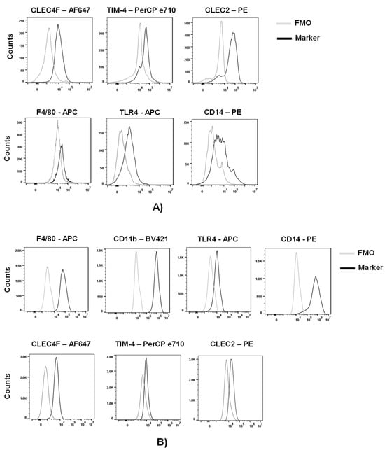
Ebola virus disease (EVD) represents a global health threat. The etiological agents of EVD are six species of Orthoebolaviruses, with Orthoebolavirus zairense (EBOV) having the greatest public health and medical significance. EVD pathogenesis occurs as a result of broad cellular tropism of the virus, robust viral replication and a potent and dysregulated production of cytokines. In vivo, tissue macrophages are some of the earliest cells infected and contribute significantly to virus load and cytokine production. While EBOV is known to infect macrophages and to generate high titer virus in the liver, EBOV infection of liver macrophages, Kupffer cells, has not previously been examined in tissue culture or experimentally manipulated in vivo. Here, we employed primary murine Kupffer cells (KC) and an immortalized murine Kupffer cell line (ImKC) to assess EBOV-eGFP replication in liver macrophages. KCs and ImKCs were highly permissive for EBOV infection and IFN-γ polarization of these cells suppressed their permissiveness to infection. The kinetics of IFN-γ-elicited antiviral responses were examined using a biologically contained model of EBOV infection termed EBOV ΔVP30. The antiviral activity of IFN-γ was transient, but a modest ~3-fold reduction of infection persisted for as long as 6 days post-treatment. To assess the interferon-stimulated gene products (ISGs) responsible for protection, the efficacy of secreted ISGs induced by IFN-γ was evaluated and secreted ISGs failed to block EBOV ΔVP30. Our studies define new cellular tools for the study of EBOV infection that can potentially aid the development of new antiviral therapies. Furthermore, our data underscore the importance of macrophages in EVD pathogenesis and those IFN-γ-elicited ISGs that help to control EBOV infection. Full article
Show Figures

Figure 1

11 pages, 1629 KiB  
Article
The Susceptibility of BALB/c Mice to a Mouse-Adapted Ebola Virus Intravaginal Infection
by Olivier Escaffre, Terry L. Juelich, Jennifer K. Smith, Lihong Zhang, Nigel Bourne and Alexander N. Freiberg
Viruses 2023, 15(7), 1590; https://doi.org/10.3390/v15071590 - 21 Jul 2023
Cited by 1 | Viewed by 1394
Abstract
Ebola virus (EBOV) causes Ebola virus disease (EVD), which is characterized by hemorrhagic fever with high mortality rates in humans. EBOV sexual transmission has been a concern since the 2014–2016 outbreak in Africa, as persistent infection in the testis and transmission to women [...] Read more.
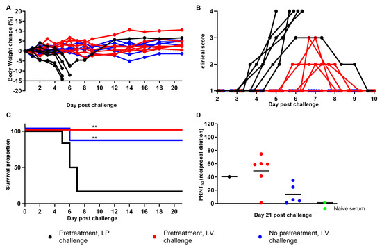
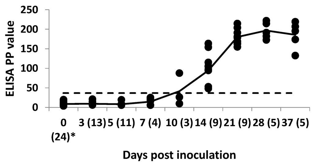
Ebola virus (EBOV) causes Ebola virus disease (EVD), which is characterized by hemorrhagic fever with high mortality rates in humans. EBOV sexual transmission has been a concern since the 2014–2016 outbreak in Africa, as persistent infection in the testis and transmission to women was demonstrated. The only study related to establishing an intravaginal small animal infection model was recently documented in IFNAR−/− mice using wild-type and mouse-adapted EBOV (maEBOV), and resulted in 80% mortality, supporting epidemiological data. However, this route of transmission is still poorly understood in women, and the resulting EVD from it is understudied. Here, we contribute to this field of research by providing data from immunocompetent BALB/c mice. We demonstrate that progesterone priming increased the likelihood of maEBOV vaginal infection and of exhibiting the symptoms of disease and seroconversion. However, our data suggest subclinical infection, regardless of the infective dose. We conclude that maEBOV can infect BALB/c mice through vaginal inoculation, but that this route of infection causes significantly less disease compared to intraperitoneal injection at a similar dose, which is consistent with previous studies using other peripheral routes of inoculation in that animal model. Our data are inconsistent with the disease severity described in female patients, therefore suggesting that BALB/c mice are unsuitable for modeling typical EVD following vaginal challenge with maEBOV. Further studies are required to determine the mechanisms by which EVD is attenuated in BALB/c mice, using maEBOV via the vaginal route, as in our experimental set-up. Full article
Show Figures

Figure 1

2022

Jump to: 2023, 2020, 2019, 2018, 2017, 2016, 2015, 2014

17 pages, 3034 KiB  
Article
Ebola Virus Activates IRE1α-Dependent XBP1u Splicing
by Cornelius Rohde, Sebastian Pfeiffer, Sara Baumgart, Stephan Becker and Verena Krähling
Viruses 2023, 15(1), 122; https://doi.org/10.3390/v15010122 - 30 Dec 2022
Cited by 2 | Viewed by 2587
Abstract
Ebola (EBOV) and Marburg virus (MARV) are highly pathogenic filoviruses that influence cellular signaling according to their own needs. MARV has been shown to regulate the IRE1α-dependent unfolded protein response (UPR) to ensure optimal virus replication. It was not known whether EBOV affects [...] Read more.
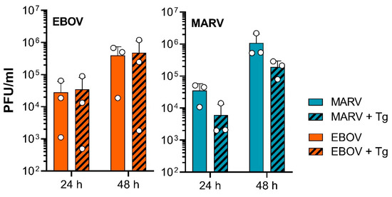
Ebola (EBOV) and Marburg virus (MARV) are highly pathogenic filoviruses that influence cellular signaling according to their own needs. MARV has been shown to regulate the IRE1α-dependent unfolded protein response (UPR) to ensure optimal virus replication. It was not known whether EBOV affects this signaling cascade, which can be beneficial or detrimental for viruses. Activation of IRE1α leads to the expression of the transcription factor XBP1s, which binds to cis-acting UPR elements (UPRE), resulting in the expression of genes aimed at restoring homeostasis in the endoplasmic reticulum. We observed that EBOV infection, in contrast to MARV infection, led to UPR activation by IRE1α-dependent but not ATF6-dependent signaling. We showed an activation of IRE1α, XBP1s and UPRE target genes upon EBOV infection. ATF6, another UPRE transcription factor, was not activated. UPRE activation was mainly attributed to the EBOV nucleoprotein NP and the soluble glycoprotein sGP. Finally, activation of UPR by thapsigargin, a potent ER-stress inducer, in parallel to infection as well as knock-out of XBP1 had no effect on EBOV growth, while MARV proliferation was affected by thapsigargin-dependent UPR activation. Taken together EBOV and MARV differ in their strategy of balancing IRE1α-dependent signaling for their own needs. Full article
Show Figures

Figure 1

2020

Jump to: 2023, 2022, 2019, 2018, 2017, 2016, 2015, 2014

20 pages, 3513 KiB  
Communication
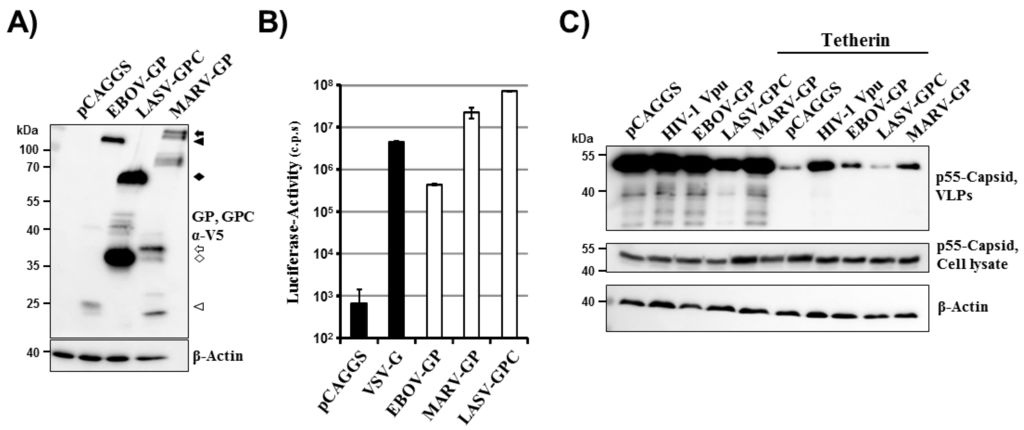
A Novel Ebola Virus VP40 Matrix Protein-Based Screening for Identification of Novel Candidate Medical Countermeasures
by Ryan P. Bennett, Courtney L. Finch, Elena N. Postnikova, Ryan A. Stewart, Yingyun Cai, Shuiqing Yu, Janie Liang, Julie Dyall, Jason D. Salter, Harold C. Smith and Jens H. Kuhn
Viruses 2021, 13(1), 52; https://doi.org/10.3390/v13010052 - 31 Dec 2020
Cited by 14 | Viewed by 4903
Abstract
Filoviruses, such as Ebola virus and Marburg virus, are of significant human health concern. From 2013 to 2016, Ebola virus caused 11,323 fatalities in Western Africa. Since 2018, two Ebola virus disease outbreaks in the Democratic Republic of the Congo resulted in 2354 [...] Read more.
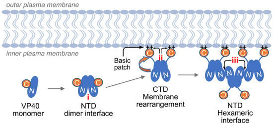
Filoviruses, such as Ebola virus and Marburg virus, are of significant human health concern. From 2013 to 2016, Ebola virus caused 11,323 fatalities in Western Africa. Since 2018, two Ebola virus disease outbreaks in the Democratic Republic of the Congo resulted in 2354 fatalities. Although there is progress in medical countermeasure (MCM) development (in particular, vaccines and antibody-based therapeutics), the need for efficacious small-molecule therapeutics remains unmet. Here we describe a novel high-throughput screening assay to identify inhibitors of Ebola virus VP40 matrix protein association with viral particle assembly sites on the interior of the host cell plasma membrane. Using this assay, we screened nearly 3000 small molecules and identified several molecules with the desired inhibitory properties. In secondary assays, one identified compound, sangivamycin, inhibited not only Ebola viral infectivity but also that of other viruses. This finding indicates that it is possible for this new VP40-based screening method to identify highly potent MCMs against Ebola virus and its relatives. Full article
Show Figures

Figure 1

16 pages, 4726 KiB  
Article
A Surrogate Animal Model for Screening of Ebola and Marburg Glycoprotein-Targeting Drugs Using Pseudotyped Vesicular Stomatitis Viruses
by Takeshi Saito, Junki Maruyama, Noriyo Nagata, Mao Isono, Kosuke Okuya, Yoshihiro Takadate, Yurie Kida, Hiroko Miyamoto, Akina Mori-Kajihara, Takanari Hattori, Wakako Furuyama, Shinya Ogawa, Shigeru Iida and Ayato Takada
Viruses 2020, 12(9), 923; https://doi.org/10.3390/v12090923 - 22 Aug 2020
Cited by 10 | Viewed by 3780
Abstract
Filoviruses, including Ebola virus (EBOV) and Marburg virus (MARV), cause severe hemorrhagic fever in humans and nonhuman primates with high mortality rates. There is no approved therapy against these deadly viruses. Antiviral drug development has been hampered by the requirement of a biosafety [...] Read more.
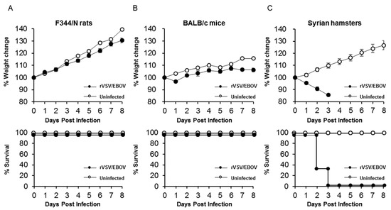
Filoviruses, including Ebola virus (EBOV) and Marburg virus (MARV), cause severe hemorrhagic fever in humans and nonhuman primates with high mortality rates. There is no approved therapy against these deadly viruses. Antiviral drug development has been hampered by the requirement of a biosafety level (BSL)-4 facility to handle infectious EBOV and MARV because of their high pathogenicity to humans. In this study, we aimed to establish a surrogate animal model that can be used for anti-EBOV and -MARV drug screening under BSL-2 conditions by focusing on the replication-competent recombinant vesicular stomatitis virus (rVSV) pseudotyped with the envelope glycoprotein (GP) of EBOV (rVSV/EBOV) and MARV (rVSV/MARV), which has been investigated as vaccine candidates and thus widely used in BSL-2 laboratories. We first inoculated mice, rats, and hamsters intraperitoneally with rVSV/EBOV and found that only hamsters showed disease signs and succumbed within 4 days post-infection. Infection with rVSV/MARV also caused lethal infection in hamsters. Both rVSV/EBOV and rVSV/MARV were detected at high titers in multiple organs including the liver, spleen, kidney, and lungs of infected hamsters, indicating acute and systemic infection resulting in fatal outcomes. Therapeutic effects of passive immunization with an anti-EBOV neutralizing antibody were specifically observed in rVSV/EBOV-infected hamsters. Thus, this animal model is expected to be a useful tool to facilitate in vivo screening of anti-filovirus drugs targeting the GP molecule. Full article
Show Figures

Figure 1

12 pages, 4680 KiB  
Article
Ebola Virus Disease Survivors Show More Efficient Antibody Immunity than Vaccinees Despite Similar Levels of Circulating Immunoglobulins
by Till Koch, Monika Rottstegge, Paula Ruibal, Sergio Gomez-Medina, Emily V. Nelson, Beatriz Escudero-Pérez, Matthias Pillny, My Linh Ly, Fara Raymond Koundouno, Joseph Akoi Bore, N’Faly Magassouba, Christine Dahlke, Stephan Günther, Miles W. Carroll, Marylyn M. Addo and César Muñoz-Fontela
Viruses 2020, 12(9), 915; https://doi.org/10.3390/v12090915 - 20 Aug 2020
Cited by 14 | Viewed by 4929
Abstract
The last seven years have seen the greatest surge of Ebola virus disease (EVD) cases in equatorial Africa, including the 2013–2016 epidemic in West Africa and the recent epidemics in the Democratic Republic of Congo (DRC). The vaccine clinical trials that took place [...] Read more.
The last seven years have seen the greatest surge of Ebola virus disease (EVD) cases in equatorial Africa, including the 2013–2016 epidemic in West Africa and the recent epidemics in the Democratic Republic of Congo (DRC). The vaccine clinical trials that took place in West Africa and the DRC, as well as follow-up studies in collaboration with EVD survivor communities, have for the first time allowed researchers to compare immune memory induced by natural infection and vaccination. These comparisons may be relevant to evaluate the putative effectiveness of vaccines and candidate medical countermeasures such as convalescent plasma transfer. In this study, we compared the long-term functionality of anti-EBOV glycoprotein (GP) antibodies from EVD survivors with that from volunteers who received the recombinant vesicular stomatitis virus vectored vaccine (rVSV-ZEBOV) during the Phase I clinical trial in Hamburg. Our study highlights important differences between EBOV vaccination and natural infection and provides a framework for comparison with other vaccine candidates. Full article
Show Figures

Figure 1

27 pages, 6039 KiB  
Article
Reporter Assays for Ebola Virus Nucleoprotein Oligomerization, Virion-Like Particle Budding, and Minigenome Activity Reveal the Importance of Nucleoprotein Amino Acid Position 111
by Aaron E. Lin, William E. Diehl, Yingyun Cai, Courtney L. Finch, Chidiebere Akusobi, Robert N. Kirchdoerfer, Laura Bollinger, Stephen F. Schaffner, Elizabeth A. Brown, Erica Ollmann Saphire, Kristian G. Andersen, Jens H. Kuhn, Jeremy Luban and Pardis C. Sabeti
Viruses 2020, 12(1), 105; https://doi.org/10.3390/v12010105 - 15 Jan 2020
Cited by 8 | Viewed by 5489
Abstract
For highly pathogenic viruses, reporter assays that can be rapidly performed are critically needed to identify potentially functional mutations for further study under maximal containment (e.g., biosafety level 4 [BSL-4]). The Ebola virus nucleoprotein (NP) plays multiple essential roles during the viral life [...] Read more.
For highly pathogenic viruses, reporter assays that can be rapidly performed are critically needed to identify potentially functional mutations for further study under maximal containment (e.g., biosafety level 4 [BSL-4]). The Ebola virus nucleoprotein (NP) plays multiple essential roles during the viral life cycle, yet few tools exist to study the protein under BSL-2 or equivalent containment. Therefore, we adapted reporter assays to measure NP oligomerization and virion-like particle (VLP) production in live cells and further measured transcription and replication using established minigenome assays. As a proof-of-concept, we examined the NP-R111C substitution, which emerged during the 2013–2016 Western African Ebola virus disease epidemic and rose to high frequency. NP-R111C slightly increased NP oligomerization and VLP budding but slightly decreased transcription and replication. By contrast, a synthetic charge-reversal mutant, NP-R111E, greatly increased oligomerization but abrogated transcription and replication. These results are intriguing in light of recent structures of NP oligomers, which reveal that the neighboring residue, K110, forms a salt bridge with E349 on adjacent NP molecules. By developing and utilizing multiple reporter assays, we find that the NP-111 position mediates a complex interplay between NP’s roles in protein structure, virion budding, and transcription and replication. Full article
Show Figures

Figure 1

2019

Jump to: 2023, 2022, 2020, 2018, 2017, 2016, 2015, 2014

12 pages, 4764 KiB  
Article
Ebola Virus Uptake into Polarized Cells from the Apical Surface
by Meng Hu, Fei Wang, Wei Li, Xiaowei Zhang, Zhiping Zhang, Xian-En Zhang and Zongqiang Cui
Viruses 2019, 11(12), 1117; https://doi.org/10.3390/v11121117 - 2 Dec 2019
Cited by 8 | Viewed by 4529
Abstract
Ebola virus (EBOV) causes severe hemorrhagic fever with high mortality rates. EBOV can infect many types of cells. During severe EBOV infection, polarized epithelial and endothelial cells are damaged, which promotes vascular instability and dysregulation. However, the mechanism causing these symptoms is largely [...] Read more.
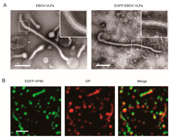
Ebola virus (EBOV) causes severe hemorrhagic fever with high mortality rates. EBOV can infect many types of cells. During severe EBOV infection, polarized epithelial and endothelial cells are damaged, which promotes vascular instability and dysregulation. However, the mechanism causing these symptoms is largely unknown. Here, we studied virus infection in polarized Vero C1008 cells grown on semipermeable Transwell by using EGFP-labeled Ebola virus-like particles (VLPs). Our results showed that Ebola VLPs preferred to enter polarized Vero cells from the apical cell surface. Furthermore, we showed that the EBOV receptors TIM-1 and Axl were distributed apically, which could be responsible for mediating efficient apical viral entry. Macropinocytosis and intracellular receptor Niemann–Pick type C1 (NPC1) had no polarized distribution, although they played roles in virus entry. This study provides a new view of EBOV uptake and cell polarization, which facilitates a further understanding of EBOV infection and pathogenesis. Full article
Show Figures

Figure 1

12 pages, 5050 KiB  
Article
Efficient Expression and Processing of Ebola Virus Glycoprotein Induces Morphological Changes in BmN Cells but Cannot Rescue Deficiency of Bombyx Mori Nucleopolyhedrovirus GP64
by Jinshan Huang, Na Liu, Fanbo Xu, Ellen Ayepa, Charles Amanze, Luping Sun, Yaqin Shen, Miao Yang, Shuwen Yang, Xingjia Shen and Bifang Hao
Viruses 2019, 11(11), 1067; https://doi.org/10.3390/v11111067 - 15 Nov 2019
Cited by 7 | Viewed by 4540
Abstract
Ebola virus (EBOV) disease outbreaks have resulted in many fatalities, yet no licensed vaccines are available to prevent infection. Recombinant glycoprotein (GP) production may contribute to finding a cure for Ebola virus disease, which is the key candidate protein for vaccine preparation. To [...] Read more.
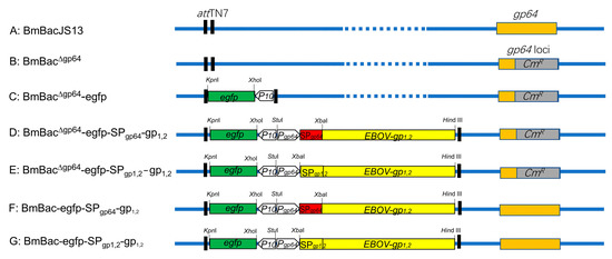
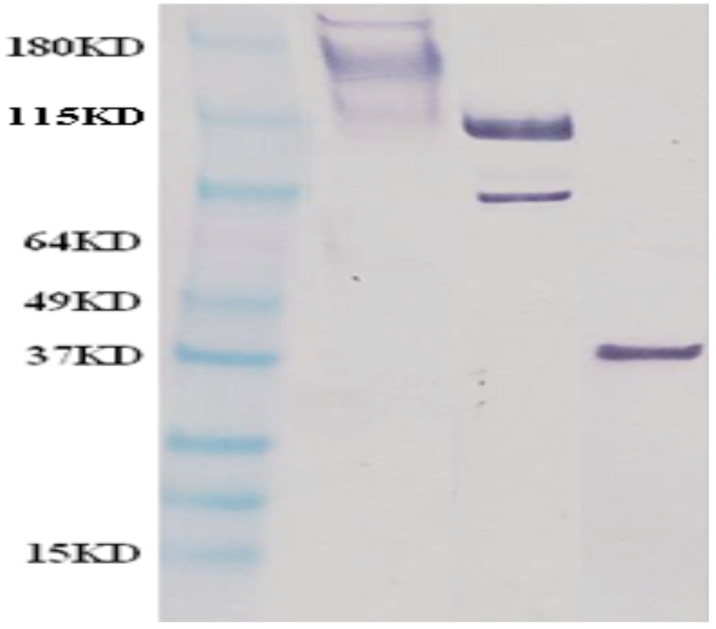
Ebola virus (EBOV) disease outbreaks have resulted in many fatalities, yet no licensed vaccines are available to prevent infection. Recombinant glycoprotein (GP) production may contribute to finding a cure for Ebola virus disease, which is the key candidate protein for vaccine preparation. To explore GP1,2 expression in BmN cells, EBOV-GP1,2 with its native signal peptide or the GP64 signal peptide was cloned and transferred into a normal or gp64 null Bombyx mori nucleopolyhedrovirus (BmNPV) bacmid via transposition. The infectivity of the recombinant bacmids was investigated after transfection, expression and localization of EBOV-GP were investigated, and cell morphological changes were analyzed by TEM. The GP64 signal peptide, but not the GP1,2 native signal peptide, caused GP1,2 localization to the cell membrane, and the differentially localized GP1,2 proteins were cleaved into GP1 and GP2 fragments in BmN cells. GP1,2 expression resulted in dramatic morphological changes in BmN cells in the early stage of infection. However, GP1,2 expression did not rescue GP64 deficiency in BmNPV infection. This study provides a better understanding of GP expression and processing in BmN cells, which may lay a foundation for EBOV-GP expression using the BmNPV baculovirus expression system. Full article
Show Figures

Figure 1

18 pages, 2531 KiB  
Article
Investigating the Cellular Transcriptomic Response Induced by the Makona Variant of Ebola Virus in Differentiated THP-1 Cells
by Andrew Bosworth, Stuart D. Dowall, Stuart Armstrong, Xuan Liu, Xiaofeng Dong, Christine B. Bruce, Lisa F. P. Ng, Miles W. Carroll, Roger Hewson and Julian A. Hiscox
Viruses 2019, 11(11), 1023; https://doi.org/10.3390/v11111023 - 4 Nov 2019
Cited by 7 | Viewed by 3304
Abstract
Recent studies have shown that transcriptomic analysis of blood samples taken from patients with acute Ebola virus disease (EVD) during the 2013–2016 West African outbreak was suggestive that a severe inflammatory response took place in acutely ill patients. The significant knowledge gained from [...] Read more.
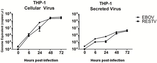
Recent studies have shown that transcriptomic analysis of blood samples taken from patients with acute Ebola virus disease (EVD) during the 2013–2016 West African outbreak was suggestive that a severe inflammatory response took place in acutely ill patients. The significant knowledge gained from studying the Makona variant, a cause of the largest known EVD outbreak, may be applicable to other species of ebolavirus, and other variants of the Ebola virus (EBOV) species. To investigate the ability of Makona to initiate an inflammatory response in human macrophages and characterise the host response in a similar manner to previously characterised EBOV variants, the human monocytic cell line THP-1 was differentiated into macrophage-like cells and infected with Makona. RNA-Seq and quantitative proteomics were used to identify and quantify host mRNA and protein abundance during infection. Data from infection with Reston virus (RESTV) were used as comparators to investigate changes that may be specific to, or enhanced in, Makona infection in relation to a less pathogenic species of ebolavirus.. This study found demonstrable induction of the inflammatory response, and increase in the activation state of THP-1 macrophages infected with Makona. NFκB and inflammation-associated transcripts displayed significant changes in abundance, reflective of what was observed in human patients during the 2013–2016 EBOV outbreak in West Africa, and demonstrated that transcriptomic changes found in Makona-infected cells were similar to that observed in Reston virus infection and that have been described in previous studies of other variants of EBOV. Full article
Show Figures

Figure 1

25 pages, 6209 KiB  
Article
Cytokine Effects on the Entry of Filovirus Envelope Pseudotyped Virus-Like Particles into Primary Human Macrophages
by Tzanko S. Stantchev, Autumn Zack-Taylor, Nicholas Mattson, Klaus Strebel, Christopher C. Broder and Kathleen A. Clouse
Viruses 2019, 11(10), 889; https://doi.org/10.3390/v11100889 - 23 Sep 2019
Cited by 4 | Viewed by 4619
Abstract
Macrophages are one of the first and also a major site of filovirus replication and, in addition, are a source of multiple cytokines, presumed to play a critical role in the pathogenesis of the viral infection. Some of these cytokines are known to [...] Read more.
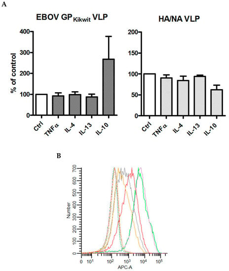
Macrophages are one of the first and also a major site of filovirus replication and, in addition, are a source of multiple cytokines, presumed to play a critical role in the pathogenesis of the viral infection. Some of these cytokines are known to induce macrophage phenotypic changes in vitro, but how macrophage polarization may affect the cell susceptibility to filovirus entry remains largely unstudied. We generated different macrophage subsets using cytokine pre-treatment and subsequently tested their ability to fuse with beta-lactamase containing virus-like particles (VLP), pseudotyped with the surface glycoprotein of Ebola virus (EBOV) or the glycoproteins of other clinically relevant filovirus species. We found that pre-incubation of primary human monocyte-derived macrophages (MDM) with interleukin-10 (IL-10) significantly enhanced filovirus entry into cells obtained from multiple healthy donors, and the IL-10 effect was preserved in the presence of pro-inflammatory cytokines found to be elevated during EBOV disease. In contrast, fusion of IL-10-treated macrophages with influenza hemagglutinin/neuraminidase pseudotyped VLPs was unchanged or slightly reduced. Importantly, our in vitro data showing enhanced virus entry are consistent with the correlation established between elevated serum IL-10 and increased mortality in filovirus infected patients and also reveal a novel mechanism that may account for the IL-10-mediated increase in filovirus pathogenicity. Full article
Show Figures

Figure 1

25 pages, 1088 KiB  
Review
Extracellular Vesicles and Ebola Virus: A New Mechanism of Immune Evasion
by Michelle L. Pleet, Catherine DeMarino, Spencer W. Stonier, John M. Dye, Steven Jacobson, M. Javad Aman and Fatah Kashanchi
Viruses 2019, 11(5), 410; https://doi.org/10.3390/v11050410 - 2 May 2019
Cited by 28 | Viewed by 6956
Abstract
Ebola virus (EBOV) disease can result in a range of symptoms anywhere from virtually asymptomatic to severe hemorrhagic fever during acute infection. Additionally, spans of asymptomatic persistence in recovering survivors is possible, during which transmission of the virus may occur. In acute infection, [...] Read more.
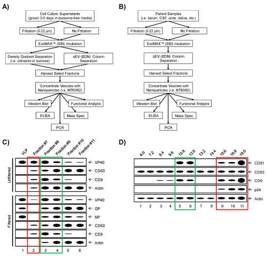
Ebola virus (EBOV) disease can result in a range of symptoms anywhere from virtually asymptomatic to severe hemorrhagic fever during acute infection. Additionally, spans of asymptomatic persistence in recovering survivors is possible, during which transmission of the virus may occur. In acute infection, substantial cytokine storm and bystander lymphocyte apoptosis take place, resulting in uncontrolled, systemic inflammation in affected individuals. Recently, studies have demonstrated the presence of EBOV proteins VP40, glycoprotein (GP), and nucleoprotein (NP) packaged into extracellular vesicles (EVs) during infection. EVs containing EBOV proteins have been shown to induce apoptosis in recipient immune cells, as well as contain pro-inflammatory cytokines. In this manuscript, we review the current field of knowledge on EBOV EVs including the mechanisms of their biogenesis, their cargo and their effects in recipient cells. Furthermore, we discuss some of the effects that may be induced by EBOV EVs that have not yet been characterized and highlight the remaining questions and future directions. Full article
Show Figures

Figure 1

12 pages, 1390 KiB  
Article
Inflammatory and Humoral Immune Response during Ebola Virus Infection in Survivor and Fatal Cases Occurred in Sierra Leone during the 2014–2016 Outbreak in West Africa
by Francesca Colavita, Mirella Biava, Concetta Castilletti, Simone Lanini, Rossella Miccio, Gina Portella, Francesco Vairo, Giuseppe Ippolito, Maria Rosaria Capobianchi, Antonino Di Caro and Eleonora Lalle
Viruses 2019, 11(4), 373; https://doi.org/10.3390/v11040373 - 23 Apr 2019
Cited by 26 | Viewed by 5926
Abstract
Ebola virus (EBOV) infection is characterized by an excessive inflammatory response, a loss of lymphocytes and a general paralysis of the immune system, however pathophysiological mechanisms are not fully understood. In a cohort of 23 fatal and 21 survivors of ebola virus disease [...] Read more.
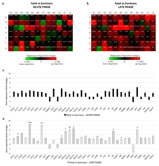
Ebola virus (EBOV) infection is characterized by an excessive inflammatory response, a loss of lymphocytes and a general paralysis of the immune system, however pathophysiological mechanisms are not fully understood. In a cohort of 23 fatal and 21 survivors of ebola virus disease (EVD) cases admitted to the Emergency Ebola-Treatment-Center in Goderich (Freetown, Sierra Leone) during the 2014 to 2016 EBOV epidemic in Western Africa, we analyzed the pathway-focused gene expression profile of secreted proteins involved in the immune response and the levels of specific anti-EBOV IgM and IgG from the time of admission till discharge or death. We observed a dysregulated inflammatory response in fatal patients as compared to survivors, mainly consisting of the upregulation of inflammatory mediators, whose extent directly correlated with viremia levels. The upregulation persisted and intensified during the late phase of infection. Relevant differences were also found in humoral immunity, as an earlier and more robust EBOV antibody response was observed in survivor patients. Full article
Show Figures

Figure 1

21 pages, 9299 KiB  
Article
Periplasmic Nanobody-APEX2 Fusions Enable Facile Visualization of Ebola, Marburg, and Mĕnglà virus Nucleoproteins, Alluding to Similar Antigenic Landscapes among Marburgvirus and Dianlovirus
by Laura J. Sherwood and Andrew Hayhurst
Viruses 2019, 11(4), 364; https://doi.org/10.3390/v11040364 - 20 Apr 2019
Cited by 12 | Viewed by 6215
Abstract
We explore evolved soybean ascorbate peroxidase (APEX2) as a reporter when fused to the C-termini of llama nanobodies (single-domain antibodies, sdAb; variable domains of heavy chain-only antibodies, VHH) targeted to the E. coli periplasm. Periplasmic expression preserves authentic antibody N-termini, intra-domain disulphide bond(s), [...] Read more.
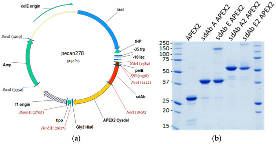
We explore evolved soybean ascorbate peroxidase (APEX2) as a reporter when fused to the C-termini of llama nanobodies (single-domain antibodies, sdAb; variable domains of heavy chain-only antibodies, VHH) targeted to the E. coli periplasm. Periplasmic expression preserves authentic antibody N-termini, intra-domain disulphide bond(s), and capitalizes on efficient haem loading through the porous E. coli outer membrane. Using monomeric and dimeric anti-nucleoprotein (NP) sdAb cross-reactive within the Marburgvirus genus and cross-reactive within the Ebolavirus genus, we show that periplasmic sdAb–APEX2 fusion proteins are easily purified at multi-mg amounts. The fusions were used in Western blotting, ELISA, and microscopy to visualize NPs using colorimetric and fluorescent imaging. Dimeric sdAb–APEX2 fusions were superior at binding NPs from viruses that were evolutionarily distant to that originally used to select the sdAb. Partial conservation of the anti-Marburgvirus sdAb epitope enabled the recognition of a novel NP encoded by the recently discovered Mĕnglà virus genome. Antibody–antigen interactions were rationalized using monovalent nanoluciferase titrations and contact mapping analysis of existing crystal structures, while molecular modelling was used to reveal the potential landscape of the Mĕnglà NP C-terminal domain. The sdAb–APEX2 fusions also enabled live Marburgvirus and Ebolavirus detection 24 h post-infection of Vero E6 cells within a BSL-4 laboratory setting. The simple and inexpensive mining of large amounts of periplasmic sdAb–APEX2 fusion proteins should help advance studies of past, contemporary, and perhaps Filovirus species yet to be discovered. Full article
Show Figures

Figure 1

21 pages, 3861 KiB  
Article
First Evidence of Antibodies Against Lloviu Virus in Schreiber’s Bent-Winged Insectivorous Bats Demonstrate a Wide Circulation of the Virus in Spain
by Eva Ramírez de Arellano, Mariano Sanchez-Lockhart, Maria J. Perteguer, Maggie Bartlett, Marta Ortiz, Pamela Campioli, Ana Hernández, Jeanette Gonzalez, Karla Garcia, Manolo Ramos, Miguel Ángel Jiménez-Clavero, Antonio Tenorio, Mª Paz Sánchez-Seco, Félix González, Juan Emilio Echevarría, Gustavo Palacios and Anabel Negredo
Viruses 2019, 11(4), 360; https://doi.org/10.3390/v11040360 - 19 Apr 2019
Cited by 15 | Viewed by 6493
Abstract
Although Lloviu virus (LLOV) was discovered in the carcasses of insectivorous Schreiber’s Bent-winged bats in the caves of Northern Spain in 2002, its infectivity and pathogenicity remain unclear. We examined the seroprevalence of LLOV in potentially exposed Schreiber’s Bent-winged bats (n = [...] Read more.
Although Lloviu virus (LLOV) was discovered in the carcasses of insectivorous Schreiber’s Bent-winged bats in the caves of Northern Spain in 2002, its infectivity and pathogenicity remain unclear. We examined the seroprevalence of LLOV in potentially exposed Schreiber’s Bent-winged bats (n = 60), common serotine bats (n = 10) as controls, and humans (n = 22) using an immunoblot assay. We found antibodies against LLOV GP2 in all of Schreiber’s Bent-winged bats serum pools, but not in any of the common serotine bats and human pools tested. To confirm this seroreactivity, 52 serums were individually tested using Domain Programmable Arrays (DPA), a phage display based-system serology technique for profiling filovirus epitopes. A serological signature against different LLOV proteins was obtained in 19/52 samples tested (36.5%). The immunodominant response was in the majority specific to LLOV-unique epitopes, confirming that the serological response detected was to LLOV. To our knowledge, this is the first serological evidence of LLOV exposure in live captured Schreiber’s Bent-winged bats, dissociating LLOV circulation as the cause of the previously reported die-offs. Full article
Show Figures

Figure 1

16 pages, 3725 KiB  
Article
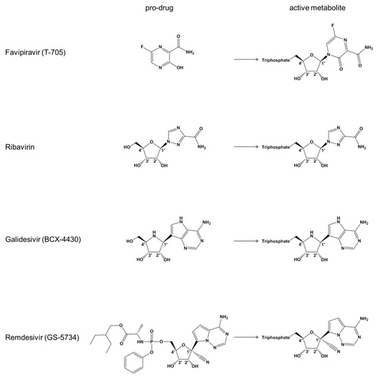
Mechanism of Inhibition of Ebola Virus RNA-Dependent RNA Polymerase by Remdesivir
by Egor P. Tchesnokov, Joy Y. Feng, Danielle P. Porter and Matthias Götte
Viruses 2019, 11(4), 326; https://doi.org/10.3390/v11040326 - 4 Apr 2019
Cited by 466 | Viewed by 40603
Abstract
Remdesivir (GS-5734) is a 1′-cyano-substituted adenosine nucleotide analogue prodrug that shows broad-spectrum antiviral activity against several RNA viruses. This compound is currently under clinical development for the treatment of Ebola virus disease (EVD). While antiviral effects have been demonstrated in cell culture and [...] Read more.
Remdesivir (GS-5734) is a 1′-cyano-substituted adenosine nucleotide analogue prodrug that shows broad-spectrum antiviral activity against several RNA viruses. This compound is currently under clinical development for the treatment of Ebola virus disease (EVD). While antiviral effects have been demonstrated in cell culture and in non-human primates, the mechanism of action of Ebola virus (EBOV) inhibition for remdesivir remains to be fully elucidated. The EBOV RNA-dependent RNA polymerase (RdRp) complex was recently expressed and purified, enabling biochemical studies with the relevant triphosphate (TP) form of remdesivir and its presumptive target. In this study, we confirmed that remdesivir-TP is able to compete for incorporation with adenosine triphosphate (ATP). Enzyme kinetics revealed that EBOV RdRp and respiratory syncytial virus (RSV) RdRp incorporate ATP and remdesivir-TP with similar efficiencies. The selectivity of ATP against remdesivir-TP is ~4 for EBOV RdRp and ~3 for RSV RdRp. In contrast, purified human mitochondrial RNA polymerase (h-mtRNAP) effectively discriminates against remdesivir-TP with a selectivity value of ~500-fold. For EBOV RdRp, the incorporated inhibitor at position i does not affect the ensuing nucleotide incorporation event at position i+1. For RSV RdRp, we measured a ~6-fold inhibition at position i+1 although RNA synthesis was not terminated. Chain termination was in both cases delayed and was seen predominantly at position i+5. This pattern is specific to remdesivir-TP and its 1′-cyano modification. Compounds with modifications at the 2′-position show different patterns of inhibition. While 2′-C-methyl-ATP is not incorporated, ara-ATP acts as a non-obligate chain terminator and prevents nucleotide incorporation at position i+1. Taken together, our biochemical data indicate that the major contribution to EBOV RNA synthesis inhibition by remdesivir can be ascribed to delayed chain termination. The long distance of five residues between the incorporated nucleotide analogue and its inhibitory effect warrant further investigation. Full article
Show Figures

Figure 1

26 pages, 4113 KiB  
Article
Characterization of the Filovirus-Resistant Cell Line SH-SY5Y Reveals Redundant Role of Cell Surface Entry Factors
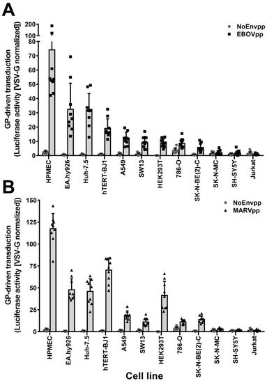
by Francisco J. Zapatero-Belinchón, Erik Dietzel, Olga Dolnik, Katinka Döhner, Rui Costa, Barbara Hertel, Barbora Veselkova, Jared Kirui, Anneke Klintworth, Michael P. Manns, Stefan Pöhlmann, Thomas Pietschmann, Thomas Krey, Sandra Ciesek, Gisa Gerold, Beate Sodeik, Stephan Becker and Thomas von Hahn
Viruses 2019, 11(3), 275; https://doi.org/10.3390/v11030275 - 19 Mar 2019
Cited by 6 | Viewed by 7369
Abstract
Filoviruses infect a wide range of cell types with the exception of lymphocytes. The intracellular proteins cathepsin B and L, two-pore channel 1 and 2, and bona fide receptor Niemann–Pick Disease C1 (NPC1) are essential for the endosomal phase of cell entry. However, [...] Read more.
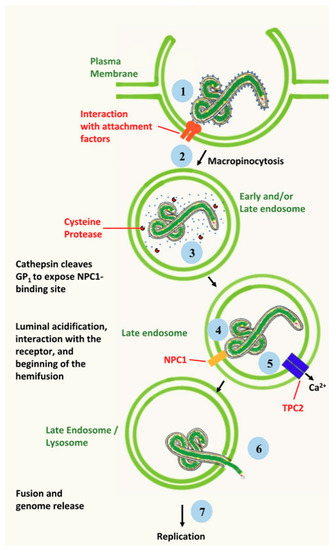
Filoviruses infect a wide range of cell types with the exception of lymphocytes. The intracellular proteins cathepsin B and L, two-pore channel 1 and 2, and bona fide receptor Niemann–Pick Disease C1 (NPC1) are essential for the endosomal phase of cell entry. However, earlier steps of filoviral infection remain poorly characterized. Numerous plasma membrane proteins have been implicated in attachment but it is still unclear which ones are sufficient for productive entry. To define a minimal set of host factors required for filoviral glycoprotein-driven cell entry, we screened twelve cell lines and identified the nonlymphocytic cell line SH-SY5Y to be specifically resistant to filovirus infection. Heterokaryons of SH-SY5Y cells fused to susceptible cells were susceptible to filoviruses, indicating that SH-SY5Y cells do not express a restriction factor but lack an enabling factor critical for filovirus entry. However, all tested cell lines expressed functional intracellular factors. Global gene expression profiling of known cell surface entry factors and protein expression levels of analyzed attachment factors did not reveal any correlation between susceptibility and expression of a specific host factor. Using binding assays with recombinant filovirus glycoprotein, we identified cell attachment as the step impaired in filovirus entry in SH-SY5Y cells. Individual overexpression of attachment factors T-cell immunoglobulin and mucin domain 1 (TIM-1), Axl, Mer, or dendritic cell-specific intercellular adhesion molecule-3-grabbing non-integrin (DC-SIGN) rendered SH-SY5Y cells susceptible to filovirus glycoprotein-driven transduction. Our study reveals that a lack of attachment factors limits filovirus entry and provides direct experimental support for a model of filoviral cell attachment where host factor usage at the cell surface is highly promiscuous. Full article
Show Figures

Figure 1

20 pages, 1865 KiB  
Review
Ebola Virus Entry: From Molecular Characterization to Drug Discovery
by Cristiano Salata, Arianna Calistri, Gualtiero Alvisi, Michele Celestino, Cristina Parolin and Giorgio Palù
Viruses 2019, 11(3), 274; https://doi.org/10.3390/v11030274 - 19 Mar 2019
Cited by 58 | Viewed by 17812
Abstract
Ebola Virus Disease (EVD) is one of the most lethal transmissible infections, characterized by a high fatality rate, and caused by a member of the Filoviridae family. The recent large outbreak of EVD in Western Africa (2013–2016) highlighted the worldwide threat represented by [...] Read more.
Ebola Virus Disease (EVD) is one of the most lethal transmissible infections, characterized by a high fatality rate, and caused by a member of the Filoviridae family. The recent large outbreak of EVD in Western Africa (2013–2016) highlighted the worldwide threat represented by the disease and its impact on global public health and the economy. The development of highly needed anti-Ebola virus antivirals has been so far hampered by the shortage of tools to study their life cycle in vitro, allowing to screen for potential active compounds outside a biosafety level-4 (BSL-4) containment. Importantly, the development of surrogate models to study Ebola virus entry in a BSL-2 setting, such as viral pseudotypes and Ebola virus-like particles, tremendously boosted both our knowledge of the viral life cycle and the identification of promising antiviral compounds interfering with viral entry. In this context, the combination of such surrogate systems with large-scale small molecule compounds and haploid genetic screenings, as well as rational drug design and drug repurposing approaches will prove priceless in our quest for the development of a treatment for EVD. Full article
Show Figures

Figure 1

14 pages, 7522 KiB  
Article
Ebola Virus Isolation Using Huh-7 Cells has Methodological Advantages and Similar Sensitivity to Isolation Using Other Cell Types and Suckling BALB/c Laboratory Mice
by James Logue, Walter Vargas Licona, Timothy K. Cooper, Becky Reeder, Russel Byrum, Jing Qin, Nicole Deiuliis Murphy, Yu Cong, Amanda Bonilla, Jennifer Sword, Wade Weaver, Gregory Kocher, Gene G. Olinger, Peter B. Jahrling, Lisa E. Hensley and Richard S. Bennett
Viruses 2019, 11(2), 161; https://doi.org/10.3390/v11020161 - 16 Feb 2019
Cited by 7 | Viewed by 5348
Abstract
Following the largest Ebola virus disease outbreak from 2013 to 2016, viral RNA has been detected in survivors from semen and breast milk long after disease recovery. However, as there have been few cases of sexual transmission, it is unclear whether every RNA [...] Read more.
Following the largest Ebola virus disease outbreak from 2013 to 2016, viral RNA has been detected in survivors from semen and breast milk long after disease recovery. However, as there have been few cases of sexual transmission, it is unclear whether every RNA positive fluid sample contains infectious virus. Virus isolation, typically using cell culture or animal models, can serve as a tool to determine the infectivity of patient samples. However, the sensitivity of these methods has not been assessed for the Ebola virus isolate, Makona. Described here is an efficiency comparison of Ebola virus Makona isolation using Vero E6, Huh-7, monocyte-derived macrophage cells, and suckling laboratory mice. Isolation sensitivity was similar in all methods tested. Laboratory mice and Huh-7 cells were less affected by toxicity from breast milk than Vero E6 and MDM cells. However, the advantages associated with isolation in Huh-7 cells over laboratory mice, including cost effectiveness, sample volume preservation, and a reduction in animal use, make Huh-7 cells the preferred substrate tested for Ebola virus Makona isolation. Full article
Show Figures

Figure 1

16 pages, 3577 KiB  
Article
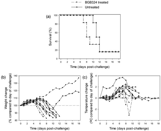
Filovirus Virulence in Interferon α/β and γ Double Knockout Mice, and Treatment with Favipiravir
by Jason E. Comer, Olivier Escaffre, Natasha Neef, Trevor Brasel, Terry L. Juelich, Jennifer K. Smith, Jeanon Smith, Birte Kalveram, David D. Perez, Shane Massey, Lihong Zhang and Alexander N. Freiberg
Viruses 2019, 11(2), 137; https://doi.org/10.3390/v11020137 - 3 Feb 2019
Cited by 15 | Viewed by 4531
Abstract
The 2014 Ebolavirus outbreak in West Africa highlighted the need for vaccines and therapeutics to prevent and treat filovirus infections. A well-characterized small animal model that is susceptible to wild-type filoviruses would facilitate the screening of anti-filovirus agents. To that end, we characterized [...] Read more.
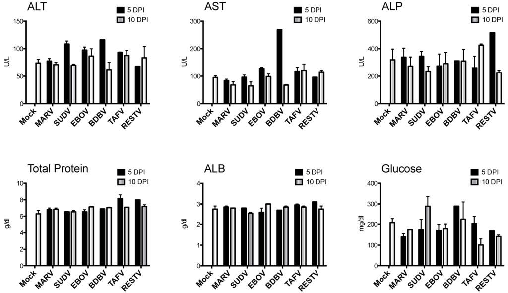
The 2014 Ebolavirus outbreak in West Africa highlighted the need for vaccines and therapeutics to prevent and treat filovirus infections. A well-characterized small animal model that is susceptible to wild-type filoviruses would facilitate the screening of anti-filovirus agents. To that end, we characterized knockout mice lacking α/β and γ interferon receptors (IFNAGR KO) as a model for wild-type filovirus infection. Intraperitoneal challenge of IFNAGR KO mice with several known human pathogenic species from the genus Ebolavirus and Marburgvirus, except Bundibugyo ebolavirus and Taï Forest ebolavirus, caused variable mortality rate. Further characterization of the prototype Ebola virus Kikwit isolate infection in this KO mouse model showed 100% lethality down to a dilution equivalent to 1.0 × 10⁻¹ pfu with all deaths occurring between 7 and 9 days post-challenge. Viral RNA was detectable in serum after challenge with 1.0 × 10² pfu as early as one day after infection. Changes in hematology and serum chemistry became pronounced as the disease progressed and mirrored the histological changes in the spleen and liver that were also consistent with those described for patients with Ebola virus disease. In a proof-of-principle study, treatment of Ebola virus infected IFNAGR KO mice with favipiravir resulted in 83% protection. Taken together, the data suggest that IFNAGR KO mice may be a useful model for early screening of anti-filovirus medical countermeasures. Full article
Show Figures

Figure 1

2018

Jump to: 2023, 2022, 2020, 2019, 2017, 2016, 2015, 2014

11 pages, 2969 KiB  
Article
Identification of Diaryl-Quinoline Compounds as Entry Inhibitors of Ebola Virus
by Qinghua Cui, Han Cheng, Rui Xiong, Gang Zhang, Ruikun Du, Manu Anantpadma, Robert A. Davey and Lijun Rong
Viruses 2018, 10(12), 678; https://doi.org/10.3390/v10120678 - 30 Nov 2018
Cited by 27 | Viewed by 5027
Abstract
Ebola virus is the causative agent of Ebola virus disease in humans. The lethality of Ebola virus infection is about 50%, supporting the urgent need to develop anti-Ebola drugs. Glycoprotein (GP) is the only surface protein of the Ebola virus, which is functionally [...] Read more.
Ebola virus is the causative agent of Ebola virus disease in humans. The lethality of Ebola virus infection is about 50%, supporting the urgent need to develop anti-Ebola drugs. Glycoprotein (GP) is the only surface protein of the Ebola virus, which is functionally critical for the virus to attach and enter the host cells, and is a promising target for anti-Ebola virus drug development. In this study, using the recombinant HIV-1/Ebola pseudovirus platform we previously established, we evaluated a small molecule library containing various quinoline compounds for anti-Ebola virus entry inhibitors. Some of the quinoline compounds specifically inhibited the entry of the Ebola virus. Among them, compound SYL1712 was the most potent Ebola virus entry inhibitor with an IC50 of ~1 μM. The binding of SYL1712 to the vial glycoprotein was computationally modeled and was predicted to interact with specific residues of GP. We used the time of the addition assay to show that compound SYL1712 blocks Ebola GP-mediated entry. Finally, consistent with being an Ebola virus entry inhibitor, compound SYL1712 inhibited infectious Ebola virus replication in tissue culture under biosafety level 4 containment, with an IC50 of 2 μM. In conclusion, we identified several related molecules with a diaryl-quinoline scaffold as potential anti-EBOV entry inhibitors, which can be further optimized for anti-Ebola drug development. Full article
Show Figures

Figure 1

14 pages, 1689 KiB  
Article
Virulence of Marburg Virus Angola Compared to Mt. Elgon (Musoke) in Macaques: A Pooled Survival Analysis
by Paul W. Blair, Maryam Keshtkar-Jahromi, Kevin J. Psoter, Ronald B. Reisler, Travis K. Warren, Sara C. Johnston, Arthur J. Goff, Lydia G. Downey, Sina Bavari and Anthony P. Cardile
Viruses 2018, 10(11), 658; https://doi.org/10.3390/v10110658 - 21 Nov 2018
Cited by 13 | Viewed by 5550
Abstract
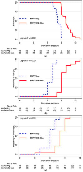
Angola variant (MARV/Ang) has replaced Mt. Elgon variant Musoke isolate (MARV/MtE-Mus) as the consensus standard variant for Marburg virus research and is regarded as causing a more aggressive phenotype of disease in animal models; however, there is a dearth of published evidence supporting [...] Read more.
Angola variant (MARV/Ang) has replaced Mt. Elgon variant Musoke isolate (MARV/MtE-Mus) as the consensus standard variant for Marburg virus research and is regarded as causing a more aggressive phenotype of disease in animal models; however, there is a dearth of published evidence supporting the higher virulence of MARV/Ang. In this retrospective study, we used data pooled from eight separate studies in nonhuman primates experimentally exposed with either 1000 pfu intramuscular (IM) MARV/Ang or MARV/MtE-Mus between 2012 and 2017 at the United States Army Medical Research Institute of Infectious Diseases (USAMRIID). Multivariable Cox proportional hazards regression was used to evaluate the association of variant type with time to death, the development of anorexia, rash, viremia, and 10 select clinical laboratory values. A total of 47 cynomolgus monkeys were included, of which 18 were exposed to MARV/Ang in three separate studies and 29 to MARV/MtE-Mus in five studies. Following universally fatal Marburg virus exposure, compared to MARV/MtE-Mus, MARV/Ang was associated with an increased risk of death (HR = 22.10; 95% CI: 7.08, 68.93), rash (HR = 5.87; 95% CI: 2.76, 12.51) and loss of appetite (HR = 35.10; 95% CI: 7.60, 162.18). Our data demonstrate an increased virulence of MARV/Ang compared to MARV/MtE-Mus variant in the 1000 pfu IM cynomolgus macaque model. Full article
Show Figures

Figure 1

18 pages, 2055 KiB  
Article
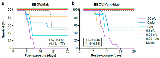
Intramuscular Exposure of Macaca fascicularis to Low Doses of Low Passage- or Cell Culture-Adapted Sudan Virus or Ebola Virus
by Kendra J. Alfson, Laura E. Avena, Michael W. Beadles, Gabriella Worwa, Melanie Amen, Jean L. Patterson, Ricardo Carrion, Jr. and Anthony Griffiths
Viruses 2018, 10(11), 642; https://doi.org/10.3390/v10110642 - 16 Nov 2018
Cited by 12 | Viewed by 4420
Abstract
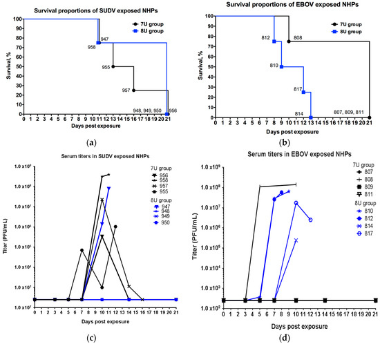
The filoviruses Ebola virus (EBOV) and Sudan virus (SUDV) can cause severe diseases, and there are currently no licensed countermeasures available for use against them. Transmission occurs frequently via contact with bodily fluids from infected individuals. However, it can be difficult to determine [...] Read more.
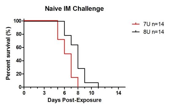
The filoviruses Ebola virus (EBOV) and Sudan virus (SUDV) can cause severe diseases, and there are currently no licensed countermeasures available for use against them. Transmission occurs frequently via contact with bodily fluids from infected individuals. However, it can be difficult to determine when or how someone became infected, or the quantity of infectious virus to which they were exposed. Evidence suggests the infectious dose is low, but the majority of published studies use high exposure doses. This study characterized the outcome of exposure to a low dose of EBOV or SUDV, using a Macaca fascicularis model. Further, because the effect of virus passage in cell culture may be more pronounced when lower exposure doses are used, viruses that possessed either the characteristics of wild type viruses (possessing predominantly 7-uridine (7U) genotype and a high particle-to-plaque forming unit (PFU) ratio) or cell culture-passaged viruses (predominantly 8-uridine (8U) genotype, a lower particle-to-PFU ratio) were used. The time to death after a low dose exposure was delayed in comparison to higher exposure doses. These data demonstrated that an extremely low dose of EBOV or SUDV is sufficient to cause lethal disease. A low dose exposure model can help inform studies on pathogenesis, transmission, and optimization of prevention strategies. Full article
Show Figures

Figure 1

13 pages, 553 KiB  
Perspective
Ebola Virus Maintenance: If Not (Only) Bats, What Else?
by Alexandre Caron, Mathieu Bourgarel, Julien Cappelle, Florian Liégeois, Hélène M. De Nys and François Roger
Viruses 2018, 10(10), 549; https://doi.org/10.3390/v10100549 - 9 Oct 2018
Cited by 34 | Viewed by 8722
Abstract
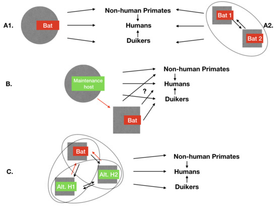
The maintenance mechanisms of ebolaviruses in African forest ecosystems are still unknown, but indirect evidences point at the involvement of some bat species. Despite intense research, the main bat-maintenance hypothesis has not been confirmed yet. The alternative hypotheses of a non-bat maintenance host [...] Read more.
The maintenance mechanisms of ebolaviruses in African forest ecosystems are still unknown, but indirect evidences point at the involvement of some bat species. Despite intense research, the main bat-maintenance hypothesis has not been confirmed yet. The alternative hypotheses of a non-bat maintenance host or a maintenance community including, or not, several bat and other species, deserves more investigation. However, African forest ecosystems host a large biodiversity and abound in potential maintenance hosts. How does one puzzle out? Since recent studies have revealed that several bat species have been exposed to ebolaviruses, the common denominator to these hypotheses is that within the epidemiological cycle, some bats species must be exposed to the viruses and infected by these potential alternative hosts. Under this constraint, and given the peculiar ecology of bats (roosting behaviour, habitat utilisation, and flight mode), we review the hosts and transmission pathways that can lead to bat exposure and infection to ebolaviruses. In contrast to the capacity of bats to transmit ebolaviruses and other pathogens to many hosts, our results indicate that only a limited number of hosts and pathways can lead to the transmission of ebolaviruses to bats, and that the alternative maintenance host, if it exists, must be amongst them. A list of these pathways is provided, along with protocols to prioritise and investigate these alternative hypotheses. In conclusion, taking into account the ecology of bats and their known involvement in ebolaviruses ecology drastically reduces the list of potential alternative maintenance hosts for ebolaviruses. Understanding the natural history of ebolaviruses is a health priority, and investigating these alternative hypotheses could complete the current effort focused on the role of bats. Full article
Show Figures

Figure 1

12 pages, 3029 KiB  
Article
Ebola Virus Causes Intestinal Tract Architectural Disruption and Bacterial Invasion in Non-Human Primates
by Ronald B. Reisler, Xiankun Zeng, Christopher W. Schellhase, Jeremy J. Bearss, Travis K. Warren, John C. Trefry, George W. Christopher, Mark G. Kortepeter, Sina Bavari and Anthony P. Cardile
Viruses 2018, 10(10), 513; https://doi.org/10.3390/v10100513 - 20 Sep 2018
Cited by 14 | Viewed by 5833
Abstract
In the 2014–2016 West Africa Ebola Virus (EBOV) outbreak, there was a significant concern raised about the potential for secondary bacterial infection originating from the gastrointestinal tract, which led to the empiric treatment of many patients with antibiotics. This retrospective pathology case series [...] Read more.
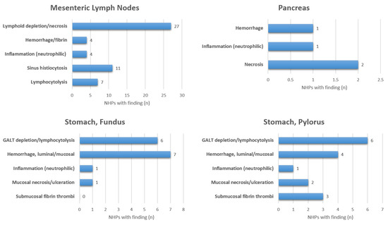
In the 2014–2016 West Africa Ebola Virus (EBOV) outbreak, there was a significant concern raised about the potential for secondary bacterial infection originating from the gastrointestinal tract, which led to the empiric treatment of many patients with antibiotics. This retrospective pathology case series summarizes the gastrointestinal pathology observed in control animals in the rhesus EBOV-Kikwit intramuscular 1000 plaque forming unit infection model. All 31 Non-human primates (NHPs) exhibited lymphoid depletion of gut-associated lymphoid tissue (GALT) but the severity and the specific location of the depletion varied. Mesenteric lymphoid depletion and necrosis were present in 87% (27/31) of NHPs. There was mucosal barrier disruption of the intestinal tract with mucosal necrosis and/or ulceration most notably in the duodenum (16%), cecum (16%), and colon (29%). In the intestinal tract, hemorrhage was noted most frequently in the duodenum (52%) and colon (45%). There were focal areas of bacterial submucosal invasion in the gastrointestinal (GI) tract in 9/31 (29%) of NHPs. Only 2/31 (6%) had evidence of pancreatic necrosis. One NHP (3%) experienced jejunal intussusception which may have been directly related to EBOV. Immunofluorescence assays demonstrated EBOV antigen in CD68+ macrophage/monocytes and endothelial cells in areas of GI vascular injury or necrosis. Full article
Show Figures

Figure 1

9 pages, 210 KiB  
Review
Catching Chances: The Movement to Be on the Ground and Research Ready before an Outbreak
by David Brett-Major and James Lawler
Viruses 2018, 10(8), 439; https://doi.org/10.3390/v10080439 - 19 Aug 2018
Cited by 6 | Viewed by 5253
Abstract
After more than 28,000 Ebola virus disease cases and at least 11,000 deaths in West Africa during the 2014–2016 epidemic, the world remains without a licensed vaccine or therapeutic broadly available and demonstrated to alleviate suffering. This deficiency has been felt acutely in [...] Read more.
After more than 28,000 Ebola virus disease cases and at least 11,000 deaths in West Africa during the 2014–2016 epidemic, the world remains without a licensed vaccine or therapeutic broadly available and demonstrated to alleviate suffering. This deficiency has been felt acutely in the two, short, following years with two Ebola virus outbreaks in the Democratic Republic of Congo (DRC), and a Marburg virus outbreak in Uganda. Despite billions of U.S. dollars invested in developing medical countermeasures for filoviruses in the antecedent decades, resulting in an array of preventative, diagnostic, and therapeutic products, none are available on commercial shelves. This paper explores why just-in-time research efforts in the field during the West Africa epidemic failed, as well as some recent initiatives to prevent similarly lost opportunities. Full article
12 pages, 1832 KiB  
Article
Post-Exposure Protection in Mice against Sudan Virus by a Two Antibody Cocktail
by Jeffrey W. Froude, Andrew S. Herbert, Thibaut Pelat, Sebastian Miethe, Samantha E. Zak, Jennifer M. Brannan, Russell R. Bakken, Alexander R. Steiner, Gang Yin, Trevor J. Hallam, Aaron K. Sato, Michael Hust, Philippe Thullier and John M. Dye
Viruses 2018, 10(6), 286; https://doi.org/10.3390/v10060286 - 26 May 2018
Cited by 15 | Viewed by 7123
Abstract
Sudan virus (SUDV) and Ebola viruses (EBOV) are both members of the Ebolavirus genus and have been sources of epidemics and outbreaks for several decades. We present here the generation and characterization of cross-reactive antibodies to both SUDV and EBOV, which were produced [...] Read more.
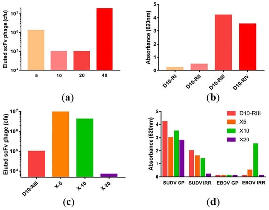
Sudan virus (SUDV) and Ebola viruses (EBOV) are both members of the Ebolavirus genus and have been sources of epidemics and outbreaks for several decades. We present here the generation and characterization of cross-reactive antibodies to both SUDV and EBOV, which were produced in a cell-free system and protective against SUDV in mice. A non-human primate, cynomolgus macaque, was immunized with viral-replicon particles expressing the glycoprotein of SUDV-Boniface (8A). Two separate antibody fragment phage display libraries were constructed after four immunogen injections. Both libraries were screened first against the SUDV and a second library was cross-selected against EBOV-Kikwit. Sequencing of 288 selected clones from the two distinct libraries identified 58 clones with distinct VH and VL sequences. Many of these clones were cross-reactive to EBOV and SUDV and able to neutralize SUDV. Three of these recombinant antibodies (X10B1, X10F3, and X10H2) were produced in the scFv-Fc format utilizing a cell-free production system. Mice that were challenged with SUDV-Boniface receiving 100µg of the X10B1/X10H2 scFv-Fc combination 6 and 48-h post-exposure demonstrated partial protection individually and complete protection as a combination. The data herein suggests these antibodies may be promising candidates for further therapeutic development. Full article
Show Figures

Figure 1

10 pages, 193 KiB  
Article
Development and Testing of a Method for Validating Chemical Inactivation of Ebola Virus
by Kendra J. Alfson and Anthony Griffiths
Viruses 2018, 10(3), 126; https://doi.org/10.3390/v10030126 - 13 Mar 2018
Cited by 15 | Viewed by 6105
Abstract
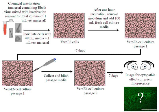
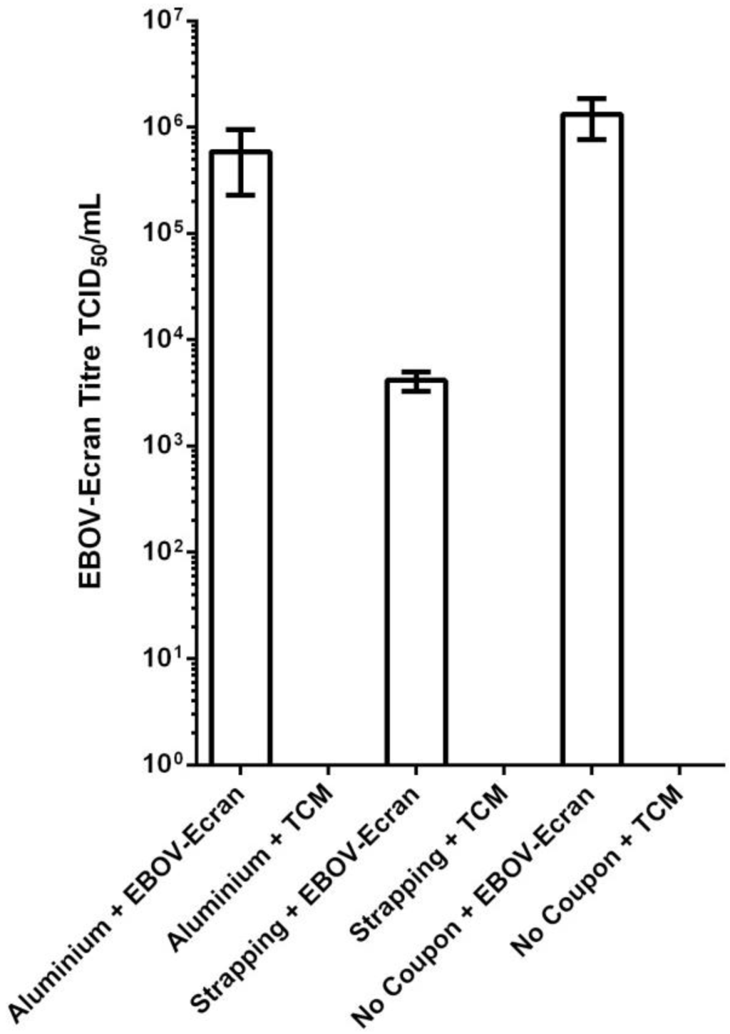
Complete inactivation of infectious Ebola virus (EBOV) is required before a sample may be removed from a Biosafety Level 4 laboratory. The United States Federal Select Agent Program regulations require that procedures used to demonstrate chemical inactivation must be validated in-house to confirm [...] Read more.
Complete inactivation of infectious Ebola virus (EBOV) is required before a sample may be removed from a Biosafety Level 4 laboratory. The United States Federal Select Agent Program regulations require that procedures used to demonstrate chemical inactivation must be validated in-house to confirm complete inactivation. The objective of this study was to develop a method for validating chemical inactivation of EBOV and then demonstrate the effectiveness of several commonly-used inactivation methods. Samples containing infectious EBOV (Zaire ebolavirus) in different matrices were treated, and the sample was diluted to limit the cytopathic effect of the inactivant. The presence of infectious virus was determined by assessing the cytopathic effect in Vero E6 cells. Crucially, this method did not result in a loss of infectivity in control samples, and we were able to detect less than five infectious units of EBOV (Zaire ebolavirus). We found that TRIzol LS reagent and RNA-Bee inactivated EBOV in serum; TRIzol LS reagent inactivated EBOV in clarified cell culture media; TRIzol reagent inactivated EBOV in tissue and infected Vero E6 cells; 10% neutral buffered formalin inactivated EBOV in tissue; and osmium tetroxide vapors inactivated EBOV on transmission electron microscopy grids. The methods described herein are easily performed and can be adapted to validate inactivation of viruses in various matrices and by various chemical methods. Full article
Show Figures

Graphical abstract

12 pages, 2186 KiB  
Article
Development and Validation of a Novel Dual Luciferase Reporter Gene Assay to Quantify Ebola Virus VP24 Inhibition of IFN Signaling
by Elisa Fanunza, Aldo Frau, Marco Sgarbanti, Roberto Orsatti, Angela Corona and Enzo Tramontano
Viruses 2018, 10(2), 98; https://doi.org/10.3390/v10020098 - 24 Feb 2018
Cited by 19 | Viewed by 10519
Abstract
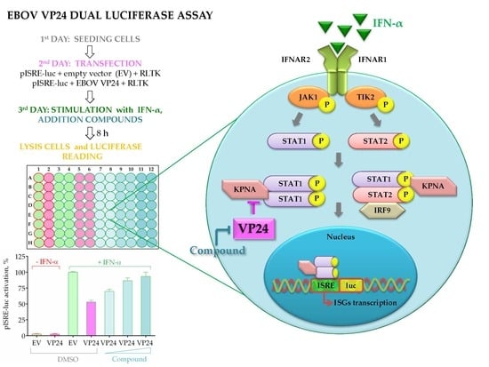
The interferon (IFN) system is the first line of defense against viral infections. Evasion of IFN signaling by Ebola viral protein 24 (VP24) is a critical event in the pathogenesis of the infection and, hence, VP24 is a potential target for drug development. [...] Read more.
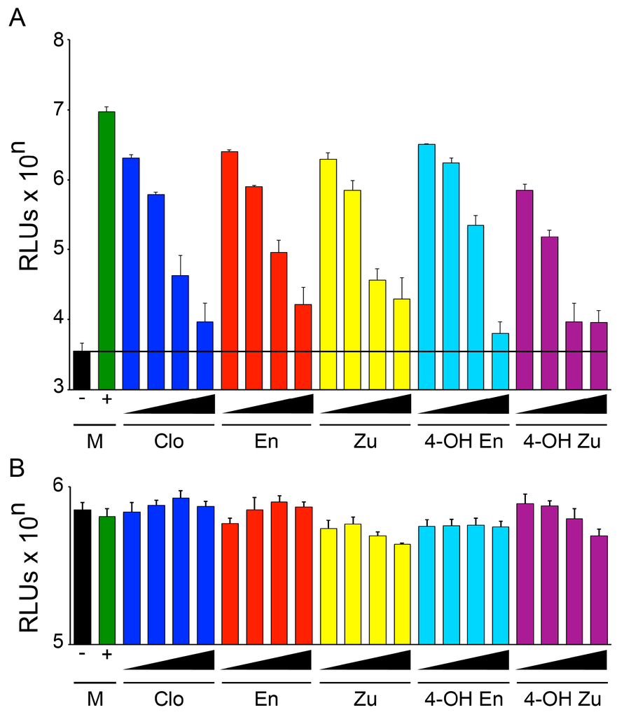
The interferon (IFN) system is the first line of defense against viral infections. Evasion of IFN signaling by Ebola viral protein 24 (VP24) is a critical event in the pathogenesis of the infection and, hence, VP24 is a potential target for drug development. Since no drugs target VP24, the identification of molecules able to inhibit VP24, restoring and possibly enhancing the IFN response, is a goal of concern. Accordingly, we developed a dual signal firefly and Renilla luciferase cell-based drug screening assay able to quantify IFN-mediated induction of Interferon Stimulated Genes (ISGs) and its inhibition by VP24. Human Embryonic Kidney 293T (HEK293T) cells were transiently transfected with a luciferase reporter gene construct driven by the promoter of ISGs, Interferon-Stimulated Response Element (ISRE). Stimulation of cells with IFN-α activated the IFN cascade leading to the expression of ISRE. Cotransfection of cells with a plasmid expressing VP24 cloned from a virus isolated during the last 2014 outbreak led to the inhibition of ISRE transcription, quantified by a luminescent signal. To adapt this system to test a large number of compounds, we performed it in 96-well plates; optimized the assay analyzing different parameters; and validated the system by calculating the Z′- and Z-factor, which showed values of 0.62 and 0.53 for IFN-α stimulation assay and VP24 inhibition assay, respectively, indicative of robust assay performance. Full article
Show Figures

Graphical abstract

12 pages, 1275 KiB  
Article
Antibody Responses to Marburg Virus in Egyptian Rousette Bats and Their Role in Protection against Infection
by Nadia Storm, Petrus Jansen Van Vuren, Wanda Markotter and Janusz T. Paweska
Viruses 2018, 10(2), 73; https://doi.org/10.3390/v10020073 - 10 Feb 2018
Cited by 26 | Viewed by 6993
Abstract
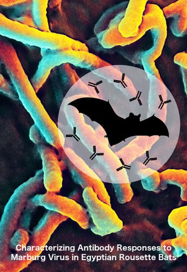
Egyptian rousette bats (ERBs) are reservoir hosts for the Marburg virus (MARV). The immune dynamics and responses to MARV infection in ERBs are poorly understood, and limited information exists on the role of antibodies in protection of ERBs against MARV infection. Here, we [...] Read more.
Egyptian rousette bats (ERBs) are reservoir hosts for the Marburg virus (MARV). The immune dynamics and responses to MARV infection in ERBs are poorly understood, and limited information exists on the role of antibodies in protection of ERBs against MARV infection. Here, we determine the duration of maternal immunity to MARV in juvenile ERBs, and evaluate the duration of the antibody response to MARV in bats naturally or experimentally infected with the virus. We further explore whether antibodies in previously naturally exposed bats is fully protective against experimental reinfection with MARV. Maternal immunity was lost in juvenile ERBs by 5 months of age. Antibodies to MARV remained detectable in 67% of experimentally infected bats approximately 4 months post inoculation (p.i.), while antibodies to MARV remained present in 84% of naturally exposed bats at least 11 months after capture. Reinfection of seropositive ERBs with MARV produced an anamnestic response from day 5 p.i. Although PCR-defined viremia was present in 73.3% of reinfected ERBs, replicating virus was recovered from the serum of only one bat on day 3 p.i. The negative PCR results in the salivary glands, intestines, bladders and reproductive tracts of reinfected bats, and the apparent absence of MARV in the majority of swabs collected from these bats suggest that reinfection may only play a minor role in the transmission and maintenance of MARV amongst ERBs in nature. Full article
Show Figures

Graphical abstract

2017

Jump to: 2023, 2022, 2020, 2019, 2018, 2016, 2015, 2014

822 KiB  
Review
Chikungunya Virus: Pathophysiology, Mechanism, and Modeling
by Vaishnavi K. Ganesan, Bin Duan and St Patrick Reid
Viruses 2017, 9(12), 368; https://doi.org/10.3390/v9120368 - 1 Dec 2017
Cited by 92 | Viewed by 15855
Abstract
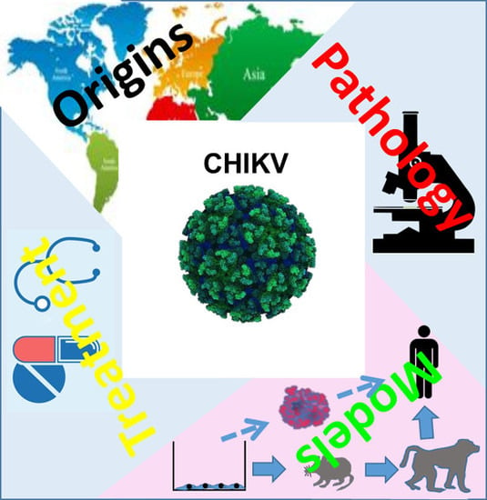
Chikungunya virus (CHIKV), a mosquito-transmitted alphavirus, is recurring in epidemic waves. In the past decade and a half, the disease has resurged in several countries around the globe, with outbreaks becoming increasingly severe. Though CHIKV was first isolated in 1952, there remain significant [...] Read more.
Chikungunya virus (CHIKV), a mosquito-transmitted alphavirus, is recurring in epidemic waves. In the past decade and a half, the disease has resurged in several countries around the globe, with outbreaks becoming increasingly severe. Though CHIKV was first isolated in 1952, there remain significant gaps in knowledge of CHIKV biology, pathogenesis, transmission, and mechanism. Diagnosis is largely simplified and based on symptoms, while treatment is supportive rather than curative. Here we present an overview of the disease, the challenges that lie ahead for future research, and what directions current studies are headed towards, with emphasis on improvement of current animal models and potential use of 3D models. Full article
Show Figures

Graphical abstract

4162 KiB  
Review
Structures and Functions of the Envelope Glycoprotein in Flavivirus Infections
by Xingcui Zhang, Renyong Jia, Haoyue Shen, Mingshu Wang, Zhongqiong Yin and Anchun Cheng
Viruses 2017, 9(11), 338; https://doi.org/10.3390/v9110338 - 13 Nov 2017
Cited by 125 | Viewed by 13191
Abstract
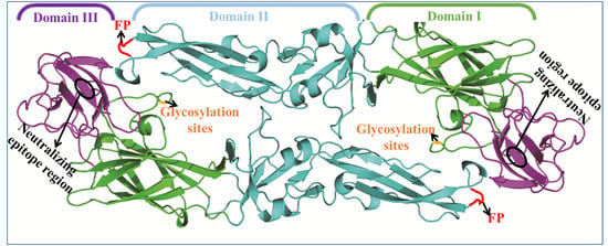
Flaviviruses are enveloped, single-stranded RNA viruses that widely infect many animal species. The envelope protein, a structural protein of flavivirus, plays an important role in host cell viral infections. It is composed of three separate structural envelope domains I, II, and III (EDI, [...] Read more.
Flaviviruses are enveloped, single-stranded RNA viruses that widely infect many animal species. The envelope protein, a structural protein of flavivirus, plays an important role in host cell viral infections. It is composed of three separate structural envelope domains I, II, and III (EDI, EDII, and EDIII). EDI is a structurally central domain of the envelope protein which stabilizes the overall orientation of the protein, and the glycosylation sites in EDI are related to virus production, pH sensitivity, and neuroinvasiveness. EDII plays an important role in membrane fusion because of the immunodominance of the fusion loop epitope and the envelope dimer epitope. Additionally, EDIII is the major target of neutralization antibodies. The envelope protein is an important target for research to develop vaccine candidates and antiviral therapeutics. This review summarizes the structures and functions of ED I/II/III, and provides practical applications for the three domains, with the ultimate goal of implementing strategies to utilize the envelope protein against flavivirus infections, thus achieving better diagnostics and developing potential flavivirus therapeutics and vaccines. Full article
Show Figures

Graphical abstract

2963 KiB  
Article
Development of a Lethal Intranasal Exposure Model of Ebola Virus in the Cynomolgus Macaque
by Kendra J. Alfson, Laura E. Avena, Gabriella Worwa, Ricardo Carrion and Anthony Griffiths
Viruses 2017, 9(11), 319; https://doi.org/10.3390/v9110319 - 29 Oct 2017
Cited by 20 | Viewed by 5637
Abstract
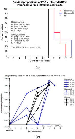
Ebola virus (EBOV) is a filovirus that can cause Ebola virus disease (EVD). No approved vaccines or therapies exist for filovirus infections, despite an urgent need. The development and testing of effective countermeasures against EBOV requires use of animal models and a thorough [...] Read more.
Ebola virus (EBOV) is a filovirus that can cause Ebola virus disease (EVD). No approved vaccines or therapies exist for filovirus infections, despite an urgent need. The development and testing of effective countermeasures against EBOV requires use of animal models and a thorough understanding of how the model aligns with EVD in humans. The majority of published studies report outcomes of parenteral exposures for emulating needle stick transmission. However, based on data from EVD outbreaks, close contact exposures to infected bodily fluid seems to be one of the primary routes of EBOV transmission. Thus, further work is needed to develop models that represent mucosal exposure. To characterize the outcome of mucosal exposure to EBOV, cynomolgus macaques were exposed to EBOV via intranasal (IN) route using the LMA® mucosal atomization device (LMA® MAD). For comparison, four non-human primates (NHPs) were exposed to EBOV via intramuscular (IM) route. This IN exposure model was uniformly lethal and correlated with a statistically significant delay in time to death when compared to exposure via the IM route. This more closely reflects the timeframes observed in human infections. An IN model of exposure offers an attractive alternative to other models as it can offer insight into the consequences of exposure via a mucosal surface and allows for screening countermeasures via a different exposure route. Full article
Show Figures

Figure 1

1247 KiB  
Review
Innate Immune Evasion Mediated by Flaviviridae Non-Structural Proteins
by Shun Chen, Zhen Wu, Mingshu Wang and Anchun Cheng
Viruses 2017, 9(10), 291; https://doi.org/10.3390/v9100291 - 7 Oct 2017
Cited by 80 | Viewed by 9998
Abstract
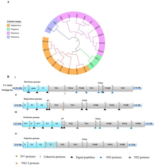
Flaviviridae-caused diseases are a critical, emerging public health problem worldwide. Flaviviridae infections usually cause severe, acute or chronic diseases, such as liver damage and liver cancer resulting from a hepatitis C virus (HCV) infection and high fever and shock caused by yellow fever. [...] Read more.
Flaviviridae-caused diseases are a critical, emerging public health problem worldwide. Flaviviridae infections usually cause severe, acute or chronic diseases, such as liver damage and liver cancer resulting from a hepatitis C virus (HCV) infection and high fever and shock caused by yellow fever. Many researchers worldwide are investigating the mechanisms by which Flaviviridae cause severe diseases. Flaviviridae can interfere with the host’s innate immunity to achieve their purpose of proliferation. For instance, dengue virus (DENV) NS2A, NS2B3, NS4A, NS4B and NS5; HCV NS2, NS3, NS3/4A, NS4B and NS5A; and West Nile virus (WNV) NS1 and NS4B proteins are involved in immune evasion. This review discusses the interplay between viral non-structural Flaviviridae proteins and relevant host proteins, which leads to the suppression of the host’s innate antiviral immunity. Full article
Show Figures

Figure 1

709 KiB  
Review
Venezuelan Equine Encephalitis Virus Capsid—The Clever Caper
by Lindsay Lundberg, Brian Carey and Kylene Kehn-Hall
Viruses 2017, 9(10), 279; https://doi.org/10.3390/v9100279 - 29 Sep 2017
Cited by 31 | Viewed by 10062
Abstract
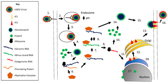
Venezuelan equine encephalitis virus (VEEV) is a New World alphavirus that is vectored by mosquitos and cycled in rodents. It can cause disease in equines and humans characterized by a febrile illness that may progress into encephalitis. Like the capsid protein of other [...] Read more.
Venezuelan equine encephalitis virus (VEEV) is a New World alphavirus that is vectored by mosquitos and cycled in rodents. It can cause disease in equines and humans characterized by a febrile illness that may progress into encephalitis. Like the capsid protein of other viruses, VEEV capsid is an abundant structural protein that binds to the viral RNA and interacts with the membrane-bound glycoproteins. It also has protease activity, allowing cleavage of itself from the growing structural polypeptide during translation. However, VEEV capsid protein has additional nonstructural roles within the host cell functioning as the primary virulence factor for VEEV. VEEV capsid inhibits host transcription and blocks nuclear import in mammalian cells, at least partially due to its complexing with the host CRM1 and importin α/β1 nuclear transport proteins. VEEV capsid also shuttles between the nucleus and cytoplasm and is susceptible to inhibitors of nuclear trafficking, making it a promising antiviral target. Herein, the role of VEEV capsid in viral replication and pathogenesis will be discussed including a comparison to proteins of other alphaviruses. Full article
Show Figures

Figure 1

837 KiB  
Letter
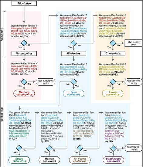
Implementation of Objective PASC-Derived Taxon Demarcation Criteria for Official Classification of Filoviruses
by Yīmíng Bào, Gaya K. Amarasinghe, Christopher F. Basler, Sina Bavari, Alexander Bukreyev, Kartik Chandran, Olga Dolnik, John M. Dye, Hideki Ebihara, Pierre Formenty, Roger Hewson, Gary P. Kobinger, Eric M. Leroy, Elke Mühlberger, Sergey V. Netesov, Jean L. Patterson, Janusz T. Paweska, Sophie J. Smither, Ayato Takada, Jonathan S. Towner, Viktor E. Volchkov, Victoria Wahl-Jensen and Jens H. Kuhnadd Show full author list remove Hide full author list
Viruses 2017, 9(5), 106; https://doi.org/10.3390/v9050106 - 11 May 2017
Cited by 20 | Viewed by 8433
Abstract
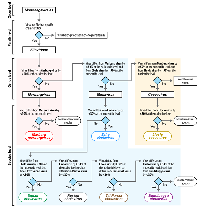
The mononegaviral family Filoviridae has eight members assigned to three genera and seven species. Until now, genus and species demarcation were based on arbitrarily chosen filovirus genome sequence divergence values (≈50% for genera, ≈30% for species) and arbitrarily chosen phenotypic virus or virion [...] Read more.
The mononegaviral family Filoviridae has eight members assigned to three genera and seven species. Until now, genus and species demarcation were based on arbitrarily chosen filovirus genome sequence divergence values (≈50% for genera, ≈30% for species) and arbitrarily chosen phenotypic virus or virion characteristics. Here we report filovirus genome sequence-based taxon demarcation criteria using the publicly accessible PAirwise Sequencing Comparison (PASC) tool of the US National Center for Biotechnology Information (Bethesda, MD, USA). Comparison of all available filovirus genomes in GenBank using PASC revealed optimal genus demarcation at the 55–58% sequence diversity threshold range for genera and at the 23–36% sequence diversity threshold range for species. Because these thresholds do not change the current official filovirus classification, these values are now implemented as filovirus taxon demarcation criteria that may solely be used for filovirus classification in case additional data are absent. A near-complete, coding-complete, or complete filovirus genome sequence will now be required to allow official classification of any novel “filovirus.” Classification of filoviruses into existing taxa or determining the need for novel taxa is now straightforward and could even become automated using a presented algorithm/flowchart rooted in RefSeq (type) sequences. Full article
Show Figures

Graphical abstract

2016

Jump to: 2023, 2022, 2020, 2019, 2018, 2017, 2015, 2014

602 KiB  
Article
Antiviral Screening of Multiple Compounds against Ebola Virus
by Stuart D. Dowall, Kevin Bewley, Robert J. Watson, Seshadri S. Vasan, Chandradhish Ghosh, Mohini M. Konai, Gro Gausdal, James B. Lorens, Jason Long, Wendy Barclay, Isabel Garcia-Dorival, Julian Hiscox, Andrew Bosworth, Irene Taylor, Linda Easterbrook, James Pitman, Sian Summers, Jenny Chan-Pensley, Simon Funnell, Julia Vipond, Sue Charlton, Jayanta Haldar, Roger Hewson and Miles W. Carrolladd Show full author list remove Hide full author list
Viruses 2016, 8(11), 277; https://doi.org/10.3390/v8110277 - 27 Oct 2016
Cited by 38 | Viewed by 9359
Abstract
In light of the recent outbreak of Ebola virus (EBOV) disease in West Africa, there have been renewed efforts to search for effective antiviral countermeasures. A range of compounds currently available with broad antimicrobial activity have been tested for activity against EBOV. Using [...] Read more.
In light of the recent outbreak of Ebola virus (EBOV) disease in West Africa, there have been renewed efforts to search for effective antiviral countermeasures. A range of compounds currently available with broad antimicrobial activity have been tested for activity against EBOV. Using live EBOV, eighteen candidate compounds were screened for antiviral activity in vitro. The compounds were selected on a rational basis because their mechanisms of action suggested that they had the potential to disrupt EBOV entry, replication or exit from cells or because they had displayed some antiviral activity against EBOV in previous tests. Nine compounds caused no reduction in viral replication despite cells remaining healthy, so they were excluded from further analysis (zidovudine; didanosine; stavudine; abacavir sulphate; entecavir; JB1a; Aimspro; celgosivir; and castanospermine). A second screen of the remaining compounds and the feasibility of appropriateness for in vivo testing removed six further compounds (ouabain; omeprazole; esomeprazole; Gleevec; D-LANA-14; and Tasigna). The three most promising compounds (17-DMAG; BGB324; and NCK-8) were further screened for in vivo activity in the guinea pig model of EBOV disease. Two of the compounds, BGB324 and NCK-8, showed some effect against lethal infection in vivo at the concentrations tested, which warrants further investigation. Further, these data add to the body of knowledge on the antiviral activities of multiple compounds against EBOV and indicate that the scientific community should invest more effort into the development of novel and specific antiviral compounds to treat Ebola virus disease. Full article
Show Figures

Figure 1

2974 KiB  
Article
Clomiphene and Its Isomers Block Ebola Virus Particle Entry and Infection with Similar Potency: Potential Therapeutic Implications
by Elizabeth A. Nelson, Alyson B. Barnes, Ronald D. Wiehle, Gregory K. Fontenot, Thomas Hoenen and Judith M. White
Viruses 2016, 8(8), 206; https://doi.org/10.3390/v8080206 - 2 Aug 2016
Cited by 36 | Viewed by 9946
Abstract
The 2014 outbreak of Ebola virus (EBOV) in Western Africa highlighted the need for anti-EBOV therapeutics. Clomiphene is a U.S. Food and Drug Administration (FDA)-approved drug that blocks EBOV entry and infection in cells and significantly protects EBOV-challenged mice. As provided, clomiphene is, [...] Read more.
The 2014 outbreak of Ebola virus (EBOV) in Western Africa highlighted the need for anti-EBOV therapeutics. Clomiphene is a U.S. Food and Drug Administration (FDA)-approved drug that blocks EBOV entry and infection in cells and significantly protects EBOV-challenged mice. As provided, clomiphene is, approximately, a 60:40 mixture of two stereoisomers, enclomiphene and zuclomiphene. The pharmacokinetic properties of the two isomers vary, but both accumulate in the eye and male reproductive tract, tissues in which EBOV can persist. Here we compared the ability of clomiphene and its isomers to inhibit EBOV using viral-like particle (VLP) entry and transcription/replication-competent VLP (trVLP) assays. Clomiphene and its isomers inhibited the entry and infection of VLPs and trVLPs with similar potencies. This was demonstrated with VLPs bearing the glycoproteins from three filoviruses (EBOV Mayinga, EBOV Makona, and Marburg virus) and in two cell lines (293T/17 and Vero E6). Visual problems have been noted in EBOV survivors, and viral RNA has been isolated from semen up to nine months post-infection. Since the clomiphene isomers accumulate in these affected tissues, clomiphene or one of its isomers warrants consideration as an anti-EBOV agent, for example, to potentially help ameliorate symptoms in EBOV survivors. Full article
Show Figures

Figure 1

869 KiB  
Article
Effectiveness of Four Disinfectants against Ebola Virus on Different Materials
by Sophie Smither, Amanda Phelps, Lin Eastaugh, Sarah Ngugi, Lyn O’Brien, Andrew Dutch and Mark Stephen Lever
Viruses 2016, 8(7), 185; https://doi.org/10.3390/v8070185 - 7 Jul 2016
Cited by 14 | Viewed by 7770
Abstract
The West Africa Ebola virus (EBOV) outbreak has highlighted the need for effective disinfectants capable of reducing viral load in a range of sample types, equipment and settings. Although chlorine-based products are widely used, they can also be damaging to equipment or apparatus [...] Read more.
The West Africa Ebola virus (EBOV) outbreak has highlighted the need for effective disinfectants capable of reducing viral load in a range of sample types, equipment and settings. Although chlorine-based products are widely used, they can also be damaging to equipment or apparatus that needs continuous use such as aircraft use for transportation of infected people. Two aircraft cleaning solutions were assessed alongside two common laboratory disinfectants in a contact kill assay with EBOV on two aircraft relevant materials representative of a porous and non-porous surface. A decimal log reduction of viral titre of 4 is required for a disinfectant to be deemed effective and two of the disinfectants fulfilled this criteria under the conditions tested. One product, Ardrox 6092, was found to perform similarly to sodium hypochlorite, but as it does not have the corrosive properties of sodium hypochlorite, it could be an alternative disinfectant solution to be used for decontamination of EBOV on sensitive apparatus. Full article
Show Figures

Figure 1

255 KiB  
Review
Aerosol Transmission of Filoviruses
by Berhanu Mekibib and Kevin K. Ariën
Viruses 2016, 8(5), 148; https://doi.org/10.3390/v8050148 - 23 May 2016
Cited by 23 | Viewed by 10518
Abstract
Filoviruses have become a worldwide public health concern because of their potential for introductions into non-endemic countries through international travel and the international transport of infected animals or animal products. Since it was first identified in 1976, in the Democratic Republic of Congo [...] Read more.
Filoviruses have become a worldwide public health concern because of their potential for introductions into non-endemic countries through international travel and the international transport of infected animals or animal products. Since it was first identified in 1976, in the Democratic Republic of Congo (formerly Zaire) and Sudan, the 2013–2015 western African Ebola virus disease (EVD) outbreak is the largest, both by number of cases and geographical extension, and deadliest, recorded so far in medical history. The source of ebolaviruses for human index case(s) in most outbreaks is presumptively associated with handling of bush meat or contact with fruit bats. Transmission among humans occurs easily when a person comes in contact with contaminated body fluids of patients, but our understanding of other transmission routes is still fragmentary. This review deals with the controversial issue of aerosol transmission of filoviruses. Full article
448 KiB  
Correction
Correction: Reynard, O.; et al. Identification of a New Ribonucleoside Inhibitor of Ebola Virus Replication. Viruses 2015, 7, 6233‒6240
by Viruses Editorial Office
Viruses 2016, 8(5), 137; https://doi.org/10.3390/v8050137 - 18 May 2016
Cited by 1 | Viewed by 3835
Abstract
The Viruses Editorial Office wishes to notify its readers of corrections in [1].[...] Full article
Show Figures

Figure 1

2201 KiB  
Article
Validation of the Filovirus Plaque Assay for Use in Preclinical Studies
by Amy C. Shurtleff, Holly A. Bloomfield, Shannon Mort, Steven A. Orr, Brian Audet, Thomas Whitaker, Michelle J. Richards and Sina Bavari
Viruses 2016, 8(4), 113; https://doi.org/10.3390/v8040113 - 21 Apr 2016
Cited by 15 | Viewed by 7316
Abstract
A plaque assay for quantitating filoviruses in virus stocks, prepared viral challenge inocula and samples from research animals has recently been fully characterized and standardized for use across multiple institutions performing Biosafety Level 4 (BSL-4) studies. After standardization studies were completed, Good Laboratory [...] Read more.
A plaque assay for quantitating filoviruses in virus stocks, prepared viral challenge inocula and samples from research animals has recently been fully characterized and standardized for use across multiple institutions performing Biosafety Level 4 (BSL-4) studies. After standardization studies were completed, Good Laboratory Practices (GLP)-compliant plaque assay method validation studies to demonstrate suitability for reliable and reproducible measurement of the Marburg Virus Angola (MARV) variant and Ebola Virus Kikwit (EBOV) variant commenced at the United States Army Medical Research Institute of Infectious Diseases (USAMRIID). The validation parameters tested included accuracy, precision, linearity, robustness, stability of the virus stocks and system suitability. The MARV and EBOV assays were confirmed to be accurate to ±0.5 log10 PFU/mL. Repeatability precision, intermediate precision and reproducibility precision were sufficient to return viral titers with a coefficient of variation (%CV) of ≤30%, deemed acceptable variation for a cell-based bioassay. Intraclass correlation statistical techniques for the evaluation of the assay’s precision when the same plaques were quantitated by two analysts returned values passing the acceptance criteria, indicating high agreement between analysts. The assay was shown to be accurate and specific when run on Nonhuman Primates (NHP) serum and plasma samples diluted in plaque assay medium, with negligible matrix effects. Virus stocks demonstrated stability for freeze-thaw cycles typical of normal usage during assay retests. The results demonstrated that the EBOV and MARV plaque assays are accurate, precise and robust for filovirus titration in samples associated with the performance of GLP animal model studies. Full article
Show Figures

Figure 1

2334 KiB  
Article
Virus-Like Particle Vaccination Protects Nonhuman Primates from Lethal Aerosol Exposure with Marburgvirus (VLP Vaccination Protects Macaques against Aerosol Challenges)
by John M. Dye, Kelly L. Warfield, Jay B. Wells, Robert C. Unfer, Sergey Shulenin, Hong Vu, Donald K. Nichols, M. Javad Aman and Sina Bavari
Viruses 2016, 8(4), 94; https://doi.org/10.3390/v8040094 - 8 Apr 2016
Cited by 19 | Viewed by 6534
Abstract
Marburg virus (MARV) was the first filovirus to be identified following an outbreak of viral hemorrhagic fever disease in Marburg, Germany in 1967. Due to several factors inherent to filoviruses, they are considered a potential bioweapon that could be disseminated via an aerosol [...] Read more.
Marburg virus (MARV) was the first filovirus to be identified following an outbreak of viral hemorrhagic fever disease in Marburg, Germany in 1967. Due to several factors inherent to filoviruses, they are considered a potential bioweapon that could be disseminated via an aerosol route. Previous studies demonstrated that MARV virus-like particles (VLPs) containing the glycoprotein (GP), matrix protein VP40 and nucleoprotein (NP) generated using a baculovirus/insect cell expression system could protect macaques from subcutaneous (SQ) challenge with multiple species of marburgviruses. In the current study, the protective efficacy of the MARV VLPs in conjunction with two different adjuvants: QS-21, a saponin derivative, and poly I:C against homologous aerosol challenge was assessed in cynomolgus macaques. Antibody responses against the GP antigen were equivalent in all groups receiving MARV VLPs irrespective of the adjuvant; adjuvant only-vaccinated macaques did not demonstrate appreciable antibody responses. All macaques were subsequently challenged with lethal doses of MARV via aerosol or SQ as a positive control. All MARV VLP-vaccinated macaques survived either aerosol or SQ challenge while animals administered adjuvant only exhibited clinical signs and lesions consistent with MARV disease and were euthanized after meeting the predetermined criteria. Therefore, MARV VLPs induce IgG antibodies recognizing MARV GP and VP40 and protect cynomolgus macaques from an otherwise lethal aerosol exposure with MARV. Full article
Show Figures

Figure 1

3537 KiB  
Article
Natural History of Aerosol Exposure with Marburg Virus in Rhesus Macaques
by Evan C. Ewers, William D. Pratt, Nancy A. Twenhafel, Joshua Shamblin, Ginger Donnelly, Heather Esham, Carly Wlazlowski, Joshua C. Johnson, Miriam Botto, Lisa E. Hensley and Arthur J. Goff
Viruses 2016, 8(4), 87; https://doi.org/10.3390/v8040087 - 30 Mar 2016
Cited by 19 | Viewed by 6876
Abstract
Marburg virus causes severe and often lethal viral disease in humans, and there are currently no Food and Drug Administration (FDA) approved medical countermeasures. The sporadic occurrence of Marburg outbreaks does not allow for evaluation of countermeasures in humans, so therapeutic and vaccine [...] Read more.
Marburg virus causes severe and often lethal viral disease in humans, and there are currently no Food and Drug Administration (FDA) approved medical countermeasures. The sporadic occurrence of Marburg outbreaks does not allow for evaluation of countermeasures in humans, so therapeutic and vaccine candidates can only be approved through the FDA animal rule—a mechanism requiring well-characterized animal models in which efficacy would be evaluated. Here, we describe a natural history study where rhesus macaques were surgically implanted with telemetry devices and central venous catheters prior to aerosol exposure with Marburg-Angola virus, enabling continuous physiologic monitoring and blood sampling without anesthesia. After a three to four day incubation period, all animals developed fever, viremia, and lymphopenia before developing tachycardia, tachypnea, elevated liver enzymes, decreased liver function, azotemia, elevated D-dimer levels and elevated pro-inflammatory cytokines suggesting a systemic inflammatory response with organ failure. The final, terminal period began with the onset of sustained hypotension, dehydration progressed with signs of major organ hypoperfusion (hyperlactatemia, acute kidney injury, hypothermia), and ended with euthanasia or death. The most significant pathologic findings were marked infection of the respiratory lymphoid tissue with destruction of the tracheobronchial and mediastinal lymph nodes, and severe diffuse infection in the liver, and splenitis. Full article
Show Figures

Figure 1

162 KiB  
Editorial
Testing New Hypotheses Regarding Ebolavirus Reservoirs
by Siv Aina Jensen Leendertz
Viruses 2016, 8(2), 30; https://doi.org/10.3390/v8020030 - 26 Jan 2016
Cited by 33 | Viewed by 10608
Abstract
Despite a relatively long search for the origin of ebolaviruses, their reservoirs remain elusive. Researchers might have to consider testing alternative hypotheses about how these viruses persist and emerge to advance ebolavirus research. This article aims to encourage researchers to bring forward such [...] Read more.
Despite a relatively long search for the origin of ebolaviruses, their reservoirs remain elusive. Researchers might have to consider testing alternative hypotheses about how these viruses persist and emerge to advance ebolavirus research. This article aims to encourage researchers to bring forward such hypotheses, to discuss them scientifically and to open alternative research avenues regarding the origin and ecology of ebolaviruses. Full article
644 KiB  
Article
Experimental Inoculation of Egyptian Fruit Bats (Rousettus aegyptiacus) with Ebola Virus
by Janusz T. Paweska, Nadia Storm, Antoinette A. Grobbelaar, Wanda Markotter, Alan Kemp and Petrus Jansen van Vuren
Viruses 2016, 8(2), 29; https://doi.org/10.3390/v8020029 - 22 Jan 2016
Cited by 61 | Viewed by 8875
Abstract
Colonized Egyptian fruit bats (Rousettus aegyptiacus), originating in South Africa, were inoculated subcutaneously with Ebola virus (EBOV). No overt signs of morbidity, mortality, or gross lesions were noted. Bats seroconverted by Day 10–16 post inoculation (p.i.), with the highest mean anti-EBOV [...] Read more.
Colonized Egyptian fruit bats (Rousettus aegyptiacus), originating in South Africa, were inoculated subcutaneously with Ebola virus (EBOV). No overt signs of morbidity, mortality, or gross lesions were noted. Bats seroconverted by Day 10–16 post inoculation (p.i.), with the highest mean anti-EBOV IgG level on Day 28 p.i. EBOV RNA was detected in blood from one bat. In 16 other tissues tested, viral RNA distribution was limited and at very low levels. No seroconversion could be demonstrated in any of the control bats up to 28 days after in-contact exposure to subcutaneously-inoculated bats. The control bats were subsequently inoculated intraperitoneally, and intramuscularly with the same dose of EBOV. No mortality, morbidity or gross pathology was observed in these bats. Kinetics of immune response was similar to that in subcutaneously-inoculated bats. Viral RNA was more widely disseminated to multiple tissues and detectable in a higher proportion of individuals, but consistently at very low levels. Irrespective of the route of inoculation, no virus was isolated from tissues which tested positive for EBOV RNA. Viral RNA was not detected in oral, nasal, ocular, vaginal, penile and rectal swabs from any of the experimental groups. Full article
Show Figures

Figure 1

2015

Jump to: 2023, 2022, 2020, 2019, 2018, 2017, 2016, 2014

1481 KiB  
Article
Ebola Virus Infections in Nonhuman Primates Are Temporally Influenced by Glycoprotein Poly-U Editing Site Populations in the Exposure Material
by John C. Trefry, Suzanne E. Wollen, Farooq Nasar, Joshua D. Shamblin, Steven J. Kern, Jeremy J. Bearss, Michelle A. Jefferson, Taylor B. Chance, Jeffery R. Kugelman, Jason T. Ladner, Anna N. Honko, Dean J. Kobs, Morgan Q.S. Wending, Carol L. Sabourin, William D. Pratt, Gustavo F. Palacios and M. Louise M. Pitt
Viruses 2015, 7(12), 6739-6754; https://doi.org/10.3390/v7122969 - 19 Dec 2015
Cited by 24 | Viewed by 7274
Abstract
Recent experimentation with the variants of the Ebola virus that differ in the glycoprotein’s poly-uridine site, which dictates the form of glycoprotein produced through a transcriptional stutter, has resulted in questions regarding the pathogenicity and lethality of the stocks used to develop products [...] Read more.
Recent experimentation with the variants of the Ebola virus that differ in the glycoprotein’s poly-uridine site, which dictates the form of glycoprotein produced through a transcriptional stutter, has resulted in questions regarding the pathogenicity and lethality of the stocks used to develop products currently undergoing human clinical trials to combat the disease. In order to address these concerns and prevent the delay of these critical research programs, we designed an experiment that permitted us to intramuscularly challenge statistically significant numbers of naïve and vaccinated cynomolgus macaques with either a 7U or 8U variant of the Ebola virus, Kikwit isolate. In naïve animals, no difference in survivorship was observed; however, there was a significant delay in the disease course between the two groups. Significant differences were also observed in time-of-fever, serum chemistry, and hematology. In vaccinated animals, there was no statistical difference in survivorship between either challenge groups, with two succumbing in the 7U group compared to 1 in the 8U challenge group. In summary, survivorship was not affected, but the Ebola virus disease course in nonhuman primates is temporally influenced by glycoprotein poly-U editing site populations. Full article
Show Figures

Graphical abstract

917 KiB  
Brief Report
Identification of a New Ribonucleoside Inhibitor of Ebola Virus Replication
by Olivier Reynard, Xuan-Nhi Nguyen, Nathalie Alazard-Dany, Véronique Barateau, Andrea Cimarelli and Viktor E. Volchkov
Viruses 2015, 7(12), 6233-6240; https://doi.org/10.3390/v7122934 - 1 Dec 2015
Cited by 83 | Viewed by 8015 | Correction
Abstract
The current outbreak of Ebola virus (EBOV) in West Africa has claimed the lives of more than 15,000 people and highlights an urgent need for therapeutics capable of preventing virus replication. In this study we screened known nucleoside analogues for their ability to [...] Read more.
The current outbreak of Ebola virus (EBOV) in West Africa has claimed the lives of more than 15,000 people and highlights an urgent need for therapeutics capable of preventing virus replication. In this study we screened known nucleoside analogues for their ability to interfere with EBOV replication. Among them, the cytidine analogue β-d-N4-hydroxycytidine (NHC) demonstrated potent inhibitory activities against EBOV replication and spread at non-cytotoxic concentrations. Thus, NHC constitutes an interesting candidate for the development of a suitable drug treatment against EBOV. Full article
Show Figures

Figure 1

5537 KiB  
Article
Requirements within the Ebola Viral Glycoprotein for Tetherin Antagonism
by Nathan H. Vande Burgt, Rachel L. Kaletsky and Paul Bates
Viruses 2015, 7(10), 5587-5602; https://doi.org/10.3390/v7102888 - 26 Oct 2015
Cited by 17 | Viewed by 6994
Abstract
Tetherin is an interferon-induced, intrinsic cellular response factor that blocks release of numerous viruses, including Ebola virus, from infected cells. As with many viruses targeted by host factors, Ebola virus employs a tetherin antagonist, the viral glycoprotein (EboGP), to counteract restriction and promote [...] Read more.
Tetherin is an interferon-induced, intrinsic cellular response factor that blocks release of numerous viruses, including Ebola virus, from infected cells. As with many viruses targeted by host factors, Ebola virus employs a tetherin antagonist, the viral glycoprotein (EboGP), to counteract restriction and promote virus release. Unlike other tetherin antagonists such as HIV-1 Vpu or KSHV K5, the features within EboGP needed to overcome tetherin are not well characterized. Here, we describe sequences within the EboGP ectodomain and membrane spanning domain (msd) as necessary to relieve tetherin restriction of viral particle budding. Fusing the EboGP msd to a normally secreted form of the glycoprotein effectively promotes Ebola virus particle release. Cellular protein or lipid anchors could not substitute for the EboGP msd. The requirement for the EboGP msd was not specific for filovirus budding, as similar results were seen with HIV particles. Furthermore trafficking of chimeric proteins to budding sites did not correlate with an ability to counter tetherin. Additionally, we find that a glycoprotein construct, which mimics the cathepsin-activated species by proteolytic removal of the EboGP glycan cap and mucin domains, is unable to counteract tetherin. Combining these results suggests an important role for the EboGP glycan cap and msd in tetherin antagonism. Full article
Show Figures

Figure 1

3974 KiB  
Review
The Role of Cytokines and Chemokines in Filovirus Infection
by Sandra L. Bixler and Arthur J. Goff 
Viruses 2015, 7(10), 5489-5507; https://doi.org/10.3390/v7102892 - 23 Oct 2015
Cited by 51 | Viewed by 13773
Abstract
Ebola- and marburgviruses are highly pathogenic filoviruses and causative agents of viral hemorrhagic fever. Filovirus disease is characterized by a dysregulated immune response, severe organ damage, and coagulation abnormalities. This includes modulation of cytokines, signaling mediators that regulate various components of the immune [...] Read more.
Ebola- and marburgviruses are highly pathogenic filoviruses and causative agents of viral hemorrhagic fever. Filovirus disease is characterized by a dysregulated immune response, severe organ damage, and coagulation abnormalities. This includes modulation of cytokines, signaling mediators that regulate various components of the immune system as well as other biological processes. Here we examine the role of cytokines in filovirus infection, with an emphasis on understanding how these molecules affect development of the antiviral immune response and influence pathology. These proteins may present targets for immune modulation by therapeutic agents and vaccines in an effort to boost the natural immune response to infection and/or reduce immunopathology. Full article
Show Figures

Graphical abstract

256 KiB  
Review
Filoviruses: One of These Things is (not) Like the Other
by Scott M. Anthony and Steven B. Bradfute
Viruses 2015, 7(10), 5172-5190; https://doi.org/10.3390/v7102867 - 29 Sep 2015
Cited by 23 | Viewed by 7727
Abstract
The family Filoviridae contains several of the most deadly pathogens known to date and the current Ebola virus disease (EVD) outbreak in Western Africa, due to Ebola virus (EBOV) infection, highlights the need for active and broad research into filovirus pathogenesis. However, in [...] Read more.
The family Filoviridae contains several of the most deadly pathogens known to date and the current Ebola virus disease (EVD) outbreak in Western Africa, due to Ebola virus (EBOV) infection, highlights the need for active and broad research into filovirus pathogenesis. However, in comparison, the seven other known filovirus family members are significantly understudied. Many of these, including Marburgviruses and Ebolaviruses other than EBOV, are also highly virulent and fully capable of causing widespread epidemics. This review places the focus on these non-EBOV filoviruses, including known immunological and pathological data. The available animal models, research tools and currently available therapeutics will also be discussed along with an emphasis in the large number of current gaps in knowledge of these less highlighted filoviruses. It is evident that much research is yet to be done in order to bring the non-EBOV filovirus field to the forefront of current research and, importantly, to the development of more effective vaccines and therapeutics to combat potential future outbreaks. Full article
99 KiB  
Essay
Learning from Ebola Virus: How to Prevent Future Epidemics
by Alexander S. Kekulé
Viruses 2015, 7(7), 3789-3797; https://doi.org/10.3390/v7072797 - 9 Jul 2015
Cited by 14 | Viewed by 12810
Abstract
The recent Ebola virus disease (EVD) epidemic in Guinea, Liberia and Sierra Leone demonstrated that the World Health Organization (WHO) is incapable to control outbreaks of infectious diseases in less developed regions of the world. This essay analyses the causes for the failure [...] Read more.
The recent Ebola virus disease (EVD) epidemic in Guinea, Liberia and Sierra Leone demonstrated that the World Health Organization (WHO) is incapable to control outbreaks of infectious diseases in less developed regions of the world. This essay analyses the causes for the failure of the international response and proposes four measures to improve resilience, early detection and response to future outbreaks of infectious diseases. Full article
1568 KiB  
Article
Experimental Inoculation of Egyptian Rousette Bats (Rousettus aegyptiacus) with Viruses of the Ebolavirus and Marburgvirus Genera
by Megan E.B. Jones, Amy J. Schuh, Brian R. Amman, Tara K. Sealy, Sherif R. Zaki, Stuart T. Nichol and Jonathan S. Towner
Viruses 2015, 7(7), 3420-3442; https://doi.org/10.3390/v7072779 - 25 Jun 2015
Cited by 106 | Viewed by 13517
Abstract
The Egyptian rousette bat (Rousettus aegyptiacus) is a natural reservoir for marburgviruses and a consistent source of virus spillover to humans. Cumulative evidence suggests various bat species may also transmit ebolaviruses. We investigated the susceptibility of Egyptian rousettes to each of [...] Read more.
The Egyptian rousette bat (Rousettus aegyptiacus) is a natural reservoir for marburgviruses and a consistent source of virus spillover to humans. Cumulative evidence suggests various bat species may also transmit ebolaviruses. We investigated the susceptibility of Egyptian rousettes to each of the five known ebolaviruses (Sudan, Ebola, Bundibugyo, Taï Forest, and Reston), and compared findings with Marburg virus. In a pilot study, groups of four juvenile bats were inoculated with one of the ebolaviruses or Marburg virus. In ebolavirus groups, viral RNA tissue distribution was limited, and no bat became viremic. Sudan viral RNA was slightly more widespread, spurring a second, 15-day Sudan virus serial euthanasia study. Low levels of Sudan viral RNA disseminated to multiple tissues at early time points, but there was no viremia or shedding. In contrast, Marburg virus RNA was widely disseminated, with viremia, oral and rectal shedding, and antigen in spleen and liver. This is the first experimental infection study comparing tissue tropism, viral shedding, and clinical and pathologic effects of six different filoviruses in the Egyptian rousette, a known marburgvirus reservoir. Our results suggest Egyptian rousettes are unlikely sources for ebolaviruses in nature, and support a possible single filovirus—single reservoir host relationship. Full article
Show Figures

Figure 1

1430 KiB  
Article
Evaluation of Signature Erosion in Ebola Virus Due to Genomic Drift and Its Impact on the Performance of Diagnostic Assays
by Shanmuga Sozhamannan, Mitchell Y. Holland, Adrienne T. Hall, Daniel A. Negrón, Mychal Ivancich, Jeffrey W. Koehler, Timothy D. Minogue, Catherine E. Campbell, Walter J. Berger, George W. Christopher, Bruce G. Goodwin and Michael A. Smith
Viruses 2015, 7(6), 3130-3154; https://doi.org/10.3390/v7062763 - 17 Jun 2015
Cited by 33 | Viewed by 10456
Abstract
Genome sequence analyses of the 2014 Ebola Virus (EBOV) isolates revealed a potential problem with the diagnostic assays currently in use; i.e., drifting genomic profiles of the virus may affect the sensitivity or even produce false-negative results. We evaluated signature erosion in ebolavirus [...] Read more.
Genome sequence analyses of the 2014 Ebola Virus (EBOV) isolates revealed a potential problem with the diagnostic assays currently in use; i.e., drifting genomic profiles of the virus may affect the sensitivity or even produce false-negative results. We evaluated signature erosion in ebolavirus molecular assays using an in silico approach and found frequent potential false-negative and false-positive results. We further empirically evaluated many EBOV assays, under real time PCR conditions using EBOV Kikwit (1995) and Makona (2014) RNA templates. These results revealed differences in performance between assays but were comparable between the old and new EBOV templates. Using a whole genome approach and a novel algorithm, termed BioVelocity, we identified new signatures that are unique to each of EBOV, Sudan virus (SUDV), and Reston virus (RESTV). Interestingly, many of the current assay signatures do not fall within these regions, indicating a potential drawback in the past assay design strategies. The new signatures identified in this study may be evaluated with real-time reverse transcription PCR (rRT-PCR) assay development and validation. In addition, we discuss regulatory implications and timely availability to impact a rapidly evolving outbreak using existing but perhaps less than optimal assays versus redesign these assays for addressing genomic changes. Full article
Show Figures

Figure 1

661 KiB  
Article
Evaluating Environmental Persistence and Disinfection of the Ebola Virus Makona Variant
by Bradley W. M. Cook, Todd A. Cutts, Aidan M. Nikiforuk, Philip Guillaume Poliquin, Deborah A. Court, James E. Strong and Steven S. Theriault
Viruses 2015, 7(4), 1975-1986; https://doi.org/10.3390/v7041975 - 14 Apr 2015
Cited by 51 | Viewed by 11849
Abstract
Background: The current disease outbreak caused by the Ebola virus Makona variant (EBOV/Mak) has led to unprecedented morbidity and lethality given its geographic reach and sustained transmission. Sodium hypochlorite and ethanol are well-accepted decontamination agents, however little published evidence supports the selection of [...] Read more.
Background: The current disease outbreak caused by the Ebola virus Makona variant (EBOV/Mak) has led to unprecedented morbidity and lethality given its geographic reach and sustained transmission. Sodium hypochlorite and ethanol are well-accepted decontamination agents, however little published evidence supports the selection of appropriate concentrations and contact times. The present study addresses the environmental robustness of EBOV/Mak and evaluates the effectiveness of sodium hypochlorite and ethanol as disinfectants. Methods: EBOV/Mak was suspended in a simulated organic soil load and dried onto surfaces. Viability was measured at 1 hour, 24 hours, 72 hours, and 192 hours. For the evaluation of disinfectants, EBOV/Mak in a simulated organic soil was dried onto stainless steel carriers and disinfected with 0.01% (v/v), 0.1% (v/v), 0.5% (v/v) and 1% (v/v) sodium hypochlorite solutions or 67% (v/v) ethanol at contact times of 1, 5 or 10 minutes. Results: EBOV/Mak persisted longer on steel and plastic surfaces (192 hours) than cotton (<24 hours). Dilute sodium hypochlorite (0.01% and 0.1%) showed little antiviral action, whereas 0.5% and 1% sodium hypochlorite solutions demonstrated recoverable virus at one minute but sterilized surfaces in five minutes. Disinfection with 67% ethanol did not fully clear infectious virions from 3/9 carriers at 1 minute but sterilized all carriers at 5 and 10 minutes. Conclusions: Sodium hypochlorite and ethanol effectively decontaminate EBOV/Mak suspended in a simulated organic load; however, selection of concentration and contact time proves critical. Full article
Show Figures

Figure 1

990 KiB  
Article
Evaluation of ViroCyt® Virus Counter for Rapid Filovirus Quantitation
by Cynthia A. Rossi, Brian J. Kearney, Scott P. Olschner, Priscilla L. Williams, Camenzind G. Robinson, Megan L. Heinrich, Ashley M. Zovanyi, Michael F. Ingram, David A. Norwood and Randal J. Schoepp
Viruses 2015, 7(3), 857-872; https://doi.org/10.3390/v7030857 - 20 Feb 2015
Cited by 37 | Viewed by 10820
Abstract
Development and evaluation of medical countermeasures for diagnostics, vaccines, and therapeutics requires production of standardized, reproducible, and well characterized virus preparations. For filoviruses this includes plaque assay for quantitation of infectious virus, transmission electron microscopy (TEM) for morphology and quantitation of virus particles, [...] Read more.
Development and evaluation of medical countermeasures for diagnostics, vaccines, and therapeutics requires production of standardized, reproducible, and well characterized virus preparations. For filoviruses this includes plaque assay for quantitation of infectious virus, transmission electron microscopy (TEM) for morphology and quantitation of virus particles, and real-time reverse transcription PCR for quantitation of viral RNA (qRT-PCR). The ViroCyt® Virus Counter (VC) 2100 (ViroCyt, Boulder, CO, USA) is a flow-based instrument capable of quantifying virus particles in solution. Using a proprietary combination of fluorescent dyes that stain both nucleic acid and protein in a single 30 min step, rapid, reproducible, and cost-effective quantification of filovirus particles was demonstrated. Using a seed stock of Ebola virus variant Kikwit, the linear range of the instrument was determined to be 2.8E+06 to 1.0E+09 virus particles per mL with coefficient of variation ranging from 9.4% to 31.5% for samples tested in triplicate. VC particle counts for various filovirus stocks were within one log of TEM particle counts. A linear relationship was established between the plaque assay, qRT-PCR, and the VC. VC results significantly correlated with both plaque assay and qRT-PCR. These results demonstrated that the VC is an easy, fast, and consistent method to quantify filoviruses in stock preparations. Full article
Show Figures

Figure 1

564 KiB  
Review
Understanding Ebola Virus Transmission
by Seth Judson, Joseph Prescott and Vincent Munster
Viruses 2015, 7(2), 511-521; https://doi.org/10.3390/v7020511 - 3 Feb 2015
Cited by 74 | Viewed by 36349
Abstract
An unprecedented number of Ebola virus infections among healthcare workers and patients have raised questions about our understanding of Ebola virus transmission. Here, we explore different routes of Ebola virus transmission between people, summarizing the known epidemiological and experimental data. From this data, [...] Read more.
An unprecedented number of Ebola virus infections among healthcare workers and patients have raised questions about our understanding of Ebola virus transmission. Here, we explore different routes of Ebola virus transmission between people, summarizing the known epidemiological and experimental data. From this data, we expose important gaps in Ebola virus research pertinent to outbreak situations. We further propose experiments and methods of data collection that will enable scientists to fill these voids in our knowledge about the transmission of Ebola virus. Full article
Show Figures

Graphical abstract

1198 KiB  
Article
Modeling of the Ebola Virus Delta Peptide Reveals a Potential Lytic Sequence Motif
by William R. Gallaher and Robert F. Garry
Viruses 2015, 7(1), 285-305; https://doi.org/10.3390/v7010285 - 20 Jan 2015
Cited by 22 | Viewed by 13036
Abstract
Filoviruses, such as Ebola and Marburg viruses, cause severe outbreaks of human infection, including the extensive epidemic of Ebola virus disease (EVD) in West Africa in 2014. In the course of examining mutations in the glycoprotein gene associated with 2014 Ebola virus (EBOV) [...] Read more.
Filoviruses, such as Ebola and Marburg viruses, cause severe outbreaks of human infection, including the extensive epidemic of Ebola virus disease (EVD) in West Africa in 2014. In the course of examining mutations in the glycoprotein gene associated with 2014 Ebola virus (EBOV) sequences, a differential level of conservation was noted between the soluble form of glycoprotein (sGP) and the full length glycoprotein (GP), which are both encoded by the GP gene via RNA editing. In the region of the proteins encoded after the RNA editing site sGP was more conserved than the overlapping region of GP when compared to a distant outlier species, Tai Forest ebolavirus. Half of the amino acids comprising the “delta peptide”, a 40 amino acid carboxy-terminal fragment of sGP, were identical between otherwise widely divergent species. A lysine-rich amphipathic peptide motif was noted at the carboxyl terminus of delta peptide with high structural relatedness to the cytolytic peptide of the non-structural protein 4 (NSP4) of rotavirus. EBOV delta peptide is a candidate viroporin, a cationic pore-forming peptide, and may contribute to EBOV pathogenesis. Full article
Show Figures

Graphical abstract

1207 KiB  
Article
Immune Memory to Sudan Virus: Comparison between Two Separate Disease Outbreaks
by Ariel Sobarzo, Yael Eskira, Andrew S. Herbert, Ana I. Kuehne, Spencer W. Stonier, David E. Ochayon, Shlomit Fedida-Metula, Steven Balinandi, Yaara Kislev, Neta Tali, Eli C. Lewis, Julius Julian Lutwama, John M. Dye, Victoria Yavelsky and Leslie Lobel
Viruses 2015, 7(1), 37-51; https://doi.org/10.3390/v7010037 - 6 Jan 2015
Cited by 18 | Viewed by 9400
Abstract
Recovery from ebolavirus infection in humans is associated with the development of both cell-mediated and humoral immune responses. According to recent studies, individuals that did not survive infection with ebolaviruses appear to have lacked a robust adaptive immune response and the expression of [...] Read more.
Recovery from ebolavirus infection in humans is associated with the development of both cell-mediated and humoral immune responses. According to recent studies, individuals that did not survive infection with ebolaviruses appear to have lacked a robust adaptive immune response and the expression of several early innate response markers. However, a comprehensive protective immune profile has yet to be described. Here, we examine cellular memory immune responses among survivors of two separate Ebolavirus outbreaks (EVDs) due to Sudan virus (SUDV) infection in Uganda—Gulu 2000–2001 and Kibaale 2012. Freshly collected blood samples were stimulated with inactivated SUDV, as well as with recombinant SUDV or Ebola virus (EBOV) GP (GP1–649). In addition, ELISA and plaque reduction neutralization assays were performed to determine anti-SUDV IgG titers and neutralization capacity. Cytokine expression was measured in whole blood cultures in response to SUDV and SUDV GP stimulation in both survivor pools, demonstrating recall responses that indicate immune memory. Cytokine responses between groups were similar but had distinct differences. Neutralizing, SUDV-specific IgG activity against irradiated SUDV and SUDV recombinant proteins were detected in both survivor cohorts. Furthermore, humoral and cell-mediated crossreactivity to EBOV and EBOV recombinant GP1–649 was observed in both cohorts. In conclusion, immune responses in both groups of survivors demonstrate persistent recognition of relevant antigens, albeit larger cohorts are required in order to reach greater statistical significance. The differing cytokine responses between Gulu and Kibaale outbreak survivors suggests that each outbreak may not yield identical memory responses and promotes the merits of studying the immune responses among outbreaks of the same virus. Finally, our demonstration of cross-reactive immune recognition suggests that there is potential for developing cross-protective vaccines for ebolaviruses. Full article
Show Figures

Figure 1

2014

Jump to: 2023, 2022, 2020, 2019, 2018, 2017, 2016, 2015

832 KiB  
Letter
Nomenclature- and Database-Compatible Names for the Two Ebola Virus Variants that Emerged in Guinea and the Democratic Republic of the Congo in 2014
by Jens H. Kuhn, Kristian G. Andersen, Sylvain Baize, Yīmíng Bào, Sina Bavari, Nicolas Berthet, Olga Blinkova, J. Rodney Brister, Anna N. Clawson, Joseph Fair, Martin Gabriel, Robert F. Garry, Stephen K. Gire, Augustine Goba, Jean-Paul Gonzalez, Stephan Günther, Christian T. Happi, Peter B. Jahrling, Jimmy Kapetshi, Gary Kobinger, Jeffrey R. Kugelman, Eric M. Leroy, Gael Darren Maganga, Placide K. Mbala, Lina M. Moses, Jean-Jacques Muyembe-Tamfum, Magassouba N'Faly, Stuart T. Nichol, Sunday A. Omilabu, Gustavo Palacios, Daniel J. Park, Janusz T. Paweska, Sheli R. Radoshitzky, Cynthia A. Rossi, Pardis C. Sabeti, John S. Schieffelin, Randal J. Schoepp, Rachel Sealfon, Robert Swanepoel, Jonathan S. Towner, Jiro Wada, Nadia Wauquier, Nathan L. Yozwiak and Pierre Formentyadd Show full author list remove Hide full author list
Viruses 2014, 6(11), 4760-4799; https://doi.org/10.3390/v6114760 - 24 Nov 2014
Cited by 76 | Viewed by 20383
Abstract
In 2014, Ebola virus (EBOV) was identified as the etiological agent of a large and still expanding outbreak of Ebola virus disease (EVD) in West Africa and a much more confined EVD outbreak in Middle Africa. Epidemiological and evolutionary analyses confirmed that all [...] Read more.
In 2014, Ebola virus (EBOV) was identified as the etiological agent of a large and still expanding outbreak of Ebola virus disease (EVD) in West Africa and a much more confined EVD outbreak in Middle Africa. Epidemiological and evolutionary analyses confirmed that all cases of both outbreaks are connected to a single introduction each of EBOV into human populations and that both outbreaks are not directly connected. Coding-complete genomic sequence analyses of isolates revealed that the two outbreaks were caused by two novel EBOV variants, and initial clinical observations suggest that neither of them should be considered strains. Here we present consensus decisions on naming for both variants (West Africa: “Makona”, Middle Africa: “Lomela”) and provide database-compatible full, shortened, and abbreviated names that are in line with recently established filovirus sub-species nomenclatures. Full article
Show Figures

Graphical abstract

782 KiB  
Article
Euthanasia Assessment in Ebola Virus Infected Nonhuman Primates
by Travis K. Warren, John C. Trefry, Shannon T. Marko, Taylor B. Chance, Jay B. Wells, William D. Pratt, Joshua C. Johnson, Eric M. Mucker, Sarah L. Norris, Mark Chappell, John M. Dye and Anna N. Honko
Viruses 2014, 6(11), 4666-4682; https://doi.org/10.3390/v6114666 - 24 Nov 2014
Cited by 21 | Viewed by 9737
Abstract
Multiple products are being developed for use against filoviral infections. Efficacy for these products will likely be demonstrated in nonhuman primate models of filoviral disease to satisfy licensure requirements under the Animal Rule, or to supplement human data. Typically, the endpoint for efficacy [...] Read more.
Multiple products are being developed for use against filoviral infections. Efficacy for these products will likely be demonstrated in nonhuman primate models of filoviral disease to satisfy licensure requirements under the Animal Rule, or to supplement human data. Typically, the endpoint for efficacy assessment will be survival following challenge; however, there exists no standardized approach for assessing the health or euthanasia criteria for filovirus-exposed nonhuman primates. Consideration of objective criteria is important to (a) ensure test subjects are euthanized without unnecessary distress; (b) enhance the likelihood that animals exhibiting mild or moderate signs of disease are not prematurely euthanized; (c) minimize the occurrence of spontaneous deaths and loss of end-stage samples; (d) enhance the reproducibility of experiments between different researchers; and (e) provide a defensible rationale for euthanasia decisions that withstands regulatory scrutiny. Historic records were compiled for 58 surviving and non-surviving monkeys exposed to Ebola virus at the US Army Medical Research Institute of Infectious Diseases. Clinical pathology parameters were statistically analyzed and those exhibiting predicative value for survival are reported. These findings may be useful for standardization of objective euthanasia assessments in rhesus monkeys exposed to Ebola virus and may serve as a useful approach for other standardization efforts. Full article
Show Figures

Figure 1

1623 KiB  
Article
A Loop Region in the N-Terminal Domain of Ebola Virus VP40 Is Important in Viral Assembly, Budding, and Egress
by Emmanuel Adu-Gyamfi, Smita P. Soni, Clara S. Jee, Michelle A. Digman, Enrico Gratton and Robert V. Stahelin
Viruses 2014, 6(10), 3837-3854; https://doi.org/10.3390/v6103837 - 17 Oct 2014
Cited by 35 | Viewed by 10459
Abstract
Ebola virus (EBOV) causes viral hemorrhagic fever in humans and can have clinical fatality rates of ~60%. The EBOV genome consists of negative sense RNA that encodes seven proteins including viral protein 40 (VP40). VP40 is the major Ebola virus matrix protein and [...] Read more.
Ebola virus (EBOV) causes viral hemorrhagic fever in humans and can have clinical fatality rates of ~60%. The EBOV genome consists of negative sense RNA that encodes seven proteins including viral protein 40 (VP40). VP40 is the major Ebola virus matrix protein and regulates assembly and egress of infectious Ebola virus particles. It is well established that VP40 assembles on the inner leaflet of the plasma membrane of human cells to regulate viral budding where VP40 can produce virus like particles (VLPs) without other Ebola virus proteins present. The mechanistic details, however, of VP40 lipid-interactions and protein-protein interactions that are important for viral release remain to be elucidated. Here, we mutated a loop region in the N-terminal domain of VP40 (Lys127, Thr129, and Asn130) and find that mutations (K127A, T129A, and N130A) in this loop region reduce plasma membrane localization of VP40. Additionally, using total internal reflection fluorescence microscopy and number and brightness analysis we demonstrate these mutations greatly reduce VP40 oligomerization. Lastly, VLP assays demonstrate these mutations significantly reduce VLP release from cells. Taken together, these studies identify an important loop region in VP40 that may be essential to viral egress. Full article
Show Figures

Graphical abstract

860 KiB  
Letter
A Call to Action to Enhance Filovirus Disease Outbreak Preparedness and Response
by Paul Roddy
Viruses 2014, 6(10), 3699-3718; https://doi.org/10.3390/v6103699 - 30 Sep 2014
Cited by 7 | Viewed by 10296
Abstract
The frequency and magnitude of recognized and declared filovirus-disease outbreaks have increased in recent years, while pathogenic filoviruses are potentially ubiquitous throughout sub-Saharan Africa. Meanwhile, the efficiency and effectiveness of filovirus-disease outbreak preparedness and response efforts are currently limited by inherent challenges and [...] Read more.
The frequency and magnitude of recognized and declared filovirus-disease outbreaks have increased in recent years, while pathogenic filoviruses are potentially ubiquitous throughout sub-Saharan Africa. Meanwhile, the efficiency and effectiveness of filovirus-disease outbreak preparedness and response efforts are currently limited by inherent challenges and persistent shortcomings. This paper delineates some of these challenges and shortcomings and provides a proposal for enhancing future filovirus-disease outbreak preparedness and response. The proposal serves as a call for prompt action by the organizations that comprise filovirus-disease outbreak response teams, namely, Ministries of Health of outbreak-prone countries, the World Health Organization, Médecins Sans Frontières, the Centers for Disease Control and Prevention—Atlanta, and others. Full article
Show Figures

Figure 1

655 KiB  
Letter
Filovirus RefSeq Entries: Evaluation and Selection of Filovirus Type Variants, Type Sequences, and Names
by Jens H. Kuhn, Kristian G. Andersen, Yīmíng Bào, Sina Bavari, Stephan Becker, Richard S. Bennett, Nicholas H. Bergman, Olga Blinkova, Steven Bradfute, J. Rodney Brister, Alexander Bukreyev, Kartik Chandran, Alexander A. Chepurnov, Robert A. Davey, Ralf G. Dietzgen, Norman A. Doggett, Olga Dolnik, John M. Dye, Sven Enterlein, Paul W. Fenimore, Pierre Formenty, Alexander N. Freiberg, Robert F. Garry, Nicole L. Garza, Stephen K. Gire, Jean-Paul Gonzalez, Anthony Griffiths, Christian T. Happi, Lisa E. Hensley, Andrew S. Herbert, Michael C. Hevey, Thomas Hoenen, Anna N. Honko, Georgy M. Ignatyev, Peter B. Jahrling, Joshua C. Johnson, Karl M. Johnson, Jason Kindrachuk, Hans-Dieter Klenk, Gary Kobinger, Tadeusz J. Kochel, Matthew G. Lackemeyer, Daniel F. Lackner, Eric M. Leroy, Mark S. Lever, Elke Mühlberger, Sergey V. Netesov, Gene G. Olinger, Sunday A. Omilabu, Gustavo Palacios, Rekha G. Panchal, Daniel J. Park, Jean L. Patterson, Janusz T. Paweska, Clarence J. Peters, James Pettitt, Louise Pitt, Sheli R. Radoshitzky, Elena I. Ryabchikova, Erica Ollmann Saphire, Pardis C. Sabeti, Rachel Sealfon, Aleksandr M. Shestopalov, Sophie J. Smither, Nancy J. Sullivan, Robert Swanepoel, Ayato Takada, Jonathan S. Towner, Guido Van der Groen, Viktor E. Volchkov, Valentina A. Volchkova, Victoria Wahl-Jensen, Travis K. Warren, Kelly L. Warfield, Manfred Weidmann and Stuart T. Nicholadd Show full author list remove Hide full author list
Viruses 2014, 6(9), 3663-3682; https://doi.org/10.3390/v6093663 - 26 Sep 2014
Cited by 48 | Viewed by 15654
Abstract
Sequence determination of complete or coding-complete genomes of viruses is becoming common practice for supporting the work of epidemiologists, ecologists, virologists, and taxonomists. Sequencing duration and costs are rapidly decreasing, sequencing hardware is under modification for use by non-experts, and software is constantly [...] Read more.
Sequence determination of complete or coding-complete genomes of viruses is becoming common practice for supporting the work of epidemiologists, ecologists, virologists, and taxonomists. Sequencing duration and costs are rapidly decreasing, sequencing hardware is under modification for use by non-experts, and software is constantly being improved to simplify sequence data management and analysis. Thus, analysis of virus disease outbreaks on the molecular level is now feasible, including characterization of the evolution of individual virus populations in single patients over time. The increasing accumulation of sequencing data creates a management problem for the curators of commonly used sequence databases and an entry retrieval problem for end users. Therefore, utilizing the data to their fullest potential will require setting nomenclature and annotation standards for virus isolates and associated genomic sequences. The National Center for Biotechnology Information’s (NCBI’s) RefSeq is a non-redundant, curated database for reference (or type) nucleotide sequence records that supplies source data to numerous other databases. Building on recently proposed templates for filovirus variant naming [<virus name> (<strain>)/<isolation host-suffix>/<country of sampling>/<year of sampling>/<genetic variant designation>-<isolate designation>], we report consensus decisions from a majority of past and currently active filovirus experts on the eight filovirus type variants and isolates to be represented in RefSeq, their final designations, and their associated sequences. Full article
Show Figures

Graphical abstract

582 KiB  
Meeting Report
Challenges, Progress, and Opportunities: Proceedings of the Filovirus Medical Countermeasures Workshop
by Rona Hirschberg, Lucy A. Ward, Nicole Kilgore, Rebecca Kurnat, Helen Schiltz, Mark T. Albrecht, George W. Christopher and Ed Nuzum
Viruses 2014, 6(7), 2673-2697; https://doi.org/10.3390/v6072673 - 9 Jul 2014
Cited by 30 | Viewed by 8910
Abstract
On August 22–23, 2013, agencies within the United States Department of Defense (DoD) and the Department of Health and Human Services (HHS) sponsored the Filovirus Medical Countermeasures (MCMs) Workshop as an extension of the activities of the Filovirus Animal Non-clinical Group (FANG). The [...] Read more.
On August 22–23, 2013, agencies within the United States Department of Defense (DoD) and the Department of Health and Human Services (HHS) sponsored the Filovirus Medical Countermeasures (MCMs) Workshop as an extension of the activities of the Filovirus Animal Non-clinical Group (FANG). The FANG is a federally-recognized multi-Agency group established in 2011 to coordinate and facilitate U.S. government (USG) efforts to develop filovirus MCMs. The workshop brought together government, academic and industry experts to consider the needs for filovirus MCMs and evaluate the status of the product development pipeline. This report summarizes speaker presentations and highlights progress and challenges remaining in the field. Full article
1763 KiB  
Article
Analysis of Determinants in Filovirus Glycoproteins Required for Tetherin Antagonism
by Kerstin Gnirß, Marie Fiedler, Annika Krämer-Kühl, Sebastian Bolduan, Eva Mittler, Stephan Becker, Michael Schindler and Stefan Pöhlmann
Viruses 2014, 6(4), 1654-1671; https://doi.org/10.3390/v6041654 - 9 Apr 2014
Cited by 23 | Viewed by 9856
Abstract
The host cell protein tetherin can restrict the release of enveloped viruses from infected cells. The HIV-1 protein Vpu counteracts tetherin by removing it from the site of viral budding, the plasma membrane, and this process depends on specific interactions between the transmembrane [...] Read more.
The host cell protein tetherin can restrict the release of enveloped viruses from infected cells. The HIV-1 protein Vpu counteracts tetherin by removing it from the site of viral budding, the plasma membrane, and this process depends on specific interactions between the transmembrane domains of Vpu and tetherin. In contrast, the glycoproteins (GPs) of two filoviruses, Ebola and Marburg virus, antagonize tetherin without reducing surface expression, and the domains in GP required for tetherin counteraction are unknown. Here, we show that filovirus GPs depend on the presence of their authentic transmembrane domains for virus-cell fusion and tetherin antagonism. However, conserved residues within the transmembrane domain were dispensable for membrane fusion and tetherin counteraction. Moreover, the insertion of the transmembrane domain into a heterologous viral GP, Lassa virus GPC, was not sufficient to confer tetherin antagonism to the recipient. Finally, mutation of conserved residues within the fusion peptide of Ebola virus GP inhibited virus-cell fusion but did not ablate tetherin counteraction, indicating that the fusion peptide and the ability of GP to drive host cell entry are not required for tetherin counteraction. These results suggest that the transmembrane domains of filoviral GPs contribute to tetherin antagonism but are not the sole determinants. Full article
Show Figures

Figure 1

510 KiB  
Article
Clinical Documentation and Data Transfer from Ebola and Marburg Virus Disease Wards in Outbreak Settings: Health Care Workers’ Experiences and Preferences
by Silja Bühler, Paul Roddy, Ellen Nolte and Matthias Borchert
Viruses 2014, 6(2), 927-937; https://doi.org/10.3390/v6020927 - 19 Feb 2014
Cited by 13 | Viewed by 12421
Abstract
Understanding human filovirus hemorrhagic fever (FHF) clinical manifestations and evaluating treatment strategies require the collection of clinical data in outbreak settings, where clinical documentation has been limited. Currently, no consensus among filovirus outbreak-response organisations guides best practice for clinical documentation and data transfer. [...] Read more.
Understanding human filovirus hemorrhagic fever (FHF) clinical manifestations and evaluating treatment strategies require the collection of clinical data in outbreak settings, where clinical documentation has been limited. Currently, no consensus among filovirus outbreak-response organisations guides best practice for clinical documentation and data transfer. Semi-structured interviews were conducted with health care workers (HCWs) involved in FHF outbreaks in sub-Saharan Africa, and with HCWs experienced in documenting and transferring data from high-risk areas (isolation wards or biosafety level 4 laboratories). Methods for data documentation and transfer were identified, described in detail and categorised by requirement for electricity and ranked by interviewee preference. Some methods involve removing paperwork and other objects from the filovirus disease ward without disinfection. We believe that if done properly, these methods are reasonably safe for certain settings. However, alternative methods avoiding the removal of objects, or involving the removal of paperwork or objects after non-damaging disinfection, are available. These methods are not only safer, they are also perceived as safer and likely more acceptable to health workers and members of the community. The use of standardised clinical forms is overdue. Experiments with by sunlight disinfection should continue, and non-damaging disinfection of impregnated paper, suitable tablet computers and underwater cameras should be evaluated under field conditions. Full article
7919 KiB  
Article
ABSL-4 Aerobiology Biosafety and Technology at the NIH/NIAID Integrated Research Facility at Fort Detrick
by Matthew G. Lackemeyer, Fabian De Kok-Mercado, Jiro Wada, Laura Bollinger, Jason Kindrachuk, Victoria Wahl-Jensen, Jens H. Kuhn and Peter B. Jahrling
Viruses 2014, 6(1), 137-150; https://doi.org/10.3390/v6010137 - 7 Jan 2014
Cited by 13 | Viewed by 14313
Abstract
The overall threat of a viral pathogen to human populations is largely determined by the modus operandi and velocity of the pathogen that is transmitted among humans. Microorganisms that can spread by aerosol are considered a more challenging enemy than those that require [...] Read more.
The overall threat of a viral pathogen to human populations is largely determined by the modus operandi and velocity of the pathogen that is transmitted among humans. Microorganisms that can spread by aerosol are considered a more challenging enemy than those that require direct body-to-body contact for transmission, due to the potential for infection of numerous people rather than a single individual. Additionally, disease containment is much more difficult to achieve for aerosolized viral pathogens than for pathogens that spread solely via direct person-to-person contact. Thus, aerobiology has become an increasingly necessary component for studying viral pathogens that are naturally or intentionally transmitted by aerosol. The goal of studying aerosol viral pathogens is to improve public health preparedness and medical countermeasure development. Here, we provide a brief overview of the animal biosafety level 4 Aerobiology Core at the NIH/NIAID Integrated Research Facility at Fort Detrick, Maryland, USA. Full article
Show Figures

Graphical abstract

Back to TopTop