Next Issue
Volume 21, February-1
Previous Issue
Volume 21, January-1
 
 
ijms-logo

Journal Browser

Journal Browser

Int. J. Mol. Sci., Volume 21, Issue 2 (January-2 2020) – 316 articles

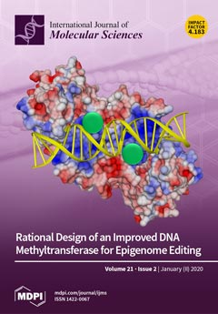
Cover Story (view full-size image): Targeted DNA methylation is a promising approach for the modulation of gene expression in basic research and in the clinic. However, it suffers from a lack of specificity, because often, off-target regions are also methylated. We have rationally designed different DNMT3A variants with reduced DNA binding and applied them for targeted methylation. Our data show that this leads to a pronounced improvement in the specificity of targeted DNA methylation, as indicated by a strongly reduced off-target methylation accompanied by an only mild reduction of on-target activity. These improved DNMT3A variants will advance the future applications of this emerging technology. View this paper.
  • Issues are regarded as officially published after their release is announced to the table of contents alert mailing list.
  • You may sign up for e-mail alerts to receive table of contents of newly released issues.
  • PDF is the official format for papers published in both, html and pdf forms. To view the papers in pdf format, click on the "PDF Full-text" link, and use the free Adobe Reader to open them.
Order results
Result details
Section
Select all
Export citation of selected articles as:
23 pages, 5336 KiB  
Article
Nitrate and Ammonium Affect the Overall Maize Response to Nitrogen Availability by Triggering Specific and Common Transcriptional Signatures in Roots
by Laura Ravazzolo, Sara Trevisan, Cristian Forestan, Serena Varotto, Stefania Sut, Stefano Dall’Acqua, Mario Malagoli and Silvia Quaggiotti
Int. J. Mol. Sci. 2020, 21(2), 686; https://doi.org/10.3390/ijms21020686 - 20 Jan 2020
Cited by 35 | Viewed by 5652
Abstract
Nitrogen (N) is an essential macronutrient for crops. Plants have developed several responses to N fluctuations, thus optimizing the root architecture in response to N availability. Nitrate and ammonium are the main inorganic N forms taken up by plants, and act as both [...] Read more.
Nitrogen (N) is an essential macronutrient for crops. Plants have developed several responses to N fluctuations, thus optimizing the root architecture in response to N availability. Nitrate and ammonium are the main inorganic N forms taken up by plants, and act as both nutrients and signals, affecting gene expression and plant development. In this study, RNA-sequencing was applied to gain comprehensive information on the pathways underlying the response of maize root, pre-treated in an N-deprived solution, to the provision of nitrate or ammonium. The analysis of the transcriptome shows that nitrate and ammonium regulate overlapping and distinct pathways, thus leading to different responses. Ammonium activates the response to stress, while nitrate acts as a negative regulator of transmembrane transport. Both the N-source repress genes related to the cytoskeleton and reactive oxygen species detoxification. Moreover, the presence of ammonium induces the accumulation of anthocyanins, while also reducing biomass and chlorophyll and flavonoids accumulation. Furthermore, the later physiological effects of these nutrients were evaluated through the assessment of shoot and root growth, leaf pigment content and the amino acid concentrations in root and shoot, confirming the existence of common and distinct features in response to the two nitrogen forms. Full article
Show Figures

Figure 1

17 pages, 6097 KiB  
Review
Dissecting Molecular Features of Gliomas: Genetic Loci and Validated Biomarkers
by Antonietta Arcella, Fiona Limanaqi, Rosangela Ferese, Francesca Biagioni, Maria Antonietta Oliva, Marianna Storto, Mirco Fanelli, Stefano Gambardella and Francesco Fornai
Int. J. Mol. Sci. 2020, 21(2), 685; https://doi.org/10.3390/ijms21020685 - 20 Jan 2020
Cited by 25 | Viewed by 12738
Abstract
Recently, several studies focused on the genetics of gliomas. This allowed identifying several germline loci that contribute to individual risk for tumor development, as well as various somatic mutations that are key for disease classification. Unfortunately, none of the germline loci clearly confers [...] Read more.
Recently, several studies focused on the genetics of gliomas. This allowed identifying several germline loci that contribute to individual risk for tumor development, as well as various somatic mutations that are key for disease classification. Unfortunately, none of the germline loci clearly confers increased risk per se. Contrariwise, somatic mutations identified within the glioma tissue define tumor genotype, thus representing valid diagnostic and prognostic markers. Thus, genetic features can be used in glioma classification and guided therapy. Such copious genomic variabilities are screened routinely in glioma diagnosis. In detail, Sanger sequencing or pyrosequencing, fluorescence in-situ hybridization, and microsatellite analyses were added to immunohistochemistry as diagnostic markers. Recently, Next Generation Sequencing was set-up as an all-in-one diagnostic tool aimed at detecting both DNA copy number variations and mutations in gliomas. This approach is widely used also to detect circulating tumor DNA within cerebrospinal fluid from patients affected by primary brain tumors. Such an approach is providing an alternative cost-effective strategy to genotype all gliomas, which allows avoiding surgical tissue collection and repeated tumor biopsies. This review summarizes available molecular features that represent solid tools for the genetic diagnosis of gliomas at present or in the next future. Full article
(This article belongs to the Special Issue Molecular Biology of Brain Tumors)
Show Figures

Figure 1

17 pages, 1355 KiB  
Review
Natural Polyphenols Targeting Senescence: A Novel Prevention and Therapy Strategy for Cancer
by Yan Bian, Juntong Wei, Changsheng Zhao and Guorong Li
Int. J. Mol. Sci. 2020, 21(2), 684; https://doi.org/10.3390/ijms21020684 - 20 Jan 2020
Cited by 60 | Viewed by 8225
Abstract
Cancer is one of the most serious diseases endangering human health. In view of the side effects caused by chemotherapy and radiotherapy, it is necessary to develop low-toxic anti-cancer compounds. Polyphenols are natural compounds with anti-cancer properties and their application is a considerable [...] Read more.
Cancer is one of the most serious diseases endangering human health. In view of the side effects caused by chemotherapy and radiotherapy, it is necessary to develop low-toxic anti-cancer compounds. Polyphenols are natural compounds with anti-cancer properties and their application is a considerable choice. Pro-senescence therapy is a recently proposed anti-cancer strategy and has been shown to effectively inhibit cancer. It is of great significance to clarify the mechanisms of polyphenols on tumor suppression by inducing senescence. In this review, we delineated the characteristics of senescent cells, and summarized the mechanisms of polyphenols targeting tumor microenvironment and inducing cancer cell senescence for cancer prevention and therapy. Although many studies have shown that polyphenols effectively inhibit cancer by targeting senescence, it warrants further investigation in preclinical and clinical studies. Full article
(This article belongs to the Special Issue Bioactive Phytochemicals for Cancer Prevention and Treatment II)
Show Figures

Figure 1

13 pages, 4182 KiB  
Article
Topical Therapy with Antisense Tumor Necrosis Factor Alpha Using Novel β-Glucan-Based Drug Delivery System Ameliorates Intestinal Inflammation
by Hideto Sakisaka, Hidetoshi Takedatsu, Keiichi Mitsuyama, Shinichi Mochizuki, Kazuo Sakurai, Shotaro Sakisaka and Fumihito Hirai
Int. J. Mol. Sci. 2020, 21(2), 683; https://doi.org/10.3390/ijms21020683 - 20 Jan 2020
Cited by 17 | Viewed by 4729
Abstract
Anti-tumor necrosis factor alpha (TNF-α) antibodies are effective in patients with inflammatory bowel disease (IBD). However, the effect is not optimal because a sufficient concentration of antibodies cannot be maintained at the site of inflammation. Thus, a macromolecular complex was developed with schizophyllan [...] Read more.
Anti-tumor necrosis factor alpha (TNF-α) antibodies are effective in patients with inflammatory bowel disease (IBD). However, the effect is not optimal because a sufficient concentration of antibodies cannot be maintained at the site of inflammation. Thus, a macromolecular complex was developed with schizophyllan (SPG) and antisense oligonucleotides. In the present study, an SPG-antisense TNF-α complex was prepared, and its therapeutic efficacy was examined using a dextran sodium sulfate (DSS)-induced colitis model. The TNF-α production in CD11b+ macrophages significantly increased in the colon of DSS-treated mice. Dectin-1, a receptor of SPG, binds with SPG and is subsequently taken into the cells via phagocytosis. The expression of dectin-1 by CD11b+ macrophages significantly increased in DSS-treated mice. Flow cytometry revealed that the uptake of SPG-antisense TNF-α in the macrophages was efficient. TNF-α production was suppressed significantly by SPG-antisense TNF-α in vitro, which was administered via enema to evaluate its efficacy. The intrarectal administration of SPG-antisense TNF-α ameliorated the intestinal inflammation. In this study, we showed that the delivery system that conjugates SPG and antisense can have higher therapeutic efficacy. Thus, the new therapeutic approach presented in this study may be used in the management of IBD. Full article
(This article belongs to the Special Issue Update on Basic and Molecular Research in Inflammatory Bowel Disease)
Show Figures

Figure 1

14 pages, 3144 KiB  
Article
STVNa Attenuates Isoproterenol-Induced Cardiac Hypertrophy Response through the HDAC4 and Prdx2/ROS/Trx1 Pathways
by Fei Liu, Hao Su, Bo Liu, Ying Mei, Qingjin Ke, Xiaoou Sun and Wen Tan
Int. J. Mol. Sci. 2020, 21(2), 682; https://doi.org/10.3390/ijms21020682 - 20 Jan 2020
Cited by 15 | Viewed by 5762
Abstract
Recent data show that cardiac hypertrophy contributes substantially to the overall heart failure burden. Mitochondrial dysfunction is a common feature of cardiac hypertrophy. Recent studies have reported that isosteviol inhibits myocardial ischemia-reperfusion injury in guinea pigs and H9c2 cells. This work investigated the [...] Read more.
Recent data show that cardiac hypertrophy contributes substantially to the overall heart failure burden. Mitochondrial dysfunction is a common feature of cardiac hypertrophy. Recent studies have reported that isosteviol inhibits myocardial ischemia-reperfusion injury in guinea pigs and H9c2 cells. This work investigated the protective mechanisms of isosteviol sodium (STVNa) against isoproterenol (Iso)-induced cardiac hypertrophy. We found that STVNa significantly inhibited H9c2 cell and rat primary cardiomyocyte cell surface, restored mitochondrial membrane potential (MMP) and morphological integrity, and decreased the expression of mitochondrial function-related proteins Fis1 and Drp1. Furthermore, STVNa decreased reactive oxygen species (ROS) levels and upregulated the expression of antioxidant factors, Thioredoxin 1 (Trx1) and Peroxiredoxin 2 (Prdx2). Moreover, STVNa restored the activity of histone deacetylase 4 (HDAC4) in the nucleus. Together, our data show that STVNa confers protection against Iso-induced myocardial hypertrophy primarily through the Prdx2/ROS/Trx1 signaling pathway. Thus, STVNA is a potentially effective treatment for cardiac hypertrophy in humans. Full article
(This article belongs to the Section Molecular Pathology, Diagnostics, and Therapeutics)
Show Figures

Figure 1

14 pages, 2693 KiB  
Article
Topical MTII Therapy Suppresses Melanoma Through PTEN Upregulation and Cyclooxygenase II Inhibition
by Jian-Ching Wu, Han-En Tsai, Yi-Hsiang Hsiao, Ji-Syuan Wu, Chieh-Shan Wu and Ming-Hong Tai
Int. J. Mol. Sci. 2020, 21(2), 681; https://doi.org/10.3390/ijms21020681 - 20 Jan 2020
Cited by 4 | Viewed by 4580
Abstract
Melanotan II (MTII), a synthetic analogue of the alpha-melanocyte stimulating hormone (α-MSH), has been applied for skin tanning in humans. However, the carcinogenic consequence of topical MTII has been equivocal. This study aims to delineate the anti-neoplastic efficacy and mechanism of MTII using [...] Read more.
Melanotan II (MTII), a synthetic analogue of the alpha-melanocyte stimulating hormone (α-MSH), has been applied for skin tanning in humans. However, the carcinogenic consequence of topical MTII has been equivocal. This study aims to delineate the anti-neoplastic efficacy and mechanism of MTII using the B16-F10 melanoma model in vitro and in vivo. It was found that, despite a lack of influence on proliferation, MTII potently inhibited the migration, invasion, and colony-forming capability of melanoma cells. Moreover, topical MTII application significantly attenuated the tumor progression in mice bearing established melanoma. Histological analysis revealed that MTII therapy induced apoptosis while inhibiting the proliferation and neovaluarization in melanoma tissues. By immunoblot and immunohistochemical analysis, it was found that MTII dose-dependently increased the phosphatase and tensin homolog (PTEN) protein level while reducing PTEN phosphorylation, which resulted in the inhibition of AKT/nuclear factor kappa B (NFκB) signaling. Consistently, MTII treatment inhibited cyclooxygenase II (COX-2) expression and prostaglandin E2 (PGE2) production in melanoma cells. Finally, studies of antibody neutralization suggest that the melanocortin 1 receptor (MC1R) plays a critical role in MTII-induced PTEN upregulation and melanoma suppression. Together, these results indicate that MTII elicits PTEN upregulation via MC1R, thereby suppressing melanoma progression through downregulating COX-2/PGE2 signaling. Hence, topical MTII therapy may facilitate a novel therapeutic strategy against melanoma. Full article
(This article belongs to the Special Issue Signaling in Melanoma)
Show Figures

Figure 1

22 pages, 3316 KiB  
Article
Genome and Karyotype Reorganization after Whole Genome Duplication in Free-Living Flatworms of the Genus Macrostomum
by Kira S. Zadesenets, Ilyas Y. Jetybayev, Lukas Schärer and Nikolay B. Rubtsov
Int. J. Mol. Sci. 2020, 21(2), 680; https://doi.org/10.3390/ijms21020680 - 20 Jan 2020
Cited by 12 | Viewed by 5060
Abstract
The genus Macrostomum represents a diverse group of rhabditophoran flatworms with >200 species occurring around the world. Earlier we uncovered karyotype instability linked to hidden polyploidy in both M. lignano (2n = 8) and its sibling species M. janickei (2n = [...] Read more.
The genus Macrostomum represents a diverse group of rhabditophoran flatworms with >200 species occurring around the world. Earlier we uncovered karyotype instability linked to hidden polyploidy in both M. lignano (2n = 8) and its sibling species M. janickei (2n = 10), prompting interest in the karyotype organization of close relatives. In this study, we investigated chromosome organization in two recently described and closely related Macrostomum species, M. mirumnovem and M. cliftonensis, and explored karyotype instability in laboratory lines and cultures of M. lignano (DV1/10, 2n = 10) and M. janickei in more detail. We revealed that three of the four studied species are characterized by karyotype instability, while M. cliftonensis showed a stable 2n = 6 karyotype. Next, we performed comparative cytogenetics of these species using fluorescent in situ hybridization (FISH) with a set of DNA probes (including microdissected DNA probes generated from M. lignano chromosomes, rDNA, and telomeric DNA). To explore the chromosome organization of the unusual 2n = 9 karyotype discovered in M. mirumnovem, we then generated chromosome-specific DNA probes for all chromosomes of this species. Similar to M. lignano and M. janickei, our findings suggest that M. mirumnovem arose via whole genome duplication (WGD) followed by considerable chromosome reshuffling. We discuss possible evolutionary scenarios for the emergence and reorganization of the karyotypes of these Macrostomum species and consider their suitability as promising animal models for studying the mechanisms and regularities of karyotype and genome evolution after a recent WGD. Full article
(This article belongs to the Special Issue Chromosome and Karyotype Variation)
Show Figures

Figure 1

19 pages, 274 KiB  
Review
Review of Alterations in Perlecan-Associated Vascular Risk Factors in Dementia
by Amanda L. Trout, Ibolya Rutkai, Ifechukwude J. Biose and Gregory J. Bix
Int. J. Mol. Sci. 2020, 21(2), 679; https://doi.org/10.3390/ijms21020679 - 20 Jan 2020
Cited by 7 | Viewed by 3781
Abstract
Perlecan is a heparan sulfate proteoglycan protein in the extracellular matrix that structurally and biochemically supports the cerebrovasculature by dynamically responding to changes in cerebral blood flow. These changes in perlecan expression seem to be contradictory, ranging from neuroprotective and angiogenic to thrombotic [...] Read more.
Perlecan is a heparan sulfate proteoglycan protein in the extracellular matrix that structurally and biochemically supports the cerebrovasculature by dynamically responding to changes in cerebral blood flow. These changes in perlecan expression seem to be contradictory, ranging from neuroprotective and angiogenic to thrombotic and linked to lipid retention. This review investigates perlecan’s influence on risk factors such as diabetes, hypertension, and amyloid that effect Vascular contributions to Cognitive Impairment and Dementia (VCID). VCID, a comorbidity with diverse etiology in sporadic Alzheimer’s disease (AD), is thought to be a major factor that drives the overall clinical burden of dementia. Accordingly, changes in perlecan expression and distribution in response to VCID appears to be injury, risk factor, location, sex, age, and perlecan domain dependent. While great effort has been made to understand the role of perlecan in VCID, additional studies are needed to increase our understanding of perlecan’s role in health and in cerebrovascular disease. Full article
(This article belongs to the Special Issue Extracellular Matrix in Development and Disease 2.0)
25 pages, 6930 KiB  
Article
Patterns of Expression of Purinergic Receptor P2RY12, a Putative Marker for Non-Activated Microglia, in Aged and Alzheimer’s Disease Brains
by Douglas G. Walker, Tiffany M. Tang, Anarmaa Mendsaikhan, Ikuo Tooyama, Geidy E. Serrano, Lucia I. Sue, Thomas G. Beach and Lih-Fen Lue
Int. J. Mol. Sci. 2020, 21(2), 678; https://doi.org/10.3390/ijms21020678 - 20 Jan 2020
Cited by 84 | Viewed by 9280
Abstract
Neuroinflammation is considered a key pathological process in neurodegenerative diseases of aging, including Alzheimer’s disease (AD). Many studies have defined phenotypes of reactive microglia, the brain-resident macrophages, with different antigenic markers to identify those potentially causing inflammatory damage. We took an alternative approach [...] Read more.
Neuroinflammation is considered a key pathological process in neurodegenerative diseases of aging, including Alzheimer’s disease (AD). Many studies have defined phenotypes of reactive microglia, the brain-resident macrophages, with different antigenic markers to identify those potentially causing inflammatory damage. We took an alternative approach with the goal of characterizing the distribution of purinergic receptor P2RY12-positive microglia, a marker previously defined as identifying homeostatic or non-activated microglia. We examined the expression of P2RY12 by dual-color light and fluorescence immunohistochemistry using sections of middle temporal gyrus from AD, high plaque and low plaque non-demented cases in relation to amyloid beta (Aβ) plaques and phosphorylated tau, markers of pathology, and HLA-DR, IBA-1, CD68, and progranulin, microglial phenotype markers. In low plaque cases, P2RY12-positive microglia mostly had non-activated morphologies, while the morphologies of P2RY12-positive microglia in AD brains were highly variable, suggesting its expression could encompass a wider range of phenotypes than originally hypothesized. P2RY12 expression by microglia differed depending on the types of plaques or tangles they were associated with. Areas of inflammation characterized by lack of P2RY12-positive microglia around mature plaques could be observed, but many diffuse plaques showed colocalization with P2RY12-positive microglia. Based on these results, P2RY12 expression by microglia should not be considered solely a marker of resting microglia as P2RY12 immunoreactivity was identifying microglia positive for CD68, progranulin and to a limited extent HLA-DR, markers of activation. Full article
Show Figures

Graphical abstract

11 pages, 4100 KiB  
Communication
Sonic Hedgehog Regulates Bone Fracture Healing
by Hiroaki Takebe, Nazmus Shalehin, Akihiro Hosoya, Tsuyoshi Shimo and Kazuharu Irie
Int. J. Mol. Sci. 2020, 21(2), 677; https://doi.org/10.3390/ijms21020677 - 20 Jan 2020
Cited by 27 | Viewed by 4434
Abstract
Bone fracture healing involves the combination of intramembranous and endochondral ossification. It is known that Indian hedgehog (Ihh) promotes chondrogenesis during fracture healing. Meanwhile, Sonic hedgehog (Shh), which is involved in ontogeny, has been reported to be involved in fracture healing, but the [...] Read more.
Bone fracture healing involves the combination of intramembranous and endochondral ossification. It is known that Indian hedgehog (Ihh) promotes chondrogenesis during fracture healing. Meanwhile, Sonic hedgehog (Shh), which is involved in ontogeny, has been reported to be involved in fracture healing, but the details had not been clarified. In this study, we demonstrated that Shh participated in fracture healing. Six-week-old Sprague–Dawley rats and Gli-CreERT2; tdTomato mice were used in this study. The right rib bones of experimental animals were fractured. The localization of Shh and Gli1 during fracture healing was examined. The localization of Gli1 progeny cells and osterix (Osx)-positive cells was similar during fracture healing. Runt-related transcription factor 2 (Runx2) and Osx, both of which are osteoblast markers, were observed on the surface of the new bone matrix and chondrocytes on day seven after fracture. Shh and Gli1 were co-localized with Runx2 and Osx. These findings suggest that Shh is involved in intramembranous and endochondral ossification during fracture healing. Full article
(This article belongs to the Special Issue Hedgehog Signaling in Organogenesis and Tumor Microenvironment)
Show Figures

Figure 1

14 pages, 954 KiB  
Article
Differential Expression of SMAD Genes and S1PR1 on Circulating CD4+ T Cells in Multiple Sclerosis and Crohn’s Disease
by Judith Abarca-Zabalía, Ma Isabel García, Alberto Lozano Ros, Ignacio Marín-Jiménez, Maria L. Martínez-Ginés, Beatriz López-Cauce, María L. Martín-Barbero, Sara Salvador-Martín, María Sanjurjo-Saez, Jose M. García-Domínguez and Luis A. López Fernández
Int. J. Mol. Sci. 2020, 21(2), 676; https://doi.org/10.3390/ijms21020676 - 20 Jan 2020
Cited by 17 | Viewed by 4056
Abstract
The Th17 immune response plays a key role in autoimmune diseases such as multiple sclerosis (MS) and inflammatory bowel disease (IBD). Expression of Th17-related genes in inflamed tissues has been reported in autoimmune diseases. However, values are frequently obtained using invasive methods. We [...] Read more.
The Th17 immune response plays a key role in autoimmune diseases such as multiple sclerosis (MS) and inflammatory bowel disease (IBD). Expression of Th17-related genes in inflamed tissues has been reported in autoimmune diseases. However, values are frequently obtained using invasive methods. We aimed to identify biomarkers of MS in an accessible sample, such as blood, by quantifying the relative expression of 91 Th17-related genes in CD4+ T lymphocytes from patients with MS during a relapse or during a remitting phase. We also compared our findings with those of healthy controls. After confirmation in a validation cohort, expression of SMAD7 and S1PR1 mRNAs was decreased in remitting disease (–2.3-fold and –1.3-fold, respectively) and relapsing disease (–2.2-fold and –1.3-fold, respectively). No differential expression was observed for other SMAD7-related genes, namely, SMAD2, SMAD3, and SMAD4. Under-regulation of SMAD7 and S1PR1 was also observed in another autoimmune disease, Crohn’s disease (CD) (−4.6-fold, -1.6-fold, respectively), suggesting the presence of common markers for autoimmune diseases. In addition, expression of TNF, SMAD2, SMAD3, and SMAD4 were also decreased in CD (–2.2-fold, –1.4-fold, –1.6-fold, and –1.6-fold, respectively). Our study suggests that expression of SMAD7 and S1PR1 mRNA in blood samples are markers for MS and CD, and TNF, SMAD2, SMAD3, and SMAD4 for CD. These genes could prove useful as markers of autoimmune diseases, thus obviating the need for invasive methods. Full article
(This article belongs to the Special Issue Peripheral Biomarkers in Neurodegenerative Diseases)
Show Figures

Figure 1

19 pages, 1592 KiB  
Review
Cerebral Cavernous Malformation Proteins in Barrier Maintenance and Regulation
by Shu Wei, Ye Li, Sean P. Polster, Christopher R. Weber, Issam A. Awad and Le Shen
Int. J. Mol. Sci. 2020, 21(2), 675; https://doi.org/10.3390/ijms21020675 - 20 Jan 2020
Cited by 24 | Viewed by 9280
Abstract
Cerebral cavernous malformation (CCM) is a disease characterized by mulberry shaped clusters of dilated microvessels, primarily in the central nervous system. Such lesions can cause seizures, headaches, and stroke from brain bleeding. Loss-of-function germline and somatic mutations of a group of genes, called [...] Read more.
Cerebral cavernous malformation (CCM) is a disease characterized by mulberry shaped clusters of dilated microvessels, primarily in the central nervous system. Such lesions can cause seizures, headaches, and stroke from brain bleeding. Loss-of-function germline and somatic mutations of a group of genes, called CCM genes, have been attributed to disease pathogenesis. In this review, we discuss the impact of CCM gene encoded proteins on cellular signaling, barrier function of endothelium and epithelium, and their contribution to CCM and potentially other diseases. Full article
(This article belongs to the Special Issue The Tight Junction and Its Proteins: More Than Just a Barrier)
Show Figures

Figure 1

13 pages, 1377 KiB  
Article
Age-Induced Differential Changes in the Central and Colonic Human Circadian Oscillators
by Cristina Camello-Almaraz, Francisco E. Martin-Cano, Francisco J. Santos, Mª Teresa Espin, Juan Antonio Madrid, Maria J. Pozo and Pedro J. Camello
Int. J. Mol. Sci. 2020, 21(2), 674; https://doi.org/10.3390/ijms21020674 - 20 Jan 2020
Cited by 5 | Viewed by 3334
Abstract
Aging modifies not only multiple cellular and homeostatic systems, but also biological rhythms. The circadian system is driven by a central hypothalamic oscillator which entrains peripheral oscillators, in both cases underlain by circadian genes. Our aim was to characterize the effect of aging [...] Read more.
Aging modifies not only multiple cellular and homeostatic systems, but also biological rhythms. The circadian system is driven by a central hypothalamic oscillator which entrains peripheral oscillators, in both cases underlain by circadian genes. Our aim was to characterize the effect of aging in the circadian expression of clock genes in the human colon. Ambulatory recordings of the circadian rhythms of skin wrist temperature, motor activity and the integrated variable TAP (temperature, activity and position) were dampened by aging, especially beyond 74 years of age. On the contrary, quantitative analysis of genes expression in the muscle layer of colonic explants during 24 h revealed that the circadian expression of Bmal1, Per1 and Clock genes, was larger beyond that age. In vitro experiments showed that aging induced a parallel increase in the myogenic contractility of the circular colonic muscle. This effect was not accompanied by enhancement of Ca2+ signals. In conclusion, we describe here for the first time the presence of a molecular oscillator in the human colon. Aging has a differential effect on the systemic circadian rhythms, that are impaired by aging, and the colonic oscillator, that is strengthened in parallel with the myogenic contractility. Full article
Show Figures

Figure 1

19 pages, 4922 KiB  
Article
Glycerol-Induced Powdery Mildew Resistance in Wheat by Regulating Plant Fatty Acid Metabolism, Plant Hormones Cross-Talk, and Pathogenesis-Related Genes
by Yinghui Li, Lina Qiu, Xinye Liu, Qiang Zhang, Xiangxi Zhuansun, Tzion Fahima, Tamar Krugman, Qixin Sun and Chaojie Xie
Int. J. Mol. Sci. 2020, 21(2), 673; https://doi.org/10.3390/ijms21020673 - 20 Jan 2020
Cited by 33 | Viewed by 5288
Abstract
Our previous study indicated that glycerol application induced resistance to powdery mildew (Bgt) in wheat by regulating two important signal molecules, glycerol-3-phosphate (G3P) and oleic acid (OA18:1). Transcriptome analysis of wheat leaves treated by glycerol and inoculated with Bgt was performed [...] Read more.
Our previous study indicated that glycerol application induced resistance to powdery mildew (Bgt) in wheat by regulating two important signal molecules, glycerol-3-phosphate (G3P) and oleic acid (OA18:1). Transcriptome analysis of wheat leaves treated by glycerol and inoculated with Bgt was performed to identify the activated immune response pathways. We identified a set of differentially expressed transcripts (e.g., TaGLI1, TaACT1, and TaSSI2) involved in glycerol and fatty acid metabolism that were upregulated in response to Bgt infection and might contribute to G3P and OA18:1 accumulation. Gene Ontology (GO) enrichment analysis revealed GO terms induced by glycerol, such as response to jasmonic acid (JA), defense response to bacterium, lipid oxidation, and growth. In addition, glycerol application induced genes (e.g., LOX, AOS, and OPRs) involved in the metabolism pathway of linolenic and alpha-linolenic acid, which are precursor molecules of JA biosynthesis. Glycerol induced JA and salicylic acid (SA) levels, while glycerol reduced the auxin (IAA) level in wheat. Glycerol treatment also induced pathogenesis related (PR) genes, including PR-1, PR-3, PR-10, callose synthase, PRMS, RPM1, peroxidase, HSP70, HSP90, etc. These results indicate that glycerol treatment regulates fatty acid metabolism and hormones cross-talk and induces the expression of PR genes that together contribute to Bgt resistance in wheat. Full article
(This article belongs to the Special Issue Abiotic Stress and Gene Networks in Plants 2020)
Show Figures

Figure 1

29 pages, 5922 KiB  
Review
Myosin and Other Energy-Transducing ATPases: Structural Dynamics Studied by Electron Paramagnetic Resonance
by Toshiaki Arata
Int. J. Mol. Sci. 2020, 21(2), 672; https://doi.org/10.3390/ijms21020672 - 20 Jan 2020
Cited by 4 | Viewed by 4778
Abstract
The objective of this article was to document the energy-transducing and regulatory interactions in supramolecular complexes such as motor, pump, and clock ATPases. The dynamics and structural features were characterized by motion and distance measurements using spin-labeling electron paramagnetic resonance (EPR) spectroscopy. In [...] Read more.
The objective of this article was to document the energy-transducing and regulatory interactions in supramolecular complexes such as motor, pump, and clock ATPases. The dynamics and structural features were characterized by motion and distance measurements using spin-labeling electron paramagnetic resonance (EPR) spectroscopy. In particular, we focused on myosin ATPase with actin–troponin–tropomyosin, neural kinesin ATPase with microtubule, P-type ion-motive ATPase, and cyanobacterial clock ATPase. Finally, we have described the relationships or common principles among the molecular mechanisms of various energy-transducing systems and how the large-scale thermal structural transition of flexible elements from one state to the other precedes the subsequent irreversible chemical reactions. Full article
(This article belongs to the Special Issue Motile Function of Myosins in Cells and Tissues)
Show Figures

Graphical abstract

22 pages, 3656 KiB  
Article
DNA Methylation Is Correlated with Gene Expression during Diapause Termination of Early Embryonic Development in the Silkworm (Bombyx mori)
by Bing Li, Pei Hu, Lin-Bao Zhu, Ling-Ling You, Hui-Hua Cao, Jie Wang, Shang-Zhi Zhang, Ming-Hui Liu, Shahzad Toufeeq, Shou-Jun Huang and Jia-Ping Xu
Int. J. Mol. Sci. 2020, 21(2), 671; https://doi.org/10.3390/ijms21020671 - 20 Jan 2020
Cited by 14 | Viewed by 4818
Abstract
DNA modification is a naturally occurring DNA modification in prokaryotic and eukaryotic organisms and is involved in several biological processes. Although genome-wide methylation has been studied in many insects, the understanding of global and genomic DNA methylation during insect early embryonic development, is [...] Read more.
DNA modification is a naturally occurring DNA modification in prokaryotic and eukaryotic organisms and is involved in several biological processes. Although genome-wide methylation has been studied in many insects, the understanding of global and genomic DNA methylation during insect early embryonic development, is lacking especially for insect diapause. In this study, we analyzed the relationship between DNA methylomes and transcriptomes in diapause-destined eggs compared to diapause-terminated eggs in the silkworm, Bombyx mori (B. mori). The results revealed that methylation was sparse in this species, as previously reported. Moreover, methylation levels in diapause-terminated eggs (HCl-treated) were 0.05% higher than in non-treated eggs, mainly due to the contribution of CG methylation sites. Methylation tends to occur in the coding sequences and promoter regions, especially at transcription initiation sites and short interspersed elements. Additionally, 364 methylome- and transcriptome-associated genes were identified, which showed significant differences in methylation and expression levels in diapause-destined eggs when compared with diapause-terminated eggs, and 74% of methylome and transcriptome associated genes showed both hypermethylation and elevated expression. Most importantly, Kyoto Encyclopaedia of Genes and Genomes (KEGG) analyses showed that methylation may be positively associated with Bombyx mori embryonic development, by regulating cell differentiation, metabolism, apoptosis pathways and phosphorylation. Through analyzing the G2/M phase-specific E3 ubiquitin-protein ligase (G2E3), we speculate that methylation may affect embryo diapause by regulating the cell cycle in Bombyx mori. These findings will help unravel potential linkages between DNA methylation and gene expression during early insect embryonic development and insect diapause. Full article
Show Figures

Figure 1

19 pages, 5211 KiB  
Article
The Soybean bZIP Transcription Factor Gene GmbZIP2 Confers Drought and Salt Resistances in Transgenic Plants
by Yan Yang, Tai-Fei Yu, Jian Ma, Jun Chen, Yong-Bin Zhou, Ming Chen, You-Zhi Ma, Wen-Liang Wei and Zhao-Shi Xu
Int. J. Mol. Sci. 2020, 21(2), 670; https://doi.org/10.3390/ijms21020670 - 20 Jan 2020
Cited by 78 | Viewed by 7477
Abstract
Abiotic stresses, such as drought and salt, are major environmental stresses, affecting plant growth and crop productivity. Plant bZIP transcription factors (bZIPs) confer stress resistances in harsh environments and play important roles in each phase of plant growth processes. In this research, 15 [...] Read more.
Abiotic stresses, such as drought and salt, are major environmental stresses, affecting plant growth and crop productivity. Plant bZIP transcription factors (bZIPs) confer stress resistances in harsh environments and play important roles in each phase of plant growth processes. In this research, 15 soybean bZIP family members were identified from drought-induced de novo transcriptomic sequences of soybean, which were unevenly distributed across 12 soybean chromosomes. Promoter analysis showed that these 15 genes were rich in ABRE, MYB and MYC cis-acting elements which were reported to be involved in abiotic stress responses. Quantitative real-time polymerase chain reaction (qRT-PCR) analysis indicated that 15 GmbZIP genes could be induced by drought and salt stress. GmbZIP2 was significantly upregulated under stress conditions and thus was selected for further study. Subcellular localization analysis revealed that the GmbZIP2 protein was located in the cell nucleus. qRT-PCR results show that GmbZIP2 can be induced by multiple stresses. The overexpression of GmbZIP2 in Arabidopsis and soybean hairy roots could improve plant resistance to drought and salt stresses. The result of differential expression gene analysis shows that the overexpression of GmbZIP2 in soybean hairy roots could enhance the expression of the stress responsive genes GmMYB48, GmWD40, GmDHN15, GmGST1 and GmLEA. These results indicate that soybean bZIPs played pivotal roles in plant resistance to abiotic stresses. Full article
(This article belongs to the Section Molecular Biology)
Show Figures

Figure 1

19 pages, 4723 KiB  
Article
Metalloporphyrin Pd(T4) Exhibits Oncolytic Activity and Cumulative Effects with 5-ALA Photodynamic Treatment against C918 Cells
by Brandon Leviskas, Tibor Valyi-Nagy, Gnanasekar Munirathinam, Matthew Bork, Klara Valyi-Nagy and Troy Skwor
Int. J. Mol. Sci. 2020, 21(2), 669; https://doi.org/10.3390/ijms21020669 - 20 Jan 2020
Cited by 6 | Viewed by 4428
Abstract
Photodynamic therapy is a non-invasive method where light activates a photosensitizer bound to cancer cells, generating reactive oxygen species and resulting in cell death. This study assessed the oncolytic potential of photodynamic therapy, comparing European Medicines Agency and United States Food and Drug [...] Read more.
Photodynamic therapy is a non-invasive method where light activates a photosensitizer bound to cancer cells, generating reactive oxygen species and resulting in cell death. This study assessed the oncolytic potential of photodynamic therapy, comparing European Medicines Agency and United States Food and Drug Administration-approved 5-aminolevulinic acid (5-ALA) to a metalloporphyrin, Pd(T4), against a highly invasive uveal melanoma cell line (C918) in two- and three-dimensional models in vitro. Epithelial monolayer studies displayed strong oncolytic effects (>70%) when utilizing Pd(T4) at a fraction of the concentration, and reduced pre-illumination time compared to 5-ALA post-405 nm irradiance. When analyzed at sub-optimal concentrations, application of Pd(T4) and 5-ALA with 405 nm displayed cumulative effects. Lethality from Pd(T4)-photodynamic therapy was maintained within a three-dimensional model, including the more resilient vasculogenic mimicry-forming cells, though at lower rates. At high concentrations, modality of cell death exhibited necrosis partially dependent on reactive oxygen species. However, sub-optimal concentrations of photosensitizer exhibited an apoptotic protein expression profile characterized by increased Bax/Bcl-2 ratio and endoplasmic stress-related proteins, along with downregulation of apoptotic inhibitors CIAP-1 and -2. Together, our results indicate Pd(T4) as a strong photosensitizer alone and in combination with 5-ALA against C918 cells. Full article
(This article belongs to the Section Molecular Pathology, Diagnostics, and Therapeutics)
Show Figures

Figure 1

145 pages, 634 KiB  
Editorial
Acknowledgement to Reviewers of International Journal of Molecular Science in 2019
by International Journal of Molecular Science Editorial Office
Int. J. Mol. Sci. 2020, 21(2), 668; https://doi.org/10.3390/ijms21020668 - 19 Jan 2020
Viewed by 11163
Abstract
The editorial team greatly appreciates the reviewers who have dedicated their considerable time and expertise to the journal’s rigorous editorial process over the past 12 months, regardless of whether the papers are finally published or not.[...] Full article
23 pages, 882 KiB  
Review
Advances of Zinc Signaling Studies in Prostate Cancer
by Dangdang Li, Daniel B. Stovall, Wenmeng Wang and Guangchao Sui
Int. J. Mol. Sci. 2020, 21(2), 667; https://doi.org/10.3390/ijms21020667 - 19 Jan 2020
Cited by 49 | Viewed by 9101
Abstract
Prostate cancer (PCa) is one of the most common cancers and the second leading cause of cancer-related death among men worldwide. Despite progresses in early diagnosis and therapeutic strategies, prognosis for patients with advanced PCa remains poor. Noteworthily, a unique feature of healthy [...] Read more.
Prostate cancer (PCa) is one of the most common cancers and the second leading cause of cancer-related death among men worldwide. Despite progresses in early diagnosis and therapeutic strategies, prognosis for patients with advanced PCa remains poor. Noteworthily, a unique feature of healthy prostate is its highest level of zinc content among all soft tissues in the human body, which dramatically decreases during prostate tumorigenesis. To date, several reviews have suggested antitumor activities of zinc and its potential as a therapeutic strategy of PCa. However, an overview about the role of zinc and its signaling in PCa is needed. Here, we review literature related to the content, biological function, compounds and clinical application of zinc in PCa. We first summarize zinc content in prostate tissue and sera of PCa patients with their clinical relevance. We then elaborate biological functions of zinc signaling in PCa on three main aspects, including cell proliferation, death and tumor metastasis. Finally, we discuss clinical applications of zinc-containing compounds and proteins involved in PCa signaling pathways. Based on currently available studies, we conclude that zinc plays a tumor suppressive role and can serve as a biomarker in PCa diagnosis and therapies. Full article
(This article belongs to the Special Issue Zinc Biology 2019)
Show Figures

Figure 1

27 pages, 11526 KiB  
Article
Mice Lacking the Matrilin Family of Extracellular Matrix Proteins Develop Mild Skeletal Abnormalities and Are Susceptible to Age-Associated Osteoarthritis
by Ping Li, Lutz Fleischhauer, Claudia Nicolae, Carina Prein, Zsuzsanna Farkas, Maximilian Michael Saller, Wolf Christian Prall, Raimund Wagener, Juliane Heilig, Anja Niehoff, Hauke Clausen-Schaumann, Paolo Alberton and Attila Aszodi
Int. J. Mol. Sci. 2020, 21(2), 666; https://doi.org/10.3390/ijms21020666 - 19 Jan 2020
Cited by 27 | Viewed by 6890
Abstract
Matrilins (MATN1, MATN2, MATN3 and MATN4) are adaptor proteins of the cartilage extracellular matrix (ECM), which bridge the collagen II and proteoglycan networks. In humans, dominant-negative mutations in MATN3 lead to various forms of mild chondrodysplasias. However, single or double matrilin knockout mice [...] Read more.
Matrilins (MATN1, MATN2, MATN3 and MATN4) are adaptor proteins of the cartilage extracellular matrix (ECM), which bridge the collagen II and proteoglycan networks. In humans, dominant-negative mutations in MATN3 lead to various forms of mild chondrodysplasias. However, single or double matrilin knockout mice generated previously in our laboratory do not show an overt skeletal phenotype, suggesting compensation among the matrilin family members. The aim of our study was to establish a mouse line, which lacks all four matrilins and analyze the consequence of matrilin deficiency on endochondral bone formation and cartilage function. Matn1-4−/− mice were viable and fertile, and showed a lumbosacral transition phenotype characterized by the sacralization of the sixth lumbar vertebra. The development of the appendicular skeleton, the structure of the growth plate, chondrocyte differentiation, proliferation, and survival were normal in mutant mice. Biochemical analysis of knee cartilage demonstrated moderate alterations in the extractability of the binding partners of matrilins in Matn1-4−/− mice. Atomic force microscopy (AFM) revealed comparable compressive stiffness but higher collagen fiber diameters in the growth plate cartilage of quadruple mutant compared to wild-type mice. Importantly, Matn1-4−/− mice developed more severe spontaneous osteoarthritis at the age of 18 months, which was accompanied by changes in the biomechanical properties of the articular cartilage. Interestingly, Matn4−/− mice also developed age-associated osteoarthritis suggesting a crucial role of MATN4 in maintaining the stability of the articular cartilage. Collectively, our data provide evidence that matrilins are important to protect articular cartilage from deterioration and are involved in the specification of the vertebral column. Full article
Show Figures

Figure 1

24 pages, 2304 KiB  
Review
Advances in Analysis of Biodistribution of Exosomes by Molecular Imaging
by Yong Weon Yi, Jun Ho Lee, Sang-Yeob Kim, Chan-Gi Pack, Dae Hyun Ha, Sang Rae Park, Jinkwon Youn and Byong Seung Cho
Int. J. Mol. Sci. 2020, 21(2), 665; https://doi.org/10.3390/ijms21020665 - 19 Jan 2020
Cited by 140 | Viewed by 12242
Abstract
Exosomes are nano-sized membranous vesicles produced by nearly all types of cells. Since exosome-like vesicles are produced in an evolutionarily conserved manner for information and function transfer from the originating cells to recipient cells, an increasing number of studies have focused on their [...] Read more.
Exosomes are nano-sized membranous vesicles produced by nearly all types of cells. Since exosome-like vesicles are produced in an evolutionarily conserved manner for information and function transfer from the originating cells to recipient cells, an increasing number of studies have focused on their application as therapeutic agents, drug delivery vehicles, and diagnostic targets. Analysis of the in vivo distribution of exosomes is a prerequisite for the development of exosome-based therapeutics and drug delivery vehicles with accurate prediction of therapeutic dose and potential side effects. Various attempts to evaluate the biodistribution of exosomes obtained from different sources have been reported. In this review, we examined the current trends and the advantages and disadvantages of the methods used to determine the biodistribution of exosomes by molecular imaging. We also reviewed 29 publications to compare the methods employed to isolate, analyze, and label exosomes as well as to determine the biodistribution of labeled exosomes. Full article
(This article belongs to the Special Issue Exosomes in Cancer Diagnosis and Therapy)
Show Figures

Figure 1

33 pages, 2184 KiB  
Article
Aggregation of Omic Data and Secretome Prediction Enable the Discovery of Candidate Plasma Biomarkers for Beef Tenderness
by Sabrina Boudon, Joelle Henry-Berger and Isabelle Cassar-Malek
Int. J. Mol. Sci. 2020, 21(2), 664; https://doi.org/10.3390/ijms21020664 - 19 Jan 2020
Cited by 11 | Viewed by 4168
Abstract
Beef quality is a complex phenotype that can be evaluated only after animal slaughtering. Previous research has investigated the potential of genetic markers or muscle-derived proteins to assess beef tenderness. Thus, the use of low-invasive biomarkers in living animals is an issue for [...] Read more.
Beef quality is a complex phenotype that can be evaluated only after animal slaughtering. Previous research has investigated the potential of genetic markers or muscle-derived proteins to assess beef tenderness. Thus, the use of low-invasive biomarkers in living animals is an issue for the beef sector. We hypothesized that publicly available data may help us discovering candidate plasma biomarkers. Thanks to a review of the literature, we built a corpus of articles on beef tenderness. Following data collection, aggregation, and computational reconstruction of the muscle secretome, the putative plasma proteins were searched by comparison with a bovine plasma proteome atlas and submitted to mining of biological information. Of the 44 publications included in the study, 469 unique gene names were extracted for aggregation. Seventy-one proteins putatively released in the plasma were revealed. Among them 13 proteins were predicted to be secreted in plasma, 44 proteins as hypothetically secreted in plasma, and 14 additional candidate proteins were detected thanks to network analysis. Among these 71 proteins, 24 were included in tenderness quantitative trait loci. The in-silico workflow enabled the discovery of candidate plasma biomarkers for beef tenderness from reconstruction of the secretome, to be examined in the cattle plasma proteome. Full article
(This article belongs to the Special Issue Cellular Secretomes)
Show Figures

Figure 1

19 pages, 2880 KiB  
Article
Molecular and Functional Characterization of Grapevine NIPs through Heterologous Expression in aqy-Null Saccharomyces cerevisiae
by Farzana Sabir, Sara Gomes, Maria C. Loureiro-Dias, Graça Soveral and Catarina Prista
Int. J. Mol. Sci. 2020, 21(2), 663; https://doi.org/10.3390/ijms21020663 - 19 Jan 2020
Cited by 16 | Viewed by 4174
Abstract
Plant Nodulin 26-like Intrinsic Proteins (NIPs) are multifunctional membrane channels of the Major Intrinsic Protein (MIP) family. Unlike other homologs, they have low intrinsic water permeability. NIPs possess diverse substrate selectivity, ranging from water to glycerol and to other small solutes, depending on [...] Read more.
Plant Nodulin 26-like Intrinsic Proteins (NIPs) are multifunctional membrane channels of the Major Intrinsic Protein (MIP) family. Unlike other homologs, they have low intrinsic water permeability. NIPs possess diverse substrate selectivity, ranging from water to glycerol and to other small solutes, depending on the group-specific amino acid composition at aromatic/Arg (ar/R) constriction. We cloned three NIPs (NIP1;1, NIP5;1, and NIP6;1) from grapevine (cv. Touriga Nacional). Their expression in the membrane of aqy-null Saccharomyces cerevisiae enabled their functional characterization for water and glycerol transport through stopped-flow spectroscopy. VvTnNIP1;1 demonstrated high water as well as glycerol permeability, whereas VvTnNIP6;1 was impermeable to water but presented high glycerol permeability. Their transport activities were declined by cytosolic acidification, implying that internal-pH can regulate NIPs gating. Furthermore, an extension of C-terminal in VvTnNIP6;1M homolog, led to improved channel activity, suggesting that NIPs gating is putatively regulated by C-terminal. Yeast growth assays in the presence of diverse substrates suggest that the transmembrane flux of metalloids (As, B, and Se) and the heavy metal (Cd) are facilitated through grapevine NIPs. This is the first molecular and functional characterization of grapevine NIPs, providing crucial insights into understanding their role for uptake and translocation of small solutes, and extrusion of toxic compounds in grapevine. Full article
(This article belongs to the Special Issue Aquaporins: Water Channels Essential for Living Organisms 3.0)
Show Figures

Graphical abstract

12 pages, 874 KiB  
Review
Proton Pump Inhibitor Use, Hypergastrinemia, and Gastric Carcinoids—What Is the Relationship?
by Denis M. McCarthy
Int. J. Mol. Sci. 2020, 21(2), 662; https://doi.org/10.3390/ijms21020662 - 19 Jan 2020
Cited by 44 | Viewed by 6239
Abstract
Neuroendocrine tumors (NETs) throughout the body are the focus of much current interest. Most occur in the gastrointestinal tract and have shown a major increase in incidence over the past 30 years, roughly paralleling the world-wide increase in the use of proton pump [...] Read more.
Neuroendocrine tumors (NETs) throughout the body are the focus of much current interest. Most occur in the gastrointestinal tract and have shown a major increase in incidence over the past 30 years, roughly paralleling the world-wide increase in the use of proton pump inhibitor (PPI) drugs. The greatest rise has occurred in gastric carcinoids (g-NETs) arising from enterochromaffin-like (ECL) cells. These tumors are long known to occur in auto-immune chronic atrophic gastritis (CAG) and Zollinger-Ellison syndrome (ZES), with or without multiple endocrine neoplasia type-1 (MEN-1), but the incidences of these conditions do not appear to have increased over the same time period. Common to these disease states is persistent hypergastrinemia, generally accepted as causing g-NETs in CAG and ZES, and postulated as having similar tumorigenic effects in PPI users. In efforts to study the increase in their occurrence, g-NETs have been classified in a number of discussed ways into different grades that differ in their incidence and apparent pathogenesis. Based on a large amount of experimental data, tumorigenesis is mediated by gastrin’s effects on the CCK2R-receptor on ECL-cells that in turn leads to hyperplasia, dysplasia, and finally neoplasia. However, in all three conditions, the extent of response of ECL-cells to gastrin is modified by a number of genetic influences and other underlying risk factors, and by the duration of exposure to the hormonal influence. Data relating to trophic effects of hypergastrinemia due to PPI use in humans are reviewed and, in an attached Appendix A, all 11 reports of g-NETs that occurred in long-term PPI users in the absence of CAG or ZES are summarized. Mention of additional suspected cases reported elsewhere are also listed. Furthermore, the risk in humans may be affected by the presence of underlying conditions or genetic factors, including their PPI-metabolizer phenotype, with slow metabolizers likely at increased risk. Other problems in estimating the true incidence of g-NETs are discussed, relating to non-reporting of small tumors and failure of the Surveillance, Epidemiology, and End Results Program (SEER) and other databases, to capture small tumors or those not accorded a T1 rating. Overall, it appears likely that the true incidence of g-NETs may be seriously underestimated: the possibility that hypergastrinemia also affects tumorigenesis in additional gastrointestinal sites or in tumors in other organ systems is briefly examined. Overall, the risk of developing a g-NET appears greatest in patients who are more than 10 years on drug and on higher doses: those affected by chronic H. pylori gastritis and/or consequent gastric atrophy may also be at increased risk. While the overall risk of g-NETs induced by PPI therapy is undoubtedly low, it is real: this necessitates caution in using PPI therapy for long periods of time, particularly when initiated in young subjects. Full article
(This article belongs to the Special Issue Proton-Pump Inhibitors (PPIs))
Show Figures

Figure 1

15 pages, 1810 KiB  
Communication
Curcumin Affects HSP60 Folding Activity and Levels in Neuroblastoma Cells
by Celeste Caruso Bavisotto, Antonella Marino Gammazza, Filippa Lo Cascio, Emanuele Mocciaro, Alessandra Maria Vitale, Giuseppe Vergilio, Andrea Pace, Francesco Cappello, Claudia Campanella and Antonio Palumbo Piccionello
Int. J. Mol. Sci. 2020, 21(2), 661; https://doi.org/10.3390/ijms21020661 - 19 Jan 2020
Cited by 28 | Viewed by 4215
Abstract
The fundamental challenge in fighting cancer is the development of protective agents able to interfere with the classical pathways of malignant transformation, such as extracellular matrix remodeling, epithelial–mesenchymal transition and, alteration of protein homeostasis. In the tumors of the brain, proteotoxic stress represents [...] Read more.
The fundamental challenge in fighting cancer is the development of protective agents able to interfere with the classical pathways of malignant transformation, such as extracellular matrix remodeling, epithelial–mesenchymal transition and, alteration of protein homeostasis. In the tumors of the brain, proteotoxic stress represents one of the main triggering agents for cell transformation. Curcumin is a natural compound with anti-inflammatory and anti-cancer properties with promising potential for the development of therapeutic drugs for the treatment of cancer as well as neurodegenerative diseases. Among the mediators of cancer development, HSP60 is a key factor for the maintenance of protein homeostasis and cell survival. High HSP60 levels were correlated, in particular, with cancer development and progression, and for this reason, we investigated the ability of curcumin to affect HSP60 expression, localization, and post-translational modifications using a neuroblastoma cell line. We have also looked at the ability of curcumin to interfere with the HSP60/HSP10 folding machinery. The cells were treated with 6, 12.5, and 25 µM of curcumin for 24 h, and the flow cytometry analysis showed that the compound induced apoptosis in a dose-dependent manner with a higher percentage of apoptotic cells at 25 µM. This dose of curcumin-induced a decrease in HSP60 protein levels and an upregulation of HSP60 mRNA expression. Moreover, 25 µM of curcumin reduced HSP60 ubiquitination and nitration, and the chaperonin levels were higher in the culture media compared with the untreated cells. Furthermore, curcumin at the same dose was able to favor HSP60 folding activity. The reduction of HSP60 levels, together with the increase in its folding activity and the secretion in the media led to the supposition that curcumin might interfere with cancer progression with a protective mechanism involving the chaperonin. Full article
Show Figures

Figure 1

13 pages, 3829 KiB  
Article
Computational Fluid Simulation of Fibrinogen around Dental Implant Surfaces
by Hiroaki Kitajima, Makoto Hirota, Toshinori Iwai, Kosuke Hamajima, Ryotaro Ozawa, Yuichiro Hayashi, Yasuharu Yajima, Masaki Iida, Toshiyuki Koizumi, Mitomu Kioi, Kenji Mitsudo and Takahiro Ogawa
Int. J. Mol. Sci. 2020, 21(2), 660; https://doi.org/10.3390/ijms21020660 - 19 Jan 2020
Cited by 16 | Viewed by 4844
Abstract
Ultraviolet treatment of titanium implants makes their surfaces hydrophilic and enhances osseointegration. However, the mechanism is not fully understood. This study hypothesizes that the recruitment of fibrinogen, a critical molecule for blood clot formation and wound healing, is influenced by the degrees of [...] Read more.
Ultraviolet treatment of titanium implants makes their surfaces hydrophilic and enhances osseointegration. However, the mechanism is not fully understood. This study hypothesizes that the recruitment of fibrinogen, a critical molecule for blood clot formation and wound healing, is influenced by the degrees of hydrophilicity/hydrophobicity of the implant surfaces. Computational fluid dynamics (CFD) implant models were created for fluid flow simulation. The hydrophilicity level was expressed by the contact angle between the implant surface and blood plasma, ranging from 5° (superhydrophilic), 30° (hydrophilic) to 50° and 70° (hydrophobic), and 100° (hydrorepellent). The mass of fibrinogen flowing into the implant interfacial zone (fibrinogen infiltration) increased in a time dependent manner, with a steeper slope for surfaces with greater hydrophilicity. The mass of blood plasma absorbed into the interfacial zone (blood plasma infiltration) was also promoted by the hydrophilic surfaces but it was rapid and non-time-dependent. There was no linear correlation between the fibrinogen infiltration rate and the blood plasma infiltration rate. These results suggest that hydrophilic implant surfaces promote both fibrinogen and blood plasma infiltration to their interface. However, the infiltration of the two components were not proportional, implying a selectively enhanced recruitment of fibrinogen by hydrophilic implant surfaces. Full article
(This article belongs to the Special Issue Functional Materials for Bone Regeneration: Biomaterials and Cells)
Show Figures

Graphical abstract

26 pages, 632 KiB  
Review
Stem Cells Therapy for Spinal Cord Injury: An Overview of Clinical Trials
by Serena Silvestro, Placido Bramanti, Oriana Trubiani and Emanuela Mazzon
Int. J. Mol. Sci. 2020, 21(2), 659; https://doi.org/10.3390/ijms21020659 - 19 Jan 2020
Cited by 72 | Viewed by 13876
Abstract
Spinal cord injury (SCI) is a traumatic lesion that causes disability with temporary or permanent sensory and/or motor deficits. The pharmacological approach still in use for the treatment of SCI involves the employment of corticosteroid drugs. However, SCI remains a very complex disorder [...] Read more.
Spinal cord injury (SCI) is a traumatic lesion that causes disability with temporary or permanent sensory and/or motor deficits. The pharmacological approach still in use for the treatment of SCI involves the employment of corticosteroid drugs. However, SCI remains a very complex disorder that needs future studies to find effective pharmacological treatments. SCI actives a strong inflammatory response that induces a loss of neurons followed by a cascade of events that lead to further spinal cord damage. Many experimental studies demonstrate the therapeutic effect of stem cells in SCI due to their capacity to differentiate into neuronal cells and by releasing neurotrophic factors. Therefore, they appear to be a valid strategy to use in the field of regenerative medicine. The purpose of this paper is to provide an overview of clinical trials, recorded in clinical trial.gov during 2005–2019, aimed to evaluate the use of stem cell-based therapy in SCI. The results available thus far show the safety and efficacy of stem cell therapy in patients with SCI. However, future trials are needed to investigate the safety and efficacy of stem cell transplantation. Full article
(This article belongs to the Special Issue Role and Application of Stem Cells in Regenerative Medicine 2.0)
Show Figures

Figure 1

14 pages, 2042 KiB  
Article
Analysis of Differentially Expressed Transcripts in Apolygus lucorum (Meyer-Dür) Exposed to Different Temperature Coefficient Insecticides
by Jingjie An, Chang Liu, Ya’nan Dou, Zhanlin Gao, Zhihong Dang, Xiu Yan, Wenliang Pan and Yaofa Li
Int. J. Mol. Sci. 2020, 21(2), 658; https://doi.org/10.3390/ijms21020658 - 19 Jan 2020
Cited by 12 | Viewed by 3503
Abstract
The existence of a temperature effect of insecticides frustrated the control of the green plant bug Apolygus lucorum (Meyer-Dür). Previous studies mostly focused on the application of insecticides, but the underlying mechanism remains incompletely understood. Here, we report a transcriptome profiling of A. [...] Read more.
The existence of a temperature effect of insecticides frustrated the control of the green plant bug Apolygus lucorum (Meyer-Dür). Previous studies mostly focused on the application of insecticides, but the underlying mechanism remains incompletely understood. Here, we report a transcriptome profiling of A. lucorum treated by three kinds of temperature coefficient insecticides (TCIs) (positive TCI: imidacloprid, negative TCI: b-cypermethrin and non-effect TCI: phoxim) at 15 °C, 25 °C and 35 °C by using next- and third-generation RNA-Seq methods. A total of 34,739 transcripts were annotated from 277.74 Gb of clean data. There were more up-regulated transcripts than down-regulated transcripts in all three kinds of TCI treatments. Further Venn diagrams indicate the regulatory transcripts and regulatory modes were different at the three temperatures. The responses to imidacloprid involved more detox and stress response transcripts such as cytochrome P450 (CYP450), carboxylesterase (CarE) and catalase (CAT) at 35 °C, which was the case for beta-cypermethrin at 15 °C. UDP-glucuronyltransferase (UGT) and heat shock protein (HSP) transcripts were heavily involved, and thus deserve particular note in the temperature effect of insecticides. This high-confidence transcriptome atlas provides improved gene information for further study on the insecticide temperature effect related physiological and biochemical processes of A. lucorum. Full article
(This article belongs to the Section Molecular Biology)
Show Figures

Figure 1

26 pages, 915 KiB  
Review
The Emergence of Human Induced Pluripotent Stem Cell-Derived Cardiomyocytes (hiPSC-CMs) as a Platform to Model Arrhythmogenic Diseases
by Marc Pourrier and David Fedida
Int. J. Mol. Sci. 2020, 21(2), 657; https://doi.org/10.3390/ijms21020657 - 19 Jan 2020
Cited by 35 | Viewed by 6346
Abstract
There is a need for improved in vitro models of inherited cardiac diseases to better understand basic cellular and molecular mechanisms and advance drug development. Most of these diseases are associated with arrhythmias, as a result of mutations in ion channel or ion [...] Read more.
There is a need for improved in vitro models of inherited cardiac diseases to better understand basic cellular and molecular mechanisms and advance drug development. Most of these diseases are associated with arrhythmias, as a result of mutations in ion channel or ion channel-modulatory proteins. Thus far, the electrophysiological phenotype of these mutations has been typically studied using transgenic animal models and heterologous expression systems. Although they have played a major role in advancing the understanding of the pathophysiology of arrhythmogenesis, more physiological and predictive preclinical models are necessary to optimize the treatment strategy for individual patients. Human induced pluripotent stem cell-derived cardiomyocytes (hiPSC-CMs) have generated much interest as an alternative tool to model arrhythmogenic diseases. They provide a unique opportunity to recapitulate the native-like environment required for mutated proteins to reproduce the human cellular disease phenotype. However, it is also important to recognize the limitations of this technology, specifically their fetal electrophysiological phenotype, which differentiates them from adult human myocytes. In this review, we provide an overview of the major inherited arrhythmogenic cardiac diseases modeled using hiPSC-CMs and for which the cellular disease phenotype has been somewhat characterized. Full article
(This article belongs to the Special Issue From hIPSCs to Adult Cells in a Dish: Promises and Pitfalls)
Show Figures

Figure 1

Previous Issue
Back to TopTop