Next Issue
Volume 9, June
Previous Issue
Volume 9, April


Viruses, Volume 9, Issue 5 (May 2017) – 32 articles

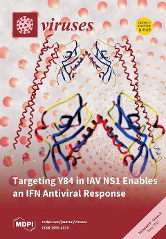
Cover Story (view full-size image): Targeting Y84 in IAV NS1 Enables an IFN Antiviral Response.
This article provides evidence that a major part of the virulence of the influenza A virus (IAV) NS1 is attributable to its ability to block an IFN response, mediated by a tyrosine residue, Y84, in a conserved SH2 binding domain that interacts with host cell proteins. Targeting Y84 reduces the virulence of the virus and permits an effective antiviral IFN response. These studies highlight the importance of the IFN response to control IAV infections and suggest that targeting the tyrosine residue in this highly conserved SH2 binding domain in IAVs may be a therapeutic strategy. View this paper
  • Issues are regarded as officially published after their release is announced to the table of contents alert mailing list.
  • You may sign up for e-mail alerts to receive table of contents of newly released issues.
  • PDF is the official format for papers published in both, html and pdf forms. To view the papers in pdf format, click on the "PDF Full-text" link, and use the free Adobe Reader to open them.
Order results
Result details
Section
Select all
Export citation of selected articles as:
3705 KiB  
Review
A Role for the Host DNA Damage Response in Hepatitis B Virus cccDNA Formation—and Beyond?
by Sabrina Schreiner and Michael Nassal
Viruses 2017, 9(5), 125; https://doi.org/10.3390/v9050125 - 22 May 2017
Cited by 75 | Viewed by 14696
Abstract
Chronic hepatitis B virus (HBV) infection puts more than 250 million people at a greatly increased risk to develop end-stage liver disease. Like all hepadnaviruses, HBV replicates via protein-primed reverse transcription of a pregenomic (pg) RNA, yielding an unusually structured, viral polymerase-linked relaxed-circular [...] Read more.
Chronic hepatitis B virus (HBV) infection puts more than 250 million people at a greatly increased risk to develop end-stage liver disease. Like all hepadnaviruses, HBV replicates via protein-primed reverse transcription of a pregenomic (pg) RNA, yielding an unusually structured, viral polymerase-linked relaxed-circular (RC) DNA as genome in infectious particles. Upon infection, RC-DNA is converted into nuclear covalently closed circular (ccc) DNA. Associating with cellular proteins into an episomal minichromosome, cccDNA acts as template for new viral RNAs, ensuring formation of progeny virions. Hence, cccDNA represents the viral persistence reservoir that is not directly targeted by current anti-HBV therapeutics. Eliminating cccDNA will thus be at the heart of a cure for chronic hepatitis B. The low production of HBV cccDNA in most experimental models and the associated problems in reliable cccDNA quantitation have long hampered a deeper understanding of cccDNA molecular biology. Recent advancements including cccDNA-dependent cell culture systems have begun to identify select host DNA repair enzymes that HBV usurps for RC-DNA to cccDNA conversion. While this list is bound to grow, it may represent just one facet of a broader interaction with the cellular DNA damage response (DDR), a network of pathways that sense and repair aberrant DNA structures and in the process profoundly affect the cell cycle, up to inducing cell death if repair fails. Given the divergent interactions between other viruses and the DDR it will be intriguing to see how HBV copes with this multipronged host system. Full article
(This article belongs to the Special Issue Recent Advances in Hepatitis B Virus Research)
Show Figures

Figure 1

2601 KiB  
Article
A Japanese Encephalitis Virus Vaccine Inducing Antibodies Strongly Enhancing In Vitro Infection Is Protective in Pigs
by Obdulio García-Nicolás, Meret E. Ricklin, Matthias Liniger, Nathalie J. Vielle, Sylvie Python, Philippe Souque, Pierre Charneau and Artur Summerfield
Viruses 2017, 9(5), 124; https://doi.org/10.3390/v9050124 - 22 May 2017
Cited by 23 | Viewed by 7623
Abstract
The Japanese encephalitis virus (JEV) is responsible for zoonotic severe viral encephalitis transmitted by Culex mosquitoes. Although birds are reservoirs, pigs play a role as amplifying hosts, and are affected in particular through reproductive failure. Here, we show that a lentiviral JEV vector, [...] Read more.
The Japanese encephalitis virus (JEV) is responsible for zoonotic severe viral encephalitis transmitted by Culex mosquitoes. Although birds are reservoirs, pigs play a role as amplifying hosts, and are affected in particular through reproductive failure. Here, we show that a lentiviral JEV vector, expressing JEV prM and E proteins (TRIP/JEV.prME), but not JEV infection induces strong antibody-dependent enhancement (ADE) activities for infection of macrophages. Such antibodies strongly promoted infection via Fc receptors. ADE was found at both neutralizing and non-neutralizing serum dilutions. Nevertheless, in vivo JEV challenge of pigs demonstrated comparable protection induced by the TRIP/JEV.prME vaccine or heterologous JEV infection. Thus, either ADE antibodies cause no harm in the presence of neutralizing antibodies or may even have protective effects in vivo in pigs. Additionally, we found that both pre-infected and vaccinated pigs were not fully protected as low levels of viral RNA were found in lymphoid and nervous system tissue in some animals. Strikingly, the virus from the pre-infection persisted in the tonsils throughout the experiment. Finally, despite the vaccination challenge, viral RNA was detected in the oronasal swabs in all vaccinated pigs. These latter data are relevant when JEV vaccination is employed in pigs. Full article
(This article belongs to the Special Issue Porcine Viruses)
Show Figures

Figure 1

2867 KiB  
Article
Distinct Contributions of Autophagy Receptors in Measles Virus Replication
by Denitsa S. Petkova, Pauline Verlhac, Aurore Rozières, Joël Baguet, Mathieu Claviere, Carole Kretz-Remy, Renaud Mahieux, Christophe Viret and Mathias Faure
Viruses 2017, 9(5), 123; https://doi.org/10.3390/v9050123 - 22 May 2017
Cited by 37 | Viewed by 6961
Abstract
Autophagy is a potent cell autonomous defense mechanism that engages the lysosomal pathway to fight intracellular pathogens. Several autophagy receptors can recognize invading pathogens in order to target them towards autophagy for their degradation after the fusion of pathogen-containing autophagosomes with lysosomes. However, [...] Read more.
Autophagy is a potent cell autonomous defense mechanism that engages the lysosomal pathway to fight intracellular pathogens. Several autophagy receptors can recognize invading pathogens in order to target them towards autophagy for their degradation after the fusion of pathogen-containing autophagosomes with lysosomes. However, numerous intracellular pathogens can avoid or exploit autophagy, among which is measles virus (MeV). This virus induces a complete autophagy flux, which is required to improve viral replication. We therefore asked how measles virus interferes with autophagy receptors during the course of infection. We report that in addition to NDP52/CALCOCO2 and OPTINEURIN/OPTN, another autophagy receptor, namely T6BP/TAXIBP1, also regulates the maturation of autophagosomes by promoting their fusion with lysosomes, independently of any infection. Surprisingly, only two of these receptors, NDP52 and T6BP, impacted measles virus replication, although independently, and possibly through physical interaction with MeV proteins. Thus, our results suggest that a restricted set of autophagosomes is selectively exploited by measles virus to replicate in the course of infection. Full article
(This article belongs to the Special Issue Viruses and Autophagy)
Show Figures

Figure 1

2622 KiB  
Article
Stumbling across the Same Phage: Comparative Genomics of Widespread Temperate Phages Infecting the Fish Pathogen Vibrio anguillarum
by Panos G. Kalatzis, Nanna Iben Rørbo, Daniel Castillo, Jesper Juel Mauritzen, Jóhanna Jørgensen, Constantina Kokkari, Faxing Zhang, Pantelis Katharios and Mathias Middelboe
Viruses 2017, 9(5), 122; https://doi.org/10.3390/v9050122 - 20 May 2017
Cited by 35 | Viewed by 8005
Abstract
Nineteen Vibrio anguillarum-specific temperate bacteriophages isolated across Europe and Chile from aquaculture and environmental sites were genome sequenced and analyzed for host range, morphology and life cycle characteristics. The phages were classified as Siphoviridae with genome sizes between 46,006 and 54,201 bp. [...] Read more.
Nineteen Vibrio anguillarum-specific temperate bacteriophages isolated across Europe and Chile from aquaculture and environmental sites were genome sequenced and analyzed for host range, morphology and life cycle characteristics. The phages were classified as Siphoviridae with genome sizes between 46,006 and 54,201 bp. All 19 phages showed high genetic similarity, and 13 phages were genetically identical. Apart from sporadically distributed single nucleotide polymorphisms (SNPs), genetic diversifications were located in three variable regions (VR1, VR2 and VR3) in six of the phage genomes. Identification of specific genes, such as N6-adenine methyltransferase and lambda like repressor, as well as the presence of a tRNAArg, suggested a both mutualistic and parasitic interaction between phages and hosts. During short term phage exposure experiments, 28% of a V. anguillarum host population was lysogenized by the temperate phages and a genomic analysis of a collection of 31 virulent V. anguillarum showed that the isolated phages were present as prophages in >50% of the strains covering large geographical distances. Further, phage sequences were widely distributed among CRISPR-Cas arrays of publicly available sequenced Vibrios. The observed distribution of these specific temperate Vibriophages across large geographical scales may be explained by efficient dispersal of phages and bacteria in the marine environment combined with a mutualistic interaction between temperate phages and their hosts which selects for co-existence rather than arms race dynamics. Full article
(This article belongs to the Special Issue Marine Viruses 2016)
Show Figures

Figure 1

2354 KiB  
Article
Evaluation and Comparison of the Pathogenicity and Host Immune Responses Induced by a G2b Taiwan Porcine Epidemic Diarrhea Virus (Strain Pintung 52) and Its Highly Cell-Culture Passaged Strain in Conventional 5-Week-Old Pigs
by Yen-Chen Chang, Chi-Fei Kao, Chia-Yu Chang, Chian-Ren Jeng, Pei-Shiue Tsai, Victor Fei Pang, Hue-Ying Chiou, Ju-Yi Peng, Ivan-Chen Cheng and Hui-Wen Chang
Viruses 2017, 9(5), 121; https://doi.org/10.3390/v9050121 - 19 May 2017
Cited by 35 | Viewed by 8319
Abstract
A genogroup 2b (G2b) porcine epidemic diarrhea virus (PEDV) Taiwan Pintung 52 (PEDVPT) strain was isolated in 2014. The pathogenicity and host antibody responses elicited by low-passage (passage 5; PEDVPT-P5) and high-passage (passage 96; PEDVPT-P96) PEDVPT strains were compared in post-weaning PEDV-seronegative pigs [...] Read more.
A genogroup 2b (G2b) porcine epidemic diarrhea virus (PEDV) Taiwan Pintung 52 (PEDVPT) strain was isolated in 2014. The pathogenicity and host antibody responses elicited by low-passage (passage 5; PEDVPT-P5) and high-passage (passage 96; PEDVPT-P96) PEDVPT strains were compared in post-weaning PEDV-seronegative pigs by oral inoculation. PEDVPT-P5-inoculation induced typical diarrhea during 1–9 days post inoculation with fecal viral shedding persisting for 26 days. Compared to PEDVPT-P5, PEDVPT-P96 inoculation induced none-to-mild diarrhea and lower, delayed fecal viral shedding. Although PEDVPT-P96 elicited slightly lower neutralizing antibodies and PEDV-specific immunoglobulin G (IgG) and immunoglobulin A (IgA) titers, a reduction in pathogenicity and viral shedding of the subsequent challenge with PEDVPT-P5 were noted in both PEDVPT-P5- and PEDVPT-P96-inoculated pigs. Alignment and comparison of full-length sequences of PEDVPT-P5 and PEDVPT-P96 revealed 23 nucleotide changes and resultant 19 amino acid substitutions in non-structure proteins 2, 3, 4, 9, 14, 15, spike, open reading frame 3 (ORF3), and membrane proteins with no detectable deletion or insertion. The present study confirmed the pathogenicity of the PEDVPT isolate in conventional post-weaning pigs. Moreover, data regarding viral attenuation and potency of induced antibodies against PEDVPT-P5 identified PEDVPT-P96 as a potential live-attenuated vaccine candidate. Full article
(This article belongs to the Special Issue Porcine Viruses)
Show Figures

Figure 1

3099 KiB  
Article
Differentiation and Structure in Sulfolobus islandicus Rod-Shaped Virus Populations
by Maria A. Bautista, Jesse A. Black, Nicholas D. Youngblut and Rachel J. Whitaker
Viruses 2017, 9(5), 120; https://doi.org/10.3390/v9050120 - 19 May 2017
Cited by 23 | Viewed by 9471
Abstract
In the past decade, molecular surveys of viral diversity have revealed that viruses are the most diverse and abundant biological entities on Earth. In culture, however, most viral isolates that infect microbes are represented by a few variants isolated on type strains, limiting [...] Read more.
In the past decade, molecular surveys of viral diversity have revealed that viruses are the most diverse and abundant biological entities on Earth. In culture, however, most viral isolates that infect microbes are represented by a few variants isolated on type strains, limiting our ability to study how natural variation affects virus-host interactions in the laboratory. We screened a set of 137 hot spring samples for viruses that infect a geographically diverse panel of the hyperthemophilic crenarchaeon Sulfolobus islandicus. We isolated and characterized eight SIRVs (Sulfolobus islandicus rod-shaped viruses) from two different regions within Yellowstone National Park (USA). Comparative genomics revealed that all SIRV sequenced isolates share 30 core genes that represent 50–60% of the genome. The core genome phylogeny, as well as the distribution of variable genes (shared by some but not all SIRVs) and the signatures of host-virus interactions recorded on the CRISPR (clustered regularly interspaced short palindromic repeats) repeat-spacer arrays of S. islandicus hosts, identify different SIRV lineages, each associated with a different geographic location. Moreover, our studies reveal that SIRV core genes do not appear to be under diversifying selection and thus we predict that the abundant and diverse variable genes govern the coevolutionary arms race between SIRVs and their hosts. Full article
(This article belongs to the Special Issue Viruses of Microbes)
Show Figures

Figure 1

1877 KiB  
Article
ATP-Driven Contraction of Phage T3 Capsids with DNA Incompletely Packaged In Vivo
by Philip Serwer and Elena T. Wright
Viruses 2017, 9(5), 119; https://doi.org/10.3390/v9050119 - 19 May 2017
Cited by 8 | Viewed by 5610
Abstract
Adenosine triphosphate (ATP) cleavage powers packaging of a double-stranded DNA (dsDNA) molecule in a pre-assembled capsid of phages that include T3. Several observations constitute a challenge to the conventional view that the shell of the capsid is energetically inert during packaging. Here, we [...] Read more.
Adenosine triphosphate (ATP) cleavage powers packaging of a double-stranded DNA (dsDNA) molecule in a pre-assembled capsid of phages that include T3. Several observations constitute a challenge to the conventional view that the shell of the capsid is energetically inert during packaging. Here, we test this challenge by analyzing the in vitro effects of ATP on the shells of capsids generated by DNA packaging in vivo. These capsids retain incompletely packaged DNA (ipDNA) and are called ipDNA-capsids; the ipDNA-capsids are assumed to be products of premature genome maturation-cleavage. They were isolated via preparative Nycodenz buoyant density centrifugation. For some ipDNA-capsids, Nycodenz impermeability increases hydration and generates density so low that shell hyper-expansion must exist to accommodate associated water. Electron microscopy (EM) confirmed hyper-expansion and low permeability and revealed that 3.0 mM magnesium ATP (physiological concentration) causes contraction of hyper-expanded, lowpermeability ipDNA-capsids to less than mature size; 5.0 mM magnesium ATP (border of supraphysiological concentration) or more disrupts them. Additionally, excess sodium ADP reverses 3.0 mM magnesium ATP-induced contraction and re-generates hyper-expansion. The Nycodenz impermeability implies assembly perfection that suggests selection for function in DNA packaging. These findings support the above challenge and can be explained via the assumption that T3 DNA packaging includes a back-up cycle of ATP-driven capsid contraction and hyper-expansion. Full article
(This article belongs to the Section Bacterial Viruses)
Show Figures

Figure 1

7252 KiB  
Article
Manipulation of Viral MicroRNAs as a Potential Antiviral Strategy for the Treatment of Cytomegalovirus Infection
by Jiang Deng, Jun Xiao, Ping Ma, Bo Gao, Feng Gong, Liping Lv, Yanyu Zhang and Jinbo Xu
Viruses 2017, 9(5), 118; https://doi.org/10.3390/v9050118 - 19 May 2017
Cited by 6 | Viewed by 4851
Abstract
Cytomegalovirus (CMV) infection leads to notable morbidity and mortality in immunosuppressed patients. Current antiviral drugs are effective but seriously limited in their long-term use due to their relatively high toxicity. In the present study, we characterized the expression of murine CMV microRNAs (MCMV [...] Read more.
Cytomegalovirus (CMV) infection leads to notable morbidity and mortality in immunosuppressed patients. Current antiviral drugs are effective but seriously limited in their long-term use due to their relatively high toxicity. In the present study, we characterized the expression of murine CMV microRNAs (MCMV miRNAs) both in vitro and in vivo. Although 29 miRNAs were detectable during in vitro infection, only 11 miRNAs (classified as Group 1) were detectable during in vivo infection, and as many as 18 viral miRNAs (classified as Group 2) were less detectable (<50% of animals) in both the liver and lungs. In addition, viral miRNA profiles in the blood revealed unstable and reduced expression. We next explored the in vitro effects of viral miRNAs on MCMV replication. The inhibition of Group 1 viral miRNAs had little effect on virus production, but transfected cells overexpressing miR-m01-3-5p, miR-M23-1-5p, miR-M55-1, and miR-m107-1-5p in Group 2 showed statistically lower viral loads than those transfected with control miRNA (29%, 29%, 39%, and 43%, respectively, versus control). Finally, we performed hydrodynamic injection of viral miRNA agomirs and observed lower levels of MCMV recurrence in the livers of animals overexpressing the miR-m01-3-5p or mcmv-miR-M23-1-5p agomirs compared with those of animals transfected with control agomir, confirming the antiviral effects of viral miRNA manipulation in vivo. Therefore, the manipulation of viral miRNA expression shows great therapeutic potential and represents a novel antiviral strategy for the miRNA-based treatment of cytomegalovirus infection. Full article
(This article belongs to the Section Viral Immunology, Vaccines, and Antivirals)
Show Figures

Figure 1

18247 KiB  
Article
Understanding the Complex Patterns Observed during Hepatitis B Virus Therapy
by Andrea Carracedo Rodriguez, Matthias Chung and Stanca M. Ciupe
Viruses 2017, 9(5), 117; https://doi.org/10.3390/v9050117 - 19 May 2017
Cited by 10 | Viewed by 5350
Abstract
Data from human clinical trials have shown that the hepatitis B virus (HBV) follows complex profiles, such as bi-phasic, tri-phasic, stepwise decay and rebound. We utilized a deterministic model of HBV kinetics following antiviral therapy to uncover the mechanistic interactions behind HBV dynamics. [...] Read more.
Data from human clinical trials have shown that the hepatitis B virus (HBV) follows complex profiles, such as bi-phasic, tri-phasic, stepwise decay and rebound. We utilized a deterministic model of HBV kinetics following antiviral therapy to uncover the mechanistic interactions behind HBV dynamics. Analytical investigation of the model was used to separate the parameter space describing virus decay and rebound. Monte Carlo sampling of the parameter space was used to determine the virological, pharmacological and immunological factors that separate the bi-phasic and tri-phasic virus profiles. We found that the level of liver infection at the start of therapy best separates the decay patterns. Moreover, drug efficacy, ratio between division of uninfected and infected cells, and the strength of cytotoxic immune response are important in assessing the amount of liver damage experienced over time and in quantifying the duration of therapy leading to virus resolution in each of the observed profiles. Full article
(This article belongs to the Special Issue Mathematical Modeling of Viral Infections)
Show Figures

Figure 1

2246 KiB  
Article
Variation in the Genetic Repertoire of Viruses Infecting Micromonas pusilla Reflects Horizontal Gene Transfer and Links to Their Environmental Distribution
by Jan F. Finke, Danielle M. Winget, Amy M. Chan and Curtis A. Suttle
Viruses 2017, 9(5), 116; https://doi.org/10.3390/v9050116 - 19 May 2017
Cited by 16 | Viewed by 6158
Abstract
Prasinophytes, a group of eukaryotic phytoplankton, has a global distribution and is infected by large double-stranded DNA viruses (prasinoviruses) in the family Phycodnaviridae. This study examines the genetic repertoire, phylogeny, and environmental distribution of phycodnaviruses infecting Micromonas pusilla, other prasinophytes and [...] Read more.
Prasinophytes, a group of eukaryotic phytoplankton, has a global distribution and is infected by large double-stranded DNA viruses (prasinoviruses) in the family Phycodnaviridae. This study examines the genetic repertoire, phylogeny, and environmental distribution of phycodnaviruses infecting Micromonas pusilla, other prasinophytes and chlorophytes. Based on comparisons among the genomes of viruses infecting M. pusilla and other phycodnaviruses, as well as the genome from a host isolate of M. pusilla, viruses infecting M. pusilla (MpVs) share a limited set of core genes, but vary strongly in their flexible pan-genome that includes numerous metabolic genes, such as those associated with amino acid synthesis and sugar manipulation. Surprisingly, few of these presumably host-derived genes are shared with M. pusilla, but rather have their closest non-viral homologue in bacteria and other eukaryotes, indicating horizontal gene transfer. A comparative analysis of full-length DNA polymerase (DNApol) genes from prasinoviruses with their overall gene content, demonstrated that the phylogeny of DNApol gene fragments reflects the gene content of the viruses; hence, environmental DNApol gene sequences from prasinoviruses can be used to infer their overall genetic repertoire. Thus, the distribution of virus ecotypes across environmental samples based on DNApol sequences implies substantial underlying differences in gene content that reflect local environmental conditions. Moreover, the high diversity observed in the genetic repertoire of prasinoviruses has been driven by horizontal gene transfer throughout their evolutionary history, resulting in a broad suite of functional capabilities and a high diversity of prasinovirus ecotypes. Full article
(This article belongs to the Special Issue Viruses of Microbes)
Show Figures

Figure 1

4093 KiB  
Article
Label-Free Quantitative Proteomic Analysis of Chitosan Oligosaccharide-Treated Rice Infected with Southern Rice Black-Streaked Dwarf Virus
by Anming Yang, Lu Yu, Zhuo Chen, Shanxue Zhang, Jing Shi, Xiaozhen Zhao, Yuanyou Yang, Deyu Hu and Baoan Song
Viruses 2017, 9(5), 115; https://doi.org/10.3390/v9050115 - 18 May 2017
Cited by 80 | Viewed by 6245
Abstract
Southern rice black-streaked dwarf virus (SRBSDV) has spread from thesouth of China to the north of Vietnam in the past few years and severelyinfluenced rice production. Its long incubation period and early symptoms are not evident; thus, controlling it is difficult. Chitosan oligosaccharide [...] Read more.
Southern rice black-streaked dwarf virus (SRBSDV) has spread from thesouth of China to the north of Vietnam in the past few years and severelyinfluenced rice production. Its long incubation period and early symptoms are not evident; thus, controlling it is difficult. Chitosan oligosaccharide (COS) is a green plant immunomodulator. Early studies showed that preventing and controlling SRBSDV have a certain effect and reduce disease infection rate, but its underlying controlling and preventing mechanism is unclear. In this study, label-free proteomics was used to analyze differentially expressed proteins in rice after COS treatment. The results showed that COS can up-regulate the plant defense-related proteins and down-regulate the protein expression levels of SRBSDV. Meanwhile, quantitative real-time PCR test results showed that COS can improve defense gene expression in rice. Moreover, COS can enhance the defense enzymatic activities of peroxidase, superoxide dismutase and catalase through mitogen-activated protein kinase signaling cascade pathway, and enhance the rice disease resistance. Full article
(This article belongs to the Section Viruses of Plants, Fungi and Protozoa)
Show Figures

Figure 1

8741 KiB  
Article
TMPRSS2 and MSPL Facilitate Trypsin-Independent Porcine Epidemic Diarrhea Virus Replication in Vero Cells
by Wen Shi, Wenlu Fan, Jing Bai, Yandong Tang, Li Wang, Yanping Jiang, Lijie Tang, Min Liu, Wen Cui, Yigang Xu and Yijing Li
Viruses 2017, 9(5), 114; https://doi.org/10.3390/v9050114 - 18 May 2017
Cited by 23 | Viewed by 7139
Abstract
Type II transmembrane serine proteases (TTSPs) facilitate the spread and replication of viruses such as influenza and human coronaviruses, although it remains unclear whether TTSPs play a role in the progression of animal coronavirus infections, such as that by porcine epidemic diarrhea virus [...] Read more.
Type II transmembrane serine proteases (TTSPs) facilitate the spread and replication of viruses such as influenza and human coronaviruses, although it remains unclear whether TTSPs play a role in the progression of animal coronavirus infections, such as that by porcine epidemic diarrhea virus (PEDV). In this study, TTSPs including TMPRSS2, HAT, DESC1, and MSPL were tested for their ability to facilitate PEDV replication in Vero cells. Our results showed that TMPRSS2 and MSPL played significant roles in the stages of cell–cell fusion and virus–cell fusion, whereas HAT and DESC1 exhibited weaker effects. This activation may be involved in the interaction between TTSPs and the PEDV S protein, as the S protein extensively co-localized with TMPRSS2 and MSPL and could be cleaved by co-expression with TMPRSS2 or MSPL. Moreover, the use of Vero cells expressing TMPRSS2 and MSPL facilitated PEDV replication in the absence of exogenous trypsin. In sum, we identified two host proteases, TMPRSS2 and MSPL, which may provide insights and a novel method for enhancing viral titers, expanding virus production, and improving the adaptability of PEDV isolates in vitro. Full article
(This article belongs to the Special Issue Porcine Viruses)
Show Figures

Figure 1

2308 KiB  
Article
Antigenic and Biological Characterization of ORF2–6 Variants at Early Times Following PRRSV Infection
by Alyssa B. Evans, Hyelee Loyd, Jenelle R. Dunkelberger, Sarah Van Tol, Marcus J. Bolton, Karin S. Dorman, Jack C. M. Dekkers and Susan Carpenter
Viruses 2017, 9(5), 113; https://doi.org/10.3390/v9050113 - 16 May 2017
Cited by 15 | Viewed by 5007
Abstract
Genetic diversity of porcine reproductive and respiratory syndrome virus (PRRSV) challenges efforts to develop effective and broadly acting vaccines. Although genetic variation in PRRSV has been extensively documented, the effects of this variation on virus phenotype are less well understood. In the present [...] Read more.
Genetic diversity of porcine reproductive and respiratory syndrome virus (PRRSV) challenges efforts to develop effective and broadly acting vaccines. Although genetic variation in PRRSV has been extensively documented, the effects of this variation on virus phenotype are less well understood. In the present study, PRRSV open reading frame (ORF)2–6 variants predominant during the first six weeks following experimental infection were characterized for antigenic and replication phenotype. There was limited genetic variation during these early times after infection; however, distinct ORF2–6 haplotypes that differed from the NVSL97-7895 inoculum were identified in each of the five pigs examined. Chimeric viruses containing all or part of predominant ORF2–6 haplotypes were constructed and tested in virus neutralization and in vitro replication assays. In two pigs, genetic variation in ORF2–6 resulted in increased resistance to neutralization by autologous sera. Mapping studies indicated that variation in either ORF2–4 or ORF5–6 could confer increased neutralization resistance, but there was no single amino acid substitution that was predictive of neutralization phenotype. Detailed analyses of the early steps in PRRSV replication in the presence and absence of neutralizing antibody revealed both significant inhibition of virion attachment and, independently, a significant delay in the appearance of newly synthesized viral RNA. In all pigs, genetic variation in ORF2–6 also resulted in significant reduction in infectivity on MARC-145 cells, suggesting variation in ORF2–6 may also be important for virus replication in vivo. Together, these data reveal that variation appearing early after infection, though limited, alters important virus phenotypes and contributes to antigenic and biologic diversity of PRRSV. Full article
(This article belongs to the Special Issue Porcine Viruses)
Show Figures

Figure 1

2455 KiB  
Article
In Silico Analysis of Epitope-Based Vaccine Candidates against Hepatitis B Virus Polymerase Protein
by Juzeng Zheng, Xianfan Lin, Xiuyan Wang, Liyu Zheng, Songsong Lan, Sisi Jin, Zhanfan Ou and Jinming Wu
Viruses 2017, 9(5), 112; https://doi.org/10.3390/v9050112 - 16 May 2017
Cited by 49 | Viewed by 8010
Abstract
Hepatitis B virus (HBV) infection has persisted as a major public health problem due to the lack of an effective treatment for those chronically infected. Therapeutic vaccination holds promise, and targeting HBV polymerase is pivotal for viral eradication. In this research, a computational [...] Read more.
Hepatitis B virus (HBV) infection has persisted as a major public health problem due to the lack of an effective treatment for those chronically infected. Therapeutic vaccination holds promise, and targeting HBV polymerase is pivotal for viral eradication. In this research, a computational approach was employed to predict suitable HBV polymerase targeting multi-peptides for vaccine candidate selection. We then performed in-depth computational analysis to evaluate the predicted epitopes’ immunogenicity, conservation, population coverage, and toxicity. Lastly, molecular docking and MHC-peptide complex stabilization assay were utilized to determine the binding energy and affinity of epitopes to the HLA-A0201 molecule. Criteria-based analysis provided four predicted epitopes, RVTGGVFLV, VSIPWTHKV, YMDDVVLGA and HLYSHPIIL. Assay results indicated the lowest binding energy and high affinity to the HLA-A0201 molecule for epitopes VSIPWTHKV and YMDDVVLGA and epitopes RVTGGVFLV and VSIPWTHKV, respectively. Regions 307 to 320 and 377 to 387 were considered to have the highest probability to be involved in B cell epitopes. The T cell and B cell epitopes identified in this study are promising targets for an epitope-focused, peptide-based HBV vaccine, and provide insight into HBV-induced immune response. Full article
(This article belongs to the Section Viral Immunology, Vaccines, and Antivirals)
Show Figures

Figure 1

3349 KiB  
Article
Human Beta Defensin 2 Selectively Inhibits HIV-1 in Highly Permissive CCR6+CD4+ T Cells
by Mark K. Lafferty, Lingling Sun, Aaron Christensen-Quick, Wuyuan Lu and Alfredo Garzino-Demo
Viruses 2017, 9(5), 111; https://doi.org/10.3390/v9050111 - 16 May 2017
Cited by 22 | Viewed by 5764
Abstract
Chemokine receptor type 6 (CCR6)+CD4+ T cells are preferentially infected and depleted during HIV disease progression, but are preserved in non-progressors. CCR6 is expressed on a heterogeneous population of memory CD4+ T cells that are critical to mucosal immunity. [...] Read more.
Chemokine receptor type 6 (CCR6)+CD4+ T cells are preferentially infected and depleted during HIV disease progression, but are preserved in non-progressors. CCR6 is expressed on a heterogeneous population of memory CD4+ T cells that are critical to mucosal immunity. Preferential infection of these cells is associated, in part, with high surface expression of CCR5, CXCR4, and α4β7. In addition, CCR6+CD4+ T cells harbor elevated levels of integrated viral DNA and high levels of proliferation markers. We have previously shown that the CCR6 ligands MIP-3α and human beta defensins inhibit HIV replication. The inhibition required CCR6 and the induction of APOBEC3G. Here, we further characterize the induction of apolipoprotein B mRNA editing enzyme (APOBEC3G) by human beta defensin 2. Human beta defensin 2 rapidly induces transcriptional induction of APOBEC3G that involves extracellular signal-regulated kinases 1/2 (ERK1/2) activation and the transcription factors NFATc2, NFATc1, and IRF4. We demonstrate that human beta defensin 2 selectively protects primary CCR6+CD4+ T cells infected with HIV-1. The selective protection of CCR6+CD4+ T cell subsets may be critical in maintaining mucosal immune function and preventing disease progression. Full article
(This article belongs to the Section Viral Immunology, Vaccines, and Antivirals)
Show Figures

Graphical abstract

3344 KiB  
Article
A Novel Roseosiphophage Isolated from the Oligotrophic South China Sea
by Yunlan Yang, Lanlan Cai, Ruijie Ma, Yongle Xu, Yigang Tong, Yong Huang, Nianzhi Jiao and Rui Zhang
Viruses 2017, 9(5), 109; https://doi.org/10.3390/v9050109 - 15 May 2017
Cited by 43 | Viewed by 6903
Abstract
The Roseobacter clade is abundant and widespread in marine environments and plays an important role in oceanic biogeochemical cycling. In this present study, a lytic siphophage (labeled vB_DshS-R5C) infecting the strain type of Dinoroseobacter shibae named DFL12T, which is part of [...] Read more.
The Roseobacter clade is abundant and widespread in marine environments and plays an important role in oceanic biogeochemical cycling. In this present study, a lytic siphophage (labeled vB_DshS-R5C) infecting the strain type of Dinoroseobacter shibae named DFL12T, which is part of the Roseobacter clade, was isolated from the oligotrophic South China Sea. Phage R5C showed a narrow host range, short latent period and low burst size. The genome length of phage R5C was 77, 874 bp with a G+C content of 61.5%. Genomic comparisons detected no genome matches in the GenBank database and phylogenetic analysis based on DNA polymerase I revealed phylogenetic features that were distinct to other phages, suggesting the novelty of R5C. Several auxiliary metabolic genes (e.g., phoH gene, heat shock protein and queuosine biosynthesis genes) were identified in the R5C genome that may be beneficial to the host and/or offer a competitive advantage for the phage. Among siphophages infecting the Roseobacter clade (roseosiphophages), four gene transfer agent-like genes were commonly located with close proximity to structural genes, suggesting that their function may be related to the tail of siphoviruses. The isolation and characterization of R5C demonstrated the high genomic and physiological diversity of roseophages as well as improved our understanding of host–phage interactions and the ecology of the marine Roseobacter. Full article
(This article belongs to the Special Issue Marine Viruses 2016)
Show Figures

Figure 1

212 KiB  
Review
HIV-Enhancing and HIV-Inhibiting Properties of Cationic Peptides and Proteins
by Alexander M. Cole and Amy L. Cole
Viruses 2017, 9(5), 108; https://doi.org/10.3390/v9050108 - 15 May 2017
Cited by 7 | Viewed by 4658
Abstract
Cationic antimicrobial peptides and proteins have historically been ascribed roles in innate immunity that infer killing of microbial and viral pathogens and protection of the host. In the context of sexually transmitted HIV-1, we take an unconventional approach that questions this paradigm. It [...] Read more.
Cationic antimicrobial peptides and proteins have historically been ascribed roles in innate immunity that infer killing of microbial and viral pathogens and protection of the host. In the context of sexually transmitted HIV-1, we take an unconventional approach that questions this paradigm. It is becoming increasingly apparent that many of the cationic polypeptides present in the human genital or anorectal mucosa, or human semen, are capable of enhancing HIV-1 infection, often in addition to other reported roles as viral inhibitors. We explore how the in vivo environment may select for or against the HIV-enhancing aspects of these cationic polypeptides by focusing on biological relevance. We stress that the distinction between enhancing and inhibiting HIV-1 infection is not mutually exclusive to specific classes of cationic polypeptides. Understanding how virally enhancing peptides and proteins act to promote sexual transmission of HIV-1 would be important for the design of topical microbicides, mucosal vaccines, and other preventative measures. Full article
(This article belongs to the Section Viral Immunology, Vaccines, and Antivirals)
13914 KiB  
Article
A Conserved Residue, Tyrosine (Y) 84, in H5N1 Influenza A Virus NS1 Regulates IFN Signaling Responses to Enhance Viral Infection
by Ben X. Wang, Lianhu Wei, Lakshmi P. Kotra, Earl G. Brown and Eleanor N. Fish
Viruses 2017, 9(5), 107; https://doi.org/10.3390/v9050107 - 12 May 2017
Cited by 8 | Viewed by 5621
Abstract
The non-structural protein, NS1, is a virulence factor encoded by influenza A viruses (IAVs). In this report, we provide evidence that the conserved residue, tyrosine (Y) 84, in a conserved putative SH2-binding domain in A/Duck/Hubei/2004/L-1 [H5N1] NS1 is critical for limiting an interferon [...] Read more.
The non-structural protein, NS1, is a virulence factor encoded by influenza A viruses (IAVs). In this report, we provide evidence that the conserved residue, tyrosine (Y) 84, in a conserved putative SH2-binding domain in A/Duck/Hubei/2004/L-1 [H5N1] NS1 is critical for limiting an interferon (IFN) response to infection. A phenylalanine (F) substitution of this Y84 residue abolishes NS1-mediated downregulation of IFN-inducible STAT phosphorylation, and surface IFNAR1 expression. Recombinant IAV (rIAV) [H1N1] expressing A/Grey Heron/Hong Kong/837/2004 [H5N1] NS1-Y84F (rWSN-GH-NS1-Y84F) replicates to lower titers in human lung epithelial cells and is more susceptible to the antiviral effects of IFN-β treatment compared with rIAV expressing the intact H5N1 NS1 (rWSN-GH-NS1-wt). Cells infected with rWSN-GH-NS1-Y84F express higher levels of IFN stimulated genes (ISGs) associated with an antiviral response compared with cells infected with rWSN-GH-NS1-wt. In mice, intranasal infection with rWSN-GH-NS1-Y84F resulted in a delay in onset of weight loss, reduced lung pathology, lower lung viral titers and higher ISG expression, compared with mice infected with rWSN-GH-NS1-wt. IFN-β treatment of mice infected with rWSN-GH-NS1-Y84F reduced lung viral titers and increased lung ISG expression, but did not alter viral titers and ISG expression in mice infected with rWSN-GH-NS1-wt. Viewed altogether, these data suggest that the virulence associated with this conserved Y84 residue in NS1 is, in part, due to its role in regulating the host IFN response. Full article
(This article belongs to the Section Animal Viruses)
Show Figures

Figure 1

837 KiB  
Letter
Implementation of Objective PASC-Derived Taxon Demarcation Criteria for Official Classification of Filoviruses
by Yīmíng Bào, Gaya K. Amarasinghe, Christopher F. Basler, Sina Bavari, Alexander Bukreyev, Kartik Chandran, Olga Dolnik, John M. Dye, Hideki Ebihara, Pierre Formenty, Roger Hewson, Gary P. Kobinger, Eric M. Leroy, Elke Mühlberger, Sergey V. Netesov, Jean L. Patterson, Janusz T. Paweska, Sophie J. Smither, Ayato Takada, Jonathan S. Towner, Viktor E. Volchkov, Victoria Wahl-Jensen and Jens H. Kuhnadd Show full author list remove Hide full author list
Viruses 2017, 9(5), 106; https://doi.org/10.3390/v9050106 - 11 May 2017
Cited by 20 | Viewed by 8421
Abstract
The mononegaviral family Filoviridae has eight members assigned to three genera and seven species. Until now, genus and species demarcation were based on arbitrarily chosen filovirus genome sequence divergence values (≈50% for genera, ≈30% for species) and arbitrarily chosen phenotypic virus or virion [...] Read more.
The mononegaviral family Filoviridae has eight members assigned to three genera and seven species. Until now, genus and species demarcation were based on arbitrarily chosen filovirus genome sequence divergence values (≈50% for genera, ≈30% for species) and arbitrarily chosen phenotypic virus or virion characteristics. Here we report filovirus genome sequence-based taxon demarcation criteria using the publicly accessible PAirwise Sequencing Comparison (PASC) tool of the US National Center for Biotechnology Information (Bethesda, MD, USA). Comparison of all available filovirus genomes in GenBank using PASC revealed optimal genus demarcation at the 55–58% sequence diversity threshold range for genera and at the 23–36% sequence diversity threshold range for species. Because these thresholds do not change the current official filovirus classification, these values are now implemented as filovirus taxon demarcation criteria that may solely be used for filovirus classification in case additional data are absent. A near-complete, coding-complete, or complete filovirus genome sequence will now be required to allow official classification of any novel “filovirus.” Classification of filoviruses into existing taxa or determining the need for novel taxa is now straightforward and could even become automated using a presented algorithm/flowchart rooted in RefSeq (type) sequences. Full article
(This article belongs to the Collection Advances in Ebolavirus, Marburgvirus, and Cuevavirus Research)
Show Figures

Graphical abstract

2563 KiB  
Article
Microbial Natural Product Alternariol 5-O-Methyl Ether Inhibits HIV-1 Integration by Blocking Nuclear Import of the Pre-Integration Complex
by Jiwei Ding, Jianyuan Zhao, Zhijun Yang, Ling Ma, Zeyun Mi, Yanbing Wu, Jiamei Guo, Jinmin Zhou, Xiaoyu Li, Ying Guo, Zonggen Peng, Tao Wei, Haisheng Yu, Liguo Zhang, Mei Ge and Shan Cen
Viruses 2017, 9(5), 105; https://doi.org/10.3390/v9050105 - 10 May 2017
Cited by 19 | Viewed by 6671
Abstract
While Highly Active Antiretroviral Therapy (HAART) has significantly decreased the mortality of human immunodeficiency virus (HIV)-infected patients, emerging drug resistance to approved HIV-1 integrase inhibitors highlights the need to develop new antivirals with novel mechanisms of action. In this study, we screened a [...] Read more.
While Highly Active Antiretroviral Therapy (HAART) has significantly decreased the mortality of human immunodeficiency virus (HIV)-infected patients, emerging drug resistance to approved HIV-1 integrase inhibitors highlights the need to develop new antivirals with novel mechanisms of action. In this study, we screened a library of microbial natural compounds from endophytic fungus Colletotrichum sp. and identified alternariol 5-O-methyl ether (AME) as a compound that inhibits HIV-1 pre-integration steps. Time-of addition analysis, quantitative real-time PCR, confocal microscopy, and WT viral replication assay were used to elucidate the mechanism. As opposed to the approved integrase inhibitor Raltegravir, AME reduced both the integrated viral DNA and the 2-long terminal repeat (2-LTR) circular DNA, which suggests that AME impairs the nuclear import of viral DNA. Further confocal microscopy studies showed that AME specifically blocks the nuclear import of HIV-1 integrase and pre-integration complex without any adverse effects on the importin α/β and importin β-mediated nuclear import pathway in general. Importantly, AME inhibited Raltegravir-resistant HIV-1 strains and exhibited a broad anti-HIV-1 activity in diverse cell lines. These data collectively demonstrate the potential of AME for further development into a new HIV inhibitor, and suggest the utility of viral DNA nuclear import as a target for anti-HIV drug discovery. Full article
Show Figures

Figure 1

3862 KiB  
Article
Nasal Infection of Enterovirus D68 Leading to Lower Respiratory Tract Pathogenesis in Ferrets (Mustela putorius furo)
by Hui-Wen Zheng, Ming Sun, Lei Guo, Jing-Jing Wang, Jie Song, Jia-Qi Li, Hong-Zhe Li, Ruo-Tong Ning, Ze-Ning Yang, Hai-Tao Fan, Zhan-Long He and Long-Ding Liu
Viruses 2017, 9(5), 104; https://doi.org/10.3390/v9050104 - 10 May 2017
Cited by 22 | Viewed by 5656
Abstract
Data from EV-D68-infected patients demonstrate that pathological changes in the lower respiratory tract are principally characterized by severe respiratory illness in children and acute flaccid myelitis. However, lack of a suitable animal model for EV-D68 infection has limited the study on the pathogenesis [...] Read more.
Data from EV-D68-infected patients demonstrate that pathological changes in the lower respiratory tract are principally characterized by severe respiratory illness in children and acute flaccid myelitis. However, lack of a suitable animal model for EV-D68 infection has limited the study on the pathogenesis of this critical pathogen, and the development of a vaccine. Ferrets have been widely used to evaluate respiratory virus infections. In the current study, we used EV-D68-infected ferrets as a potential animal to identify impersonal indices, involving clinical features and histopathological changes in the upper and lower respiratory tract (URT and LRT). The research results demonstrate that the EV-D68 virus leads to minimal clinical symptoms in ferrets. According to the viral load detection in the feces, nasal, and respiratory tracts, the infection and shedding of EV-D68 in the ferret model was confirmed, and these results were supported by the EV-D68 VP1 immunofluorescence confocal imaging with α2,6-linked sialic acid (SA) in lung tissues. Furthermore, we detected the inflammatory cytokine/chemokine expression level, which implied high expression levels of interleukin (IL)-1a, IL-8, IL-5, IL-12, IL-13, and IL-17a in the lungs. These data indicate that systemic observation of responses following infection with EV-D68 in ferrets could be used as a model for EV-D68 infection and pathogenesis. Full article
Show Figures

Figure 1

561 KiB  
Review
African Swine Fever Virus: A Review
by Inmaculada Galindo and Covadonga Alonso
Viruses 2017, 9(5), 103; https://doi.org/10.3390/v9050103 - 10 May 2017
Cited by 441 | Viewed by 36556
Abstract
African swine fever (ASF) is a highly contagious viral disease of swine which causes high mortality, approaching 100%, in domestic pigs. ASF is caused by a large, double stranded DNA virus, ASF virus (ASFV), which replicates predominantly in the cytoplasm of macrophages and [...] Read more.
African swine fever (ASF) is a highly contagious viral disease of swine which causes high mortality, approaching 100%, in domestic pigs. ASF is caused by a large, double stranded DNA virus, ASF virus (ASFV), which replicates predominantly in the cytoplasm of macrophages and is the only member of the Asfarviridae family, genus Asfivirus. The natural hosts of this virus include wild suids and arthropod vectors of the Ornithodoros genus. The infection of ASFV in its reservoir hosts is usually asymptomatic and develops a persistent infection. In contrast, infection of domestic pigs leads to a lethal hemorrhagic fever for which there is no effective vaccine. Identification of ASFV genes involved in virulence and the characterization of mechanisms used by the virus to evade the immune response of the host are recognized as critical steps in the development of a vaccine. Moreover, the interplay of the viral products with host pathways, which are relevant for virus replication, provides the basic information needed for the identification of potential targets for the development of intervention strategies against this disease. Full article
(This article belongs to the Special Issue Porcine Viruses)
Show Figures

Figure 1

1234 KiB  
Review
The Broad Host Range and Genetic Diversity of Mammalian and Avian Astroviruses
by Celeste Donato and Dhanasekaran Vijaykrishna
Viruses 2017, 9(5), 102; https://doi.org/10.3390/v9050102 - 10 May 2017
Cited by 125 | Viewed by 9829
Abstract
Astroviruses are a diverse family of viruses that infect a wide range of mammalian and avian hosts. Here we describe the phylogenetic diversity and current classification methodology of astroviruses based on the ORF1b and ORF2 genes, highlighting the propensity of astroviruses to undergo [...] Read more.
Astroviruses are a diverse family of viruses that infect a wide range of mammalian and avian hosts. Here we describe the phylogenetic diversity and current classification methodology of astroviruses based on the ORF1b and ORF2 genes, highlighting the propensity of astroviruses to undergo interspecies transmission and genetic recombination which greatly increase diversity and complicate attempts at a unified and comprehensive classification strategy. Full article
(This article belongs to the Special Issue Astroviruses)
Show Figures

Figure 1

1791 KiB  
Article
Cowpox virus: What’s in a Name?
by Matthew R. Mauldin, Markus Antwerpen, Ginny L. Emerson, Yu Li, Gudrun Zoeller, Darin S. Carroll and Hermann Meyer
Viruses 2017, 9(5), 101; https://doi.org/10.3390/v9050101 - 9 May 2017
Cited by 38 | Viewed by 9756
Abstract
Traditionally, virus taxonomy relied on phenotypic properties; however, a sequence-based virus taxonomy has become essential since the recent requirement of a species to exhibit monophyly. The species Cowpox virus has failed to meet this requirement, necessitating a reexamination of this species. Here, we [...] Read more.
Traditionally, virus taxonomy relied on phenotypic properties; however, a sequence-based virus taxonomy has become essential since the recent requirement of a species to exhibit monophyly. The species Cowpox virus has failed to meet this requirement, necessitating a reexamination of this species. Here, we report the genomic sequences of nine Cowpox viruses and, by combining them with the available data of 37 additional genomes, confirm polyphyly of Cowpox viruses and find statistical support based on genetic data for more than a dozen species. These results are discussed in light of the current International Committee on Taxonomy of Viruses species definition, as well as immediate and future implications for poxvirus taxonomic classification schemes. Data support the recognition of five monophyletic clades of Cowpox viruses as valid species. Full article
(This article belongs to the Special Issue Smallpox and Emerging Zoonotic Orthopoxviruses: What Is Coming Next?)
Show Figures

Figure 1

752 KiB  
Review
Regulated Entry of Hepatitis C Virus into Hepatocytes
by Zhijiang Miao, Zhenrong Xie, Jing Miao, Jieyu Ran, Yue Feng and Xueshan Xia
Viruses 2017, 9(5), 100; https://doi.org/10.3390/v9050100 - 9 May 2017
Cited by 33 | Viewed by 11338
Abstract
Hepatitis C virus (HCV) is a model for the study of virus–host interaction and host cell responses to infection. Virus entry into hepatocytes is the first step in the HCV life cycle, and this process requires multiple receptors working together. The scavenger receptor [...] Read more.
Hepatitis C virus (HCV) is a model for the study of virus–host interaction and host cell responses to infection. Virus entry into hepatocytes is the first step in the HCV life cycle, and this process requires multiple receptors working together. The scavenger receptor class B type I (SR-BI) and claudin-1 (CLDN1), together with human cluster of differentiation (CD) 81 and occludin (OCLN), constitute the minimal set of HCV entry receptors. Nevertheless, HCV entry is a complex process involving multiple host signaling pathways that form a systematic regulatory network; this network is centrally controlled by upstream regulators epidermal growth factor receptor (EGFR) and transforming growth factor β receptor (TGFβ-R). Further feedback regulation and cell-to-cell spread of the virus contribute to the chronic maintenance of HCV infection. A comprehensive and accurate disclosure of this critical process should provide insights into the viral entry mechanism, and offer new strategies for treatment regimens and targets for HCV therapeutics. Full article
(This article belongs to the Section Animal Viruses)
Show Figures

Figure 1

256 KiB  
Review
Porcine Circovirus Type 2 (PCV2) Vaccines in the Context of Current Molecular Epidemiology
by Anbu K. Karuppannan and Tanja Opriessnig
Viruses 2017, 9(5), 99; https://doi.org/10.3390/v9050099 - 6 May 2017
Cited by 124 | Viewed by 13405
Abstract
Porcine circovirus type 2 (PCV2) is an economically important swine pathogen and, although small, it has the highest evolution rate among DNA viruses. Since the discovery of PCV2 in the late 1990s, this minimalistic virus with a 1.7 kb single-stranded DNA genome and [...] Read more.
Porcine circovirus type 2 (PCV2) is an economically important swine pathogen and, although small, it has the highest evolution rate among DNA viruses. Since the discovery of PCV2 in the late 1990s, this minimalistic virus with a 1.7 kb single-stranded DNA genome and two indispensable genes has become one of the most important porcine pathogens, and presently is subjected to the highest volume of prophylactic intervention in the form of vaccines in global swine production. PCV2 can currently be divided into five different genotypes, PCV2a through PCV2e. It is well documented that PCV2 continues to evolve, which is reflected by changes in the prevalence of genotypes. During 2006, commercial vaccines for PCV2 were introduced on a large scale in a pig population mainly infected with PCV2b. Since 2012, the PCV2d genotype has essentially replaced the previously predominant PCV2b genotype in North America and similar trends are also documented in other geographic regions such as China and South Korea. This is the second major PCV2 genotype shift since the discovery of the virus. The potential increase in virulence of the emergent PCV2 genotype and the efficacy of the current vaccines derived from PCV2a genotype against the PCV2d genotype viruses has received considerable attention. This review attempts to synthesize the understanding of PCV2 biology, experimental studies on the antigenic variability, and molecular epidemiological analysis of the evolution of PCV2 genotypes. Full article
(This article belongs to the Special Issue Porcine Viruses)
246 KiB  
Review
Harnessing Local Immunity for an Effective Universal Swine Influenza Vaccine
by Elma Tchilian and Barbara Holzer
Viruses 2017, 9(5), 98; https://doi.org/10.3390/v9050098 - 5 May 2017
Cited by 18 | Viewed by 6859
Abstract
Influenza A virus infections are a global health threat to humans and are endemic in pigs, contributing to decreased weight gain and suboptimal reproductive performance. Pigs are also a source of new viruses of mixed swine, avian, and human origin, potentially capable of [...] Read more.
Influenza A virus infections are a global health threat to humans and are endemic in pigs, contributing to decreased weight gain and suboptimal reproductive performance. Pigs are also a source of new viruses of mixed swine, avian, and human origin, potentially capable of initiating human pandemics. Current inactivated vaccines induce neutralising antibody against the immunising strain but rapid escape occurs through antigenic drift of the surface glycoproteins. However, it is known that prior infection provides a degree of cross-protective immunity mediated by cellular immune mechanisms directed at the more conserved internal viral proteins. Here we review new data that emphasises the importance of local immunity in cross-protection and the role of the recently defined tissue-resident memory T cells, as well as locally-produced, and sometimes cross-reactive, antibody. Optimal induction of local immunity may require aerosol delivery of live vaccines, but it remains unclear how long protective local immunity persists. Nevertheless, a universal vaccine might be extremely useful for disease prevention in the face of a pandemic. As a natural host for influenza A viruses, pigs are both a target for a universal vaccine and an excellent model for developing human influenza vaccines. Full article
(This article belongs to the Special Issue Porcine Viruses)
1282 KiB  
Review
Historical Perspectives on Flavivirus Research
by Michael R. Holbrook
Viruses 2017, 9(5), 97; https://doi.org/10.3390/v9050097 - 30 Apr 2017
Cited by 115 | Viewed by 17440
Abstract
The flaviviruses are small single-stranded RNA viruses that are typically transmitted by mosquito or tick vectors. These “arboviruses” are found around the world and account for a significant number of cases of human disease. The flaviviruses cause diseases ranging from mild or sub-clinical [...] Read more.
The flaviviruses are small single-stranded RNA viruses that are typically transmitted by mosquito or tick vectors. These “arboviruses” are found around the world and account for a significant number of cases of human disease. The flaviviruses cause diseases ranging from mild or sub-clinical infections to lethal hemorrhagic fever or encephalitis. In many cases, survivors of neurologic flavivirus infections suffer long-term debilitating sequelae. Much like the emergence of West Nile virus in the United States in 1999, the recent emergence of Zika virus in the Americas has significantly increased the awareness of mosquito-borne viruses. The diseases caused by several flaviviruses have been recognized for decades, if not centuries. However, there is still a lot that is unknown about the flaviviruses as the recent experience with Zika virus has taught us. The objective of this review is to provide a general overview and some historical perspective on several flaviviruses that cause significant human disease. In addition, available medical countermeasures and significant gaps in our understanding of flavivirus biology are also discussed. Full article
(This article belongs to the Special Issue Advances in Flavivirus Research)
Show Figures

Figure 1

1024 KiB  
Review
Immune Tolerant Chronic Hepatitis B: The Unrecognized Risks
by Patrick T. F. Kennedy, Samuel Litwin, Grace E. Dolman, Antonio Bertoletti and William S. Mason
Viruses 2017, 9(5), 96; https://doi.org/10.3390/v9050096 - 29 Apr 2017
Cited by 61 | Viewed by 8027
Abstract
Chronic infection with hepatitis B virus (HBV) progresses through multiple phases, including immune tolerant, immune active, immune control, and, in a subset of patients who achieve immune control, reactivation. The first, the immune tolerant phase, is considered to be prolonged in duration but [...] Read more.
Chronic infection with hepatitis B virus (HBV) progresses through multiple phases, including immune tolerant, immune active, immune control, and, in a subset of patients who achieve immune control, reactivation. The first, the immune tolerant phase, is considered to be prolonged in duration but essentially benign in nature, lacking long-term consequences, and thus not recommended for antiviral therapy. This review challenges the notion that the immune tolerant phase is truly benign and considers the possibility that events during this phase may contribute significantly to cirrhosis, hepatocellular carcinoma (HCC), and the premature death of 25% of HBV carriers worldwide. Thus, earlier treatment than recommended by current guidelines should be considered. Low therapeutic coverage exacerbated by restrictive treatment guidelines may facilitate disease progression in many patients but also increase the risk of neonatal and horizontal transmission from untreated mothers to their children. While a prophylactic vaccine exists, there are many areas worldwide where the treatment of adults and the delivery of an effective vaccination course to newborns present difficult challenges. Full article
(This article belongs to the Special Issue Recent Advances in Hepatitis B Virus Research)
Show Figures

Figure 1

1386 KiB  
Review
Interplay between the Hepatitis B Virus and Innate Immunity: From an Understanding to the Development of Therapeutic Concepts
by Suzanne Faure-Dupuy, Julie Lucifora and David Durantel
Viruses 2017, 9(5), 95; https://doi.org/10.3390/v9050095 - 28 Apr 2017
Cited by 47 | Viewed by 9139
Abstract
The hepatitis B virus (HBV) infects hepatocytes, which are the main cell type composing a human liver. However, the liver is enriched with immune cells, particularly innate cells (e.g., myeloid cells, natural killer and natural killer T-cells (NK/NKT), dendritic cells (DCs)), in resting [...] Read more.
The hepatitis B virus (HBV) infects hepatocytes, which are the main cell type composing a human liver. However, the liver is enriched with immune cells, particularly innate cells (e.g., myeloid cells, natural killer and natural killer T-cells (NK/NKT), dendritic cells (DCs)), in resting condition. Hence, the study of the interaction between HBV and innate immune cells is instrumental to: (1) better understand the conditions of establishment and maintenance of HBV infections in this secondary lymphoid organ; (2) define the role of these innate immune cells in treatment failure and pathogenesis; and (3) design novel immune-therapeutic concepts based on the activation/restoration of innate cell functions and/or innate effectors. This review will summarize and discuss the current knowledge we have on this interplay between HBV and liver innate immunity. Full article
(This article belongs to the Special Issue Recent Advances in Hepatitis B Virus Research)
Show Figures

Figure 1

Previous Issue
Next Issue
Back to TopTop