Cancer Biomarkers in Body Fluids (Closed)

A topical collection in Cancers (ISSN 2072-6694). This collection belongs to the section "Cancer Biomarkers".

Viewed by 44602

Editor


E-Mail Website1 Website2
Guest Editor
Unit of Cancer Biomarkers, ISBReMIT, Fondazione IRCCS Casa Sollievo della Sofferenza, 71013 San Giovanni Rotondo, Italy
Interests: lung cancer; molecular oncology; gene expression; cancer biomarkers; microRNA; exosomes
Special Issues, Collections and Topics in MDPI journals

Topical Collection Information

Dear Colleagues,

Over the next 20 years, a sharp rise in cancer cases is expected, increasing from 18.1 million people diagnosed in 2018 to an expected 29.5 million people in 2040 (Global Cancer Observatory; WHO). Cancer burden can be reduced by promoting prevention campaigns, increasing early detection, and implementing personalized cancer therapies. In such a scenario, the identification of circulating biomarkers in body fluids is emerging as a breakthrough in cancer diagnostics for the relative ease of obtaining biological samples using minimally invasive procedures before, during, and after cancer treatment; additionally, the availability of groundbreaking technologies which perform high- throughput and informative biomolecular analyses on limiting sample amounts is boosting biomarkers screening studies. Recently, several scientific publications have provided proof of principle studies which show the great advantage of using circulating biomarkers to monitor exposure to cancer risk factors, increase the accuracy of cancer screening protocols, and detect actionable therapeutic targets. Liquid biopsy shows promise for cancer screening and diagnostics, though some technical challenges still remain.

This Collection will highlight the state of the art in cancer biomarkers in body fluids and their potential application for cancer prevention/screening programs.

Dr. Fabrizio Bianchi
Guest Editor

Manuscript Submission Information

Manuscripts should be submitted online at www.mdpi.com by registering and logging in to this website. Once you are registered, click here to go to the submission form. Manuscripts can be submitted until the deadline. All submissions that pass pre-check are peer-reviewed. Accepted papers will be published continuously in the journal (as soon as accepted) and will be listed together on the collection website. Research articles, review articles as well as communications are invited. For planned papers, a title and short abstract (about 100 words) can be sent to the Editorial Office for announcement on this website.

Submitted manuscripts should not have been published previously, nor be under consideration for publication elsewhere (except conference proceedings papers). All manuscripts are thoroughly refereed through a single-blind peer-review process. A guide for authors and other relevant information for submission of manuscripts is available on the Instructions for Authors page. Cancers is an international peer-reviewed open access semimonthly journal published by MDPI.

Please visit the Instructions for Authors page before submitting a manuscript. The Article Processing Charge (APC) for publication in this open access journal is 2900 CHF (Swiss Francs). Submitted papers should be well formatted and use good English. Authors may use MDPI's English editing service prior to publication or during author revisions.

Keywords

  • biomarkers
  • circulating biomarkers
  • cancer
  • early detection
  • personalized therapy
  • liquid biopsy

Published Papers (18 papers)

2024

Jump to: 2023, 2022

13 pages, 2576 KiB  
Article
Salivary Interleukin-13 and Transforming Growth Factor Beta as Potential Biomarkers of Cancer Cachexia
by Borislav Belev, Ivan Vičić, Filip Sedlić, Matko Prtorić, Majana Soče, Juraj Prejac, Slavica Potočki, Tajana Silovski, Davorin Herceg and Ana Kulić
Cancers 2024, 16(17), 3035; https://doi.org/10.3390/cancers16173035 - 30 Aug 2024
Viewed by 625
Abstract
Cancer cachexia is a syndrome characterized by weight and muscle loss and functional impairment, strongly influencing survival in cancer patients. In this study, we aimed to establish the role of saliva cytokine measurement in cancer cachexia investigation and define two potential independent salivary [...] Read more.
Cancer cachexia is a syndrome characterized by weight and muscle loss and functional impairment, strongly influencing survival in cancer patients. In this study, we aimed to establish the role of saliva cytokine measurement in cancer cachexia investigation and define two potential independent salivary biomarkers of the condition. Methods: serum and saliva specimens were obtained from 78 patients. Forty-six patients were non-cachectic, and 32 patients were cachectic (per SCRINIO group criteria), all with metastatic solid tumors. Commercial ELISA kits were used to determine the salivary and serum concentrations of interleukin 13 (IL-13) and transforming growth factor beta (TGF-β) in two patient groups and healthy controls. Laboratory values were obtained from the hospital information system, and weight and height were measured at the time of sampling. Results: A statistically significant difference was observed between the groups in saliva IL-13 concentrations but no difference in serum concentrations. Statistically significant differences were also observed between the groups in saliva and serum concentrations of TGF-β. Logistic regression analysis has identified salivary IL-13 and TGF-β as independent factors for cancer cachexia. Conclusions: We demonstrated saliva as a valuable specimen for cachexia investigation and established IL-13 and TGF-β as potential cancer cachexia biomarkers. Further research is needed to evaluate these findings. Full article
Show Figures

Figure 1

26 pages, 2043 KiB  
Review
Decoding the Dynamics of Circulating Tumor DNA in Liquid Biopsies
by Khadija Turabi, Kelsey Klute and Prakash Radhakrishnan
Cancers 2024, 16(13), 2432; https://doi.org/10.3390/cancers16132432 - 1 Jul 2024
Viewed by 1750
Abstract
Circulating tumor DNA (ctDNA), a fragment of tumor DNA found in the bloodstream, has emerged as a revolutionary tool in cancer management. This review delves into the biology of ctDNA, examining release mechanisms, including necrosis, apoptosis, and active secretion, all of which offer [...] Read more.
Circulating tumor DNA (ctDNA), a fragment of tumor DNA found in the bloodstream, has emerged as a revolutionary tool in cancer management. This review delves into the biology of ctDNA, examining release mechanisms, including necrosis, apoptosis, and active secretion, all of which offer information about the state and nature of the tumor. Comprehensive DNA profiling has been enabled by methods such as whole genome sequencing and methylation analysis. The low abundance of the ctDNA fraction makes alternative techniques, such as digital PCR and targeted next-generation exome sequencing, more valuable and accurate for mutation profiling and detection. There are numerous clinical applications for ctDNA analysis, including non-invasive liquid biopsies for minimal residual disease monitoring to detect cancer recurrence, personalized medicine by mutation profiling for targeted therapy identification, early cancer detection, and real-time evaluation of therapeutic response. Integrating ctDNA analysis into routine clinical practice creates promising avenues for successful and personalized cancer care, from diagnosis to treatment and follow-up. Full article
Show Figures

Figure 1

20 pages, 2681 KiB  
Article
Exosomal Serum Biomarkers as Predictors for Laryngeal Carcinoma
by Johannes Schuster, Olaf Wendler, Vanessa-Vivien Pesold, Michael Koch, Matti Sievert, Matthias Balk, Robin Rupp and Sarina Katrin Mueller
Cancers 2024, 16(11), 2028; https://doi.org/10.3390/cancers16112028 - 27 May 2024
Cited by 1 | Viewed by 972
Abstract
Background: The lack of screening methods for LSCC is a critical issue, as treatment options and the treatment outcome greatly depend on the stage of LSCC at initial diagnosis. Therefore, the objective of this study was to identify potential exosomal serum biomarkers that [...] Read more.
Background: The lack of screening methods for LSCC is a critical issue, as treatment options and the treatment outcome greatly depend on the stage of LSCC at initial diagnosis. Therefore, the objective of this study was to identify potential exosomal serum biomarkers that can diagnose LSCC and distinguish between early- and late-stage disease. Methods: A multiplexed proteomic array was used to identify differentially expressed proteins in exosomes isolated from the serum samples of LSCC patients compared to the control group (septorhinoplasty, SRP). The most promising proteins for diagnosis and differentiation were calculated using biostatistical methods and were validated by immunohistochemistry (IHC), Western blots (WB), and ELISA. Results: Exosomal insulin-like growth factor binding protein 7 (IGFBP7) and Annexin A1 (ANXA1) were the most promising exosomal biomarkers for distinguishing between control and LSCC patients and also between different stages of LSCC (fold change up to 15.9, p < 0.001 for all). Conclusion: The identified proteins represent potentially novel non-invasive biomarkers. However, these results need to be validated in larger cohorts with a long-term follow-up. Exosomal biomarkers show a superior signal-to-noise ratio compared to whole serum and may therefore be an important tool for non-invasive biomarker profiling for laryngeal carcinoma in the future. Full article
Show Figures

Figure 1

2023

Jump to: 2024, 2022

15 pages, 3713 KiB  
Article
Plasma Immune Proteins and Circulating Tumor DNA Predict the Clinical Outcome for Non-Small-Cell Lung Cancer Treated with an Immune Checkpoint Inhibitor
by Simone Stensgaard, Astrid Thomsen, Sofie Helstrup, Peter Meldgaard and Boe S. Sorensen
Cancers 2023, 15(23), 5628; https://doi.org/10.3390/cancers15235628 - 29 Nov 2023
Cited by 1 | Viewed by 1744
Abstract
Immunotherapy has altered the therapeutic landscape for patients with non-small-cell lung cancer (NSCLC). The immune checkpoint inhibitor pembrolizumab targets the PD-1/PD-L1 signaling axis and produces durable clinical responses, but reliable biomarkers are lacking. Using 115 plasma samples from 42 pembrolizumab-treated patients with NSCLC, [...] Read more.
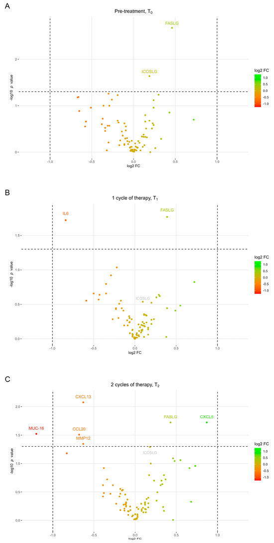
Immunotherapy has altered the therapeutic landscape for patients with non-small-cell lung cancer (NSCLC). The immune checkpoint inhibitor pembrolizumab targets the PD-1/PD-L1 signaling axis and produces durable clinical responses, but reliable biomarkers are lacking. Using 115 plasma samples from 42 pembrolizumab-treated patients with NSCLC, we were able to identify predictive biomarkers. In the plasma samples, we quantified the level of 92 proteins using the Olink proximity extension assay and circulating tumor DNA (ctDNA) using targeted next-generation sequencing. Patients with an above-median progression-free survival (PFS) had significantly higher expressions of Fas ligand (FASLG) and inducible T-cell co-stimulator ligand (ICOSLG) at baseline than patients with a PFS below the median. A Kaplan–Meier analysis demonstrated that high levels of FASLG and ICOSLG were predictive of longer PFS and overall survival (OS) (PFS: 10.83 vs. 4.49 months, OS: 27.13 vs. 18.0 months). Furthermore, we identified a subgroup with high expressions of FASLG and ICOSLG who also had no detectable ctDNA mutations after treatment initiation. This subgroup had significantly longer PFS and OS rates compared to the rest of the patients (PFS: 25.71 vs. 4.52 months, OS: 34.62 vs. 18.0 months). These findings suggest that the expressions of FASLG and ICOSLG at baseline and the absence of ctDNA mutations after the start of treatment have the potential to predict clinical outcomes. Full article
Show Figures

Figure 1

13 pages, 3144 KiB  
Article
Absolute Quantification of Pan-Cancer Plasma Proteomes Reveals Unique Signature in Multiple Myeloma
by David Kotol, Jakob Woessmann, Andreas Hober, María Bueno Álvez, Khue Hua Tran Minh, Fredrik Pontén, Linn Fagerberg, Mathias Uhlén and Fredrik Edfors
Cancers 2023, 15(19), 4764; https://doi.org/10.3390/cancers15194764 - 28 Sep 2023
Cited by 1 | Viewed by 1756
Abstract
Mass spectrometry based on data-independent acquisition (DIA) has developed into a powerful quantitative tool with a variety of implications, including precision medicine. Combined with stable isotope recombinant protein standards, this strategy provides confident protein identification and precise quantification on an absolute scale. Here, [...] Read more.
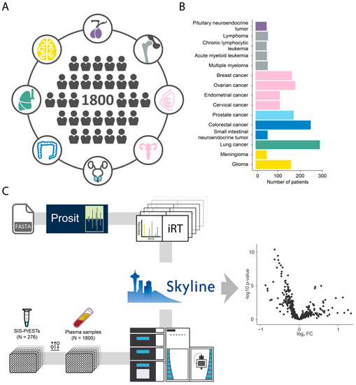
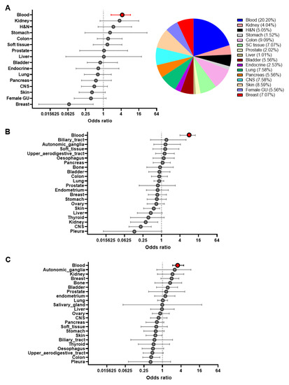
Mass spectrometry based on data-independent acquisition (DIA) has developed into a powerful quantitative tool with a variety of implications, including precision medicine. Combined with stable isotope recombinant protein standards, this strategy provides confident protein identification and precise quantification on an absolute scale. Here, we describe a comprehensive targeted proteomics approach to profile a pan-cancer cohort consisting of 1800 blood plasma samples representing 15 different cancer types. We successfully performed an absolute quantification of 253 proteins in multiplex. The assay had low intra-assay variability with a coefficient of variation below 20% (CV = 17.2%) for a total of 1013 peptides quantified across almost two thousand injections. This study identified a potential biomarker panel of seven protein targets for the diagnosis of multiple myeloma patients using differential expression analysis and machine learning. The combination of markers, including the complement C1 complex, JCHAIN, and CD5L, resulted in a prediction model with an AUC of 0.96 for the identification of multiple myeloma patients across various cancer patients. All these proteins are known to interact with immunoglobulins. Full article
Show Figures

Figure 1

17 pages, 1303 KiB  
Review
The Current Landscape of Glioblastoma Biomarkers in Body Fluids
by Saba Zanganeh, Elham Abbasgholinejad, Mohammad Doroudian, Nazanin Esmaelizad, Fatemeh Farjadian and Soumya Rahima Benhabbour
Cancers 2023, 15(15), 3804; https://doi.org/10.3390/cancers15153804 - 26 Jul 2023
Cited by 11 | Viewed by 2874
Abstract
Glioblastoma (GBM) is a highly aggressive and lethal primary brain cancer that necessitates early detection and accurate diagnosis for effective treatment and improved patient outcomes. Traditional diagnostic methods, such as imaging techniques and tissue biopsies, have limitations in providing real-time information and distinguishing [...] Read more.
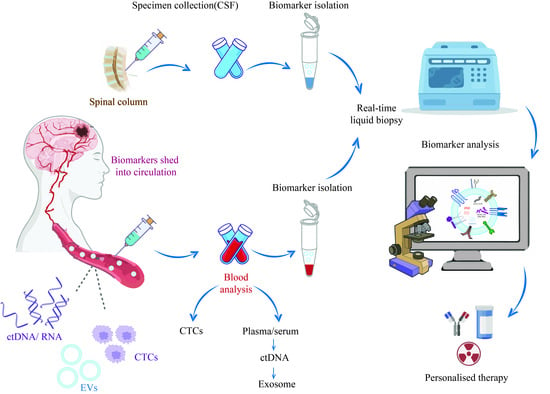
Glioblastoma (GBM) is a highly aggressive and lethal primary brain cancer that necessitates early detection and accurate diagnosis for effective treatment and improved patient outcomes. Traditional diagnostic methods, such as imaging techniques and tissue biopsies, have limitations in providing real-time information and distinguishing treatment-related changes from tumor progression. Liquid biopsies, used to analyze biomarkers in body fluids, offer a non-invasive and dynamic approach to detecting and monitoring GBM. This article provides an overview of GBM biomarkers in body fluids, including circulating tumor cells (CTCs), cell-free DNA (cfDNA), cell-free RNA (cfRNA), microRNA (miRNA), and extracellular vesicles. It explores the clinical utility of these biomarkers for GBM detection, monitoring, and prognosis. Challenges and limitations in implementing liquid biopsy strategies in clinical practice are also discussed. The article highlights the potential of liquid biopsies as valuable tools for personalized GBM management but underscores the need for standardized protocols and further research to optimize their clinical utility. Full article
Show Figures

Graphical abstract

21 pages, 396 KiB  
Review
The Importance of Detecting, Quantifying, and Characterizing Exosomes as a New Diagnostic/Prognostic Approach for Tumor Patients
by Mariantonia Logozzi, Nicola Salvatore Orefice, Rossella Di Raimo, Davide Mizzoni and Stefano Fais
Cancers 2023, 15(11), 2878; https://doi.org/10.3390/cancers15112878 - 23 May 2023
Cited by 9 | Viewed by 2592
Abstract
Exosomes are extracellular vesicles (EVs) of nanometric size studied for their role in tumor pathogenesis and progression and as a new source of tumor biomarkers. The clinical studies have provided encouraging but probably unexpected results, including the exosome plasmatic levels’ clinical relevance and [...] Read more.
Exosomes are extracellular vesicles (EVs) of nanometric size studied for their role in tumor pathogenesis and progression and as a new source of tumor biomarkers. The clinical studies have provided encouraging but probably unexpected results, including the exosome plasmatic levels’ clinical relevance and well-known biomarkers’ overexpression on the circulating EVs. The technical approach to obtaining EVs includes methods to physically purify EVs and characterize EVs, such as Nanosight Tracking Analysis (NTA), immunocapture-based ELISA, and nano-scale flow cytometry. Based on the above approaches, some clinical investigations have been performed on patients with different tumors, providing exciting and promising results. Here we emphasize data showing that exosome plasmatic levels are consistently higher in tumor patients than in controls and that plasmatic exosomes express well-known tumor markers (e.g., PSA and CEA), proteins with enzymatic activity, and nucleic acids. However, we also know that tumor microenvironment acidity is a key factor in influencing both the amount and the characteristics of the exosome released by tumor cells. In fact, acidity significantly increases exosome release by tumor cells, which correlates with the number of exosomes that circulate through the body of a tumor patient. Full article
16 pages, 1339 KiB  
Article
Cell-Free RNA from Plasma in Patients with Neuroblastoma: Exploring the Technical and Clinical Potential
by Nathalie S. M. Lak, Anne Seijger, Lieke M. J. van Zogchel, Nina U. Gelineau, Ahmad Javadi, Lily Zappeij-Kannegieter, Laura Bongiovanni, Anneloes Andriessen, Janine Stutterheim, C. Ellen van der Schoot, Alain de Bruin and Godelieve A. M. Tytgat
Cancers 2023, 15(7), 2108; https://doi.org/10.3390/cancers15072108 - 31 Mar 2023
Cited by 5 | Viewed by 2944
Abstract
Neuroblastoma affects mostly young children, bearing a high morbidity and mortality. Liquid biopsies, e.g., molecular analysis of circulating tumor-derived nucleic acids in blood, offer a minimally invasive diagnostic modality. Cell-free RNA (cfRNA) is released by all cells, especially cancer. It circulates in blood [...] Read more.
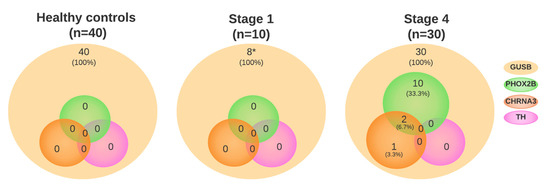
Neuroblastoma affects mostly young children, bearing a high morbidity and mortality. Liquid biopsies, e.g., molecular analysis of circulating tumor-derived nucleic acids in blood, offer a minimally invasive diagnostic modality. Cell-free RNA (cfRNA) is released by all cells, especially cancer. It circulates in blood packed in extracellular vesicles (EV) or attached to proteins. We studied the feasibility of analyzing cfRNA and EV, isolated by size exclusion chromatography (SEC), from platelet-poor plasma from healthy controls (n = 40) and neuroblastoma patients with localized (n = 10) and metastatic disease (n = 30). The mRNA content was determined using several multiplex droplet digital PCR (ddPCR) assays for a neuroblastoma-specific gene panel (PHOX2B, TH, CHRNA3) and a cell cycle regulation panel (E2F1, CDC6, ATAD2, H2AFZ, MCM2, DHFR). We applied corrections for the presence of platelets. We demonstrated that neuroblastoma-specific markers were present in plasma from 14/30 patients with metastatic disease and not in healthy controls and patients with localized disease. Most cell cycle markers had a higher expression in patients. The mRNA markers were mostly present in the EV-enriched SEC fractions. In conclusion, cfRNA can be isolated from plasma and EV and analyzed using multiplex ddPCR. cfRNA is an interesting novel liquid biopsy-based target to explore further. Full article
Show Figures

Figure 1

19 pages, 3196 KiB  
Article
Integrative Analyses Reveal the Anticancer Mechanisms and Sensitivity Markers of the Next-Generation Hypomethylating Agent NTX-301
by Byungho Lim, Dabin Yoo, Younghwa Chun, Areum Go, Ji Yeon Kim, Ha Young Lee, Rebecca J. Boohaker, Kyung-Jin Cho, Sunjoo Ahn, Jin Soo Lee, DooYoung Jung and Gildon Choi
Cancers 2023, 15(6), 1737; https://doi.org/10.3390/cancers15061737 - 13 Mar 2023
Cited by 2 | Viewed by 1986
Abstract
Epigenetic dysregulation characterized by aberrant DNA hypermethylation is a hallmark of cancer, and it can be targeted by hypomethylating agents (HMAs). Recently, we described the superior therapeutic efficacy of a novel HMA, namely, NTX-301, when used as a monotherapy and in combination with [...] Read more.
Epigenetic dysregulation characterized by aberrant DNA hypermethylation is a hallmark of cancer, and it can be targeted by hypomethylating agents (HMAs). Recently, we described the superior therapeutic efficacy of a novel HMA, namely, NTX-301, when used as a monotherapy and in combination with venetoclax in the treatment of acute myeloid leukemia. Following a previous study, we further explored the therapeutic properties of NTX-301 based on experimental investigations and integrative data analyses. Comprehensive sensitivity profiling revealed that NTX-301 primarily exerted anticancer effects against blood cancers and exhibited improved potency against a wide range of solid cancers. Subsequent assays showed that the superior efficacy of NTX-301 depended on its strong effects on cell cycle arrest, apoptosis, and differentiation. Due to its superior efficacy, low doses of NTX-301 achieved sufficiently substantial tumor regression in vivo. Multiomics analyses revealed the mechanisms of action (MoAs) of NTX-301 and linked these MoAs to markers of sensitivity to NTX-301 and to the demethylation activity of NTX-301 with high concordance. In conclusion, our findings provide a rationale for currently ongoing clinical trials of NTX-301 and will help guide the development of novel therapeutic options for cancer patients. Full article
Show Figures

Figure 1

19 pages, 2203 KiB  
Article
Canine-Inspired Chemometric Analysis of Volatile Organic Compounds in Urine Headspace to Distinguish Prostate Cancer in Mice and Men
by Mark Woollam, Amanda P. Siegel, Adam Munshi, Shengzhi Liu, Sunil Tholpady, Thomas Gardner, Bai-Yan Li, Hiroki Yokota and Mangilal Agarwal
Cancers 2023, 15(4), 1352; https://doi.org/10.3390/cancers15041352 - 20 Feb 2023
Cited by 10 | Viewed by 2644
Abstract
Canines can identify prostate cancer with high accuracy by smelling volatile organic compounds (VOCs) in urine. Previous studies have identified VOC biomarkers for prostate cancer utilizing solid phase microextraction (SPME) gas chromatography–mass spectrometry (GC-MS) but have not assessed the ability of VOCs to [...] Read more.
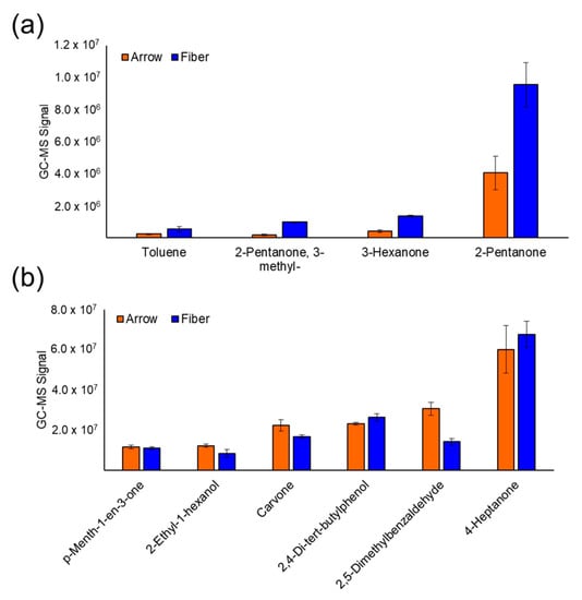
Canines can identify prostate cancer with high accuracy by smelling volatile organic compounds (VOCs) in urine. Previous studies have identified VOC biomarkers for prostate cancer utilizing solid phase microextraction (SPME) gas chromatography–mass spectrometry (GC-MS) but have not assessed the ability of VOCs to distinguish aggressive cancers. Additionally, previous investigations have utilized murine models to identify biomarkers but have not determined if the results are translatable to humans. To address these challenges, urine was collected from mice with prostate cancer and men undergoing prostate cancer biopsy and VOCs were analyzed by SPME GC-MS. Prior to analysis, SPME fibers/arrows were compared, and the fibers had enhanced sensitivity toward VOCs with a low molecular weight. The analysis of mouse urine demonstrated that VOCs could distinguish tumor-bearing mice with 100% accuracy. Linear discriminant analysis of six VOCs in human urine distinguished prostate cancer with sensitivity = 75% and specificity = 69%. Another panel of seven VOCs could classify aggressive cancer with sensitivity = 78% and specificity = 85%. These results show that VOCs have moderate accuracy in detecting prostate cancer and a superior ability to stratify aggressive tumors. Furthermore, the overlap in the structure of VOCs identified in humans and mice shows the merit of murine models for identifying biomarker candidates. Full article
Show Figures

Figure 1

18 pages, 1249 KiB  
Article
Protein Signatures and Individual Circulating Proteins, including IL-6 and IL-15, Associated with Prognosis in Patients with Biliary Tract Cancer
by Troels D. Christensen, Kasper Madsen, Emil Maag, Ole Larsen, Lars Henrik Jensen, Carsten P. Hansen, Alice Markussen, Dan T. S. Høgdall, Inna M. Chen, Dorte Nielsen and Julia S. Johansen
Cancers 2023, 15(4), 1062; https://doi.org/10.3390/cancers15041062 - 7 Feb 2023
Cited by 5 | Viewed by 2572
Abstract
Biliary tract cancer (BTC) is a rare gastrointestinal cancer with a dismal prognosis. Biomarkers with clinical utility are needed. In this study, we investigated the association between survival and 89 immuno-oncology-related proteins, with the aim of identifying prognostic biomarkers for BTC. The study [...] Read more.
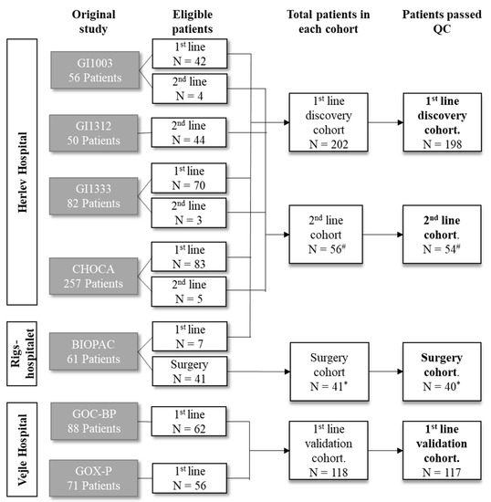
Biliary tract cancer (BTC) is a rare gastrointestinal cancer with a dismal prognosis. Biomarkers with clinical utility are needed. In this study, we investigated the association between survival and 89 immuno-oncology-related proteins, with the aim of identifying prognostic biomarkers for BTC. The study included patients with BTC (n = 394) treated at three Danish hospitals. Patients were divided into four cohorts: the first-line discovery cohort (n = 202), first-line validation cohort (n = 118), second-line cohort (n = 56), and surgery cohort (n = 41). Plasma protein levels were measured using a proximity extension assay (Olink Proteomics). Twenty-seven proteins were associated with overall survival (OS) in a multivariate analysis in the discovery cohort. In the first-line validation cohort, high levels of interleukin (IL)-6, IL-15, mucin 16, hepatocyte growth factor, programmed cell death ligand 1, and placental growth factor were significantly associated with poor OS in univariate Cox regression analyses. When adjusting for performance status, location, and stage, the association was significant only for IL-6 (hazard ratio (HR) = 1.25, 95% confidence interval (CI) 1.08–1.46) and IL-15 (HR = 2.23, 95% CI 1.48–3.35). Receiver operating characteristic analyses confirmed IL-6 and IL-15 as the strongest predictors of survival. Combining several proteins into signatures further improved the ability to distinguish between patients with short (<6 months) and long survival (>18 months). The study identified several circulating proteins as prognostic biomarkers in patients, with BTC, IL-6, and IL-15 being the most promising markers. Combining proteins in a prognostic signature improved prognostic performance, but future studies are needed to determine the optimal combination and thresholds. Full article
Show Figures

Figure 1

2022

Jump to: 2024, 2023

16 pages, 1153 KiB  
Article
Evolution of RAS Mutations in Cell-Free DNA of Patients with Tissue RAS Wild-Type Metastatic Colorectal Cancer Receiving First-Line Treatment: The PERSEIDA Study
by Manuel Valladares-Ayerbes, Pilar Garcia-Alfonso, Jorge Muñoz Luengo, Paola Patricia Pimentel Caceres, Oscar Alfredo Castillo Trujillo, Rosario Vidal-Tocino, Marta Llanos, Beatriz Llorente Ayala, Maria Luisa Limon Miron, Antonieta Salud, Luis Cirera Nogueras, Rocio Garcia-Carbonero, Maria Jose Safont, Esther Falco Ferrer, Jorge Aparicio, Maria Angeles Vicente Conesa, Carmen Guillén-Ponce, Paula Garcia-Teijido, Maria Begoña Medina Magan, Isabel Busquier, Mercedes Salgado and Ariadna Lloansí Vilaadd Show full author list remove Hide full author list
Cancers 2022, 14(24), 6075; https://doi.org/10.3390/cancers14246075 - 9 Dec 2022
Cited by 4 | Viewed by 2335
Abstract
The serial analysis of cell-free DNA (cfDNA) enables minimally invasive monitoring of tumor evolution, providing continuous genetic information. PERSEIDA was an observational, prospective study assessing the cfDNA RAS (KRAS/NRAS) mutational status evolution in first-line, metastatic CRC, RAS wild-type (according [...] Read more.
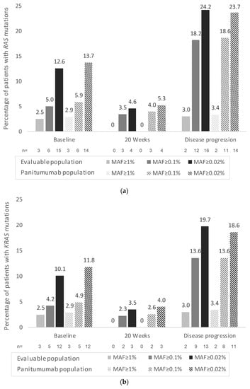
The serial analysis of cell-free DNA (cfDNA) enables minimally invasive monitoring of tumor evolution, providing continuous genetic information. PERSEIDA was an observational, prospective study assessing the cfDNA RAS (KRAS/NRAS) mutational status evolution in first-line, metastatic CRC, RAS wild-type (according to baseline tumor tissue biopsy) patients. Plasma samples were collected before first-line treatment, after 20 ± 2 weeks, and at disease progression. One hundred and nineteen patients were included (102 received panitumumab and chemotherapy as first-line treatment—panitumumab subpopulation). Fifteen (12.6%) patients presented baseline cfDNA RAS mutations (n = 14 [13.7%], panitumumab subpopulation) (mutant allele fraction ≥0.02 for all results). No patients presented emergent mutations (cfDNA RAS mutations not present at baseline) at 20 weeks. At disease progression, 11 patients (n = 9; panitumumab subpopulation) presented emergent mutations (RAS conversion rate: 19.0% [11/58]; 17.7% [9/51], panitumumab subpopulation). In contrast, three (5.2%) patients presenting baseline cfDNA RAS mutations were RAS wild-type at disease progression. No significant associations were observed between overall response rate or progression-free survival and cfDNA RAS mutational status in the total panitumumab subpopulation. Although, in patients with left-sided tumors, a significantly longer progression-free survival was observed in cfDNA RAS wild-type patients compared to those presenting cfDNA RAS mutations at any time. Continuous evaluation of RAS mutations may provide valuable insights on tumor molecular dynamics that can help clinical practice. Full article
Show Figures

Figure 1

36 pages, 3054 KiB  
Review
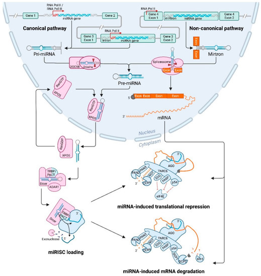
The microRNA Lifecycle in Health and Cancer
by Laura Adriana de Rooij, Dirk Jan Mastebroek, Nicky ten Voorde, Elsken van der Wall, Paul Joannes van Diest and Cathy Beatrice Moelans
Cancers 2022, 14(23), 5748; https://doi.org/10.3390/cancers14235748 - 23 Nov 2022
Cited by 17 | Viewed by 3850
Abstract
MicroRNAs (miRNAs) are small non-coding RNAs of ~22 nucleotides that regulate gene expression at the post-transcriptional level. They can bind to around 60% of all protein-coding genes with an average of 200 targets per miRNA, indicating their important function within physiological and pathological [...] Read more.
MicroRNAs (miRNAs) are small non-coding RNAs of ~22 nucleotides that regulate gene expression at the post-transcriptional level. They can bind to around 60% of all protein-coding genes with an average of 200 targets per miRNA, indicating their important function within physiological and pathological cellular processes. miRNAs can be quickly produced in high amounts through canonical and non-canonical pathways that involve a multitude of steps and proteins. In cancer, miRNA biogenesis, availability and regulation of target expression can be altered to promote tumour progression. This can be due to genetic causes, such as single nucleotide polymorphisms, epigenetic changes, differences in host gene expression, or chromosomal remodelling. Alternatively, post-transcriptional changes in miRNA stability, and defective or absent components and mediators of the miRNA-induced silencing complex can lead to altered miRNA function. This review provides an overview of the current knowledge on the lifecycle of miRNAs in health and cancer. Understanding miRNA function and regulation is fundamental prior to potential future application of miRNAs as cancer biomarkers. Full article
Show Figures

Figure 1

11 pages, 288 KiB  
Review
UroVysionTM Fluorescence In Situ Hybridization in Urological Cancers: A Narrative Review and Future Perspectives
by Chunjin Ke, Zhiquan Hu and Chunguang Yang
Cancers 2022, 14(21), 5423; https://doi.org/10.3390/cancers14215423 - 3 Nov 2022
Cited by 5 | Viewed by 2077
Abstract
UroVysionTM is a fluorescence in situ hybridization assay that was developed for the detection of bladder cancer (UC accounted for 90%) in urine specimens. It consists of fluorescently labeled DNA probes to the pericentromeric regions of chromosomes 3, 7, 17 and to [...] Read more.
UroVysionTM is a fluorescence in situ hybridization assay that was developed for the detection of bladder cancer (UC accounted for 90%) in urine specimens. It consists of fluorescently labeled DNA probes to the pericentromeric regions of chromosomes 3, 7, 17 and to the 9p21 band location of the P16 tumor suppressor gene, which was approved by the Food and Drug Administration (FDA) in 2001 and 2005, respectively, for urine detection in patients with suspected bladder cancer and postoperative recurrence monitoring. Furthermore, recent studies also demonstrated that U-FISH was useful for assessing superficial bladder cancer patients’ response to Bacillus Calmette–Guérin therapy and in detecting upper tract urothelial carcinoma. Therefore, positive U-FISH was well known to urologists as a molecular cytogenetic technique for the detection of UC. However, with the continuous enrichment of clinical studies at home and abroad, U-FISH has shown a broader application space in the detection of various urinary primary tumors and even metastatic tumors. This review focuses on summarizing the research status of U-FISH in UC, non-urothelial carcinoma and metastatic tumor, so as to strengthen urologists’ more comprehensive understanding of the application value of U-FISH and better complete the accurate diagnosis and treatment of urological cancers. Full article
22 pages, 1051 KiB  
Review
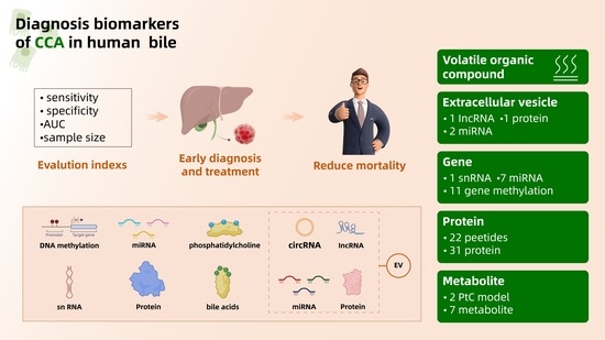
Diagnosis Biomarkers of Cholangiocarcinoma in Human Bile: An Evidence-Based Study
by Fang Bao, Jiayue Liu, Haiyang Chen, Lu Miao, Zhaochao Xu and Guixin Zhang
Cancers 2022, 14(16), 3921; https://doi.org/10.3390/cancers14163921 - 13 Aug 2022
Cited by 8 | Viewed by 3350
Abstract
Cholangiocarcinoma (CCA) is a multifactorial malignant tumor of the biliary tract, and the incidence of CCA is increasing in recent years. At present, the diagnosis of CCA mainly depends on imaging and invasive examination, with limited specificity and sensitivity and late detection. The [...] Read more.
Cholangiocarcinoma (CCA) is a multifactorial malignant tumor of the biliary tract, and the incidence of CCA is increasing in recent years. At present, the diagnosis of CCA mainly depends on imaging and invasive examination, with limited specificity and sensitivity and late detection. The early diagnosis of CCA always faces the dilemma of lacking specific diagnostic biomarkers. Non-invasive methods to assess the degree of CAA have been developed throughout the last decades. Among the many specimens looking for CCA biomarkers, bile has gotten a lot of attention lately. This paper mainly summarizes the recent developments in the current research on the diagnostic biomarkers for CCA in human bile at the levels of the gene, protein, metabolite, extracellular vesicles and volatile organic compounds. Full article
Show Figures

Graphical abstract

14 pages, 1362 KiB  
Article
Monitoring of Measurable Residual Disease Using Circulating DNA after Allogeneic Hematopoietic Cell Transplantation
by Miguel Waterhouse, Sandra Pennisi, Dietmar Pfeifer, Florian Scherer, Robert Zeiser, Justus Duyster, Hartmut Bertz, Jürgen Finke and Jesús Duque-Afonso
Cancers 2022, 14(14), 3307; https://doi.org/10.3390/cancers14143307 - 7 Jul 2022
Cited by 2 | Viewed by 2107
Abstract
Relapse of the underlying disease is a frequent complication after allogeneic hematopoietic stem cell transplantation (allo-HSCT). In this study, we describe the clinical utility of measurable residual disease (MRD) and mixed chimerism (MC) assessment in circulating cell-free DNA (cfDNA) analysis to detect earlier [...] Read more.
Relapse of the underlying disease is a frequent complication after allogeneic hematopoietic stem cell transplantation (allo-HSCT). In this study, we describe the clinical utility of measurable residual disease (MRD) and mixed chimerism (MC) assessment in circulating cell-free DNA (cfDNA) analysis to detect earlier relapse in patients with hematological malignancies after allo-HSCT. A total of 326 plasma and peripheral blood mononuclear cell (PBMCs) samples obtained from 62 patients with myeloid malignancies were analyzed by droplet-digital PCR (median follow-up: 827 days). Comparison of MC in patients at relapse and in complete remission identified an optimal discriminating threshold of 18% of recipient-derived cfDNA. After performing a targeted next-generation sequencing (NGS) panel, 136 mutations in 58 patients were detected. In a total of 119 paired samples, the putative mutations were detected in both cfDNA and PBMCs in 73 samples (61.3%). In 45 samples (37.8%) they were detected only in cfDNA, and in only one patient (0.9%) were they detected solely in DNA from PBMCs. Hence, in 6 out of 23 patients (26%) with relapse after allo-HSCT, MRD positivity was detected earlier in cfDNA (mean 397 days) than in DNA derived from PBMCs (mean 451 days). In summary, monitoring of MRD and MC in cfDNA might be useful for earlier relapse detection in patients with myeloid malignancies after allo-HSCT. Full article
Show Figures

Figure 1

12 pages, 984 KiB  
Article
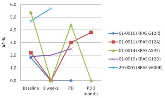
Dynamics of RAS/BRAF Mutations in cfDNA from Metastatic Colorectal Carcinoma Patients Treated with Polychemotherapy and Anti-EGFR Monoclonal Antibodies
by Anna Maria Rachiglio, Laura Forgione, Raffaella Pasquale, Carlo Antonio Barone, Evaristo Maiello, Lorenzo Antonuzzo, Antonino Cassata, Giuseppe Tonini, Roberto Bordonaro, Gerardo Rosati, Alberto Zaniboni, Sara Lonardi, Daris Ferrari, Giovanni Luca Frassineti, Stefano Tamberi, Salvatore Pisconti, Francesca Di Fabio, Cristin Roma, Armando Orlandi, Tiziana Latiano, Angela Damato, Giampaolo Tortora, Carmine Pinto and Nicola Normannoadd Show full author list remove Hide full author list
Cancers 2022, 14(4), 1052; https://doi.org/10.3390/cancers14041052 - 18 Feb 2022
Cited by 4 | Viewed by 2573
Abstract
Analysis of plasma-derived cell-free DNA (cfDNA) might allow for the early identification of resistance in metastatic colorectal carcinoma (mCRC) patients receiving anti-EGFR monoclonal antibodies. We tested plasma samples from the Erbitux Metastatic Colorectal Cancer Strategy (ERMES) phase III trial of FOLFIRI+Cetuximab in first-line [...] Read more.
Analysis of plasma-derived cell-free DNA (cfDNA) might allow for the early identification of resistance in metastatic colorectal carcinoma (mCRC) patients receiving anti-EGFR monoclonal antibodies. We tested plasma samples from the Erbitux Metastatic Colorectal Cancer Strategy (ERMES) phase III trial of FOLFIRI+Cetuximab in first-line treatment of RAS/BRAF wild-type mCRC. Samples were collected at baseline (n = 37), at 8 weeks of treatment (n = 32), progressive disease (PD; n = 36) and 3 months after PD (n = 21). cfDNA testing was performed using the Idylla™ ctKRAS and ctNRAS-BRAF tests and the Oncomine Pan-Cancer Cell-Free Assay. Analysis of basal samples revealed RAS/BRAF mutations in 6/37 cases. A transient RAS positivity not associated with PD was observed at 8 weeks in five cases that showed no mutations at baseline and PD. The frequency of mutant cases increased at PD (33.3%) and decreased again at 3 months after PD (9.5%). The median progression-free survival (mPFS) of patients RAS/BRAF mutant at PD was 7.13 months versus 7.71 months in wild-type patients (p = 0.3892). These data confirm that the occurrence of RAS/BRAF mutations in mCRC patients receiving anti-EGFR agents is relatively frequent. However, the cfDNA dynamics of RAS mutations in patients treated with anti-EGFR agents plus polychemotherapy are complex and might not be directly associated with resistance to treatment. Full article
Show Figures

Figure 1

15 pages, 1776 KiB  
Article
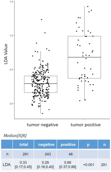
The Role of New Technologies in the Diagnosis and Surveillance of Non-Muscle Invasive Bladder Carcinoma: A Prospective, Double-Blinded, Monocentric Study of the XPERT© Bladder Cancer Monitor and Narrow Band Imaging© Cystoscopy
by Gad Singer, Venkat M. Ramakrishnan, Uwe Rogel, Andreas Schötzau, Daniel Disteldorf, Philipp Maletzki, Jean-Pascal Adank, Marc Hofmann, Tilo Niemann, Sylvia Stadlmann, Antonio Nocito, Kurt Lehmann and Lukas J. Hefermehl
Cancers 2022, 14(3), 618; https://doi.org/10.3390/cancers14030618 - 26 Jan 2022
Cited by 10 | Viewed by 3404
Abstract
Follow-up is essential for the early detection of recurrent non-muscle invasive bladder cancers (NMIBC). This study investigates the clinical relevance of new diagnostic tools such as an mRNA-based urine test (XPERT© Bladder Cancer Monitor, XBCM) and Narrow Band Imaging© (NBI) and compares them [...] Read more.
Follow-up is essential for the early detection of recurrent non-muscle invasive bladder cancers (NMIBC). This study investigates the clinical relevance of new diagnostic tools such as an mRNA-based urine test (XPERT© Bladder Cancer Monitor, XBCM) and Narrow Band Imaging© (NBI) and compares them with the established follow-up diagnostics (white-light cystoscopy (WLC) and urine cytology). This was a prospective, double-blind, single-center study that involved patients undergoing NMIBC screening at a tertiary care center. Enrollment occurred between January 2018 and March 2020. In addition to standard care (WLC, cytology, and ultrasound), patients underwent XBCM urine testing and NBI cystoscopy. In total, 301 WLCs were performed; through this, 49 patients demonstrated NMIBC recurrence. NBI cystoscopy was congruent with WLC in all patients. Cytology showed a sensitivity (SE) and specificity (SP) of 27% and 97% (PPV: 65%; NPV 87%), respectively, whereas XBCM showed SE and SP of 58% and 89%, respectively (PPV: 51%; NPV: 92%; AUC: 0.79 (0.716–0.871)). Subgroup analysis showed improved SE and similar SP (PPV, NPV) for high grade (HG) recurrence, with a SE of 74% and SP of 89% (39%, 97%). NBI cystoscopy does not necessarily provide additional benefit over standard WLC. However, the XBCM may provide better SE and a diagnostic advantage in instances of HG disease recurrence. Full article
Show Figures

Figure 1

Back to TopTop