Next Issue
Volume 13, January-1
Previous Issue
Volume 12, December-1


Cells, Volume 12, Issue 24 (December-2 2023) – 92 articles

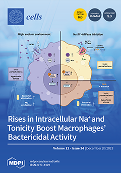
Cover Story (view full-size image): Inflamed and infected tissues can display increased local sodium (Na+) levels. In macrophages, high salt (HS) triggers osmoprotective signaling and proinflammatory activation. Pharmacological inhibition of Na+/K+-ATPase with cardiac glycosides (CGs) raised intracellular Na+ without increasing extracellular Na+ levels. HS and CGs displayed divergent effects on the osmoprotective signaling and antimicrobial activity of macrophages. While HS causes hypertonic stress and ionic perturbations, CGs only induce the latter. Cotreatment with a CG and the non-ionic osmolyte mannitol (MAN) partially mimicked HS. We suggest that intracellular Na+ accumulation and hypertonic stress are required but not sufficient to mimic the HS-boosted macrophage function. (Graph was created with Biorender.com). View this paper
  • Issues are regarded as officially published after their release is announced to the table of contents alert mailing list.
  • You may sign up for e-mail alerts to receive table of contents of newly released issues.
  • PDF is the official format for papers published in both, html and pdf forms. To view the papers in pdf format, click on the "PDF Full-text" link, and use the free Adobe Reader to open them.
Order results
Result details
Section
Select all
Export citation of selected articles as:
16 pages, 13881 KiB  
Article
Single-Cell Transcriptomics and In Vitro Lineage Tracing Reveals Differential Susceptibility of Human iPSC-Derived Midbrain Dopaminergic Neurons in a Cellular Model of Parkinson’s Disease
by Lucia F. Cardo, Jimena Monzón-Sandoval, Zongze Li, Caleb Webber and Meng Li
Cells 2023, 12(24), 2860; https://doi.org/10.3390/cells12242860 - 18 Dec 2023
Cited by 4 | Viewed by 2755
Abstract
Advances in stem cell technologies open up new avenues for modelling development and diseases. The success of these pursuits, however, relies on the use of cells most relevant to those targeted by the disease of interest, for example, midbrain dopaminergic neurons for Parkinson’s [...] Read more.
Advances in stem cell technologies open up new avenues for modelling development and diseases. The success of these pursuits, however, relies on the use of cells most relevant to those targeted by the disease of interest, for example, midbrain dopaminergic neurons for Parkinson’s disease. In the present study, we report the generation of a human induced pluripotent stem cell (iPSC) line capable of purifying and tracing nascent midbrain dopaminergic progenitors and their differentiated progeny via the expression of a Blue Fluorescent Protein (BFP). This was achieved by CRISPR/Cas9-assisted knock-in of BFP and Cre into the safe harbour locus AAVS1 and an early midbrain dopaminergic lineage marker gene LMX1A, respectively. Immunocytochemical analysis and single-cell RNA sequencing of iPSC-derived neural cultures confirm developmental recapitulation of the human fetal midbrain and high-quality midbrain cells. By modelling Parkinson’s disease-related drug toxicity using 1-Methyl-4-phenylpyridinium (MPP+), we showed a preferential reduction of BFP+ cells, a finding demonstrated independently by cell death assays and single-cell transcriptomic analysis of MPP+ treated neural cultures. Together, these results highlight the importance of disease-relevant cell types in stem cell modelling. Full article
Show Figures

Figure 1

16 pages, 3904 KiB  
Article
Repurposing Metabolic Inhibitors in the Treatment of Colon Adenocarcinoma Patient-Derived Models
by Bora Lee, ChuHee Lee, Hae-Min Moon, Se-Young Jo, Se Jin Jang and Young-Ah Suh
Cells 2023, 12(24), 2859; https://doi.org/10.3390/cells12242859 - 18 Dec 2023
Cited by 2 | Viewed by 2092
Abstract
The effect of agonists on AMP-activated protein kinase (AMPK), mainly metformin and phenformin, has been appreciated in the treatment of multiple types of tumors. Specifically, the antitumor activity of phenformin has been demonstrated in melanomas containing the v-Raf murine sarcoma viral oncogene homolog [...] Read more.
The effect of agonists on AMP-activated protein kinase (AMPK), mainly metformin and phenformin, has been appreciated in the treatment of multiple types of tumors. Specifically, the antitumor activity of phenformin has been demonstrated in melanomas containing the v-Raf murine sarcoma viral oncogene homolog B1 (BRAF) activating mutation. In this report, we elucidated the synergistic antitumor effects of biguanides with metabolism inhibitors on colon tumors. Phenformin with 2-deoxy-D-glucose (2DG) inhibited tumor cell growth in cancer cell lines, including HT29 cells harboring BRAF- and p53-mutations. Biochemical analyses showed that two chemotherapeutics exerted cooperative effects to reduce tumor growth through cell cycle arrest, apoptosis, and autophagy. The drugs demonstrated activity against phosphorylated ERK and the gain-of-function p53 mutant protein. To demonstrate tumor regressive effects in vivo, we established patient-derived models, including xenograft (PDX) and organoids (PDO). Co-treatment of biguanides with chemotherapeutics efficiently reduced the growth of patient-derived colon models in comparison to treatment with a single agent. These results strongly suggest that significant therapeutic advantages would be achieved by combining AMPK activators such as phenformin and cancer metabolic inhibitors such as 2DG. Full article
Show Figures

Figure 1

12 pages, 1486 KiB  
Article
Amplification of Signal on Cell Surfaces in Molecular Cascades
by Sergei Rudchenko, Steven Taylor, Nenad Milosavic, Maria Rudchenko, Betina Wedderhoff Tissi, Markus Y. Mapara and Milan N. Stojanovic
Cells 2023, 12(24), 2858; https://doi.org/10.3390/cells12242858 - 18 Dec 2023
Cited by 1 | Viewed by 1507
Abstract
We can formulate mixtures of oligonucleotide–antibody conjugates to act as molecular cascade-based automata that analyze pairs of cell surface markers (CD markers) on individual cells in a manner consistent with the implementation of Boolean logic—for example, by producing a fluorescent label only if [...] Read more.
We can formulate mixtures of oligonucleotide–antibody conjugates to act as molecular cascade-based automata that analyze pairs of cell surface markers (CD markers) on individual cells in a manner consistent with the implementation of Boolean logic—for example, by producing a fluorescent label only if two markers are present. While traditional methods to characterize cells are based on transducing signals from individual cell surface markers, these cascades can be used to combine into a single signal the presence of two or even more CDs. In our original design, oligonucleotide components irreversibly flowed from one antibody to another, driven by increased hybridizations, leading to the magnitude of the final signal on each cell being determined by the surface marker that was the least abundant. This is a significant limitation to the precise labeling of narrow subpopulations, and, in order to overcome it, we changed our design to accomplish signal amplification to a more abundant cell surface marker. We show the AMPLIFY function on two examples: (1) we amplify the fluorescent label from the CD19 marker onto a fivefold more abundant CD45, and (2) we amplify broadly distributed CD45RA to a more constant marker, CD3. We expect this new function to enable the increasingly complex Boolean analysis of cell surfaces. Full article
Show Figures

Figure 1

28 pages, 390 KiB  
Correction
Correction: Balaphas et al. Cell Therapy for Anal Sphincter Incontinence: Where Do We Stand? Cells 2021, 10, 2086
by Alexandre Balaphas, Jeremy Meyer, Raphael P. H. Meier, Emilie Liot, Nicolas C. Buchs, Bruno Roche, Christian Toso, Leo H. Bühler, Carmen Gonelle-Gispert and Frédéric Ris
Cells 2023, 12(24), 2857; https://doi.org/10.3390/cells12242857 - 18 Dec 2023
Viewed by 1170
Abstract
The authors would like to add a new reference to the section “3 [...] Full article
(This article belongs to the Collection Stem Cells in Tissue Engineering and Regeneration)
20 pages, 7224 KiB  
Article
Targeting Cx43 to Reduce the Severity of Pressure Ulcer Progression
by Milton Sheng Yi Kwek, Moogaambikai Thangaveloo, Leigh E. Madden, Anthony R. J. Phillips and David L. Becker
Cells 2023, 12(24), 2856; https://doi.org/10.3390/cells12242856 - 18 Dec 2023
Cited by 2 | Viewed by 1925
Abstract
In the skin, repeated incidents of ischemia followed by reperfusion can result in the breakdown of the skin and the formation of a pressure ulcer. Here we gently applied paired magnets to the backs of mice to cause ischemia for 1.5 h and [...] Read more.
In the skin, repeated incidents of ischemia followed by reperfusion can result in the breakdown of the skin and the formation of a pressure ulcer. Here we gently applied paired magnets to the backs of mice to cause ischemia for 1.5 h and then removed them to allow reperfusion. The sterile inflammatory response generated within 4 h causes a stage 1 pressure ulcer with an elevation of the gap junction protein Cx43 in the epidermis. If this process is repeated the insult will result in a more severe stage 2 pressure ulcer with a breakdown of the epidermis 2–3 days later. After a single pinch, the elevation of Cx43 in the epidermis is associated with the inflammatory response with an increased number of neutrophils, HMGB1 (marker of necrosis) and RIP3 (responsible for necroptosis). Delivering Cx43 specific antisense oligonucleotides sub-dermally after a single insult, was able to significantly reduce the elevation of epidermal Cx43 protein expression and reduce the number of neutrophils and prevent the elevation of HMGB1 and RIP3. In a double pinch model, the Cx43 antisense treatment was able to reduce the level of inflammation, necroptosis, and the extent of tissue damage and progression to an open wound. This approach may be useful in reducing the progression of stage 1 pressure ulcers to stage 2. Full article
Show Figures

Figure 1

27 pages, 569 KiB  
Review
Autologous Stem Cell Transplant in Hodgkin’s and Non-Hodgkin’s Lymphoma, Multiple Myeloma, and AL Amyloidosis
by Sulaiman Mohammed Alnasser, Khalid Saad Alharbi, Ali F. Almutairy, Sulaiman Mohammed Almutairi and Abdulmalik Mohammed Alolayan
Cells 2023, 12(24), 2855; https://doi.org/10.3390/cells12242855 - 18 Dec 2023
Cited by 1 | Viewed by 3061
Abstract
Human body cells are stem cell (SC) derivatives originating from bone marrow. Their special characteristics include their capacity to support the formation and self-repair of the cells. Cancer cells multiply uncontrollably and invade healthy tissues, making stem cell transplants a viable option for [...] Read more.
Human body cells are stem cell (SC) derivatives originating from bone marrow. Their special characteristics include their capacity to support the formation and self-repair of the cells. Cancer cells multiply uncontrollably and invade healthy tissues, making stem cell transplants a viable option for cancer patients undergoing high-dose chemotherapy (HDC). When chemotherapy is used at very high doses to eradicate all cancer cells from aggressive tumors, blood-forming cells and leukocytes are either completely or partially destroyed. Autologous stem cell transplantation (ASCT) is necessary for patients in those circumstances. The patients who undergo autologous transplants receive their own stem cells (SCs). The transplanted stem cells first come into contact with the bone marrow and then undergo engraftment, before differentiating into blood cells. ASCT is one of the most significant and innovative strategies for treating diseases. Here we focus on the treatment of Hodgkin’s lymphoma, non-Hodgkin’s lymphoma, multiple myeloma, and AL amyloidosis, using ASCT. This review provides a comprehensive picture of the effectiveness and the safety of ASCT as a therapeutic approach for these diseases, based on the currently available evidence. Full article
Show Figures

Figure 1

19 pages, 1094 KiB  
Review
Natural Killer T (NKT) Cells in Autoimmune Hepatitis: Current Evidence from Basic and Clinical Research.
by Dimitri Poddighe, Tilektes Maulenkul, Gulsamal Zhubanova, Lyudmila Akhmaldtinova and Kuanysh Dossybayeva
Cells 2023, 12(24), 2854; https://doi.org/10.3390/cells12242854 - 18 Dec 2023
Cited by 2 | Viewed by 2370
Abstract
Natural killer T (NKT) cells are unconventional T cells that are activated by glycolipid antigens. They can produce a variety of inflammatory and regulatory cytokines and, therefore, modulate multiple aspects of the immune response in different pathological settings, including autoimmunity. NKT cells have [...] Read more.
Natural killer T (NKT) cells are unconventional T cells that are activated by glycolipid antigens. They can produce a variety of inflammatory and regulatory cytokines and, therefore, modulate multiple aspects of the immune response in different pathological settings, including autoimmunity. NKT cells have also been implicated in the immunopathogenesis of autoimmune hepatitis, and in this review we summarize and analyze the main studies investigating the involvement and/or homeostasis of NKT cells in this disease. In detail, the evidence from both basic and clinical research has been specifically analyzed. Even though the experimental murine models supported a relevant role of NKT cells in immune-mediated hepatic injury, very few studies specifically investigated NKT cell homeostasis in patients with autoimmune hepatitis; however, these initial studies reported some alterations of NKT cells in these patients, which may also correlate with the disease activity to some extent. Further clinical studies are needed to investigate the potential role and use of NKT cell analysis as a disease marker of clinical relevance, and to better understand the precise cellular and molecular mechanisms by which NKT cells contribute to the pathogenesis of autoimmune hepatitis. Full article
(This article belongs to the Section Cellular Immunology)
Show Figures

Figure 1

19 pages, 4902 KiB  
Article
MiR-630 Promotes Radioresistance by Induction of Anti-Apoptotic Effect via Nrf2–GPX2 Molecular Axis in Head–Neck Cancer
by Guo-Rung You, Ann-Joy Cheng, Eric Yi-Liang Shen, Kang-Hsing Fan, Yi-Fang Huang, Yu-Chen Huang, Kai-Ping Chang and Joseph T. Chang
Cells 2023, 12(24), 2853; https://doi.org/10.3390/cells12242853 - 17 Dec 2023
Cited by 2 | Viewed by 1907
Abstract
Head and neck cancer (HNC) ranks among the top ten prevalent cancers worldwide. Radiotherapy stands as a pivotal treatment component for HNC; however, radioresistance in cancerous cells often leads to local recurrence, becoming a substantial factor in treatment failure. MicroRNAs (miRNAs) are compact, [...] Read more.
Head and neck cancer (HNC) ranks among the top ten prevalent cancers worldwide. Radiotherapy stands as a pivotal treatment component for HNC; however, radioresistance in cancerous cells often leads to local recurrence, becoming a substantial factor in treatment failure. MicroRNAs (miRNAs) are compact, non-coding RNAs that regulate gene expression by targeting mRNAs to inhibit protein translation. Although several studies have indicated that the dysregulation of miRNAs is intricately linked with malignant transformation, understanding this molecular family’s role in radioresistance remains limited. This study determined the role of miR-630 in regulating radiosensitivity in HNC. We discovered that miR-630 functions as an oncomiR, marked by its overexpression in HNC patients, correlating with a poorer prognosis. We further delineated the malignant function of miR-630 in HNC cells. While it had a minimal impact on cell growth, the miR-630 contributed to radioresistance in HNC cells. This result was supported by decreased cellular apoptosis and caspase enzyme activities. Moreover, miR-630 overexpression mitigated irradiation-induced DNA damage, evidenced by the reduced levels of the γ-H2AX histone protein, a marker for double-strand DNA breaks. Mechanistically, the overexpression of miR-630 decreased the cellular ROS levels and initiated Nrf2 transcriptional activity, resulting in the upregulation of the antioxidant enzyme GPX2. Thus, this study elucidates that miR-630 augments radioresistance by inducing an anti-apoptotic effect via the Nrf2–GPX2 molecular axis in HNC. The modulation of miR-630 may serve as a novel radiosensitizing target for HNC. Full article
Show Figures

Figure 1

15 pages, 5072 KiB  
Article
Enhanced Osteocyte Differentiation: Cathepsin D and L Secretion by Human Adipose-Derived Mesenchymal Stem Cells
by Jung-Won Choi, Soyeon Lim, Seung Eun Jung, Seongtae Jeong, Hanbyeol Moon, Byeong-Wook Song, Il-Kwon Kim, Seahyoung Lee, Ki-Chul Hwang and Sang Woo Kim
Cells 2023, 12(24), 2852; https://doi.org/10.3390/cells12242852 - 17 Dec 2023
Cited by 1 | Viewed by 1821
Abstract
Adipose-derived mesenchymal stem cells (ASCs) have the potential to differentiate into bone, cartilage, fat, and neural cells and promote tissue regeneration and healing. It is known that they can have variable responses to hypoxic conditions. In the present study, we aimed to explore [...] Read more.
Adipose-derived mesenchymal stem cells (ASCs) have the potential to differentiate into bone, cartilage, fat, and neural cells and promote tissue regeneration and healing. It is known that they can have variable responses to hypoxic conditions. In the present study, we aimed to explore diverse changes in the cells and secretome of ASCs under a hypoxic environment over time and to present the possibility of ASCs as therapeutic agents from a different perspective. The expression differences of proteins between normoxic and hypoxic conditions (6, 12, or 24 h) were specifically investigated in human ASCs using 2-DE combined with MALDI-TOF MS analysis, and secreted proteins in ASC-derived conditioned media (ASC-derived CM) were examined by an adipokine array. In addition, genetic and/or proteomic interactions were assessed using a DAVID and miRNet functional annotation bioinformatics analysis. We found that 64 and 5 proteins were differentially expressed in hypoxic ASCs and in hypoxic ASC-derived CM, respectively. Moreover, 7 proteins among the 64 markedly changed spots in hypoxic ASCs were associated with bone-related diseases. We found that two proteins, cathepsin D (CTSD) and cathepsin L (CTSL), identified through an adipokine array independently exhibited significant efficacy in promoting osteocyte differentiation in bone-marrow-derived mesenchymal stem cells (BM-MSCs). This finding introduces a promising avenue for utilizing hypoxia-preconditioned ASC-derived CM as a potential therapeutic approach for bone-related diseases. Full article
Show Figures

Figure 1

25 pages, 6258 KiB  
Article
HIF-1α/MMP-9 Axis Is Required in the Early Phases of Skeletal Myoblast Differentiation under Normoxia Condition In Vitro
by Flaminia Chellini, Alessia Tani, Martina Parigi, Francesco Palmieri, Rachele Garella, Sandra Zecchi-Orlandini, Roberta Squecco and Chiara Sassoli
Cells 2023, 12(24), 2851; https://doi.org/10.3390/cells12242851 - 16 Dec 2023
Cited by 2 | Viewed by 1671
Abstract
Hypoxia-inducible factor (HIF)-1α represents an oxygen-sensitive subunit of HIF transcriptional factor, which is usually degraded in normoxia and stabilized in hypoxia to regulate several target gene expressions. Nevertheless, in the skeletal muscle satellite stem cells (SCs), an oxygen level-independent regulation of HIF-1α has [...] Read more.
Hypoxia-inducible factor (HIF)-1α represents an oxygen-sensitive subunit of HIF transcriptional factor, which is usually degraded in normoxia and stabilized in hypoxia to regulate several target gene expressions. Nevertheless, in the skeletal muscle satellite stem cells (SCs), an oxygen level-independent regulation of HIF-1α has been observed. Although HIF-1α has been highlighted as a SC function regulator, its spatio-temporal expression and role during myogenic progression remain controversial. Herein, using biomolecular, biochemical, morphological and electrophysiological analyses, we analyzed HIF-1α expression, localization and role in differentiating murine C2C12 myoblasts and SCs under normoxia. In addition, we evaluated the role of matrix metalloproteinase (MMP)-9 as an HIF-1α effector, considering that MMP-9 is involved in myogenesis and is an HIF-1α target in different cell types. HIF-1α expression increased after 24/48 h of differentiating culture and tended to decline after 72 h/5 days. Committed and proliferating mononuclear myoblasts exhibited nuclear HIF-1α expression. Differently, the more differentiated elongated and parallel-aligned cells, which are likely ready to fuse with each other, show a mainly cytoplasmic localization of the factor. Multinucleated myotubes displayed both nuclear and cytoplasmic HIF-1α expression. The MMP-9 and MyoD (myogenic activation marker) expression synchronized with that of HIF-1α, increasing after 24 h of differentiation. By means of silencing HIF-1α and MMP-9 by short-interfering RNA and MMP-9 pharmacological inhibition, this study unraveled MMP-9’s role as an HIF-1α downstream effector and the fact that the HIF-1α/MMP-9 axis is essential in morpho-functional cell myogenic commitment. Full article
(This article belongs to the Special Issue Regulatory Programs of Skeletal Muscle Repair and Regeneration)
Show Figures

Graphical abstract

16 pages, 2783 KiB  
Article
Infrapatellar Fat Pad Modulates Osteoarthritis-Associated Cytokine and MMP Expression in Human Articular Chondrocytes
by Ewa Wisniewska, Dominik Laue, Jacob Spinnen, Leonard Kuhrt, Benjamin Kohl, Patricia Bußmann, Carola Meier, Gundula Schulze-Tanzil, Wolfgang Ertel and Michal Jagielski
Cells 2023, 12(24), 2850; https://doi.org/10.3390/cells12242850 - 15 Dec 2023
Cited by 3 | Viewed by 1611
Abstract
Osteoarthritis (OA) most frequently affects the knee joint and is associated with an elevated expression of cytokines and extracellular cartilage matrix (ECM), degrading enzymes such as matrix metalloproteinases (MMPs). Differences in gene expression of the intra-articularly located infrapatellar fat pad (IPFP) and other [...] Read more.
Osteoarthritis (OA) most frequently affects the knee joint and is associated with an elevated expression of cytokines and extracellular cartilage matrix (ECM), degrading enzymes such as matrix metalloproteinases (MMPs). Differences in gene expression of the intra-articularly located infrapatellar fat pad (IPFP) and other fatty tissue suggest its autonomous function, yet its role in OA pathogenesis remains unknown. Human IPFPs and articular cartilage were collected from OA patients undergoing total knee arthroplasty, and biopsies from the IPFP of healthy patients harvested during knee arthroscopy served as controls (CO). Isolated chondrocytes were co-cultured with either osteoarthritic (OA) or CO-IPFPs in a transwell system. Chondrocyte expression of MMP1, -3, -13, type 1 and 2 collagens, interleukin IL1β, IL6, IL10, and tumor necrosis factor TNFα was analyzed by RTD-PCR at day 0 and day 2, and TNFα secretion was analyzed by ELISA. The cytokine release in IPFPs was assessed by an array. Results: Both IPFPs (CO, OA) significantly reduced the expression of type 2 collagen and TNFα in chondrocytes. On the other hand, only CO-IPFP suppressed the expression of type 1 collagen and significantly induced the MMP13 expression. On the contrary, IL1β and IL6 were significantly induced when exposed to OA-IPFP. Conclusions: The partial loss of the suppressive effect on type 1 collagen gene expression found for OA-IPFP shows the pathological remodeling and dedifferentiation potential of the OA-IPFP on the chondrocytes. However, the significant suppression of TNFα implies that the OA- and CO-IPFP could also exhibit a protective role in the knee joint, preventing the progress of inflammation. Full article
(This article belongs to the Special Issue Cell and Molecular Causes of Joint Inflammation and Damage)
Show Figures

Graphical abstract

23 pages, 3189 KiB  
Article
Coagulation Factor Xa Has No Effects on the Expression of PAR1, PAR2, and PAR4 and No Proinflammatory Effects on HL-1 Cells
by Lukas Ruf, Alicja Bukowska, Andreas Gardemann and Andreas Goette
Cells 2023, 12(24), 2849; https://doi.org/10.3390/cells12242849 - 15 Dec 2023
Cited by 3 | Viewed by 1555
Abstract
Atrial fibrillation (AF), characterised by irregular high-frequency contractions of the atria of the heart, is of increasing clinical importance. The reasons are the increasing prevalence and thromboembolic complications caused by AF. So-called atrial remodelling is characterised, among other things, by atrial dilatation and [...] Read more.
Atrial fibrillation (AF), characterised by irregular high-frequency contractions of the atria of the heart, is of increasing clinical importance. The reasons are the increasing prevalence and thromboembolic complications caused by AF. So-called atrial remodelling is characterised, among other things, by atrial dilatation and fibrotic remodelling. As a result, AF is self-sustaining and forms a procoagulant state. But hypercoagulation not only appears to be the consequence of AF. Coagulation factors can exert influence on cells via protease-activated receptors (PAR) and thereby the procoagulation state could contribute to the development and maintenance of AF. In this work, the influence of FXa on Heart Like-1 (HL-1) cells, which are murine adult atrial cardiomyocytes (immortalized), was investigated. PAR1, PAR2, and PAR4 expression was detected. After incubations with FXa (5–50 nM; 4–24 h) or PAR1- and PAR2-agonists (20 µM; 4–24 h), no changes occurred in PAR expression or in the inflammatory signalling cascade. There were no time- or concentration-dependent changes in the phosphorylation of the MAP kinases ERK1/2 or the p65 subunit of NF-κB. In addition, there was no change in the mRNA expression of the cell adhesion molecules (ICAM-1, VCAM-1, fibronectin). Thus, FXa has no direct PAR-dependent effects on HL-1 cells. Future studies should investigate the influence of FXa on human cardiomyocytes or on other cardiac cell types like fibroblasts. Full article
(This article belongs to the Special Issue Molecular Biology of Atrial Myocardium)
Show Figures

Figure 1

24 pages, 4763 KiB  
Article
Evaluation of Mono- and Bi-Functional GLOBE-Based Vectors for Therapy of β-Thalassemia by HBBAS3 Gene Addition and Mutation-Specific RNA Interference
by Lola Koniali, Christina Flouri, Markela I. Kostopoulou, Nikoletta Y. Papaioannou, Panayiota L. Papasavva, Basma Naiisseh, Coralea Stephanou, Anthi Demetriadou, Maria Sitarou, Soteroula Christou, Michael N. Antoniou, Marina Kleanthous, Petros Patsali and Carsten W. Lederer
Cells 2023, 12(24), 2848; https://doi.org/10.3390/cells12242848 - 15 Dec 2023
Cited by 1 | Viewed by 1935
Abstract
Therapy via the gene addition of the anti-sickling βAS3-globin transgene is potentially curative for all β-hemoglobinopathies and therefore of particular clinical and commercial interest. This study investigates GLOBE-based lentiviral vectors (LVs) for βAS3-globin addition and evaluates strategies for an [...] Read more.
Therapy via the gene addition of the anti-sickling βAS3-globin transgene is potentially curative for all β-hemoglobinopathies and therefore of particular clinical and commercial interest. This study investigates GLOBE-based lentiviral vectors (LVs) for βAS3-globin addition and evaluates strategies for an increased β-like globin expression without vector dose escalation. First, we report the development of a GLOBE-derived LV, GLV2-βAS3, which, compared to its parental vector, adds anti-sickling action and a transcription-enhancing 848-bp transcription terminator element, retains high vector titers and allows for superior β-like globin expression in primary patient-derived hematopoietic stem and progenitor cells (HSPCs). Second, prompted by our previous correction of HBBIVSI−110(G>A) thalassemia based on RNApol(III)-driven shRNAs in mono- and combination therapy, we analyzed a series of novel LVs for the RNApol(II)-driven constitutive or late-erythroid expression of HBBIVSI−110(G>A)-specific miRNA30-embedded shRNAs (shRNAmiR). This included bifunctional LVs, allowing for concurrent βAS3-globin expression. LVs were initially compared for their ability to achieve high β-like globin expression in HBBIVSI−110(G>A)-transgenic cells, before the evaluation of shortlisted candidate LVs in HBBIVSI−110(G>A)-homozygous HSPCs. The latter revealed that β-globin promoter-driven designs for monotherapy with HBBIVSI−110(G>A)-specific shRNAmiRs only marginally increased β-globin levels compared to untransduced cells, whereas bifunctional LVs combining miR30-shRNA with βAS3-globin expression showed disease correction similar to that achieved by the parental GLV2-βAS3 vector. Our results establish the feasibility of high titers for LVs containing the full HBB transcription terminator, emphasize the importance of the HBB terminator for the high-level expression of HBB-like transgenes, qualify the therapeutic utility of late-erythroid HBBIVSI−110(G>A)-specific miR30-shRNA expression and highlight the exceptional potential of GLV2-βAS3 for the treatment of severe β-hemoglobinopathies. Full article
(This article belongs to the Special Issue Gene and Cell Therapy in Regenerative Medicine—Second Edition)
Show Figures

Figure 1

20 pages, 4467 KiB  
Article
Modeling RET-Rearranged Non-Small Cell Lung Cancer (NSCLC): Generation of Lung Progenitor Cells (LPCs) from Patient-Derived Induced Pluripotent Stem Cells (iPSCs)
by Paul Marcoux, Jin Wook Hwang, Christophe Desterke, Jusuf Imeri, Annelise Bennaceur-Griscelli and Ali G. Turhan
Cells 2023, 12(24), 2847; https://doi.org/10.3390/cells12242847 - 15 Dec 2023
Viewed by 1554
Abstract
REarranged during Transfection (RET) oncogenic rearrangements can occur in 1–2% of lung adenocarcinomas. While RET-driven NSCLC models have been developed using various approaches, no model based on patient-derived induced pluripotent stem cells (iPSCs) has yet been described. Patient-derived iPSCs hold great promise for [...] Read more.
REarranged during Transfection (RET) oncogenic rearrangements can occur in 1–2% of lung adenocarcinomas. While RET-driven NSCLC models have been developed using various approaches, no model based on patient-derived induced pluripotent stem cells (iPSCs) has yet been described. Patient-derived iPSCs hold great promise for disease modeling and drug screening. However, generating iPSCs with specific oncogenic drivers, like RET rearrangements, presents challenges due to reprogramming efficiency and genotypic variability within tumors. To address this issue, we aimed to generate lung progenitor cells (LPCs) from patient-derived iPSCs carrying the mutation RETC634Y, commonly associated with medullary thyroid carcinoma. Additionally, we established a RETC634Y knock-in iPSC model to validate the effect of this oncogenic mutation during LPC differentiation. We successfully generated LPCs from RETC634Y iPSCs using a 16-day protocol and detected an overexpression of cancer-associated markers as compared to control iPSCs. Transcriptomic analysis revealed a distinct signature of NSCLC tumor repression, suggesting a lung multilineage lung dedifferentiation, along with an upregulated signature associated with RETC634Y mutation, potentially linked to poor NSCLC prognosis. These findings were validated using the RETC634Y knock-in iPSC model, highlighting key cancerous targets such as PROM2 and C1QTNF6, known to be associated with poor prognostic outcomes. Furthermore, the LPCs derived from RETC634Y iPSCs exhibited a positive response to the RET inhibitor pralsetinib, evidenced by the downregulation of the cancer markers. This study provides a novel patient-derived off-the-shelf iPSC model of RET-driven NSCLC, paving the way for exploring the molecular mechanisms involved in RET-driven NSCLC to study disease progression and to uncover potential therapeutic targets. Full article
(This article belongs to the Section Stem Cells)
Show Figures

Figure 1

17 pages, 3373 KiB  
Article
Epithelial Barrier Dysfunction in Diarrhea-Predominant Irritable Bowel Syndrome (IBS-D) via Downregulation of Claudin-1
by Karem Awad, Christian Barmeyer, Christian Bojarski, Oliver Nagel, In-Fah M. Lee, Michal R. Schweiger, Jörg-Dieter Schulzke and Roland Bücker
Cells 2023, 12(24), 2846; https://doi.org/10.3390/cells12242846 - 15 Dec 2023
Cited by 3 | Viewed by 3082
Abstract
Background: In patients with diarrhea-predominant irritable bowel syndrome (IBS-D), the diarrheal mechanisms are largely unknown, and they were examined in this study on colon biopsies. Methods: Electrophysiological measurements were used for monitoring functional changes in the diarrheic colon specimens. In parallel, tight junction [...] Read more.
Background: In patients with diarrhea-predominant irritable bowel syndrome (IBS-D), the diarrheal mechanisms are largely unknown, and they were examined in this study on colon biopsies. Methods: Electrophysiological measurements were used for monitoring functional changes in the diarrheic colon specimens. In parallel, tight junction protein expression was analyzed by Western blot and confocal laser-scanning microscopy, and signaling pathway analysis was performed using RNA sequencing and bioinformatics. Results: Epithelial resistance was decreased, indicating an epithelial leak flux diarrheal mechanism with a molecular correlate of decreased claudin-1 expression, while induction of active anion secretion and impairment of active sodium absorption via the epithelial sodium channel, ENaC, were not detected. The pathway analysis revealed activation of barrier-affecting cytokines TNF-α, IFN-γ, IL-1β and IL-4. Conclusions: Barrier dysfunction as a result of epithelial tight junction changes plays a role in IBS-D as a pathomechanism inducing a leak flux type of diarrhea. Full article
(This article belongs to the Special Issue Structure and Function of Tight Junctions)
Show Figures

Figure 1

17 pages, 8375 KiB  
Article
Resveratrol’s Impact on the Chondrogenic Reagents’ Effects in Cell Sheet Cultures of Wharton’s Jelly-Derived MSCs
by Anastasiia D. Kurenkova, Viktoria S. Presniakova, Zlata A. Mosina, Pavel D. Kibirskiy, Irina A. Romanova, Gilyana K. Tugaeva, Nastasia V. Kosheleva, Kirill S. Vinogradov, Sergei V. Kostjuk, Svetlana L. Kotova, Yury A. Rochev, Ekaterina V. Medvedeva and Peter S. Timashev
Cells 2023, 12(24), 2845; https://doi.org/10.3390/cells12242845 - 15 Dec 2023
Viewed by 1319
Abstract
Human Wharton’s jelly mesenchymal stem cells (hWJ-MSCs) are of great interest in tissue engineering. We obtained hWJ-MSCs from four patients, and then we stimulated their chondrogenic phenotype formation in vitro by adding resveratrol (during cell expansion) and a canonical Wnt pathway activator, LiCl, [...] Read more.
Human Wharton’s jelly mesenchymal stem cells (hWJ-MSCs) are of great interest in tissue engineering. We obtained hWJ-MSCs from four patients, and then we stimulated their chondrogenic phenotype formation in vitro by adding resveratrol (during cell expansion) and a canonical Wnt pathway activator, LiCl, as well as a Rho-associated protein kinase inhibitor, Y27632 (during differentiation). The effects of the added reagents on the formation of hWJ-MSC sheets destined to repair osteochondral injuries were investigated. Three-dimensional hWJ-MSC sheets grown on P(NIPAM-co-NtBA)-based matrices were characterized in vitro and in vivo. The combination of resveratrol and LiCl showed effects on hWJ-MSC sheets similar to those of the basal chondrogenic medium. Adding Y27632 decreased both the proportion of hypertrophied cells and the expression of the hyaline cartilage markers. In vitro, DMSO was observed to impede the effects of the chondrogenic factors. The mouse knee defect model experiment revealed that hWJ-MSC sheets grown with the addition of resveratrol and Y27632 were well integrated with the surrounding tissues; however, after 3 months, the restored tissue was identical to that of the naturally healed cartilage injury. Thus, the combination of chondrogenic supplements may not always have additive effects on the progress of cell culture and could be neutralized by the microenvironment after transplantation. Full article
(This article belongs to the Special Issue 3D Stem Cell Culture—Series 2)
Show Figures

Graphical abstract

19 pages, 1726 KiB  
Article
Trans-Regulation of Alternative PD-L1 mRNA Processing by CDK12 in Non-Small-Cell Lung Cancer Cells
by Trine V. Larsen, Christoffer T. Maansson, Tina F. Daugaard, Brage S. Andresen, Boe S. Sorensen and Anders L. Nielsen
Cells 2023, 12(24), 2844; https://doi.org/10.3390/cells12242844 - 15 Dec 2023
Viewed by 1798
Abstract
Immunotherapy using checkpoint inhibitors targeting the interaction between PD-1 on T cells and PD-L1 on cancer cells has shown significant results in non-small-cell lung cancer (NSCLC). Not all patients respond to the therapy, and PD-L1 expression heterogeneity is proposed to be one determinant [...] Read more.
Immunotherapy using checkpoint inhibitors targeting the interaction between PD-1 on T cells and PD-L1 on cancer cells has shown significant results in non-small-cell lung cancer (NSCLC). Not all patients respond to the therapy, and PD-L1 expression heterogeneity is proposed to be one determinant for this. The alternative processing of PD-L1 RNA, which depends on an alternative poly-A site in intron 4, generates a shorter mRNA variant (PD-L1v4) encoding soluble PD-L1 (sPD-L1), relative to the canonical PD-L1v1 mRNA encoding membrane-associated PD-L1 (mPD-L1). This study aimed to identify factors influencing the ratio between these two PD-L1 mRNAs in NSCLC cells. First, we verified the existence of the alternative PD-L1 RNA processing in NSCLC cells, and from in silico analyses, we identified a candidate list of regulatory factors. Examining selected candidates showed that CRISPR/Cas9-generated loss-of-function mutations in CDK12 increased the PD-L1v4/PD-L1v1 mRNA ratio and, accordingly, the sPD-L1/mPD-L1 balance. The CDK12/13 inhibitor THZ531 could also increase the PD-L1v4/PD-L1v1 mRNA ratio and impact the PD-L1 transcriptional response to IFN-γ stimulation. The fact that CDK12 regulates PD-L1 transcript variant formation in NSCLC cells is consistent with CDK12’s role in promoting transcriptional elongation over intron-located poly-A sites. This study lays the groundwork for clinical investigations to delineate the implications of the CDK12-mediated balancing of sPD-L1 relative to mPD-L1 for immunotherapeutic responses in NSCLC. Full article
(This article belongs to the Section Cellular Immunology)
Show Figures

Figure 1

4 pages, 218 KiB  
Editorial
Immunogenic Cell Stress and Death Sensitize Tumors to Immunotherapy
by Oliver Kepp and Guido Kroemer
Cells 2023, 12(24), 2843; https://doi.org/10.3390/cells12242843 - 15 Dec 2023
Cited by 4 | Viewed by 1484
Abstract
The efficacy of chemotherapy with cytotoxicants and that of targeted therapies with more sophisticated agents is limited due to the plasticity of malignant cells, which leads to the inevitable development of resistance [...] Full article
(This article belongs to the Special Issue Immunogenic Cell Stress and Death)
14 pages, 1030 KiB  
Review
Regulatory TR3-56 Cells in the Complex Panorama of Immune Activation and Regulation
by Flavia Carriero, Valentina Rubino, Stefania Leone, Rosangela Montanaro, Vincenzo Brancaleone, Giuseppina Ruggiero and Giuseppe Terrazzano
Cells 2023, 12(24), 2841; https://doi.org/10.3390/cells12242841 - 15 Dec 2023
Cited by 4 | Viewed by 1601
Abstract
The interplay between immune activation and immune regulation is a fundamental aspect of the functional harmony of the immune system. This delicate balance is essential to triggering correct and effective immune responses against pathogens while preventing excessive inflammation and the immunopathogenic mechanisms of [...] Read more.
The interplay between immune activation and immune regulation is a fundamental aspect of the functional harmony of the immune system. This delicate balance is essential to triggering correct and effective immune responses against pathogens while preventing excessive inflammation and the immunopathogenic mechanisms of autoimmunity. The knowledge of all the mechanisms involved in immune regulation is not yet definitive, and, probably, the overall picture is much broader than what has been described in the scientific literature so far. Given the plasticity of the immune system and the diversity of organisms, it is highly probable that numerous other cells and molecules are still to be ascribed to the immune regulation process. Here, we report a general overview of how immune activation and regulation interact, based on the involvement of molecules and cells specifically dedicated to these processes. In addition, we discuss the role of TR3-56 lymphocytes as a new cellular candidate in the immune regulation landscape. Full article
Show Figures

Figure 1

21 pages, 726 KiB  
Review
Signaling Pathways Involved in Manganese-Induced Neurotoxicity
by Hong Cheng, Beatriz Ferrer Villahoz, Romina Deza Ponzio, Michael Aschner and Pan Chen
Cells 2023, 12(24), 2842; https://doi.org/10.3390/cells12242842 - 14 Dec 2023
Cited by 5 | Viewed by 2310
Abstract
Manganese (Mn) is an essential trace element, but insufficient or excessive bodily amounts can induce neurotoxicity. Mn can directly increase neuronal insulin and activate insulin-like growth factor (IGF) receptors. As an important cofactor, Mn regulates signaling pathways involved in various enzymes. The IGF [...] Read more.
Manganese (Mn) is an essential trace element, but insufficient or excessive bodily amounts can induce neurotoxicity. Mn can directly increase neuronal insulin and activate insulin-like growth factor (IGF) receptors. As an important cofactor, Mn regulates signaling pathways involved in various enzymes. The IGF signaling pathway plays a protective role in the neurotoxicity of Mn, reducing apoptosis in neurons and motor deficits by regulating its downstream protein kinase B (Akt), mitogen-activated protein kinase (MAPK), and mammalian target of rapamycin (mTOR). In recent years, some new mechanisms related to neuroinflammation have been shown to also play an important role in Mn-induced neurotoxicity. For example, DNA-sensing receptor cyclic GMP–AMP synthase (cCAS) and its downstream signal efficient interferon gene stimulator (STING), NOD-like receptor family pyrin domain containing 3(NLRP3)-pro-caspase1, cleaves to the active form capase1 (CASP1), nuclear factor κB (NF-κB), sirtuin (SIRT), and Janus kinase (JAK) and signal transducers and activators of the transcription (STAT) signaling pathway. Moreover, autophagy, as an important downstream protein degradation pathway, determines the fate of neurons and is regulated by these upstream signals. Interestingly, the role of autophagy in Mn-induced neurotoxicity is bidirectional. This review summarizes the molecular signaling pathways of Mn-induced neurotoxicity, providing insight for further understanding of the mechanisms of Mn. Full article
(This article belongs to the Special Issue Molecular Signaling Pathways Associated with Neurotoxicity)
Show Figures

Figure 1

22 pages, 4759 KiB  
Article
Amyloid Beta Oligomers Activate Death Receptors and Mitochondria-Mediated Apoptotic Pathways in Cerebral Vascular Smooth Muscle Cells; Protective Effects of Carbonic Anhydrase Inhibitors
by Amy Anzovino, Elisa Canepa, Micaelly Alves, Nicole L. Lemon, Roxana O. Carare and Silvia Fossati
Cells 2023, 12(24), 2840; https://doi.org/10.3390/cells12242840 - 14 Dec 2023
Cited by 3 | Viewed by 1995
Abstract
Amyloid beta (Aβ) deposition within the brain vasculature is an early hallmark of Alzheimer’s disease (AD), which triggers loss of brain vascular smooth muscle cells (BVSMCs) in cerebral arteries, via poorly understood mechanisms, altering cerebral blood flow, brain waste clearance, and promoting cognitive [...] Read more.
Amyloid beta (Aβ) deposition within the brain vasculature is an early hallmark of Alzheimer’s disease (AD), which triggers loss of brain vascular smooth muscle cells (BVSMCs) in cerebral arteries, via poorly understood mechanisms, altering cerebral blood flow, brain waste clearance, and promoting cognitive impairment. We have previously shown that, in brain endothelial cells (ECs), vasculotropic Aβ species induce apoptosis through death receptors (DRs) DR4 and DR5 and mitochondria-mediated mechanisms, while FDA-approved carbonic anhydrase inhibitors (CAIs) prevent mitochondria-mediated EC apoptosis in vitro and in vivo. In this study, we analyzed Aβ-induced extrinsic and intrinsic (DR- and mitochondria-mediated) apoptotic pathways in BVSMC, aiming to unveil new therapeutic targets to prevent BVSMC stress and death. We show that both apoptotic pathways are activated in BVSMCs by oligomeric Aβ42 and Aβ40-Q22 (AβQ22) and mitochondrial respiration is severely impaired. Importantly, the CAIs methazolamide (MTZ) and acetazolamide (ATZ) prevent the pro-apoptotic effects in BVSMCs, while reducing caspase 3 activation and Aβ deposition in the arterial walls of TgSwDI animals, a murine model of cerebral amyloid angiopathy (CAA). This study reveals new molecular targets and a promising therapeutic strategy against BVSMC dysfunction in AD, CAA, and ARIA (amyloid-related imaging abnormalities) complications of recently FDA-approved anti-Aβ antibodies. Full article
(This article belongs to the Special Issue Mitochondria at the Crossroad of Health and Disease)
Show Figures

Figure 1

25 pages, 3813 KiB  
Article
The Sleep Quality- and Myopia-Linked PDE11A-Y727C Variant Impacts Neural Physiology by Reducing Catalytic Activity and Altering Subcellular Compartmentalization of the Enzyme
by Irina Sbornova, Emilie van der Sande, Snezana Milosavljevic, Elvis Amurrio, Steven D. Burbano, Prosun K. Das, Helen H. Do, Janet L. Fisher, Porschderek Kargbo, Janvi Patel, Latarsha Porcher, Chris I. De Zeeuw, Magda A. Meester-Smoor, Beerend H. J. Winkelman, Caroline C. W. Klaver, Ana Pocivavsek and Michy P. Kelly
Cells 2023, 12(24), 2839; https://doi.org/10.3390/cells12242839 - 14 Dec 2023
Cited by 1 | Viewed by 1725
Abstract
Recently, a Y727C variant in the dual-specific 3′,5′-cyclic nucleotide phosphodiesterase 11A (PDE11A-Y727C) was linked to increased sleep quality and reduced myopia risk in humans. Given the well-established role that the PDE11 substrates cAMP and cGMP play in eye physiology and sleep, we determined [...] Read more.
Recently, a Y727C variant in the dual-specific 3′,5′-cyclic nucleotide phosphodiesterase 11A (PDE11A-Y727C) was linked to increased sleep quality and reduced myopia risk in humans. Given the well-established role that the PDE11 substrates cAMP and cGMP play in eye physiology and sleep, we determined if (1) PDE11A protein is expressed in the retina or other eye segments in mice, (2) PDE11A-Y7272C affects catalytic activity and/or subcellular compartmentalization more so than the nearby suicide-associated PDE11A-M878V variant, and (3) Pde11a deletion alters eye growth or sleep quality in male and female mice. Western blots show distinct protein expression of PDE11A4, but not PDE11A1-3, in eyes of Pde11a WT, but not KO mice, that vary by eye segment and age. In HT22 and COS-1 cells, PDE11A4-Y727C reduces PDE11A4 catalytic activity far more than PDE11A4-M878V, with both variants reducing PDE11A4-cAMP more so than PDE11A4-cGMP activity. Despite this, Pde11a deletion does not alter age-related changes in retinal or lens thickness or axial length, nor vitreous or anterior chamber depth. Further, Pde11a deletion only minimally changes refractive error and sleep quality. That said, both variants also dramatically alter the subcellular compartmentalization of human and mouse PDE11A4, an effect occurring independently of dephosphorylating PDE11A4-S117/S124 or phosphorylating PDE11A4-S162. Rather, re-compartmentalization of PDE11A4-Y727C is due to the loss of the tyrosine changing how PDE11A4 is packaged/repackaged via the trans-Golgi network. Therefore, the protective impact of the Y727C variant may reflect a gain-of-function (e.g., PDE11A4 displacing another PDE) that warrants further investigation in the context of reversing/preventing sleep disturbances or myopia. Full article
Show Figures

Figure 1

26 pages, 8547 KiB  
Article
A Novel Approach: Investigating the Intracellular Clearance Mechanism of Glyceraldehyde-Derived Advanced Glycation End-Products Using the Artificial Checkpoint Kinase 1 d270KD Mutant as a Substrate Model
by Kenji Takeda, Akiko Sakai-Sakasai, Kouji Kajinami and Masayoshi Takeuchi
Cells 2023, 12(24), 2838; https://doi.org/10.3390/cells12242838 - 14 Dec 2023
Cited by 4 | Viewed by 1448
Abstract
Advanced glycation end-products (AGEs), formed through glyceraldehyde (GA) as an intermediate in non-enzymatic reactions with intracellular proteins, are cytotoxic and have been implicated in the pathogenesis of various diseases. Despite their significance, the mechanisms underlying the degradation of GA-derived AGEs (GA-AGEs) remain unclear. [...] Read more.
Advanced glycation end-products (AGEs), formed through glyceraldehyde (GA) as an intermediate in non-enzymatic reactions with intracellular proteins, are cytotoxic and have been implicated in the pathogenesis of various diseases. Despite their significance, the mechanisms underlying the degradation of GA-derived AGEs (GA-AGEs) remain unclear. In the present study, we found that N-terminal checkpoint kinase 1 cleavage products (CHK1-CPs) and their mimic protein, d270WT, were degraded intracellularly post-GA exposure. Notably, a kinase-dead d270WT variant (d270KD) underwent rapid GA-induced degradation, primarily via the ubiquitin–proteasome pathway. The high-molecular-weight complexes formed by the GA stimulation of d270KD were abundant in the RIPA-insoluble fraction, which also contained high levels of GA-AGEs. Immunoprecipitation experiments indicated that the high-molecular-weight complexes of d270KD were modified by GA-AGEs and that p62/SQSTM1 was one of its components. The knockdown of p62 or treatment with chloroquine reduced the amount of high-molecular-weight complexes in the RIPA-insoluble fraction, indicating its involvement in the formation of GA-AGE aggregates. The present results suggest that the ubiquitin–proteasome pathway and p62 play a role in the degradation and aggregation of intracellular GA-AGEs. This study provides novel insights into the mechanisms underlying GA-AGE metabolism and may lead to the development of novel therapeutic strategies for diseases associated with the accumulation of GA-AGEs. Full article
Show Figures

Graphical abstract

21 pages, 3047 KiB  
Review
Therapeutic Monoclonal Antibodies against Cancer: Present and Future
by Marisa Delgado and Jose A. Garcia-Sanz
Cells 2023, 12(24), 2837; https://doi.org/10.3390/cells12242837 - 14 Dec 2023
Cited by 9 | Viewed by 4038
Abstract
A series of monoclonal antibodies with therapeutic potential against cancer have been generated and developed. Ninety-one are currently used in the clinics, either alone or in combination with chemotherapeutic agents or other antibodies, including immune checkpoint antibodies. These advances helped to coin the [...] Read more.
A series of monoclonal antibodies with therapeutic potential against cancer have been generated and developed. Ninety-one are currently used in the clinics, either alone or in combination with chemotherapeutic agents or other antibodies, including immune checkpoint antibodies. These advances helped to coin the term personalized medicine or precision medicine. However, it seems evident that in addition to the current work on the analysis of mechanisms to overcome drug resistance, the use of different classes of antibodies (IgA, IgE, or IgM) instead of IgG, the engineering of the Ig molecules to increase their half-life, the acquisition of additional effector functions, or the advantages associated with the use of agonistic antibodies, to allow a broad prospective usage of precision medicine successfully, a strategy change is required. Here, we discuss our view on how these strategic changes should be implemented and consider their pros and cons using therapeutic antibodies against cancer as a model. The same strategy can be applied to therapeutic antibodies against other diseases, such as infectious or autoimmune diseases. Full article
Show Figures

Graphical abstract

21 pages, 3197 KiB  
Article
Lipophilic Statins Eliminate Senescent Endothelial Cells by inducing Anoikis-Related Cell Death
by Barbora Belakova, Nicholas K. Wedige, Ezzat M. Awad, Simon Hess, André Oszwald, Marlene Fellner, Shafaat Y. Khan, Ulrike Resch, Markus Lipovac, Karel Šmejkal, Pavel Uhrin and Johannes M. Breuss
Cells 2023, 12(24), 2836; https://doi.org/10.3390/cells12242836 - 14 Dec 2023
Cited by 4 | Viewed by 2548
Abstract
Pre-clinical studies from the recent past have indicated that senescent cells can negatively affect health and contribute to premature aging. Targeted eradication of these cells has been shown to improve the health of aged experimental animals, leading to a clinical interest in finding [...] Read more.
Pre-clinical studies from the recent past have indicated that senescent cells can negatively affect health and contribute to premature aging. Targeted eradication of these cells has been shown to improve the health of aged experimental animals, leading to a clinical interest in finding compounds that selectively eliminate senescent cells while sparing non-senescent ones. In our study, we identified a senolytic capacity of statins, which are lipid-lowering drugs prescribed to patients at high risk of cardiovascular events. Using two different models of senescence in human vascular endothelial cells (HUVECs), we found that statins preferentially eliminated senescent cells, while leaving non-senescent cells unharmed. We observed that the senolytic effect of statins could be negated with the co-administration of mevalonic acid and that statins induced cell detachment leading to anoikis-like apoptosis, as evidenced by real-time visualization of caspase-3/7 activation. Our findings suggest that statins possess a senolytic property, possibly also contributing to their described beneficial cardiovascular effects. Further studies are needed to explore the potential of short-term, high-dose statin treatment as a candidate senolytic therapy. Full article
(This article belongs to the Special Issue Senescence in the Cardiovascular System)
Show Figures

Graphical abstract

24 pages, 9357 KiB  
Article
The Effect of Visfatin on the Functioning of the Porcine Pituitary Gland: An In Vitro Study
by Karolina Szymanska, Edyta Rytelewska, Ewa Zaobidna, Marta Kiezun, Marlena Gudelska, Grzegorz Kopij, Kamil Dobrzyn, Ewa Mlyczynska, Patrycja Kurowska, Barbara Kaminska, Anna Nynca, Nina Smolinska, Agnieszka Rak and Tadeusz Kaminski
Cells 2023, 12(24), 2835; https://doi.org/10.3390/cells12242835 - 14 Dec 2023
Cited by 7 | Viewed by 1923 | Correction
Abstract
Visfatin (VIS), also known as nicotinamide phosphoribosyltransferase (NAMPT), is the rate-limiting enzyme in the biosynthesis of nicotinamide adenine dinucleotide (NAD+). Recently, VIS has been also recognized as an adipokine. Our previous study revealed that VIS is produced in the anterior and posterior lobes [...] Read more.
Visfatin (VIS), also known as nicotinamide phosphoribosyltransferase (NAMPT), is the rate-limiting enzyme in the biosynthesis of nicotinamide adenine dinucleotide (NAD+). Recently, VIS has been also recognized as an adipokine. Our previous study revealed that VIS is produced in the anterior and posterior lobes of the porcine pituitary. Moreover, the expression and secretion of VIS are dependent on the phase of the estrous cycle and/or the stage of early pregnancy. Based on this, we hypothesized that VIS may regulate porcine pituitary function. This study was conducted on anterior pituitary (AP) glands harvested from pigs during specific phases of the estrous cycle. We have shown the modulatory effect of VIS in vitro on LH and FSH secretion by porcine AP cells (determined by ELISA). VIS was also found to stimulate cell proliferation (determined by Alamar Blue) without affecting apoptosis in these cells (determined using flow cytometry technique). Moreover, it was indicated that VIS may act in porcine AP cells through the INSR, AKT/PI3K, MAPK/ERK1/2, and AMPK signaling pathways (determined by ELISA or Western Blot). This observation was further supported by the finding that simultaneous treatment of cells with VIS and inhibitors of these pathways abolished the observed VIS impact on LH and FSH secretion (determined by ELISA). In addition, our research indicated that VIS affected the mentioned processes in a manner that was dependent on the dose of VIS and/or the phase of the estrous cycle. Thus, these findings suggest that VIS may regulate the functioning of the porcine pituitary gland during the estrous cycle. Full article
Show Figures

Graphical abstract

16 pages, 2257 KiB  
Review
Protein Kinase CK2α’, More than a Backup of CK2α
by Mathias Montenarh and Claudia Götz
Cells 2023, 12(24), 2834; https://doi.org/10.3390/cells12242834 - 14 Dec 2023
Cited by 2 | Viewed by 1340
Abstract
The serine/threonine protein kinase CK2 is implicated in the regulation of fundamental processes in eukaryotic cells. CK2 consists of two catalytic α or α’ isoforms and two regulatory CK2β subunits. These three proteins exist in a free form, bound to other cellular proteins, [...] Read more.
The serine/threonine protein kinase CK2 is implicated in the regulation of fundamental processes in eukaryotic cells. CK2 consists of two catalytic α or α’ isoforms and two regulatory CK2β subunits. These three proteins exist in a free form, bound to other cellular proteins, as tetrameric holoenzymes composed of CK2α22, CK2αα’/β2, or CK2α’22 as well as in higher molecular forms of the tetramers. The catalytic domains of CK2α and CK2α’ share a 90% identity. As CK2α contains a unique C-terminal sequence. Both proteins function as protein kinases. These properties raised the question of whether both isoforms are just backups of each other or whether they are regulated differently and may then function in an isoform-specific manner. The present review provides observations that the regulation of both CK2α isoforms is partly different concerning the subcellular localization, post-translational modifications, and aggregation. Up to now, there are only a few isoform-specific cellular binding partners. The expression of both CK2α isoforms seems to vary in different cell lines, in tissues, in the cell cycle, and with differentiation. There are different reports about the expression and the functions of the CK2α isoforms in tumor cells and tissues. In many cases, a cell-type-specific expression and function is known, which raises the question about cell-specific regulators of both isoforms. Another future challenge is the identification or design of CK2α’-specific inhibitors. Full article
Show Figures

Figure 1

21 pages, 5794 KiB  
Article
Separable Roles for Neur and Ubiquitin in Delta Signalling in the Drosophila CNS Lineages
by Konstantina Kalodimou, Margarita Stapountzi, Nicole Vüllings, Ekaterina Seib, Thomas Klein and Christos Delidakis
Cells 2023, 12(24), 2833; https://doi.org/10.3390/cells12242833 - 14 Dec 2023
Cited by 1 | Viewed by 1269
Abstract
The execution of a Notch signal at the plasma membrane relies on the mechanical force exerted onto Notch by its ligand. It has been appreciated that the DSL ligands need to collaborate with a ubiquitin (Ub) ligase, either Neuralized or Mindbomb1, in order [...] Read more.
The execution of a Notch signal at the plasma membrane relies on the mechanical force exerted onto Notch by its ligand. It has been appreciated that the DSL ligands need to collaborate with a ubiquitin (Ub) ligase, either Neuralized or Mindbomb1, in order to exert this pulling force, but the role of ubiquitylation per se is uncertain. Regarding the Delta–Neur pair, it is documented that neither the Neur catalytic domain nor the Delta intracellular lysines (putative Ub acceptors) are needed for activity. Here, we present a dissection of the Delta activity using the Delta–Notch-dependent expression of Hey in newborn Drosophila neurons as a sensitive in vivo assay. We show that the Delta–Neur interaction per se, rather than ubiquitylation, is needed for activity, pointing to the existence of a Delta–Neur signaling complex. The Neur catalytic domain, although not strictly needed, greatly improves Delta–Neur complex functionality when the Delta lysines are mutated, suggesting that the ubiquitylation of some component of the complex, other than Delta, can enhance signaling. Since Hey expression is sensitive to the perturbation of endocytosis, we propose that the Delta–Neur complex triggers a force-generating endocytosis event that activates Notch in the adjacent cell. Full article
(This article belongs to the Special Issue Molecular Studies of Drosophila Signaling Pathways)
Show Figures

Figure 1

14 pages, 2909 KiB  
Article
CILP-1 Is a Biomarker for Backward Failure and Right Ventricular Dysfunction in HFrEF
by Annika Weidenhammer, Suriya Prausmüller, Clemens Partsch, Georg Spinka, Bianca Luckerbauer, Mirella Larch, Henrike Arfsten, Ramy Abdel Mawgoud, Philipp E. Bartko, Georg Goliasch, Stefan Kastl, Christian Hengstenberg, Martin Hülsmann and Noemi Pavo
Cells 2023, 12(24), 2832; https://doi.org/10.3390/cells12242832 - 13 Dec 2023
Cited by 2 | Viewed by 1265
Abstract
Background: CILP-1 regulates myocardial fibrotic response and remodeling and was reported to indicate right ventricular dysfunction (RVD) in pulmonary hypertension (PH) and heart failure (HF). This study examines CILP-1 as a potential biomarker for RVD and prognosis in heart failure with reduced ejection [...] Read more.
Background: CILP-1 regulates myocardial fibrotic response and remodeling and was reported to indicate right ventricular dysfunction (RVD) in pulmonary hypertension (PH) and heart failure (HF). This study examines CILP-1 as a potential biomarker for RVD and prognosis in heart failure with reduced ejection fraction (HFrEF) patients on guideline-directed medical therapy. Methods: CILP-1 levels were measured in 610 HFrEF patients from a prospective registry with biobanking (2016–2022). Correlations with echocardiographic and hemodynamic data and its association with RVD and prognosis were analyzed. Results: The median age was 62 years (Q1–Q3: 52–72), 77.7% of patients were male, and the median NT-proBNP was 1810 pg/mL (Q1–Q3: 712–3962). CILP-1 levels increased with HF severity, as indicated by NT-proBNP and NYHA class (p < 0.0001, for both). CILP-1 showed a weak–moderate direct association with increased left ventricular filling pressures and its sequalae, i.e., backward failure (LA diameter rs = 0.15, p = 0.001; sPAP rs = 0.28, p = 0.010; RVF rs = 0.218, p < 0.0001), but not with cardiac index (CI) and systemic vascular resistance (SVR). CILP-1 trended as a risk factor for all-cause mortality (crude HR for 500 pg/mL increase: 1.03 (95%CI: 1.00–1.06), p = 0.053) but lost significance when it was adjusted for NT-proBNP (adj. HR: 1.00 (95%CI: 1.00–1.00), p = 0.770). No association with cardiovascular hospitalization was observed. Conclusions: CILP-1 correlates with HFrEF severity and may indicate an elevated risk for all-cause mortality, though it is not independent from NT-proBNP. Increased CILP-1 is associated with backward failure and RVD rather than forward failure. Whether CILP-1 release in this context is based on elevated pulmonary pressures or is specific to RVD needs to be further investigated. Full article
(This article belongs to the Special Issue Understanding Biomarkers in Cardiology Volume II)
Show Figures

Figure 1

27 pages, 3773 KiB  
Review
The Emerging Roles of γ-Glutamyl Peptides Produced by γ-Glutamyltransferase and the Glutathione Synthesis System
by Yoshitaka Ikeda and Junichi Fujii
Cells 2023, 12(24), 2831; https://doi.org/10.3390/cells12242831 - 13 Dec 2023
Cited by 7 | Viewed by 2906
Abstract
L-γ-Glutamyl-L-cysteinyl-glycine is commonly referred to as glutathione (GSH); this ubiquitous thiol plays essential roles in animal life. Conjugation and electron donation to enzymes such as glutathione peroxidase (GPX) are prominent functions of GSH. Cellular glutathione balance is robustly maintained via regulated synthesis, which [...] Read more.
L-γ-Glutamyl-L-cysteinyl-glycine is commonly referred to as glutathione (GSH); this ubiquitous thiol plays essential roles in animal life. Conjugation and electron donation to enzymes such as glutathione peroxidase (GPX) are prominent functions of GSH. Cellular glutathione balance is robustly maintained via regulated synthesis, which is catalyzed via the coordination of γ-glutamyl-cysteine synthetase (γ-GCS) and glutathione synthetase, as well as by reductive recycling by glutathione reductase. A prevailing short supply of L-cysteine (Cys) tends to limit glutathione synthesis, which leads to the production of various other γ-glutamyl peptides due to the unique enzymatic properties of γ-GCS. Extracellular degradation of glutathione by γ-glutamyltransferase (GGT) is a dominant source of Cys for some cells. GGT catalyzes the hydrolytic removal of the γ-glutamyl group of glutathione or transfers it to amino acids or to dipeptides outside cells. Such processes depend on an abundance of acceptor substrates. However, the physiological roles of extracellularly preserved γ-glutamyl peptides have long been unclear. The identification of γ-glutamyl peptides, such as glutathione, as allosteric modulators of calcium-sensing receptors (CaSRs) could provide insights into the significance of the preservation of γ-glutamyl peptides. It is conceivable that GGT could generate a new class of intercellular messaging molecules in response to extracellular microenvironments. Full article
(This article belongs to the Section Cell Signaling)
Show Figures

Figure 1

Previous Issue
Back to TopTop