Next Issue
Volume 45, November
Previous Issue
Volume 45, September
 
 
cimb-logo

Journal Browser

Journal Browser

Curr. Issues Mol. Biol., Volume 45, Issue 10 (October 2023) – 53 articles

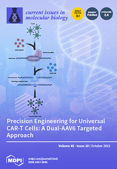
Cover Story (view full-size image): In this study, the researchers utilized AAV6-mediated dual transduction to precisely integrate an anti-CD19 CAR into human T cells at a specific genomic location. They employed two viral vectors, one expressing Cas9 endonuclease and a guide RNA targeting the T-cell receptor alpha constant locus, while the other carried the DNA template for CAR insertion. The molecular analysis results confirmed the targeted integration of the CAR transgene into the desired genomic location, offering a promising alternative to the conventional random integration methods for CAR T cell generation. This method not only reduces the risk of insertional mutagenesis, but also knocks out the endogenous T-cell receptor, enabling the use of allogeneic donor cells and potentially paving the way for "off-the-shelf" universal CAR-T cell immunotherapies, thus simplifying production and administration. View this paper
  • Issues are regarded as officially published after their release is announced to the table of contents alert mailing list.
  • You may sign up for e-mail alerts to receive table of contents of newly released issues.
  • PDF is the official format for papers published in both, html and pdf forms. To view the papers in pdf format, click on the "PDF Full-text" link, and use the free Adobe Reader to open them.
Order results
Result details
Section
Select all
Export citation of selected articles as:
13 pages, 1961 KiB  
Review
Functional Roles of DYRK2 as a Tumor Regulator
by Yuta Mochimaru and Kiyotsugu Yoshida
Curr. Issues Mol. Biol. 2023, 45(10), 8539-8551; https://doi.org/10.3390/cimb45100538 - 23 Oct 2023
Cited by 2 | Viewed by 1431
Abstract
The dual-specificity tyrosine phosphorylation-regulated kinase 2 (DYRK2) regulates the induction of apoptosis and DNA repair, metastasis inhibition, cell cycle G1/S transition, protein scaffold stability for E3 ligase complexes, and embryogenesis. Owing to these functions, DYRK2 is thought to regulate tumorigenesis, and its function [...] Read more.
The dual-specificity tyrosine phosphorylation-regulated kinase 2 (DYRK2) regulates the induction of apoptosis and DNA repair, metastasis inhibition, cell cycle G1/S transition, protein scaffold stability for E3 ligase complexes, and embryogenesis. Owing to these functions, DYRK2 is thought to regulate tumorigenesis, and its function in cancer has been investigated. Notably, DYRK2 has been reported to function as a tumor suppressor; however, it has also been reported to act as an oncogene in some cancers. This discrepancy makes it difficult to elucidate the conserved functions of DYRK2 in cancer. Here, we reviewed the functions of DYRK2 in various cancers. Patient tissue samples were evaluated for each cancer type. Although some studies have used cell lines and/or xenografts to elucidate the mechanism of DYRK2 function, these studies are not sufficient to understand the role of DYRK2 in cancers. In particular, studies using genetically modified mice would help us to understand the reported functional duality of DYRK2 in cancer. Full article
(This article belongs to the Special Issue Advances in Molecular Pathogenesis Regulation in Cancer, 2nd Edition)
Show Figures

Figure 1

20 pages, 6096 KiB  
Article
Hyperthermia Enhances Adeno-Associated Virus Vector Transduction Efficiency in Melanoma Cells
by Alicja Bieńkowska-Tokarczyk, Anna Stelmaszczyk-Emmel, Urszula Demkow and Maciej Małecki
Curr. Issues Mol. Biol. 2023, 45(10), 8519-8538; https://doi.org/10.3390/cimb45100537 - 23 Oct 2023
Cited by 1 | Viewed by 1477
Abstract
Gene therapy perfectly fits in the current needs of medicine for patients with melanoma. One of the major challenges of gene therapy is to increase gene transfer. The role of hyperthermia in the improvement of AAV (adeno-associated virus) transduction efficiency has been indicated. [...] Read more.
Gene therapy perfectly fits in the current needs of medicine for patients with melanoma. One of the major challenges of gene therapy is to increase gene transfer. The role of hyperthermia in the improvement of AAV (adeno-associated virus) transduction efficiency has been indicated. The aim of the present study was to assess the transduction efficacy of melanoma cell lines (A375, G-361, and SK-MEL-1) with the use of the rAAV/DJ mosaic vector under hyperthermia conditions. The analysis of changes in the transduction efficacy and expression of HSPs (heat shock proteins) and receptors for AAV was performed. The transduction was performed at 37 °C and at 43 °C (1 h). Hyperthermia enhanced gene transfer in all the tested cell lines. The most efficient transducing cell line under hyperthermia was A375 (increase by 17%). G361 and SK-MEL-1 cells showed an increase of 7%. The changes in the expression of the AAV receptors and HSPs after hyperthermia were observed. A key role in the improvement of gene transfer may be played by AAVR, HSPB1, HSP6, DNAJC4, HSPD1, HSPA8, HSPA9, HSP90AB1, and AHSA1. This study showed the possibility of the use of hyperthermia as a factor enabling the stimulation of cell transduction with rAAV vectors, thereby providing tools for the improvement in the efficacy of gene therapy based on rAAV. Full article
(This article belongs to the Special Issue Adhesion, Metastasis and Inhibition of Cancer Cells)
Show Figures

Figure 1

17 pages, 6763 KiB  
Article
Validation of a Novel Cuproptosis–Related Prognostic Gene Marker and Differential Expression Associated with Lung Adenocarcinoma
by Tingting Liu and Jianshe Wei
Curr. Issues Mol. Biol. 2023, 45(10), 8502-8518; https://doi.org/10.3390/cimb45100536 - 22 Oct 2023
Cited by 2 | Viewed by 2047
Abstract
Background: Cuproptosis induction is seen as a promising alternative for immunotherapies and targeted therapies in breast cancer. The objective of this research was to examine the prognostic and biological importance of cuproptosis-related genes (CRGs) in lung adenocarcinoma (LUAD). Methods: The following methods were [...] Read more.
Background: Cuproptosis induction is seen as a promising alternative for immunotherapies and targeted therapies in breast cancer. The objective of this research was to examine the prognostic and biological importance of cuproptosis-related genes (CRGs) in lung adenocarcinoma (LUAD). Methods: The following methods were used: GSE10072 dataset and TCGA database analysis, differential expression analysis of CRGs, and biological function (BP) and signaling pathway enrichment analysis, prognostic analysis and clinical analysis of CRGs, construction of the prognostic signature and RNA modified genes and miRNA analysis of CRGs in LUAD, immunoinfiltration analysis and immunohistochemical staining of DβH, UBE2D3, SOD1, UBE2D1 and LOXL2. Results: AOC1, ATOX1, CCL8, CCS, COX11, CP, LOXL2, MAP2K2, PDK1, SCO2, SOD1, UBE2D1, UBE2D3 and VEGFA showed significantly higher expression, while ATP7B, DβH, PDE3B, SLC31A2, UBE2D2, UBE2D4 and ULK2 showed lower expression in LUAD tissues than normal tissues. We also found that ATP7B (4%), AOC1 (3%) PDE3B (2%), DβH (2%), CP (1%), ULK2 (1%), PDK1 (1%), LOXL2 (1%) and UBE2D3 (1%) showed higher mutation frequencies. The univariate Cox analysis was used to identify CRGs that have prognostic value. It identified 21 genes that showed significant prognostic value, containing DβH, UBE2D3, SOD1, UBE2D1 and LOXL2. Patients with DβH up–expression have a longer survival time and patients with UBE2D3, SOD1, UBE2D1 and LOXL2 down–expression also have a longer survival time. hsa–miR–29c–3p, hsa–miR–29a–3p, hsa–miR–181c–5p, hsa–miR–1245a, etc., play an important role in the miRNA regulatory network, and in LUAD, miR–29a, miR–29c and miR–181c high expression survival was longer, and miR–1245a low expression survival was longer. We also performed an analysis to examine the relationships between DβH, LOXL2, SOD1, UBE2D1 and UBE2D3 and immune infiltration in LUAD, including B cells, CD8+ T cells, CD4+ T cells, macrophages, neutrophils and DCs. Conclusion: DβH, UBE2D3, SOD1, UBE2D1, and LOXL2 are potential candidates implicated in LUAD and can be further explored for their application as diagnostic, prognostic, and therapeutic biomarkers for LUAD. Full article
(This article belongs to the Special Issue Targeting Tumor Microenvironment for Cancer Therapy, 2nd Edition)
Show Figures

Figure 1

10 pages, 3319 KiB  
Article
Presynaptic Purinergic Modulation of the Rat Neuro-Muscular Transmission
by Adel E. Khairullin, Sergey N. Grishin and Ayrat U. Ziganshin
Curr. Issues Mol. Biol. 2023, 45(10), 8492-8501; https://doi.org/10.3390/cimb45100535 - 19 Oct 2023
Cited by 2 | Viewed by 1367
Abstract
ATP, being a well-known universal high-energy compound, plays an important role as a signaling molecule and together with its metabolite adenosine they both attenuate the release of acetylcholine in the neuro-muscular synapse acting through membrane P2 and P1 receptors, respectively. In this work, [...] Read more.
ATP, being a well-known universal high-energy compound, plays an important role as a signaling molecule and together with its metabolite adenosine they both attenuate the release of acetylcholine in the neuro-muscular synapse acting through membrane P2 and P1 receptors, respectively. In this work, using a mechanomyographic method, we analyzed the presynaptic mechanisms by which ATP and adenosine can modulate the transduction in the rat m. soleus and m. extensor digitorum longus. N-ethylmaleimide, a G-protein antagonist, prevents the modulating effects of both ATP and adenosine. The action of ATP is abolished by chelerythrin, a specific phospholipase C inhibitor, while the inhibitory effect of adenosine is slightly increased by Rp-cAMPS, an inhibitor of protein kinase A, and by nitrendipine, a blocker of L-type Ca2+ channels. The addition of DPCPX, an A1 receptor antagonist, fully prevents the inhibitory action of adenosine in both muscles. Our data indicate that the inhibitory action of ATP involves metabotropic P2Y receptors and is mediated by phospholipase C dependent processes in rat motor neuron terminals. We suggest that the presynaptic effect of adenosine consists of negative and positive actions. The negative action occurs by stimulation of adenosine A1 receptors while the positive action is associated with the stimulation of adenosine A2A receptors, activation of protein kinase A and opening of L-type calcium channels. The combined mechanism of the modulating action of ATP and adenosine provides fine tuning of the synapse to fast changing conditions in the skeletal muscles. Full article
(This article belongs to the Special Issue Molecular Insights into Skeletal Muscle Homeostasis and Metabolism)
Show Figures

Figure 1

16 pages, 2150 KiB  
Review
The Role of p16/Ki67 Dual Staining in Cervical Cancer Screening
by Andraž Dovnik and Alenka Repše Fokter
Curr. Issues Mol. Biol. 2023, 45(10), 8476-8491; https://doi.org/10.3390/cimb45100534 - 19 Oct 2023
Cited by 7 | Viewed by 4229
Abstract
Cervical cancer screening has enabled a decrease in the incidence and mortality of cervical cancer. Various screening modalities have been studied to date. In many countries, screening is still based on cervical cytology, where cervical cells obtained either on glass or in a [...] Read more.
Cervical cancer screening has enabled a decrease in the incidence and mortality of cervical cancer. Various screening modalities have been studied to date. In many countries, screening is still based on cervical cytology, where cervical cells obtained either on glass or in a liquid medium are examined under a microscope. However, the fact that the vast majority of cervical cancers are a result of persistent infection with high-risk human papillomaviruses (hr-HPV) has led to the implementation of primary HPV screening in many countries. Taking into consideration the fact that the majority of HPV infections are transient and do not cause cervical precancer, effective triage methods are needed to prevent an increase in colposcopy referrals. Among these, the most extensively investigated are HPV genotyping, HPV methylation, and p16/Ki67 dual staining. In this manuscript, we briefly summarize the current knowledge regarding different screening strategies for the prevention of cervical cancer, with a focus on p16/Ki67 dual staining. In addition, we provide an explanation regarding the rationale for the use of various screening modalities based on the molecular biology of cervical cancer and cervical precancerous lesions. Full article
(This article belongs to the Special Issue Molecular Research on Female Reproductive Diseases)
Show Figures

Figure 1

15 pages, 4480 KiB  
Review
Recent Trends in the Antidiabetic Prominence of Natural and Synthetic Analogues of Aurones
by Rammohan Aluru, Anindita Mukherjee, Grigory V. Zyryanov, Adinath Majee and Sougata Santra
Curr. Issues Mol. Biol. 2023, 45(10), 8461-8475; https://doi.org/10.3390/cimb45100533 - 19 Oct 2023
Cited by 3 | Viewed by 1976
Abstract
Natural products are a boundless source for the development of pharmaceutical agents against a wide range of human diseases. Accordingly, naturally occurring aurones possess various biological benefits, such as anticancer, antioxidant, antimicrobial, antidiabetic, anti-inflammatory, antiviral and neuroprotective effects. In addition, various studies have [...] Read more.
Natural products are a boundless source for the development of pharmaceutical agents against a wide range of human diseases. Accordingly, naturally occurring aurones possess various biological benefits, such as anticancer, antioxidant, antimicrobial, antidiabetic, anti-inflammatory, antiviral and neuroprotective effects. In addition, various studies have revealed that aurones are potential templates for the regulation of diabetes mellitus and its associated complications. Likewise, certain aurones and their analogues have been found to be remarkable kinase inhibitors of DARK2, PPAR-γ, PTPM1, AGE, α-amylase and α-glucosidase, which represents a promising approach for the treatment of chronic metabolic disorders such as diabetes. Therefore, in our present study, we provide a detailed account of the advances in aurones as antidiabetic agents over the past decade. Full article
(This article belongs to the Special Issue Natural Products in Biomedicine and Pharmacotherapy)
Show Figures

Figure 1

17 pages, 7311 KiB  
Article
Protein Lactylation Modification and Proteomics Features in Cirrhosis Patients after UC-MSC Treatment
by Ye Xie, Ying Li, Jia Yao, Xiaojing Song, Haiping Wang, Jianjun Zhang and Xun Li
Curr. Issues Mol. Biol. 2023, 45(10), 8444-8460; https://doi.org/10.3390/cimb45100532 - 18 Oct 2023
Viewed by 2394
Abstract
Umbilical cord mesenchymal stem cell (UC-MSC) therapy improves liver function in liver cirrhosis patients. This study aimed to elucidate the therapeutic mechanism underlying cell therapy by analyzing changes in the modification and expression of proteins 1 month post-treatment with UC-MSCs. This prospective study [...] Read more.
Umbilical cord mesenchymal stem cell (UC-MSC) therapy improves liver function in liver cirrhosis patients. This study aimed to elucidate the therapeutic mechanism underlying cell therapy by analyzing changes in the modification and expression of proteins 1 month post-treatment with UC-MSCs. This prospective study included 11 cirrhosis patients who received MSC injection. The laboratory indexes before and after treatment were collected to evaluate the clinical treatment effect of UC-MSCs, and the protein expression and lactylation modification in the liver were comprehensively revealed. Meanwhile, weighted gene co-expression network analysis was used to analyze the co-expression protein modules and their relationship with clinical features. The patients with liver cirrhosis showed an improvement trend after receiving UC-MSC treatment; specifically, the liver protein synthesis function was significantly improved and the coagulation function was also significantly improved. Proteomics combined with lactic acid proteomics revealed 160 lysine lactylation (Kla) sites of 119 proteins. Functional analysis showed that the lactylation-modified proteins were enriched in the pathway of glucose and other substances’ metabolism, and many key enzymes of glycolysis and gluconeogenesis were lactated. UC-MSC therapy has a certain clinical effect in the treatment of liver cirrhosis and may act by regulating material metabolism, because the lactylation protein points to energy metabolism. Full article
(This article belongs to the Special Issue Predicting Drug Targets Using Bioinformatics Methods)
Show Figures

Figure 1

17 pages, 5617 KiB  
Article
Neuroprotective Effects of Water Extract from Brown Algae Petalonia binghamiae in an Experimental Model of Focal Cerebral Ischemia In Vitro and In Vivo
by Sun Ho Eom, Geum-Lan Hong, Hyun Bae Kang, Nam-Seob Lee, Do Kyung Kim, Young Gil Jeong, Chun-Sung Kim, Yung Choon Yoo, Bong Ho Lee, Ju-Young Jung, Dong-Sub Kim and Seung Yun Han
Curr. Issues Mol. Biol. 2023, 45(10), 8427-8443; https://doi.org/10.3390/cimb45100531 - 17 Oct 2023
Cited by 2 | Viewed by 1631
Abstract
Focal cerebral ischemia (fCI) can result in brain injury and sensorimotor deficits. Brown algae are currently garnering scientific attention as potential therapeutic candidates for fCI. This study investigated the therapeutic effects of the hot water extract of Petalonia binghamiae (wPB), a brown alga, [...] Read more.
Focal cerebral ischemia (fCI) can result in brain injury and sensorimotor deficits. Brown algae are currently garnering scientific attention as potential therapeutic candidates for fCI. This study investigated the therapeutic effects of the hot water extract of Petalonia binghamiae (wPB), a brown alga, in in vitro and in vivo models of fCI. The neuroprotective efficacy of wPB was evaluated in an in vitro excitotoxicity model established using HT-22 cells challenged with glutamate. Afterward, C57/BL6 mice were administered wPB for 7 days (10 or 100 mg/kg, intragastric) and subjected to middle cerebral artery occlusion and reperfusion (MCAO/R) operation, which was used as an in vivo fCI model. wPB co-incubation significantly inhibited cell death, oxidative stress, and apoptosis, as well as stimulated the expression of heme oxygenase-1 (HO-1), an antioxidant enzyme, and the nuclear translocation of its upstream regulator, nuclear factor erythroid 2-related factor 2 (Nrf2) in HT-22 cells challenged with glutamate-induced excitotoxicity. Pretreatment with either dose of wPB significantly attenuated infarction volume, neuronal death, and sensorimotor deficits in an in vivo fCI model. Furthermore, the attenuation of oxidative stress and apoptosis in the ischemic lesion accompanied the wPB-associated protection. This study suggests that wPB can counteract fCI via an antioxidative effect, upregulating the Nrf2/HO-1 pathway. Full article
(This article belongs to the Special Issue Neuropathology: From Molecular Mechanisms to Therapeutic Solutions)
Show Figures

Figure 1

15 pages, 3520 KiB  
Article
Effects of Melatonin on Liver of D-Galactose-Induced Aged Mouse Model
by Ran Lee, Won-Yong Lee and Hyun-Jung Park
Curr. Issues Mol. Biol. 2023, 45(10), 8412-8426; https://doi.org/10.3390/cimb45100530 - 17 Oct 2023
Cited by 3 | Viewed by 2641
Abstract
Melatonin, a hormone secreted by the pineal gland of vertebrates, regulates sleep, blood pressure, and circadian and seasonal rhythms, and acts as an antioxidant and anti-inflammatory agent. We investigated the protective effects of melatonin against markers of D-galactose (D-Gal)-induced hepatocellular aging, including liver [...] Read more.
Melatonin, a hormone secreted by the pineal gland of vertebrates, regulates sleep, blood pressure, and circadian and seasonal rhythms, and acts as an antioxidant and anti-inflammatory agent. We investigated the protective effects of melatonin against markers of D-galactose (D-Gal)-induced hepatocellular aging, including liver inflammation, hepatocyte structural damage, and non-alcoholic fatty liver. Mice were divided into four groups: phosphate-buffered saline (PBS, control), D-Gal (200 mg/kg/day), melatonin (20 mg/kg), and D-Gal (200 mg/kg) and melatonin (20 mg) cotreatment. The treatments were administered once daily for eight consecutive weeks. Melatonin treatment alleviated D-Gal-induced hepatocyte impairment. The AST level was significantly increased in the D-Gal-treated groups compared to that in the control group, while the ALT level was decreased compared to the melatonin and D-Gal cotreated group. Inflammatory genes, such as IL1-β, NF-κB, IL-6, TNFα, and iNOS, were significantly increased in the D-Gal aging model, whereas the expression levels of these genes were low in the D-Gal and melatonin cotreated group. Interestingly, the expression levels of hepatic steatosis-related genes, such as LXRα, C/EBPα, PPARα, ACC, ACOX1, and CPT-1, were markedly decreased in the D-Gal and melatonin cotreated group. These results suggest that melatonin suppresses hepatic steatosis and inflammation in a mouse model of D-Gal-induced aging. Full article
(This article belongs to the Special Issue Cellular and Molecular Mechanisms of Nonalcoholic Fatty Liver Disease)
Show Figures

Figure 1

17 pages, 6485 KiB  
Article
Mitochondrial Dysfunction in Dopaminergic Neurons Derived from Patients with LRRK2- and SNCA-Associated Genetic Forms of Parkinson’s Disease
by Anna S. Vetchinova, Marina R. Kapkaeva, Mikhail V. Ivanov, Kristina A. Kutukova, Natalia M. Mudzhiri, Lydia E. Frumkina, Anatoly V. Brydun, Vladimir S. Sukhorukov and Sergey N. Illarioshkin
Curr. Issues Mol. Biol. 2023, 45(10), 8395-8411; https://doi.org/10.3390/cimb45100529 - 17 Oct 2023
Cited by 2 | Viewed by 2072
Abstract
Parkinson’s disease (PD) is the second most common neurodegenerative disease. Some cases of PD may be caused by genetic factors, among which mutations in the LRRK2 and SNCA genes play an important role. To develop effective neuroprotective strategies for PD, it is important [...] Read more.
Parkinson’s disease (PD) is the second most common neurodegenerative disease. Some cases of PD may be caused by genetic factors, among which mutations in the LRRK2 and SNCA genes play an important role. To develop effective neuroprotective strategies for PD, it is important to diagnose the disease at the earliest stages of the neurodegenerative process. Therefore, the detection of diagnostic and prognostic markers of Parkinson’s disease (PD) is an urgent medical need. Advances in induced pluripotent stem cell (iPSC) culture technology provide new opportunities for the search for new biomarkers of PD and its modeling in vitro. In our work, we used a new technology for multiplex profiling of gene expression using barcoding on the Nanostring platform to assess the activity of mitochondrial genes on iPSC-derived cultures of dopaminergic neurons obtained from patients with LRRK2- and SNCA-associated genetic forms PD and a healthy donor. Electron microscopy revealed ultrastructural changes in mitochondria in both LRRK2 and SNCA mutant cells, whereas mitochondria in cells from a healthy donor were normal. In a culture with the SNCA gene mutation, the ratio of the area occupied by mitochondria to the total area of the cytoplasm was significantly lower than in the control and in the line with the LRRK2 gene mutation. Transcriptome analysis of 105 mitochondria proteome genes using the Nanostring platform revealed differences between the diseased and normal cells in the activity of genes involved in respiratory complex function, the tricarboxylic acid cycle, ATP production, mitochondria–endoplasmic reticulum interaction, mitophagy, regulation of calcium concentration, and mitochondrial DNA replication. Full article
Show Figures

Figure 1

23 pages, 8308 KiB  
Article
Extremophilic Solutions: The Role of Deinoxanthin in Counteracting UV-Induced Skin Harm
by Mehmet Kuzucu
Curr. Issues Mol. Biol. 2023, 45(10), 8372-8394; https://doi.org/10.3390/cimb45100528 - 16 Oct 2023
Viewed by 1815
Abstract
This research delved into the protective capacities of deinoxanthin, a carotenoid present in Deinococcus radiodurans, against UVA- and UVB-mediated skin damage using human fibroblast foreskin cells (HFF-1). Using the MTT assay, HFF-1 cells treated with 10 µM DNX displayed 20% and 31.7% higher [...] Read more.
This research delved into the protective capacities of deinoxanthin, a carotenoid present in Deinococcus radiodurans, against UVA- and UVB-mediated skin damage using human fibroblast foreskin cells (HFF-1). Using the MTT assay, HFF-1 cells treated with 10 µM DNX displayed 20% and 31.7% higher viability than the positive (Vitamin C-treated) and negative (DNX-untreated) control groups, respectively, upon 100 mJ/cm2 UVB exposure. At 24 J/cm2 UVA, 20 µM DNX-treated cells showed 80.6% viability, exceeding the positive and negative control groups by 28.6% and 33.6%, respectively. Flow cytometry analysis revealed that cells treated with DNX and exposed to 24 J/cm2 UVA exhibited a 69.32% reduction in apoptotic processes compared to untreated cells. Similarly, when exposed to 100 mJ/cm2 UVB, DNX-treated cells demonstrated a 72.35% decrease in apoptotic processes relative to their untreated counterparts. DNX also displayed dose-dependent inhibition on tyrosinase activity. The study emphasized DNX’s antioxidative capacity, evident in its modulation of superoxide dismutase activity and measurements of Malondialdehyde and intracellular reactive oxygen species levels. DNX-treated cells exhibited higher hydroxyproline levels, suggesting healthier collagen production. Additionally, the wound-healing assay method confirmed an accelerated healing rate in DNX-treated cells. Conclusively, DNX offers significant protection against UV-induced skin damage, emphasizing its potential for skincare and therapeutics. Full article
(This article belongs to the Special Issue Natural Products and Their Biological Activities)
Show Figures

Figure 1

13 pages, 1228 KiB  
Article
Association of OXTR, AVPR1a, LNPEP, and CD38 Genes’ Expression with the Clinical Presentation of Autism Spectrum Disorder
by Krzysztof Maria Wilczyński, Aleksandra Auguściak-Duma, Aleksandra Stasik, Lena Cichoń, Alicja Kawalec and Małgorzata Janas-Kozik
Curr. Issues Mol. Biol. 2023, 45(10), 8359-8371; https://doi.org/10.3390/cimb45100527 - 16 Oct 2023
Viewed by 1451
Abstract
Autism spectrum disorder (ASD) is a complex neurodevelopmental disorder that affects social interactions, communication, and behavior. Although the predominant genetic predisposition to ASD seems beyond doubt, its exact nature remains unclear. In the context of social cognition disorders and the basis of ASD, [...] Read more.
Autism spectrum disorder (ASD) is a complex neurodevelopmental disorder that affects social interactions, communication, and behavior. Although the predominant genetic predisposition to ASD seems beyond doubt, its exact nature remains unclear. In the context of social cognition disorders and the basis of ASD, the oxytocinergic and vasopresynergic systems arouse great interest among researchers. The aim of the present study was to analyze gene expression levels for oxytocin and vasopressin receptors, as well as CD38 protein and oxytocinase, in the context of the clinical picture of autism spectrum disorders. The study included 90 people, of whom 63 were diagnosed with ASD based on anamnesis, mental status testing, and the ADOS-2 protocol. The results obtained in the presented study indicate that the balance between the levels of expression of the CD38 gene and the oxytocinase gene plays a key role in the risk and clinical presentation of ASD. In a hypothetical scenario, an imbalance in the expression of CD38 and LNPEP could potentially lead to alterations in the concentrations of oxytocin and vasopressin. At the same time, the most frequently studied genes—AVPR1a and OXTR—seem to be at best of marginal importance for the risk of ASD. Full article
Show Figures

Figure 1

22 pages, 954 KiB  
Review
Cardiac Toxicities in Oncology: Elucidating the Dark Box in the Era of Precision Medicine
by Younan Samuel, Aswin Babu, Foteini Karagkouni, Ayden Ismail, Sunyoung Choi and Stergios Boussios
Curr. Issues Mol. Biol. 2023, 45(10), 8337-8358; https://doi.org/10.3390/cimb45100526 - 15 Oct 2023
Cited by 3 | Viewed by 2639
Abstract
Despite current advancements in chemotherapy, immunotherapy and targeted treatments, the potential for major adverse cardiovascular events, regardless of previous cardiac history, persists. Scoring systems, such as the Heart Failure Association-International Cardio-Oncology Society (HFA-ICOS) risk assessment tool, can be utilized to evaluate several factors [...] Read more.
Despite current advancements in chemotherapy, immunotherapy and targeted treatments, the potential for major adverse cardiovascular events, regardless of previous cardiac history, persists. Scoring systems, such as the Heart Failure Association-International Cardio-Oncology Society (HFA-ICOS) risk assessment tool, can be utilized to evaluate several factors including prior cardiac history, risk factors and cardiac biomarkers to categorize patients into low, moderate, high, and very high-risk groups. Common cardiotoxicity complications include new or worsening left ventricular ejection fraction (LVEF), QT interval prolongation, myocardial ischaemia, hypertension, thromboembolic disease, cardiac device malfunction and valve disease. Baseline electrocardiogram (ECG) and transthoracic echocardiogram (TTE) are routinely performed for all patients commenced on cardiotoxic treatment, while other imaging modalities and biochemical markers have proven useful for monitoring. Management mainly includes early risk stratification and prompt identification of cardiovascular complications, with patient-specific surveillance throughout treatment. A multidisciplinary approach is crucial in determining the relationship between potential treatment benefits and cardiotoxicity, and whether the continuation of treatment is appropriate on a case-by-case basis. Early risk stratification, optimizing the patient’s cardiovascular status prior to treatment, and prompt identification of suspected cardiotoxicity are key in significantly reducing risk. This article provides a comprehensive review of the various types of treatment-related cardiotoxicity, offering guidance on identifying high-risk patients, recognizing early signs of cardiotoxicity, and outlining appropriate treatment approaches and follow-up care for such cases. Full article
(This article belongs to the Special Issue Focus on Molecular Basis of Cardiac Diseases)
Show Figures

Figure 1

16 pages, 3496 KiB  
Article
6-Hydroxy-2,2,4-trimethyl-1,2-dihydroquinoline Demonstrates Anti-Inflammatory Properties and Reduces Oxidative Stress in Acetaminophen-Induced Liver Injury in Rats
by Evgenii D. Kryl’skii, Svetlana E. Kravtsova, Tatyana N. Popova, Larisa V. Matasova, Khidmet S. Shikhaliev and Svetlana M. Medvedeva
Curr. Issues Mol. Biol. 2023, 45(10), 8321-8336; https://doi.org/10.3390/cimb45100525 - 12 Oct 2023
Cited by 1 | Viewed by 1510
Abstract
We examined the effects of 6-hydroxy-2,2,4-trimethyl-1,2-dihydroquinoline on markers of liver injury, oxidative status, and the extent of inflammatory and apoptotic processes in rats with acetaminophen-induced liver damage. The administration of acetaminophen caused the accumulation of 8-hydroxy-2-deoxyguanosine and 8-isoprostane in the liver and serum, [...] Read more.
We examined the effects of 6-hydroxy-2,2,4-trimethyl-1,2-dihydroquinoline on markers of liver injury, oxidative status, and the extent of inflammatory and apoptotic processes in rats with acetaminophen-induced liver damage. The administration of acetaminophen caused the accumulation of 8-hydroxy-2-deoxyguanosine and 8-isoprostane in the liver and serum, as well as an increase in biochemiluminescence indicators. Oxidative stress resulted in the activation of pro-inflammatory cytokine and NF-κB factor mRNA synthesis and increased levels of immunoglobulin G, along with higher activities of caspase-3, caspase-8, and caspase-9. The administration of acetaminophen also resulted in the development of oxidative stress, leading to a decrease in the level of reduced glutathione and an imbalance in the function of antioxidant enzymes. This study discovered that 6-hydroxy-2,2,4-trimethyl-1,2-dihydroquinoline reduced oxidative stress by its antioxidant activity, hence reducing the level of pro-inflammatory cytokine and NF-κB mRNA, as well as decreasing the concentration of immunoglobulin G. These changes resulted in a reduction in the activity of caspase-8 and caspase-9, which are involved in the activation of ligand-induced and mitochondrial pathways of apoptosis and inhibited the effector caspase-3. In addition, 6-hydroxy-2,2,4-trimethyl-1,2-dihydroquinoline promoted the normalization of antioxidant system function in animals treated with acetaminophen. As a result, the compound being tested alleviated inflammation and apoptosis by decreasing oxidative stress, which led to improved liver marker indices and ameliorated histopathological alterations. Full article
(This article belongs to the Special Issue Advances in Understanding Molecular Basis of Inflammatory Diseases)
Show Figures

Figure 1

12 pages, 3221 KiB  
Article
Hyperbaric Oxygen Therapy Adjuvant Chemotherapy and Radiotherapy through Inhibiting Stemness in Glioblastoma
by Chun-Man Yuen, Hung-Pei Tsai, Tzu-Ting Tseng, Yu-Lung Tseng, Ann-Shung Lieu, Aij-Lie Kwan and Alice Y. W. Chang
Curr. Issues Mol. Biol. 2023, 45(10), 8309-8320; https://doi.org/10.3390/cimb45100524 - 12 Oct 2023
Cited by 3 | Viewed by 2892
Abstract
Glioblastoma multiforme (GBM) is the most common and deadliest primary brain tumor in adults. Despite the advances in GBM treatment, outcomes remain poor, with a 2-year survival rate of less than 5%. Hyperbaric oxygen (HBO) therapy is an intermittent, high-concentration, short-term oxygen therapy [...] Read more.
Glioblastoma multiforme (GBM) is the most common and deadliest primary brain tumor in adults. Despite the advances in GBM treatment, outcomes remain poor, with a 2-year survival rate of less than 5%. Hyperbaric oxygen (HBO) therapy is an intermittent, high-concentration, short-term oxygen therapy used to increase cellular oxygen content. In this study, we evaluated the effects of HBO therapy, alone or combined with other treatment modalities, on GBM in vitro and in vivo. In the in vitro analysis, we used a 3-(4,5-Dimethylthiazol-2-yl)-2,5-diphenyltetrazolium bromide (MTT) assay to assess the effects of HBO therapy alone, a colony formation assay to analyze the effects of HBO therapy combined with radiotherapy and with temozolomide (TMZ), and a neurosphere assay to assess GBM stemness. In the in vivo analysis, we used immunohistochemical staining and in vivo bioluminescence imaging to assess GBM stemness and the therapeutic effect of HBO therapy alone or combined with TMZ or radiotherapy, respectively. HBO therapy did not affect GBM cell viability, but it did reduce the analyzed tumors’ ability to form cancer stem cells. In addition, HBO therapy increased GBM sensitivity to TMZ and radiotherapy both in vitro and in vivo. HBO therapy did not enhance tumor growth and exhibited adjuvant effects to chemotherapy and radiotherapy through inhibiting GBM stemness. In conclusion, HBO therapy shows promise as an adjuvant treatment for GBM by reducing cancer stem cell formation and enhancing sensitivity to chemotherapy and radiotherapy. Full article
(This article belongs to the Special Issue Understanding Cellular Radiation Responses for Radiation Therapy)
Show Figures

Graphical abstract

20 pages, 1674 KiB  
Review
Changes of Signaling Pathways in Hypothalamic Neurons with Aging
by Petr M. Masliukov
Curr. Issues Mol. Biol. 2023, 45(10), 8289-8308; https://doi.org/10.3390/cimb45100523 - 12 Oct 2023
Cited by 3 | Viewed by 2049
Abstract
The hypothalamus is an important regulator of autonomic and endocrine functions also involved in aging regulation. The aging process in the hypothalamus is accompanied by disturbed intracellular signaling including insulin/insulin-like growth factor-1 (IGF-1)/growth hormone (GH), phosphatidylinositol-4,5-bisphosphate 3-kinase (PI3K)/protein kinase B (AKT)/the mammalian target [...] Read more.
The hypothalamus is an important regulator of autonomic and endocrine functions also involved in aging regulation. The aging process in the hypothalamus is accompanied by disturbed intracellular signaling including insulin/insulin-like growth factor-1 (IGF-1)/growth hormone (GH), phosphatidylinositol-4,5-bisphosphate 3-kinase (PI3K)/protein kinase B (AKT)/the mammalian target of rapamycin (mTOR), mitogen activated protein kinase (MAPK), janus kinase (JAK)/signal transducer and activator of transcription (STAT), AMP-activated protein kinase (AMPK), nuclear factor kappa-light-chain-enhancer of activated B cells (NF-ĸB), and nitric oxide (NO). In the current review, I have summarized the current understanding of the changes in the above-mentioned pathways in aging with a focus on hypothalamic alterations. Full article
(This article belongs to the Collection Feature Papers in Current Issues in Molecular Biology)
Show Figures

Graphical abstract

12 pages, 1789 KiB  
Article
Hypoxia and HIF-1α Regulate the Activity and Expression of Na,K-ATPase Subunits in H9c2 Cardiomyoblasts
by Beyza Gurler, Gizem Gencay and Emel Baloglu
Curr. Issues Mol. Biol. 2023, 45(10), 8277-8288; https://doi.org/10.3390/cimb45100522 - 12 Oct 2023
Cited by 2 | Viewed by 2179
Abstract
The optimal function of the Na,K-ATPase (NKA) pump is essential for the heart. In ischemic heart disease, NKA activity decreases due to the decreased expression of the pump subunits. Here, we tested whether the hypoxia-inducible transcription factor (HIF-1α), the key signaling molecule regulating [...] Read more.
The optimal function of the Na,K-ATPase (NKA) pump is essential for the heart. In ischemic heart disease, NKA activity decreases due to the decreased expression of the pump subunits. Here, we tested whether the hypoxia-inducible transcription factor (HIF-1α), the key signaling molecule regulating the adaptation of cells to hypoxia, is involved in controlling the expression and cellular dynamics of α1- and β1-NKA isoforms and of NKA activity in in-vitro hypoxic H9c2 cardiomyoblasts. HIF-1α was silenced through adenoviral infection, and cells were kept in normoxia (19% O2) or hypoxia (1% O2) for 24 h. We investigated the mRNA and protein expression of α1-, β1-NKA using RT-qPCR and Western blot in whole-cell lysates, cell membranes, and cytoplasmic fractions after labeling the cell surface with NHS-SS-biotin and immunoprecipitation. NKA activity and intracellular ATP levels were also measured. We found that in hypoxia, silencing HIF-1α prevented the decreased mRNA expression of α1-NKA but not of β1-NKA. Hypoxia decreased the plasma membrane expression of α1-NKA and β1- NKA compared to normoxic cells. In hypoxic cells, HIF-1α silencing prevented this effect by inhibiting the internalization of α1-NKA. Total protein expression was not affected. The decreased activity of NKA in hypoxic cells was fully prevented by silencing HIF-1α independent of cellular ATP levels. This study is the first to show that in hypoxic H9c2 cardiomyoblasts, HIF-1α controls the internalization and membrane insertion of α1-NKA subunit and of NKA activity. The mechanism behind this regulation needs further investigation. Full article
(This article belongs to the Special Issue A Focus on Molecular Basis in Cardiac Diseases)
Show Figures

Figure 1

22 pages, 1350 KiB  
Review
A Synopsis of Hepatitis C Virus Treatments and Future Perspectives
by Christian Medina, Alexis Hipólito García, Francis Isamarg Crespo, Félix Isidro Toro, Soriuska José Mayora and Juan Bautista De Sanctis
Curr. Issues Mol. Biol. 2023, 45(10), 8255-8276; https://doi.org/10.3390/cimb45100521 - 11 Oct 2023
Cited by 4 | Viewed by 3915
Abstract
Hepatitis C virus (HCV) infection is a worldwide public health problem. Chronic infection with HCV can lead to liver cirrhosis or cancer. Although some immune-competent individuals can clear the virus, others develop chronic HCV disease due to viral mutations or an impaired immune [...] Read more.
Hepatitis C virus (HCV) infection is a worldwide public health problem. Chronic infection with HCV can lead to liver cirrhosis or cancer. Although some immune-competent individuals can clear the virus, others develop chronic HCV disease due to viral mutations or an impaired immune response. IFNs type I and III and the signal transduction induced by them are essential for a proper antiviral effect. Research on the viral cycle and immune escape mechanisms has formed the basis of therapeutic strategies to achieve a sustained virological response (SVR). The first therapies were based on IFNα; then, IFNα plus ribavirin (IFN–RBV); and then, pegylated-IFNα-RBV (PEGIFNα-RIV) to improve cytokine pharmacokinetics. However, the maximum SVR was 60%, and several significant side effects were observed, decreasing patients’ treatment adherence. The development of direct-acting antivirals (DAAs) significantly enhanced the SVR (>90%), and the compounds were able to inhibit HCV replication without significant side effects, even in paediatric populations. The management of coinfected HBV–HCV and HCV–HIV patients has also improved based on DAA and PEG-IFNα-RBV (HBV–HCV). CD4 cells are crucial for an effective antiviral response. The IFNλ3, IL28B, TNF-α, IL-10, TLR-3, and TLR-9 gene polymorphisms are involved in viral clearance, therapeutic responses, and hepatic pathologies. Future research should focus on searching for strategies to circumvent resistance-associated substitution (RAS) to DAAs, develop new therapeutic schemes for different medical conditions, including organ transplant, and develop vaccines for long-lasting cellular and humoral responses with cross-protection against different HCV genotypes. The goal is to minimise the probability of HCV infection, HCV chronicity and hepatic carcinoma. Full article
(This article belongs to the Collection Molecular Mechanisms in Human Diseases)
Show Figures

Graphical abstract

16 pages, 15319 KiB  
Article
Melatonin Reduces Aggravation of Renal Ischemia–Reperfusion Injury in Obese Rats by Maintaining Mitochondrial Homeostasis and Integrity through AMPK/PGC-1α/SIRT3/SOD2 Activation
by Anongporn Kobroob, Aphisek Kongkaew and Orawan Wongmekiat
Curr. Issues Mol. Biol. 2023, 45(10), 8239-8254; https://doi.org/10.3390/cimb45100520 - 11 Oct 2023
Cited by 2 | Viewed by 1676
Abstract
This study examined the potential benefits of melatonin against renal ischemia and reperfusion (IR) injury in obesity and explored the underlying mechanisms. Obesity was induced in Wistar rats by feeding a high-fat diet for 16 weeks. Three obese groups that underwent renal IR [...] Read more.
This study examined the potential benefits of melatonin against renal ischemia and reperfusion (IR) injury in obesity and explored the underlying mechanisms. Obesity was induced in Wistar rats by feeding a high-fat diet for 16 weeks. Three obese groups that underwent renal IR induction (30-min renal ischemia followed by 24-h reperfusion) were randomly assigned to receive melatonin at ischemic onset, reperfusion onset, or pretreatment for 4 weeks before IR induction. Groups of vehicle-treated obese and normal-diet-fed rats that underwent sham or IR induction were also included in the study. The results showed that renal functional and structural impairments after IR incidence were aggravated in obese rats compared to normal-diet-fed rats. The obese-IR rats also exhibited oxidative stress, mitochondrial dysfunction, apoptosis, and mitochondrial dynamics and mitophagy imbalances, which were all considerably improved upon melatonin treatment, irrespective of the treatment time. This study suggests the prophylactic and therapeutic efficacy of melatonin in IR-induced acute kidney injury (AKI) in obese individuals, which may improve the prognosis of AKI in these populations. The benefits of melatonin are likely mediated by the modification of various signaling molecules within the mitochondria that maintain mitochondrial redox balance and lead to the protection of mitochondrial homeostasis and integrity. Full article
(This article belongs to the Special Issue Mitochondrial Function and Dysfunction)
Show Figures

Figure 1

12 pages, 2943 KiB  
Article
Biodistribution of Fluorescent Albumin Nanoparticles among Organs of Laboratory Animals after Intranasal and Peroral Administration
by Olga Morozova, Elena Isaeva and Dmitry Klinov
Curr. Issues Mol. Biol. 2023, 45(10), 8227-8238; https://doi.org/10.3390/cimb45100519 - 11 Oct 2023
Cited by 1 | Viewed by 1383
Abstract
Natural, environmental and engineered nanoparticles (NP) penetrate into cells by endocytosis and induce innate immunity. The behaviour of the nanomaterials both in vitro and in vivo should be assessed. Our goal was to study protein NP stability in biological fluids and distribution in [...] Read more.
Natural, environmental and engineered nanoparticles (NP) penetrate into cells by endocytosis and induce innate immunity. The behaviour of the nanomaterials both in vitro and in vivo should be assessed. Our goal was to study protein NP stability in biological fluids and distribution in organs of animals after intranasal and oral administration. Bovine serum albumin (BSA) was labelled with the fluorescent dye RhoB and NP were fabricated by nanoprecipitation. The fluorescent protein NPwere administered intranasally and orally in laboratory-outbred mice ICR and rabbits. RhoB-BSA NP distribution in organs was detected using spectrofluorometry and fluorescent microscopy. Innate immunity was evaluated using reverse transcription with random hexanucleotide primer and subsequent real-time PCR with specific fluorescent hydrolysis probes. The labelled BSA NP were shown to remain stable in blood sera and nasopharyngeal swabs for 5 days at +37 °C. In vivo the maximal accumulation was found in the brain in 2 days posttreatment without prevalent accumulation in olfactory bulbs. For the intestine, heart and liver, the BSA NP accumulation was similar in 1 and 2 days, whereas for kidney samples even decreased after 1 day. Both intranasal and peroral administration of RhoB-BSA NP did not induce innate immunity. Thus, after intranasal or oral instillation RhoB-BSA NP were found mainly in the brain and intestine without interferon gene expression. Full article
(This article belongs to the Special Issue Effects of Nanoparticles on Living Organisms 2.0)
Show Figures

Graphical abstract

12 pages, 2638 KiB  
Article
Screening of Tnfaip1-Interacting Proteins in Zebrafish Embryonic cDNA Libraries Using a Yeast Two-Hybrid System
by Shulan Huang, Hongning Zhang, Wen Chen, Jiawei Wang, Zhen Wu, Meiqi He, Jian Zhang, Xiang Hu and Shuanglin Xiang
Curr. Issues Mol. Biol. 2023, 45(10), 8215-8226; https://doi.org/10.3390/cimb45100518 - 10 Oct 2023
Viewed by 1625
Abstract
TNFAIP1 regulates cellular biological functions, including DNA replication, DNA repair, and cell cycle, by binding to target proteins. Identification of Tnfaip1-interacting proteins contributes to the understanding of the molecular regulatory mechanisms of their biological functions. In this study, 48 hpf, 72 hpf, and [...] Read more.
TNFAIP1 regulates cellular biological functions, including DNA replication, DNA repair, and cell cycle, by binding to target proteins. Identification of Tnfaip1-interacting proteins contributes to the understanding of the molecular regulatory mechanisms of their biological functions. In this study, 48 hpf, 72 hpf, and 96 hpf wild-type zebrafish embryo mRNAs were used to construct yeast cDNA library. The library titer was 1.12 × 10⁷ CFU/mL, the recombination rate was 100%, and the average length of the inserted fragments was greater than 1000 bp. A total of 43 potential interacting proteins of Tnfaip1 were identified using zebrafish Tnfaip1 as a bait protein. Utilizing GO functional annotation and KEGG signaling pathway analysis, we found that these interacting proteins are mainly involved in translation, protein catabolic process, ribosome assembly, cytoskeleton formation, amino acid metabolism, and PPAR signaling pathway. Further yeast spotting analyses identified four interacting proteins of Tnfaip1, namely, Ubxn7, Tubb4b, Rpl10, and Ybx1. The Tnfaip1-interacting proteins, screened from zebrafish embryo cDNA in this study, increased our understanding of the network of Tnfaip1-interacting proteins during the earliest embryo development and provided a molecular foundation for the future exploration of tnfaip1’s biological functions. Full article
(This article belongs to the Special Issue Microbial Engineering: Gene Expression Regulation and Its Application)
Show Figures

Figure 1

14 pages, 669 KiB  
Review
Ferroptosis: An Emerging Target for Bladder Cancer Therapy
by Zhengda Shan, Wenbin Tang, Zhiyuan Shi and Tao Shan
Curr. Issues Mol. Biol. 2023, 45(10), 8201-8214; https://doi.org/10.3390/cimb45100517 - 10 Oct 2023
Cited by 3 | Viewed by 1952
Abstract
Bladder cancer (BC), as one of the main urological cancers in the world, possesses the abilities of multiple-drug resistance and metastasis. However, there remains a significant gap in the understanding and advancement of prognosis and therapeutic strategies for BC. Ferroptosis, a novel type [...] Read more.
Bladder cancer (BC), as one of the main urological cancers in the world, possesses the abilities of multiple-drug resistance and metastasis. However, there remains a significant gap in the understanding and advancement of prognosis and therapeutic strategies for BC. Ferroptosis, a novel type of iron-dependent regulated cell death, depends on lipid peroxidation, which has been proven to have a strong correlation with the development and treatment of BC. Its mechanism mainly includes three pathways, namely, lipid peroxidation, the antioxidant system, and the iron overload pathway. In this review, we reviewed the mechanism of ferroptosis, along with the related therapeutic targets and drugs for BC, as it might become a new anticancer treatment in the future. Full article
(This article belongs to the Special Issue Iron Metabolism: From Molecular Mechanisms to Molecular Imaging)
Show Figures

Figure 1

28 pages, 8576 KiB  
Article
In Vitro and In Silico Antiviral Activity of Di-Halogenated Compounds Derived from L-Tyrosine against Human Immunodeficiency Virus 1 (HIV-1)
by Maria S. Serna-Arbeláez, Valentina García-Cárcamo, Daniel S. Rincón-Tabares, Diego Guerra, Vanessa Loaiza-Cano, Marlen Martinez-Gutierrez, Jaime A. Pereañez, Manuel Pastrana-Restrepo, Elkin Galeano and Wildeman Zapata
Curr. Issues Mol. Biol. 2023, 45(10), 8173-8200; https://doi.org/10.3390/cimb45100516 - 9 Oct 2023
Cited by 2 | Viewed by 1806
Abstract
HIV-1 infection is considered one of the major public health problems worldwide. Due to the limited access to antiretroviral therapy, the associated side effects, and the resistance that the virus can generate, it has become necessary to continue the development of new antiviral [...] Read more.
HIV-1 infection is considered one of the major public health problems worldwide. Due to the limited access to antiretroviral therapy, the associated side effects, and the resistance that the virus can generate, it has become necessary to continue the development of new antiviral agents. The study aimed to identify potential antiviral agents for HIV-1 by evaluating the in vitro and in silico activity of 16 synthetic di-halogenated compounds derived from L-Tyrosine. The compounds were tested for cytotoxicity, which was determined using MTT, and a combined antiviral screening strategy (pre- and post-infection treatment) was performed against R5 and X4 strains of HIV-1. The most promising compounds were evaluated against a pseudotyped virus (HIV-GFP-VSV-G), and the effectiveness of these compounds was measured through GFP flow cytometry. Also, the antiviral effect of these compounds was evaluated in PBMCs using flow cytometry and ELISA for p24. The TODB-2M, TODC-2M, TODC-3M, and YDC-3M compounds showed low toxicity and significant inhibitory activity against HIV-1. In silico docking and molecular dynamics assays suggest that the compounds’ antiviral activity may be due to interaction with reverse transcriptase, viral protease, or envelope gp120. Full article
(This article belongs to the Special Issue Advanced Research in Antimicrobial and Antiviral Drugs)
Show Figures

Figure 1

21 pages, 2500 KiB  
Article
Microscopic Analysis of Heterochromatin, Euchromatin and Cohesin in Cancer Cell Models and under Anti-Cancer Treatment
by Elias Ferdinand Fischer, Götz Pilarczyk and Michael Hausmann
Curr. Issues Mol. Biol. 2023, 45(10), 8152-8172; https://doi.org/10.3390/cimb45100515 - 9 Oct 2023
Viewed by 2230
Abstract
The spatial organization of euchromatin (EC) and heterochromatin (HC) appears as a cell-type specific network, which seems to have an impact on gene regulation and cell fate. The spatial organization of cohesin should thus also be characteristic for a cell type since it [...] Read more.
The spatial organization of euchromatin (EC) and heterochromatin (HC) appears as a cell-type specific network, which seems to have an impact on gene regulation and cell fate. The spatial organization of cohesin should thus also be characteristic for a cell type since it is involved in a TAD (topologically associating domain) formation, and thus in gene regulation or DNA repair processes. Based on the previous hypotheses and results on the general importance of heterochromatin organization on genome functions in particular, the configurations of these organizational units (EC represented by H3K4me3-positive regions, HC represented by H3K9me3-positive regions, cohesins) are investigated in the cell nuclei of different cancer and non-cancerous cell types and under different anti-cancer treatments. Confocal microscopic images of the model cell systems were used and analyzed using analytical processes of quantification created in Fiji, an imaging tool box well established in different fields of science. Human fibroblasts, breast cancer and glioblastoma cells as well as murine embryonal terato-carcinoma cells were used as these cell models and compared according to the different parameters of spatial arrangements. In addition, proliferating, quiescent and from the quiescent state reactivated fibroblasts were analyzed. In some selected cases, the cells were treated with X-rays or azacitidine. Heterogeneous results were obtained by the analyses of the configurations of the three different organizational units: granulation and a loss of H3K4me3-positive regions (EC) occurred after irradiation with 4 Gy or azacitidine treatment. While fibroblasts responded to irradiation with an increase in cohesin and granulation, in breast cancer cells, it resulted in decreases in cohesin and changes in granulation. H3K9me3-positive regions (HC) in fibroblasts experienced increased granulation, whereas in breast cancer cells, the amount of such regions increased. After azacitidine treatment, murine stem cells showed losses of cohesin and granulation and an increase in the granulation of H3K9me3-positive regions. Fibroblasts that were irradiated with 2 Gy only showed irregularities in structural amounts and granulation. Quiescent fibroblasts contained less euchromatin-related H3K4me3-positive signals and cohesin levels as well as higher heterochromatin-related H3K9me3-positive signals than non-quiescent ones. In general, fibroblasts responded more intensely to X-ray irradiation than breast cancer cells. The results indicate the usefulness of model cell systems and show that, in general, characteristic differences initially existing in chromatin and cohesin organizations result in specific responses to anti-cancer treatment. Full article
(This article belongs to the Special Issue Advances in Molecular Pathogenesis Regulation in Cancer, 2nd Edition)
Show Figures

Figure 1

14 pages, 3798 KiB  
Article
Cucurbitacin E Exerts Anti-Proliferative Activity via Promoting p62-Dependent Apoptosis in Human Non-Small-Cell Lung Cancer A549 Cells
by Han-Lin Hsu, Bo-Jyun Lin, Yu-Chen Lin, Chih-Chieh Tu, Nham-Linh Nguyen, Ching-Chiung Wang, Mei-Chuan Chen and Chun-Han Chen
Curr. Issues Mol. Biol. 2023, 45(10), 8138-8151; https://doi.org/10.3390/cimb45100514 - 7 Oct 2023
Cited by 3 | Viewed by 1817
Abstract
EGFR tyrosine kinase inhibitors (TKIs) are the first-line treatment for advanced EGFR-mutated non-small-cell lung cancer (NSCLC). However, NSCLC patients with wild-type EGFR and KRAS mutation are ineligible for EGFR-TKIs. Therefore, the discovery of new therapeutic agents is urgently needed for NSCLC patients who [...] Read more.
EGFR tyrosine kinase inhibitors (TKIs) are the first-line treatment for advanced EGFR-mutated non-small-cell lung cancer (NSCLC). However, NSCLC patients with wild-type EGFR and KRAS mutation are ineligible for EGFR-TKIs. Therefore, the discovery of new therapeutic agents is urgently needed for NSCLC patients who cannot receive targeted therapies. Natural products possess tremendous chemical diversity and have been extensively investigated for their anticancer activity. In this study, we found that Cucurbitacin E (Cu E), a triterpene of cucurbitacins widely presented in the edible plants of the Cucurbitaceae family, significantly inhibits the viability and proliferation of A549 cells that harbor wild-type EGFR and KRAS mutation. Our results revealed that Cu E increases cell-cycle arrest at G2/M and subG1 phase. Mechanistically, Cu E significantly inhibits the phosphorylation and protein levels of regulatory proteins and hinders G2/M cell-cycle progression. Meanwhile, the treatment of Cu E resulted in DNA damage response and apoptosis. For the first time, we observed that Cu E induces incomplete autophagy as evidenced by increased LC3B-II expression and p62-accumulation. Knockdown of p62 rescued the cells from Cu E-mediated anti-proliferative effect, apoptosis, DNA damage, and ROS production. These findings suggest that Cu E is a promising drug candidate for NSCLC. Full article
(This article belongs to the Special Issue Molecular Mechanisms of Cancer Cell Death)
Show Figures

Figure 1

12 pages, 5474 KiB  
Article
Inhibition of Phagocytosis by Silibinin in Mouse Macrophages
by Kyung-Hoon Sun, Min-Young Lee and Young-Jin Jeon
Curr. Issues Mol. Biol. 2023, 45(10), 8126-8137; https://doi.org/10.3390/cimb45100513 - 6 Oct 2023
Viewed by 1744
Abstract
This study investigated the effects of silibinin, derived from milk thistle (Silybum marianum), on lipopolysaccharide (LPS)-induced morphological changes in mouse macrophages. Silibinin was treated at various doses and time points to assess its effects on macrophage activation, including morphological changes and [...] Read more.
This study investigated the effects of silibinin, derived from milk thistle (Silybum marianum), on lipopolysaccharide (LPS)-induced morphological changes in mouse macrophages. Silibinin was treated at various doses and time points to assess its effects on macrophage activation, including morphological changes and phagocytosis. Silibinin effectively inhibited LPS-induced pseudopodia formation and size increase, while unstimulated cells remained round. Silibinin’s impact on phagocytosis was dose- and time-dependent, showing a decrease. We explored its mechanism of action on kinases using a MAPK array. Among the three MAPK family members tested, silibinin had a limited effect on JNK and p38 but significantly inhibited ERK1/2 and related RSK1/2. Silibinin also inhibited MKK6, AKT3, MSK2, p70S6K, and GSK-3β. These findings highlight silibinin’s potent inhibitory effects on phagocytosis and morphological changes in macrophages. We suggest its potential as an anti-inflammatory agent due to its ability to target key inflammatory pathways involving ERK1/2 and related kinases. Overall, this study demonstrates the promising therapeutic properties of silibinin in modulating macrophage function and inflammation. Full article
(This article belongs to the Special Issue The Role of Bioactives in Inflammation)
Show Figures

Figure 1

14 pages, 1563 KiB  
Article
Targeting GD2-Positive Tumor Cells by Pegylated scFv Fragment–Drug Conjugates Carrying Maytansinoids DM1 and DM4
by Daniel V. Kalinovsky, Irina V. Kholodenko, Elena V. Svirshchevskaya, Alexey V. Kibardin, Dmitry Yu. Ryazantsev, Fedor N. Rozov, Sergey S. Larin, Sergey M. Deyev and Roman V. Kholodenko
Curr. Issues Mol. Biol. 2023, 45(10), 8112-8125; https://doi.org/10.3390/cimb45100512 - 5 Oct 2023
Cited by 3 | Viewed by 2096
Abstract
Oligomerization of antibody fragments via modification with polyethylene glycol (pegylation) may alter their function and properties, leading to a multivalent interaction of the resulting constructs with the target antigen. In a recent study, we generated pegylated monomers and multimers of scFv fragments of [...] Read more.
Oligomerization of antibody fragments via modification with polyethylene glycol (pegylation) may alter their function and properties, leading to a multivalent interaction of the resulting constructs with the target antigen. In a recent study, we generated pegylated monomers and multimers of scFv fragments of GD2-specific antibodies using maleimide–thiol chemistry. Multimerization enhanced the antigen-binding properties and demonstrated a more efficient tumor uptake in a syngeneic GD2-positive mouse cancer model compared to monomeric antibody fragments, thereby providing a rationale for improving the therapeutic characteristics of GD2-specific antibody fragments. In this work, we obtained pegylated conjugates of scFv fragments of GD2-specific antibodies with maytansinoids DM1 or DM4 using tetravalent PEG-maleimide (PEG4). The protein products from the two-stage thiol–maleimide reaction resolved by gel electrophoresis indicated that pegylated scFv fragments constituted the predominant part of the protein bands, and most of the scFv formed pegylated monomers and dimers. The conjugates retained the ability to bind ganglioside GD2 comparable to that of the parental scFv fragment and to specifically interact with GD2-positive cells. Both induced significant inhibitory effects in the GD2-positive B78-D14 cell line, in contrast to the GD2-negative B16 cell line. The decrease in the B78-D14 cell viability when treated with scFv-PEG4-DM4 was more prominent than that for scFv-PEG4-DM1, and was characterized by a twofold lower half-maximal inhibitory concentration (IC50). Unlike the parental scFv fragment, the product of scFv and PEG4 conjugation (scFv–PEG4), consisting predominantly of pegylated scFv multimers and monomers, induced direct cell death in the GD2-positive B78-D14 cells. However, the potency of scFv–PEG4 was low in the selected concentration range, thus demonstrating that the cytotoxic effect of DM1 and DM4 within the antibody fragment–drug conjugates was primary. The suggested approach may contribute to development of novel configurations of antibody fragment–drug conjugates for cancer treatment. Full article
(This article belongs to the Special Issue Advances in Molecular Pathogenesis Regulation in Cancer, 2nd Edition)
Show Figures

Figure 1

21 pages, 779 KiB  
Review
Disrupted Balance of the Oxidant–Antioxidant System in the Pathophysiology of Female Reproduction: Oxidative Stress and Adverse Pregnancy Outcomes
by József Gábor Joó, Endre Sulyok, József Bódis and László Kornya
Curr. Issues Mol. Biol. 2023, 45(10), 8091-8111; https://doi.org/10.3390/cimb45100511 - 4 Oct 2023
Cited by 7 | Viewed by 2493
Abstract
The significance of oxidative stress in the pathophysiology of male reproductive processes has been closely studied in the last two decades. Recently, it has become clear that oxidative stress can lead to numerous pathological conditions during female reproductive processes as well, contributing to [...] Read more.
The significance of oxidative stress in the pathophysiology of male reproductive processes has been closely studied in the last two decades. Recently, it has become clear that oxidative stress can lead to numerous pathological conditions during female reproductive processes as well, contributing to the development of endometriosis, polycystic ovary syndrome and various forms of infertility. During pregnancy, physiological generation of reactive oxygen species (ROS) occurs in association with several developmental processes including oocyte maturation and implantation. An overproduction of ROS can lead to disturbances in fetal development and increases the risk for missed abortion, intrauterine growth restriction, pre-eclampsia, premature delivery and gestational diabetes. Our review focuses on the etiological role of the disrupted oxidant–antioxidant system during human gestation as it relates to adverse pregnancy outcomes. Full article
(This article belongs to the Special Issue Molecular Research in Reproductive Biology)
Show Figures

Figure 1

20 pages, 3781 KiB  
Article
Lactic Acid Bacteria-Derived Exopolysaccharides Mitigate the Oxidative Response via the NRF2-KEAP1 Pathway in PC12 Cells
by Seda Şirin
Curr. Issues Mol. Biol. 2023, 45(10), 8071-8090; https://doi.org/10.3390/cimb45100510 - 2 Oct 2023
Cited by 2 | Viewed by 1551
Abstract
Parabiotics, including L-EPSs, have been administered to patients with neurodegenerative disorders. However, the antioxidant properties of L-EPSs against H2O2-induced oxidative stress in PC12 cells have not been studied. Herein, we aimed to investigate the antioxidant properties of the L-EPSs, [...] Read more.
Parabiotics, including L-EPSs, have been administered to patients with neurodegenerative disorders. However, the antioxidant properties of L-EPSs against H2O2-induced oxidative stress in PC12 cells have not been studied. Herein, we aimed to investigate the antioxidant properties of the L-EPSs, their plausible targets, and their mechanism of action. We first determined the amount of L-EPSs in Lactobacillus delbrueckii ssp. bulgaricus B3 and Lactiplantibacillus plantarum GD2 using spectrophotometry. Afterwards, we studied their effects on TDH, TOS/TAS, antioxidant enzyme activities, and intracellular ROS level. Finally, we used qRT-PCR and ELISA to determine the effects of L-EPSs on the NRF2-KEAP1 pathway. According to our results, the L-EPS groups exhibited significantly higher total thiol activity, native thiol activity, disulfide activity, TAS levels, antioxidant enzyme levels, and gene expression levels (GCLC, HO-1, NRF2, and NQO1) than did the H2O2 group. Additionally, the L-EPS groups caused significant reductions in TOS levels and KEAP1 gene expression levels compared with those in the H2O2 group. Our results indicate that H2O2-induced oxidative stress was modified by L-EPSs. Thus, we revealed that L-EPSs, which regulate H2O2-induced oxidative stress, could have applications in the field of neurochemistry. Full article
(This article belongs to the Special Issue Molecular Research on Oxidative Stress and Health)
Show Figures

Figure 1

18 pages, 1336 KiB  
Review
The Potential of Dendritic Cell Subsets in the Development of Personalized Immunotherapy for Cancer Treatment
by Anna Valerevna Gorodilova, Kristina Viktorovna Kitaeva, Ivan Yurevich Filin, Yuri Pavlovich Mayasin, Chulpan Bulatovna Kharisova, Shaza S. Issa, Valeriya Vladimirovna Solovyeva and Albert Anatolyevich Rizvanov
Curr. Issues Mol. Biol. 2023, 45(10), 8053-8070; https://doi.org/10.3390/cimb45100509 - 1 Oct 2023
Cited by 2 | Viewed by 2820
Abstract
Since the discovery of dendritic cells (DCs) in 1973 by Ralph Steinman, a tremendous amount of knowledge regarding these innate immunity cells has been accumulating. Their role in regulating both innate and adaptive immune processes is gradually being uncovered. DCs are proficient antigen-presenting [...] Read more.
Since the discovery of dendritic cells (DCs) in 1973 by Ralph Steinman, a tremendous amount of knowledge regarding these innate immunity cells has been accumulating. Their role in regulating both innate and adaptive immune processes is gradually being uncovered. DCs are proficient antigen-presenting cells capable of activating naive T-lymphocytes to initiate and generate effective anti-tumor responses. Although DC-based immunotherapy has not yielded significant results, the substantial number of ongoing clinical trials underscores the relevance of DC vaccines, particularly as adjunctive therapy or in combination with other treatment options. This review presents an overview of current knowledge regarding human DCs, their classification, and the functions of distinct DC populations. The stepwise process of developing therapeutic DC vaccines to treat oncological diseases is discussed, along with speculation on the potential of combined therapy approaches and the role of DC vaccines in modern immunotherapy. Full article
(This article belongs to the Special Issue Advanced Molecular Solutions for Cancer Therapy)
Show Figures

Figure 1

Previous Issue
Next Issue
Back to TopTop