Targeting Solid Tumors

A topical collection in Cancers (ISSN 2072-6694). This collection belongs to the section "Tumor Microenvironment".

Viewed by 278916

Editors


E-Mail Website
Collection Editor
Dipartimento di Medicina Sperimentale, Università di Roma "La Sapienza", Viale Regina Elena, 291, 00161 Rome, Italy
Interests: precision medicine; biomarkers; liquid biopsy; brain tumours; Hedgehog-Gli signaling; colorectal cancer; thyroid cancer; melanoma; obesity; diabetes
Special Issues, Collections and Topics in MDPI journals

E-Mail Website
Collection Editor

Topical Collection Information

Dear Colleagues,

Despite the fact that scientific achievements in the studies and therapies for cancer have reached levels that were unthinkable a few years ago, we are very far from an effective cure for solid tumors. The reason for this may be ascribed to many different aspects of such tumors, which are unraveling as we speak.

In particular, tumor microenvironment, metabolic and epigenetic changes, and immune evasion and adaptation are some of the aspects that render solid tumor eradication a real challenge.

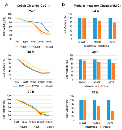
In that respect, the current Topical Collection invites contributions aimed at exploring the different characteristics of solid tumors, focused on, but not limited to, the microenvironment, such as hypoxia, alteration of tumor vasculature; extracellular matrix; pH; epigenetic changes, such as methylation, acetylation, and so on; metabolic changes, such as glucose uptake, glutamine metabolism, and so on; modification of microRNA signature; and interaction with the immune system. At the same time, contributions should provide the state-of-the-art of the therapies associated to or proposed for each solid tumor aspect discussed.

Prof. Dr. Elisabetta Ferretti
Dr. Marco Tafani
Collection Editors

Manuscript Submission Information

Manuscripts should be submitted online at www.mdpi.com by registering and logging in to this website. Once you are registered, click here to go to the submission form. Manuscripts can be submitted until the deadline. All submissions that pass pre-check are peer-reviewed. Accepted papers will be published continuously in the journal (as soon as accepted) and will be listed together on the collection website. Research articles, review articles as well as communications are invited. For planned papers, a title and short abstract (about 100 words) can be sent to the Editorial Office for announcement on this website.

Submitted manuscripts should not have been published previously, nor be under consideration for publication elsewhere (except conference proceedings papers). All manuscripts are thoroughly refereed through a single-blind peer-review process. A guide for authors and other relevant information for submission of manuscripts is available on the Instructions for Authors page. Cancers is an international peer-reviewed open access semimonthly journal published by MDPI.

Please visit the Instructions for Authors page before submitting a manuscript. The Article Processing Charge (APC) for publication in this open access journal is 2900 CHF (Swiss Francs). Submitted papers should be well formatted and use good English. Authors may use MDPI's English editing service prior to publication or during author revisions.

Keywords

  • solid tumors
  • targeted therapies
  • tumor microenvironment (TME)
  • immune evasion
  • epigenetic changes
  • microRNA signature
  • metabolic features
  • precision therapy
  • drug resistance
Published Papers (62 papers)

2024

Jump to: 2023, 2022, 2020, 2019

21 pages, 1704 KiB  
Review
Therapeutic Landscape of FOXM1 in Triple-Negative Breast Cancer and Aggressive Solid Cancers
by Sayra Dilmac, Zuhal Hamurcu and Bulent Ozpolat
Cancers 2024, 16(22), 3823; https://doi.org/10.3390/cancers16223823 - 14 Nov 2024
Viewed by 608
Abstract
Triple-negative breast cancer (TNBC) is one of the most aggressive forms of breast cancer, lacking common treatment targets such as estrogen (ER), progesterone (PR), and HER2 receptors. This subtype is associated with significant heterogeneity, chemoresistance, early recurrence, metastasis, and poor patient survival. FOXM1 [...] Read more.
Triple-negative breast cancer (TNBC) is one of the most aggressive forms of breast cancer, lacking common treatment targets such as estrogen (ER), progesterone (PR), and HER2 receptors. This subtype is associated with significant heterogeneity, chemoresistance, early recurrence, metastasis, and poor patient survival. FOXM1 is a cancer-promoting transcription factor that plays a critical role in TNBC and other highly aggressive cancers by driving cell proliferation, invasion, metastasis, and drug resistance. In TNBC, mutations in the TP53 gene—detected in approximately 80% of patients—lead to the overexpression of FOXM1, making it a promising therapeutic target. Beyond TNBC, FOXM1 is implicated in other solid cancers, such as brain (glioblastoma), lung, and pancreatic cancers, and is considered an Achilles’ heel of aggressive cancers. Despite its potential as a therapeutic target, there are currently no FDA-approved FOXM1 inhibitors, and none have advanced to clinical trials. This review explores the role of FOXM1 in cancer progression and highlights the current status of efforts to develop effective FOXM1 inhibitors. Full article
Show Figures

Figure 1

12 pages, 1016 KiB  
Article
Prospective Study of Non-Contrast, Abbreviated MRI for Hepatocellular Carcinoma Surveillance in Patients with Suboptimal Hepatic Visualisation on Ultrasound
by Mathew Vithayathil, Maria Qurashi, Pedro Rente Vicente, Ali Alsafi, Mitesh Naik, Alison Graham, Shahid Khan, Heather Lewis, Ameet Dhar, Belinda Smith, Nowlan Selvapatt, Pinelopi Manousou, Lucia Possamai, Hooshang Izadi, Adrian Lim, Paul Tait and Rohini Sharma
Cancers 2024, 16(15), 2709; https://doi.org/10.3390/cancers16152709 - 30 Jul 2024
Viewed by 1455
Abstract
Background: Biannual ultrasound (US) is recommended for hepatocellular carcinoma (HCC) surveillance in patients with cirrhosis. However, US has limited sensitivity for early-stage HCC, particularly in overweight cohorts, where hepatic visualisation is often inadequate. Currently there are no robust imaging surveillance strategies in patients [...] Read more.
Background: Biannual ultrasound (US) is recommended for hepatocellular carcinoma (HCC) surveillance in patients with cirrhosis. However, US has limited sensitivity for early-stage HCC, particularly in overweight cohorts, where hepatic visualisation is often inadequate. Currently there are no robust imaging surveillance strategies in patients with inadequate US visualisation. We investigated the ability of non-contrast, abbreviated magnetic resonance imaging (aMRI) to adequately visualise the liver for HCC surveillance in patients with previously inadequate US. Methods: Patients undergoing US surveillance, where liver visualisation was inadequate (LI-RADS VIS-B and VIS-C), were prospectively recruited. Patients underwent non-contrast T2-weighted and diffusion-weighted aMRI. The images were reviewed and reported by an expert liver radiologist. Three independent, blinded radiologists assessed the aMRI visualisation quality using a binary score assessing five parameters (parenchymal definition, vascular definition, coverage of the liver, uniformity of liver appearance and signal-to-noise ratio). Results: Thirty patients completed the aMRI protocol. The majority (90%) had underlying cirrhosis and were overweight (93.3%), with 50% obese and 20% severely obese. A total of 93.3% of the aMRI scans were of satisfactory quality. Six patients (20%) had hepatic abnormalities detected with aMRI that were not seen on their US: one HCC, one haemangioma and three clinically insignificant lesions. For the aMRI visualisation quality assessment, the coverage of the liver, vascular definition and parenchymal definition were consistently rated to be of sufficient quality by all three radiologists. Conclusions: Non-contrast aMRI provided good visualisation of the liver and detection of abnormalities in patients with inadequate US. aMRI should be further explored in a larger, prospective study as an alternative surveillance strategy in patients with inadequate US. Full article
Show Figures

Figure 1

29 pages, 986 KiB  
Systematic Review
A Molecular Hypothesis on Malignant Transformation of Oral Lichen Planus: A Systematic Review and Meta-Analysis of Cancer Hallmarks Expression in This Oral Potentially Malignant Disorder
by Carmen Keim-del Pino, Pablo Ramos-García and Miguel Ángel González-Moles
Cancers 2024, 16(15), 2614; https://doi.org/10.3390/cancers16152614 - 23 Jul 2024
Cited by 1 | Viewed by 1056
Abstract
We aimed to qualitatively and quantitatively analyze, through a systematic review and meta-analysis, the current evidence on the differential expression of the hallmarks of cancer in oral lichen planus (OLP) samples, in order to know the earliest molecular mechanisms that could be involved [...] Read more.
We aimed to qualitatively and quantitatively analyze, through a systematic review and meta-analysis, the current evidence on the differential expression of the hallmarks of cancer in oral lichen planus (OLP) samples, in order to know the earliest molecular mechanisms that could be involved in the malignant transformation of this oral potentially malignant disorder. We searched MEDLINE/PubMed, Embase, Web of Science, and Scopus for studies published before November 2023. We evaluated the methodological quality of studies and carried out meta-analyses to fulfill our objectives. Inclusion criteria were met by 110 primary-level studies, with 7065 OLP samples, in which the expression of 104 biomarkers were analyzed through immunohistochemistry. Most OLP samples showed sustained cell proliferation signaling (65.48%, 95%CI = 51.87–78.02), anti-apoptotic pathways (55.93%, 95%CI = 35.99–75.0), genome instability (48.44%, 95%CI = 13.54–84.19), and tumor-promoting inflammation events (83.10%, 95%CI = 73.93–90.74). Concurrently, OLP samples also harbored tumor growth suppressor mechanisms (64.00%, 95%CI = 53.27–74.12). In conclusion, current evidence indicates that molecular mechanisms promoting hyperproliferative signaling, an antiapoptotic state with genomic instability, and an escape of epithelial cells from immune destruction, are developed in LP-affected oral mucosa. It is plausible that these events are due to the actions exerted by the chronic inflammatory infiltrate. Malignant transformation appears to be prevented by tumor suppressor genes, which showed consistent upregulation in OLP samples. Full article
Show Figures

Figure 1

20 pages, 3316 KiB  
Review
Therapeutic Implications of Targeting YY1 in Glioblastoma
by Inesa Navasardyan, Apostolos Zaravinos and Benjamin Bonavida
Cancers 2024, 16(11), 2074; https://doi.org/10.3390/cancers16112074 - 30 May 2024
Viewed by 1115
Abstract
The transcription factor Yin Yang 1 (YY1) plays a pivotal role in the pathogenesis of glioblastoma multiforme (GBM), an aggressive form of brain tumor. This review systematically explores the diverse roles of YY1 overexpression and activities in GBM, including its impact on the [...] Read more.
The transcription factor Yin Yang 1 (YY1) plays a pivotal role in the pathogenesis of glioblastoma multiforme (GBM), an aggressive form of brain tumor. This review systematically explores the diverse roles of YY1 overexpression and activities in GBM, including its impact on the tumor microenvironment (TME) and immune evasion mechanisms. Due to the poor response of GBM to current therapies, various findings of YY1-associated pathways in the literature provide valuable insights into novel potential targeted therapeutic strategies. Moreover, YY1 acts as a significant regulator of immune checkpoint molecules and, thus, is a candidate therapeutic target in combination with immune checkpoint inhibitors. Different therapeutic implications targeting YY1 in GBM and its inherent associated challenges encompass the use of nanoparticles, YY1 inhibitors, targeted gene therapy, and exosome-based delivery systems. Despite the inherent complexities of such methods, the successful targeting of YY1 emerges as a promising avenue for reshaping GBM treatment strategies, presenting opportunities for innovative therapeutic approaches and enhanced patient outcomes. Full article
Show Figures

Figure 1

3 pages, 894 KiB  
Correction
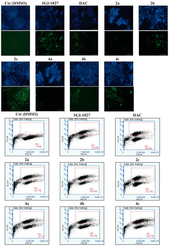
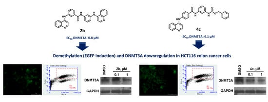
Correction: Zwergel et al. Novel Quinoline Compounds Active in Cancer Cells through Coupled DNA Methyltransferase Inhibition and Degradation. Cancers 2020, 12, 447
by Clemens Zwergel, Rossella Fioravanti, Giulia Stazi, Federica Sarno, Cecilia Battistelli, Annalisa Romanelli, Angela Nebbioso, Eduarda Mendes, Alexandra Paulo, Raffaele Strippoli, Marco Tripodi, Dany Pechalrieu, Paola B. Arimondo, Teresa De Luca, Donatella Del Bufalo, Daniela Trisciuoglio, Lucia Altucci, Sergio Valente and Antonello Mai
Cancers 2024, 16(6), 1230; https://doi.org/10.3390/cancers16061230 - 21 Mar 2024
Viewed by 990
Abstract
In the original publication [...] Full article
Show Figures

Figure 7

2023

Jump to: 2024, 2022, 2020, 2019

13 pages, 1520 KiB  
Article
Detecting Microsatellite Instability in Endometrial, Colon, and Stomach Cancers Using Targeted NGS
by Ulyana Boyarskikh, Andrey Kechin, Evgeniy Khrapov, Mikhail Fedyanin, Grigory Raskin, Marina Mukhina, Elena Kravtsova, Aleksey Tsukanov, Sergey Achkasov and Maksim Filipenko
Cancers 2023, 15(20), 5065; https://doi.org/10.3390/cancers15205065 - 20 Oct 2023
Viewed by 1660
Abstract
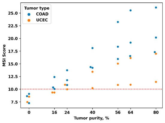
Purpose: To develop a method for testing the MSI based on targeted NGS. Methods: Based on the results of previous studies, 81 microsatellite loci with high variability in MSI-H tumors were selected, and a method for calculating the MSI score was developed. Using [...] Read more.
Purpose: To develop a method for testing the MSI based on targeted NGS. Methods: Based on the results of previous studies, 81 microsatellite loci with high variability in MSI-H tumors were selected, and a method for calculating the MSI score was developed. Using the MSI score, we defined the MSI status in endometral (162), colon (153), and stomach (190) cancers. Accuracy of the MSI scores was evaluated by comparison with MMR immunohistochemistry for 137 endometrium (63 dMMR and 74 pMMR), 76 colon (29 dMMR and 47 pMMR), and 81 stomach (8 dMMR and 73 pMMR) cancers. Results: Classification of MSS and MSI-H tumors was performed with AUC (0.99), sensitivity (92%), and specificity (98%) for all tumors without division into types. The accuracy of MSI testing in endometrial cancer was lower than for stomach and colon cancer (0.98, 87%, and 100%, respectively). The use of 27 loci only, the most informative for endometrial cancer, increased the overall accuracy (1.00, 99%, and 99%). Comparison of MSI score values in 505 tumors showed that MSI score is significantly higher in colon (p < 10−5) and stomach (p = 0.008) cancer compared with endometrial cancer. Conclusion: The MSI score accurately determines MSI status for endometrial, colon, and stomach cancers and can be used to quantify the degree of MSI. Full article
Show Figures

Figure 1

20 pages, 4917 KiB  
Article
Methotrexate Chemotherapy Causes Growth Impairments, Vitamin D Deficiency, Bone Loss, and Altered Intestinal Metabolism—Effects of Calcitriol Supplementation
by Yu-Wen Su, Alice M. C. Lee, Xukang Xu, Belinda Hua, Heather Tapp, Xue-Sen Wen and Cory J. Xian
Cancers 2023, 15(17), 4367; https://doi.org/10.3390/cancers15174367 - 1 Sep 2023
Cited by 3 | Viewed by 1949
Abstract
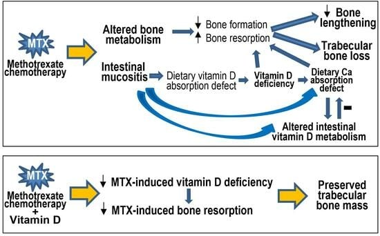
Vitamin D deficiency or insufficiency is prevalent in childhood cancer patients and survivors after chemotherapy; further studies are needed to investigate the underlying aetiology and effectiveness of vitamin D supplementation in preventing chemotherapy-induced bone loss. This study used a rat model of treatment [...] Read more.
Vitamin D deficiency or insufficiency is prevalent in childhood cancer patients and survivors after chemotherapy; further studies are needed to investigate the underlying aetiology and effectiveness of vitamin D supplementation in preventing chemotherapy-induced bone loss. This study used a rat model of treatment with antimetabolite methotrexate to investigate whether methotrexate chemotherapy causes vitamin D deficiency and if vitamin D supplementation attenuates the resultant bone loss. Methotrexate treatment (five daily injections) decreased serum vitamin D levels (from 52 to <30 ng/mL), reduced body and bone lengthening and tibial trabecular bone volume, and altered intestinal vitamin D metabolism, which was associated with intestinal mucosal damage known to cause malabsorption of nutrients, including dietary vitamin D and calcium. During the early stage after chemotherapy, mRNA expression increased for vitamin D activation enzyme CYP27B1 and for calcium-binding protein TRPV6 in the intestine. During the intestinal healing stage, expression of vitamin D catabolism enzyme CYP24 increased, and that of TRPV6 was normalised. Furthermore, subcutaneous calcitriol supplementation diminished methotrexate-induced bone loss due to its effect suppressing methotrexate-induced increased bone resorption. Thus, in young rats, methotrexate chemotherapy causes vitamin D deficiency, growth impairments, bone loss, and altered intestinal vitamin D metabolism, which are associated with intestinal damage, and vitamin D supplementation inhibits methotrexate-induced bone loss. Full article
Show Figures

Graphical abstract

15 pages, 1207 KiB  
Review
Applications of CRISPR Technology to Breast Cancer and Triple Negative Breast Cancer Research
by Mariona Pont, Marta Marqués, Maria Alba Sorolla, Eva Parisi, Izaskun Urdanibia, Serafín Morales, Antonieta Salud and Anabel Sorolla
Cancers 2023, 15(17), 4364; https://doi.org/10.3390/cancers15174364 - 1 Sep 2023
Cited by 5 | Viewed by 2668
Abstract
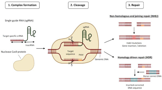
Clustered Regularly Interspaced Short Palindromic Repeats (CRISPR) technology has transformed oncology research in many ways. Breast cancer is the most prevalent malignancy globally and triple negative breast cancer (TNBC) is one of the most aggressive subtypes with numerous challenges still to be faced. [...] Read more.
Clustered Regularly Interspaced Short Palindromic Repeats (CRISPR) technology has transformed oncology research in many ways. Breast cancer is the most prevalent malignancy globally and triple negative breast cancer (TNBC) is one of the most aggressive subtypes with numerous challenges still to be faced. In this work, we have explained what CRISPR consists of and listed its applications in breast cancer while focusing on TNBC research. These are disease modelling, the search for novel genes involved in tumour progression, sensitivity to drugs and immunotherapy response, tumour fitness, diagnosis, and treatment. Additionally, we have listed the current delivery methods employed for the delivery of CRISPR systems in vivo. Lastly, we have highlighted the limitations that CRISPR technology is subject to and the future directions that we envisage. Overall, we have provided a round summary of the aspects concerning CRISPR in breast cancer/TNBC research. Full article
Show Figures

Figure 1

19 pages, 819 KiB  
Article
Changes in Gastric Pathology after H. pylori Treatment in Community-Driven Research Aimed at Gastric Cancer Prevention
by Ting Wang, Safwat Girgis, Hsiu-Ju Chang, Ali Assi, Katharine Fagan-Garcia, Taylor Cromarty, Rachel Munday, Karen J. Goodman, Sander Veldhuyzen van Zanten and the CANHelp Working Group
Cancers 2023, 15(15), 3950; https://doi.org/10.3390/cancers15153950 - 3 Aug 2023
Cited by 1 | Viewed by 1517
Abstract
Community-driven projects have characterized Helicobacter pylori (Hp) infection in Indigenous communities in the Northwest Territories (NT) and Yukon (YT), Canada. These projects address concerns about the frequent diagnosis of Hp infection among community members and its relation to gastric cancer deaths, [...] Read more.
Community-driven projects have characterized Helicobacter pylori (Hp) infection in Indigenous communities in the Northwest Territories (NT) and Yukon (YT), Canada. These projects address concerns about the frequent diagnosis of Hp infection among community members and its relation to gastric cancer deaths, perceived to occur with alarming frequency in this region. Projects included breath-test screening for Hp infection, gastroscopy with gastric biopsies, and treatment to eliminate Hp infection. Previous project results showed a high prevalence of stomach pathologies associated with increased cancer risk among Hp-positive participants at baseline. This analysis describes changes in precancerous gastric pathologies in project participants who had gastroscopy before baseline treatment during 2008–2013 and again in 2017. Throughout the study period, the same pathologist graded Hp density, active gastritis, chronic gastritis, atrophic gastritis, and intestinal metaplasia using the updated Sydney System. Of 310 participants from three communities with baseline pathology data, 69 had follow-up pathology data. Relative to baseline, the prevalence of Hp infection and precancerous gastric pathology was substantially lower at follow-up; most participants who were Hp-positive at baseline and Hp-negative at follow-up had reduced severity of active, chronic, and/or atrophic gastritis at follow-up. Though follow-up numbers are small, these results yield evidence that successful Hp treatment has the potential to reduce the risk of gastric cancer in Arctic Indigenous communities. Full article
Show Figures

Figure 1

21 pages, 3742 KiB  
Article
iPSC-Derived Glioblastoma Cells Have Enhanced Stemness Wnt/β-Catenin Activity Which Is Negatively Regulated by Wnt Antagonist sFRP4
by Ishmat Ara Yasmin, Arun Dharmarajan and Sudha Warrier
Cancers 2023, 15(14), 3622; https://doi.org/10.3390/cancers15143622 - 14 Jul 2023
Cited by 3 | Viewed by 2242
Abstract
Growing evidence indicates that cancer stem cells (CSCs) endow the tumor with stem-like properties. Recently, induced pluripotent stem cells (iPSCs) have gained increased attention because of their easy derivation and availability and their potential to differentiate into any cell type. A CSC model [...] Read more.
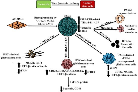
Growing evidence indicates that cancer stem cells (CSCs) endow the tumor with stem-like properties. Recently, induced pluripotent stem cells (iPSCs) have gained increased attention because of their easy derivation and availability and their potential to differentiate into any cell type. A CSC model derived from iPSCs of human origin would help understand the driving force of tumor initiation and early progression. We report the efficient generation of feeder-free SSEA4, TRA-1-60 and TRA-1-81 positive iPSCs from amniotic membrane-derived mesenchymal stem cells (AMMSCs), which successfully differentiated into three germ layers. We then developed human iPSC-derived glioblastoma multiforme (GBM) model using conditioned media (CM) from U87MG cell line and CSCs derived from U87MG, which confer iPSCs with GBM and GSC-like phenotypes within five days. Both cell types overexpress MGMT and GLI2, but only GSCs overexpress CD133, CD44, ABCG2 and ABCC2. We also observed overexpression of LEF1 and β-catenin in both cell types. Down-regulation of Wnt antagonist secreted frizzled-related protein 4 (sFRP4) in GBM and GSCs, indicating activation of the Wnt/β-catenin pathway, which could be involved in the conversion of iPSCs to CSCs. From future perspectives, our study will help in the creation of a rapid cell-based platform for understanding the complexity of GBM. Full article
Show Figures

Graphical abstract

19 pages, 2444 KiB  
Article
The Tumor Suppressor DAB2IP Is Regulated by Cell Contact and Contributes to YAP/TAZ Inhibition in Confluent Cells
by Mattia Apollonio, Arianna Bellazzo, Nicoletta Franco, Silvia Lombardi, Beatrice Senigagliesi, Loredana Casalis, Pietro Parisse, Agnes Thalhammer, Gabriele Baj, Rossella De Florian Fania, Giannino Del Sal and Licio Collavin
Cancers 2023, 15(13), 3379; https://doi.org/10.3390/cancers15133379 - 27 Jun 2023
Cited by 1 | Viewed by 2326
Abstract
External and internal mechanical forces modulate cell morphology, movement, proliferation and metabolism, and represent crucial inputs for tissue homeostasis. The transcriptional regulators YAP and TAZ are important effectors of mechanical signaling and are frequently activated in solid tumors, correlating with metastasis, chemoresistance, and [...] Read more.
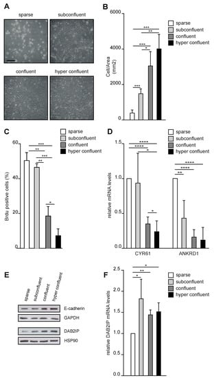
External and internal mechanical forces modulate cell morphology, movement, proliferation and metabolism, and represent crucial inputs for tissue homeostasis. The transcriptional regulators YAP and TAZ are important effectors of mechanical signaling and are frequently activated in solid tumors, correlating with metastasis, chemoresistance, and shorter patient survival. YAP/TAZ activity is controlled by various pathways that sense cell shape, polarity, contacts, and mechanical tension. In tumors, aberrant YAP/TAZ activation may result from cancer-related alterations of such regulatory networks. The tumor suppressor DAB2IP is a Ras-GAP and scaffold protein that negatively modulates multiple oncogenic pathways and is frequently downregulated or inactivated in solid tumors. Here, we provide evidence that DAB2IP expression is sustained by cell confluency. We also find that DAB2IP depletion in confluent cells alters their morphology, reducing cell packing while increasing cell stiffness. Finally, we find that DAB2IP depletion in confluent cells favors YAP/TAZ nuclear localization and transcriptional activity, while its ectopic expression in subconfluent cells increases YAP/TAZ retention in the cytoplasm. Together, these data suggest that DAB2IP may function as a sensor of cell interactions, contributing to dampening cellular responses to oncogenic inputs in confluent cells and that DAB2IP loss-of-function would facilitate YAP/TAZ activation in intact epithelia, accelerating oncogenic transformation. Full article
Show Figures

Figure 1

17 pages, 2810 KiB  
Review
The Role of MRI and PET/CT in Radiotherapy Target Volume Determination in Gastrointestinal Cancers—Review of the Literature
by Ajra Secerov Ermenc and Barbara Segedin
Cancers 2023, 15(11), 2967; https://doi.org/10.3390/cancers15112967 - 29 May 2023
Cited by 3 | Viewed by 1843
Abstract
Positron emission tomography with computed tomography (PET/CT) and magnetic resonance imaging (MRI) could improve accuracy in target volume determination for gastrointestinal cancers. A systematic search of the PubMed database was performed, focusing on studies published within the last 20 years. Articles were considered [...] Read more.
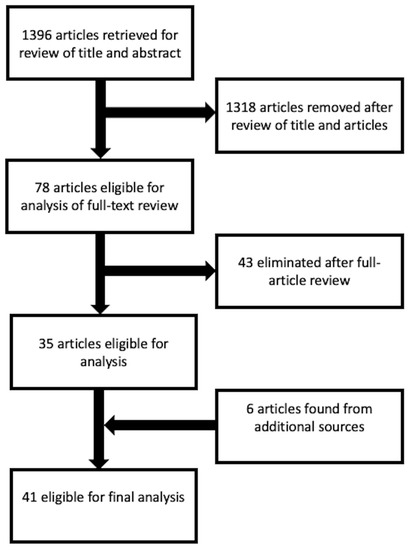
Positron emission tomography with computed tomography (PET/CT) and magnetic resonance imaging (MRI) could improve accuracy in target volume determination for gastrointestinal cancers. A systematic search of the PubMed database was performed, focusing on studies published within the last 20 years. Articles were considered eligible for the review if they included patients with anal canal, esophageal, rectal or pancreatic cancer, as well as PET/CT or MRI for radiotherapy treatment planning, and if they reported interobserver variability or changes in treatment planning volume due to different imaging modalities or correlation between the imaging modality and histopathologic specimen. The search of the literature retrieved 1396 articles. We retrieved six articles from an additional search of the reference lists of related articles. Forty-one studies were included in the final review. PET/CT seems indispensable for target volume determination of pathological lymph nodes in esophageal and anal canal cancer. MRI seems appropriate for the delineation of primary tumors in the pelvis as rectal and anal canal cancer. Delineation of the target volumes for radiotherapy of pancreatic cancer remains challenging, and additional studies are needed. Full article
Show Figures

Figure 1

16 pages, 2592 KiB  
Article
COL7A1 Expression Improves Prognosis Prediction for Patients with Clear Cell Renal Cell Carcinoma Atop of Stage
by Dzenis Koca, Irinka Séraudie, Rémy Jardillier, Claude Cochet, Odile Filhol and Laurent Guyon
Cancers 2023, 15(10), 2701; https://doi.org/10.3390/cancers15102701 - 10 May 2023
Cited by 5 | Viewed by 2420
Abstract
Clear-cell renal cell carcinoma (ccRCC) accounts for 75% of kidney cancers. Due to the high recurrence rate and treatment options that come with high costs and potential side effects, a correct prognosis of patient survival is essential for the successful and effective treatment [...] Read more.
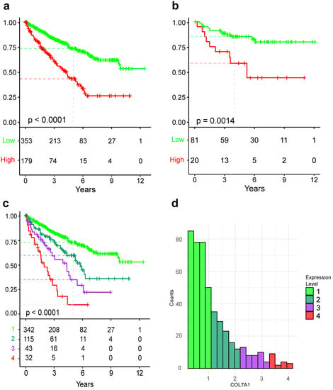
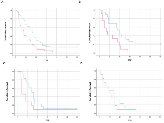
Clear-cell renal cell carcinoma (ccRCC) accounts for 75% of kidney cancers. Due to the high recurrence rate and treatment options that come with high costs and potential side effects, a correct prognosis of patient survival is essential for the successful and effective treatment of patients. Novel biomarkers could play an important role in the assessment of the overall survival of patients. COL7A1 encodes for collagen type VII, a constituent of the basal membrane. COL7A1 is associated with survival in many cancers; however, the prognostic value of COL7A1 expression as a standalone biomarker in ccRCC has not been investigated. With five publicly available independent cohorts, we used Kaplan–Meier curves and the Cox proportional hazards model to investigate the prognostic value of COL7A1, as well as gene set enrichment analysis to investigate genes co-expressed with COL7A1. COL7A1 expression stratifies patients in terms of aggressiveness, where the 5-year survival probability of each of the four groups was 72.4%, 59.1%, 34.15%, and 8.6% in order of increasing expression. Additionally, COL7A1 expression was successfully used to further divide patients of each stage and histological grade into groups of high and low risk. Similar results were obtained in independent cohorts. In vitro knockdown of COL7A1 expression significantly affected ccRCC cells’ ability to migrate, leading to the hypothesis that COL7A1 may have a role in cancer aggressiveness. To conclude, we identified COL7A1 as a new prognosis marker that can stratify ccRCC patients. Full article
Show Figures

Figure 1

21 pages, 623 KiB  
Review
Radiotherapy for Mobile Spine and Sacral Chordoma: A Critical Review and Practical Guide from the Spine Tumor Academy
by Kristin J. Redmond, Stephanie K. Schaub, Sheng-fu Larry Lo, Majid Khan, Daniel Lubelski, Mark Bilsky, Yoshiya Yamada, Michael Fehlings, Emile Gogineni, Peter Vajkoczy, Florian Ringel, Bernhard Meyer, Anubhav G. Amin, Stephanie E. Combs and Simon S. Lo
Cancers 2023, 15(8), 2359; https://doi.org/10.3390/cancers15082359 - 18 Apr 2023
Cited by 7 | Viewed by 3502
Abstract
Chordomas are rare tumors of the embryologic spinal cord remnant. They are locally aggressive and typically managed with surgery and either adjuvant or neoadjuvant radiation therapy. However, there is great variability in practice patterns including radiation type and fractionation regimen, and limited high-level [...] Read more.
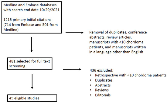
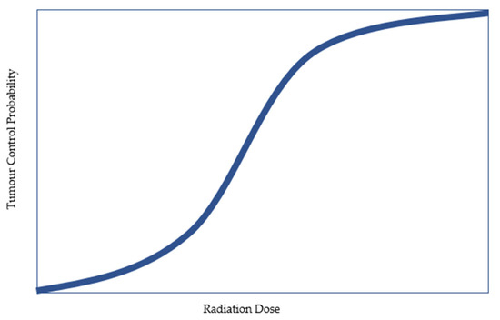
Chordomas are rare tumors of the embryologic spinal cord remnant. They are locally aggressive and typically managed with surgery and either adjuvant or neoadjuvant radiation therapy. However, there is great variability in practice patterns including radiation type and fractionation regimen, and limited high-level data to drive decision making. The purpose of this manuscript was to summarize the current literature specific to radiotherapy in the management of spine and sacral chordoma and to provide practice recommendations on behalf of the Spine Tumor Academy. A systematic review of the literature was performed using the Preferred Reporting Items for Systematic reviews and Meta-Analyses (PRISMA) approach. Medline and Embase databases were utilized. The primary outcome measure was the rate of local control. A detailed review and interpretation of eligible studies is provided in the manuscript tables and text. Recommendations were defined as follows: (1) consensus: approved by >75% of experts; (2) predominant: approved by >50% of experts; (3) controversial: not approved by a majority of experts. Expert consensus supports dose escalation as critical in optimizing local control following radiation therapy for chordoma. In addition, comprehensive target volumes including sites of potential microscopic involvement improve local control compared with focal targets. Level I and high-quality multi-institutional data comparing treatment modalities, sequencing of radiation and surgery, and dose/fractionation schedules are needed to optimize patient outcomes in this locally aggressive malignancy. Full article
Show Figures

Figure 1

21 pages, 1032 KiB  
Review
PARP Inhibitors in Breast and Ovarian Cancer
by Samuel S. Y. Wang, Yeo Ee Jie, Sim Wey Cheng, Goh Liuh Ling and Heong Valerie Yue Ming
Cancers 2023, 15(8), 2357; https://doi.org/10.3390/cancers15082357 - 18 Apr 2023
Cited by 18 | Viewed by 3375
Abstract
Poly (ADP-ribose) polymerase (PARP) inhibitors are one of the most successful examples of clinical translation of targeted therapies in medical oncology, and this has been demonstrated by their effective management of BRCA1/BRCA2 mutant cancers, most notably in breast and ovarian cancers. PARP inhibitors [...] Read more.
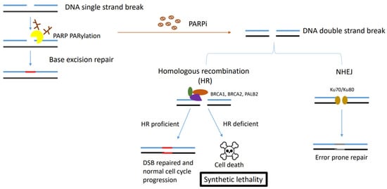
Poly (ADP-ribose) polymerase (PARP) inhibitors are one of the most successful examples of clinical translation of targeted therapies in medical oncology, and this has been demonstrated by their effective management of BRCA1/BRCA2 mutant cancers, most notably in breast and ovarian cancers. PARP inhibitors target DNA repair pathways that BRCA1/2-mutant tumours are dependent upon. Inhibition of the key components of these pathways leads to DNA damage triggering subsequent critical levels of genomic instability, mitotic catastrophe and cell death. This ultimately results in a synthetic lethal relationship between BRCA1/2 and PARP, which underpins the effectiveness of PARP inhibitors. Despite the early and dramatic response seen with PARP inhibitors, patients receiving them often develop treatment resistance. To date, data from both clinical and preclinical studies have highlighted multiple resistance mechanisms to PARP inhibitors, and only by understanding these mechanisms are we able to overcome the challenges. The focus of this review is to summarise the underlying mechanisms underpinning treatment resistance to PARP inhibitors and to aid both clinicians and scientists to develop better clinically applicable assays to better select patients who would derive the greatest benefit as well as develop new novel/combination treatment strategies to overcome these mechanisms of resistance. With a better understanding of PARP inhibitor resistance mechanisms, we would not only be able to identify a subset of patients who are unlikely to benefit from therapy but also to sequence our treatment paradigm to avoid and overcome these resistance mechanisms. Full article
Show Figures

Figure 1

18 pages, 320 KiB  
Review
NEK Family Review and Correlations with Patient Survival Outcomes in Various Cancer Types
by Khoa Nguyen, Julia Boehling, Minh N. Tran, Thomas Cheng, Andrew Rivera, Bridgette M. Collins-Burow, Sean B. Lee, David H. Drewry and Matthew E. Burow
Cancers 2023, 15(7), 2067; https://doi.org/10.3390/cancers15072067 - 30 Mar 2023
Cited by 7 | Viewed by 2646
Abstract
The Never in Mitosis Gene A (NIMA)–related kinases (NEKs) are a group of serine/threonine kinases that are involved in a wide array of cellular processes including cell cycle regulation, DNA damage repair response (DDR), apoptosis, and microtubule organization. Recent studies have identified the [...] Read more.
The Never in Mitosis Gene A (NIMA)–related kinases (NEKs) are a group of serine/threonine kinases that are involved in a wide array of cellular processes including cell cycle regulation, DNA damage repair response (DDR), apoptosis, and microtubule organization. Recent studies have identified the involvement of NEK family members in various diseases such as autoimmune disorders, malignancies, and developmental defects. Despite the existing literature exemplifying the importance of the NEK family of kinases, this family of protein kinases remains understudied. This report seeks to provide a foundation for investigating the role of different NEKs in malignancies. We do this by evaluating the 11 NEK family kinase gene expression associations with patients’ overall survival (OS) from various cancers using the Kaplan–Meier Online Tool (KMPlotter) to correlate the relationship between mRNA expression of NEK1-11 in various cancers and patient survival. Furthermore, we use the Catalog of Somatic Mutations in Cancer (COSMIC) database to identify NEK family mutations in cancers of different tissues. Overall, the data suggest that the NEK family has varying associations with patient survival in different cancers with tumor-suppressive and tumor-promoting effects being tissue-dependent. Full article
29 pages, 1826 KiB  
Review
Virtual HDR Boost for Prostate Cancer: Rebooting a Classic Treatment Using Modern Tech
by Eric Wegener, Justin Samuels, Mark Sidhom, Yuvnik Trada, Swetha Sridharan, Samuel Dickson, Nicholas McLeod and Jarad M. Martin
Cancers 2023, 15(7), 2018; https://doi.org/10.3390/cancers15072018 - 28 Mar 2023
Cited by 1 | Viewed by 1947
Abstract
Prostate cancer (PC) is the most common malignancy in men. Internal radiotherapy (brachytherapy) has been used to treat PC successfully for over a century. In particular, there is level-one evidence of the benefits of using brachytherapy to escalate the dose of radiotherapy compared [...] Read more.
Prostate cancer (PC) is the most common malignancy in men. Internal radiotherapy (brachytherapy) has been used to treat PC successfully for over a century. In particular, there is level-one evidence of the benefits of using brachytherapy to escalate the dose of radiotherapy compared with standard external beam radiotherapy approaches. However, the use of PC brachytherapy is declining, despite strong evidence for its improved cancer outcomes. A method using external beam radiotherapy known as virtual high-dose-rate brachytherapy boost (vHDRB) aims to noninvasively mimic a brachytherapy boost radiation dose plan. In this review, we consider the evidence supporting brachytherapy boosts for PC and the continuing evolution of vHDRB approaches, culminating in the current generation of clinical trials, which will help define the role of this emerging modality. Full article
Show Figures

Figure 1

29 pages, 8922 KiB  
Article
Polyphenolic Boronates Inhibit Tumor Cell Proliferation: Potential Mitigators of Oxidants in the Tumor Microenvironment
by Gang Cheng, Hakim Karoui, Micael Hardy and Balaraman Kalyanaraman
Cancers 2023, 15(4), 1089; https://doi.org/10.3390/cancers15041089 - 8 Feb 2023
Cited by 2 | Viewed by 2365
Abstract
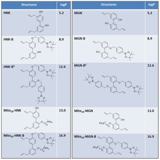
Boronate-based compounds have been used in brain cancer therapy, either as prodrugs or in combination with other modalities. Boronates containing pro-luminescent and fluorescent probes have been used in mouse models of cancer. In this study, we synthesized and developed polyphenolic boronates and mitochondria-targeted [...] Read more.
Boronate-based compounds have been used in brain cancer therapy, either as prodrugs or in combination with other modalities. Boronates containing pro-luminescent and fluorescent probes have been used in mouse models of cancer. In this study, we synthesized and developed polyphenolic boronates and mitochondria-targeted polyphenolic phytochemicals (e.g., magnolol [MGN] and honokiol [HNK]) and tested their antiproliferative effects in brain cancer cells. Results show that mitochondria-targeted (Mito) polyphenolic boronates (Mito-MGN-B and Mito-HNK-B) were slightly more potent than Mito-MGN and Mito-HNK in inhibiting proliferation of the U87MG cell line. Similar proliferation results also were observed in other cancer cell lines, such as MiaPaCa-2, A549 and UACC-62. Independent in vitro experiments indicated that reactive nitrogen species (e.g., peroxynitrite) and reactive oxygen species (e.g., hydrogen peroxide) stoichiometrically react with polyphenolic boronates and Mito-polphenolic boronates, forming polyphenols and Mito-polyphenols as major products. Previous reports suggest that both Mito-MGN and Mito-HNK activate cytotoxic T cells and inhibit immunosuppressive immune cells. We propose that Mito-polyphenolic boronate-based prodrugs may be used to inhibit tumor proliferation and mitigate oxidant formation in the tumor microenvironment, thereby generating Mito-polyphenols in situ, as well as showing activity in the tumor microenvironment. Full article
Show Figures

Figure 1

25 pages, 884 KiB  
Review
Intraoperative Imaging Techniques to Improve Surgical Resection Margins of Oropharyngeal Squamous Cell Cancer: A Comprehensive Review of Current Literature
by Bertram J. de Kleijn, Gijs T. N. Heldens, Jasmijn M. Herruer, Cornelis F. M. Sier, Cesare Piazza, Remco de Bree, Orlando Guntinas-Lichius, Luiz P. Kowalski, Vincent Vander Poorten, Juan P. Rodrigo, Nina Zidar, Cherie-Ann Nathan, Raymond K. Tsang, Pawel Golusinski, Ashok R. Shaha, Alfio Ferlito and Robert P. Takes
Cancers 2023, 15(3), 896; https://doi.org/10.3390/cancers15030896 - 31 Jan 2023
Cited by 10 | Viewed by 2675
Abstract
Inadequate resection margins in head and neck squamous cell carcinoma surgery necessitate adjuvant therapies such as re-resection and radiotherapy with or without chemotherapy and imply increasing morbidity and worse prognosis. On the other hand, taking larger margins by extending the resection also leads [...] Read more.
Inadequate resection margins in head and neck squamous cell carcinoma surgery necessitate adjuvant therapies such as re-resection and radiotherapy with or without chemotherapy and imply increasing morbidity and worse prognosis. On the other hand, taking larger margins by extending the resection also leads to avoidable increased morbidity. Oropharyngeal squamous cell carcinomas (OPSCCs) are often difficult to access; resections are limited by anatomy and functionality and thus carry an increased risk for close or positive margins. Therefore, there is a need to improve intraoperative assessment of resection margins. Several intraoperative techniques are available, but these often lead to prolonged operative time and are only suitable for a subgroup of patients. In recent years, new diagnostic tools have been the subject of investigation. This study reviews the available literature on intraoperative techniques to improve resection margins for OPSCCs. A literature search was performed in Embase, PubMed, and Cochrane. Narrow band imaging (NBI), high-resolution microendoscopic imaging, confocal laser endomicroscopy, frozen section analysis (FSA), ultrasound (US), computed tomography scan (CT), (auto) fluorescence imaging (FI), and augmented reality (AR) have all been used for OPSCC. NBI, FSA, and US are most commonly used and increase the rate of negative margins. Other techniques will become available in the future, of which fluorescence imaging has high potential for use with OPSCC. Full article
Show Figures

Figure 1

19 pages, 1749 KiB  
Article
Prognostic Implications of the Residual Tumor Microenvironment after Neoadjuvant Chemotherapy in Triple-Negative Breast Cancer Patients without Pathological Complete Response
by Marylène Lejeune, Laia Reverté, Esther Sauras, Noèlia Gallardo, Ramon Bosch, Albert Roso, Anna Petit, Vicente Peg, Francisco Riu, Joan García-Fontgivell, José Ibáñez, Fernanda Relea, Begoña Vieites, Catherine Bor, Luis de la Cruz-Merino, Meritxell Arenas, Valerie Rodriguez, Juana Galera, Anna Korzynska, Philippe Belhomme, Benoît Plancoulaine, Tomás Álvaro and Carlos Lópezadd Show full author list remove Hide full author list
Cancers 2023, 15(3), 597; https://doi.org/10.3390/cancers15030597 - 18 Jan 2023
Cited by 6 | Viewed by 3066
Abstract
With a high risk of relapse and death, and a poor or absent response to therapeutics, the triple-negative breast cancer (TNBC) subtype is particularly challenging, especially in patients who cannot achieve a pathological complete response (pCR) after neoadjuvant chemotherapy (NAC). Although the tumor [...] Read more.
With a high risk of relapse and death, and a poor or absent response to therapeutics, the triple-negative breast cancer (TNBC) subtype is particularly challenging, especially in patients who cannot achieve a pathological complete response (pCR) after neoadjuvant chemotherapy (NAC). Although the tumor microenvironment (TME) is known to influence disease progression and the effectiveness of therapeutics, its predictive and prognostic potential remains uncertain. This work aimed to define the residual TME profile after NAC of a retrospective cohort with 96 TNBC patients by immunohistochemical staining (cell markers) and chromogenic in situ hybridization (genetic markers). Kaplan–Meier curves were used to estimate the influence of the selected TME markers on five-year overall survival (OS) and relapse-free survival (RFS) probabilities. The risks of each variable being associated with relapse and death were determined through univariate and multivariate Cox analyses. We describe a unique tumor-infiltrating immune profile with high levels of lymphocytes (CD4, FOXP3) and dendritic cells (CD21, CD1a and CD83) that are valuable prognostic factors in post-NAC TNBC patients. Our study also demonstrates the value of considering not only cellular but also genetic TME markers such as MUC-1 and CXCL13 in routine clinical diagnosis to refine prognosis modelling. Full article
Show Figures

Figure 1

16 pages, 1226 KiB  
Systematic Review
Helicobacter Species and Hepato-Biliary Tract Malignancies: A Systematic Review and Meta-Analysis
by Beatriz Gros, Alberto Gómez Pérez, María Pleguezuelo, Francisco Javier Serrano Ruiz, Manuel de la Mata and Manuel Rodríguez-Perálvarez
Cancers 2023, 15(3), 595; https://doi.org/10.3390/cancers15030595 - 18 Jan 2023
Cited by 4 | Viewed by 2301
Abstract
Helicobacter species may cause chronic inflammation of the biliary tract, but its relationship with cancer is controversial. We performed a systematic review and meta-analysis to evaluate the association between Helicobacter species and hepatobiliary tract malignancies. Twenty-six studies (4083 patients) were included in qualitative [...] Read more.
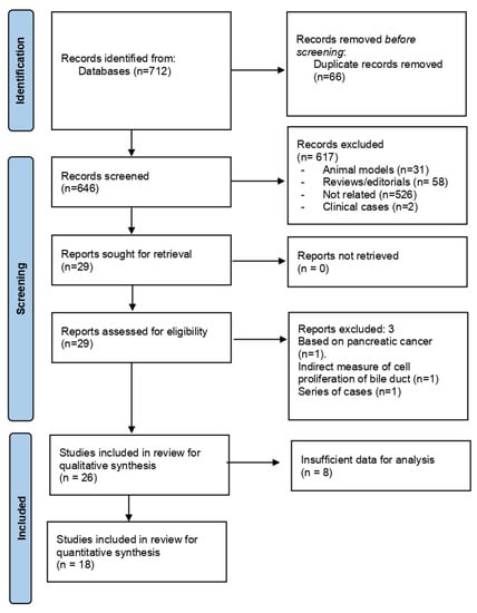
Helicobacter species may cause chronic inflammation of the biliary tract, but its relationship with cancer is controversial. We performed a systematic review and meta-analysis to evaluate the association between Helicobacter species and hepatobiliary tract malignancies. Twenty-six studies (4083 patients) were included in qualitative synthesis, and 18 studies (n = 1895 qualified for meta-analysis. All studies were at high-intermediate risk of bias. Most studies combined several direct microbiological methods, mostly PCR (23 studies), culture (8 studies), and/or CLOtest (5 studies). Different specimens alone or in combination were investigated, most frequently bile (16 studies), serum (7 studies), liver/biliary tissue (8 studies), and gastric tissue (3 studies). Patients with Helicobacter species infection had an increased risk of hepatobiliary tract malignancies (OR = 3.61 [95% CI 2.18–6.00]; p < 0.0001), with high heterogeneity in the analysis (I2 = 61%; p = 0.0003). This effect was consistent when Helicobacter was assessed in bile (OR = 3.57 [95% CI 1.73–7.39]; p = 0.0006), gastric tissue (OR = 42.63 [95% CI 5.25–346.24]; p = 0.0004), liver/biliary tissue (OR = 4.92 [95% CI 1.90–12.76]; p = 0.001) and serum (OR = 1.38 [95% CI 1.00–1.90]; p = 0.05). Heterogeneity was reduced in these sub-analyses (I2 = 0–27%; p = ns), except for liver/biliary tissue (I2 = 57%; p = 0.02). In conclusion, based on low-certainty data, Helicobacter species chronic infection is associated with a tripled risk of hepatobiliary tract malignancy. Prospective studies are required to delineate public health interventions. Full article
Show Figures

Figure 1

12 pages, 1559 KiB  
Article
Genetic Alterations in Members of the Proteasome 26S Subunit, AAA-ATPase (PSMC) Gene Family in the Light of Proteasome Inhibitor Resistance in Multiple Myeloma
by Larissa Haertle, Natalia Buenache, Hipólito Nicolás Cuesta Hernández, Michal Simicek, Renata Snaurova, Inmaculada Rapado, Nerea Martinez, Nieves López-Muñoz, José María Sánchez-Pina, Umair Munawar, Seungbin Han, Yanira Ruiz-Heredia, Rafael Colmenares, Miguel Gallardo, Margarita Sanchez-Beato, Miguel Angel Piris, Mehmet Kemal Samur, Nikhil C. Munshi, Rosa Ayala, Klaus Martin Kortüm, Santiago Barrio and Joaquín Martínez-Lópezadd Show full author list remove Hide full author list
Cancers 2023, 15(2), 532; https://doi.org/10.3390/cancers15020532 - 15 Jan 2023
Cited by 5 | Viewed by 3101
Abstract
For the treatment of Multiple Myeloma, proteasome inhibitors are highly efficient and widely used, but resistance is a major obstacle to successful therapy. Several underlying mechanisms have been proposed but were only reported for a minority of resistant patients. The proteasome is a [...] Read more.
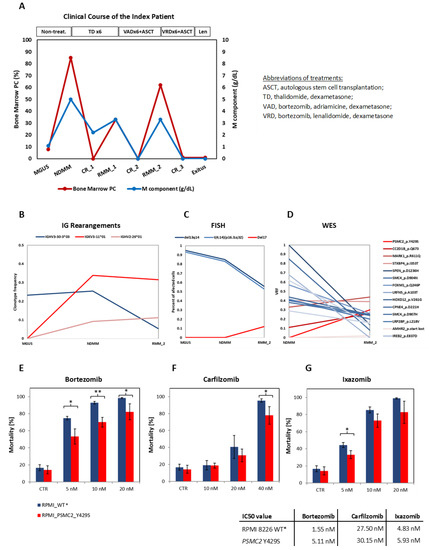
For the treatment of Multiple Myeloma, proteasome inhibitors are highly efficient and widely used, but resistance is a major obstacle to successful therapy. Several underlying mechanisms have been proposed but were only reported for a minority of resistant patients. The proteasome is a large and complex machinery. Here, we focus on the AAA ATPases of the 19S proteasome regulator (PSMC1-6) and their implication in PI resistance. As an example of cancer evolution and the acquisition of resistance, we conducted an in-depth analysis of an index patient by applying FISH, WES, and immunoglobulin-rearrangement sequencing in serial samples, starting from MGUS to newly diagnosed Multiple Myeloma to a PI-resistant relapse. The WES analysis uncovered an acquired PSMC2 Y429S mutation at the relapse after intensive bortezomib-containing therapy, which was functionally confirmed to mediate PI resistance. A meta-analysis comprising 1499 newly diagnosed and 447 progressed patients revealed a total of 36 SNVs over all six PSMC genes that were structurally accumulated in regulatory sites for activity such as the ADP/ATP binding pocket. Other alterations impact the interaction between different PSMC subunits or the intrinsic conformation of an individual subunit, consequently affecting the folding and function of the complex. Interestingly, several mutations were clustered in the central channel of the ATPase ring, where the unfolded substrates enter the 20S core. Our results indicate that PSMC SNVs play a role in PI resistance in MM. Full article
Show Figures

Figure 1

23 pages, 6512 KiB  
Article
Arachidin-1, a Prenylated Stilbenoid from Peanut, Enhances the Anticancer Effects of Paclitaxel in Triple-Negative Breast Cancer Cells
by Sepideh Mohammadhosseinpour, Alexx Weaver, Meenakshi Sudhakaran, Linh-Chi Ho, Tra Le, Andrea I. Doseff and Fabricio Medina-Bolivar
Cancers 2023, 15(2), 399; https://doi.org/10.3390/cancers15020399 - 7 Jan 2023
Cited by 7 | Viewed by 3093
Abstract
Triple-negative breast cancer (TNBC) is one of the deadliest forms of breast cancer. Investigating alternative therapies to increase survival rates for this disease is essential. To this end, the cytotoxic effects of the prenylated stilbenoids arachidin-1 (A-1) and arachidin-3 (A-3), and non-prenylated resveratrol [...] Read more.
Triple-negative breast cancer (TNBC) is one of the deadliest forms of breast cancer. Investigating alternative therapies to increase survival rates for this disease is essential. To this end, the cytotoxic effects of the prenylated stilbenoids arachidin-1 (A-1) and arachidin-3 (A-3), and non-prenylated resveratrol (RES) were evaluated in human TNBC cell lines as potential adjuvants for paclitaxel (Pac). A-1, alone or in combination with Pac, showed the highest cytotoxicity in TNBC cells. Apoptosis was further evaluated by measuring key apoptosis marker proteins, cell cycle arrest, and intracellular reactive oxygen species (ROS) generation. Furthermore, the cytotoxic effect of A-1 combined with Pac was also evaluated in a 3D spheroid TNBC model. The results showed that A-1 decreased the Pac IC50 approximately 2-fold in TNBC cells. The synergistic combination of A-1 and Pac arrested cells in G2/M phase and activated p53 expression. In addition, the combined treatment increased intracellular ROS generation and induced apoptosis. Importantly, the combination of A-1 with Pac inhibited TNBC spheroid growth. Our results demonstrated that A-1 in combination with Pac inhibited cell proliferation, induced apoptosis through mitochondrial oxidative stress, and reduced TNBC spheroid growth. These findings underscore the impactful effects of the prenylated stilbenoid A-1 as a novel adjuvant for Pac chemotherapy in TNBC treatment. Full article
Show Figures

Graphical abstract

18 pages, 3347 KiB  
Article
Prognostic Value of CD8+ Lymphocytes in Hepatocellular Carcinoma and Perineoplastic Parenchyma Assessed by Interface Density Profiles in Liver Resection Samples
by Rokas Stulpinas, Dovile Zilenaite-Petrulaitiene, Allan Rasmusson, Aiste Gulla, Agne Grigonyte, Kestutis Strupas and Arvydas Laurinavicius
Cancers 2023, 15(2), 366; https://doi.org/10.3390/cancers15020366 - 5 Jan 2023
Cited by 6 | Viewed by 2428
Abstract
Hepatocellular carcinoma (HCC) often emerges in the setting of long-standing inflammatory liver disease. CD8 lymphocytes are involved in both the antitumoral response and hepatocyte damage in the remaining parenchyma. We investigated the dual role of CD8 lymphocytes by assessing density profiles at the [...] Read more.
Hepatocellular carcinoma (HCC) often emerges in the setting of long-standing inflammatory liver disease. CD8 lymphocytes are involved in both the antitumoral response and hepatocyte damage in the remaining parenchyma. We investigated the dual role of CD8 lymphocytes by assessing density profiles at the interfaces of both HCC and perineoplastic liver parenchyma with surrounding stroma in whole-slide immunohistochemistry images of surgical resection samples. We applied a hexagonal grid-based digital image analysis method to sample the interface zones and compute the CD8 density profiles within them. The prognostic value of the indicators was explored in the context of clinicopathological, peripheral blood testing, and surgery data. Independent predictors of worse OS were a low standard deviation of CD8+ density along the tumor edge, high mean CD8+ density within the epithelial aspect of the perineoplastic liver-stroma interface, longer duration of surgery, a higher level of aspartate transaminase (AST), and a higher basophil count in the peripheral blood. A combined score, derived from these five independent predictors, enabled risk stratification of the patients into three prognostic categories with a 5-year OS probability of 76%, 40%, and 8%. Independent predictors of longer RFS were stage pT1, shorter duration of surgery, larger tumor size, wider tumor-free margin, and higher mean CD8+ density in the epithelial aspect of the tumor-stroma interface. We conclude that (1) our computational models reveal independent and opposite prognostic impacts of CD8+ cell densities at the interfaces of the malignant and non-malignant epithelium interfaces with the surrounding stroma; and (2) together with pathology, surgery, and laboratory data, comprehensive prognostic models can be constructed to predict patient outcomes after liver resection due to HCC. Full article
Show Figures

Figure 1

12 pages, 2450 KiB  
Article
Evaluation of Microsatellite Instability Molecular Analysis versus Immuno-Histochemical Interpretation in Malignant Neoplasms with Different Localizations
by Maria Sfakianaki, Maria Tzardi, Konstantina Tsantaki, Chara Koutoulaki, Ippokratis Messaritakis, Galateia Datseri, Eleni Moustou, Dimitrios Mavroudis and John Souglakos
Cancers 2023, 15(2), 353; https://doi.org/10.3390/cancers15020353 - 5 Jan 2023
Cited by 1 | Viewed by 2461
Abstract
MMR gene germline mutations are considered a major genetic disorder in patients with hereditary nonpolyposis colon cancer (HNPCC) or Lynch syndrome; A total of 15% of sporadic colon carcinomas are MSI-High. MSI has also been observed in other cancers, such as endometrial, gastric, [...] Read more.
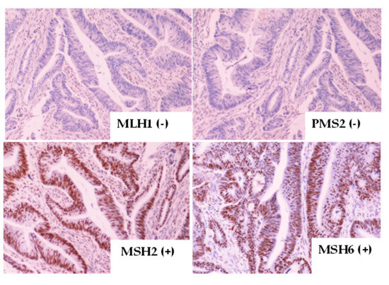
MMR gene germline mutations are considered a major genetic disorder in patients with hereditary nonpolyposis colon cancer (HNPCC) or Lynch syndrome; A total of 15% of sporadic colon carcinomas are MSI-High. MSI has also been observed in other cancers, such as endometrial, gastric, and ovarian cancer. The aim of the current study was to correlate and outline the optimal method between the molecular testing of the instability of microsatellite DNA regions (MSI status) and the loss of protein expression by immunehistochemistry (MMR). A total of 242 paraffin-embedded tissues from gastrointestinal, gynecological, genitourinary, lung, breast, and unknown primary cancer patients were analyzed for the expression of MLH1/MSH2/MSH6/PMS2 by immunohistochemistry, as well as for the molecular analysis of MSI status using PCR-based molecular fragment analysis. A total of 29 MSI-High patients were detected molecularly, while 23 patients were detected by immunohistochemistry, with rates that are comparable according to the literature. Based on the agreement coefficient of the two methods, a substantial agreement emerged (Kappa = 0.675 with standard error = 0.081, p < 0.001). Despite the substantial agreement, both methods ought to be established to determine MSI-H/dMMR status in all cancer types as a first-line screening test. Full article
Show Figures

Figure 1

2022

Jump to: 2024, 2023, 2020, 2019

18 pages, 6808 KiB  
Article
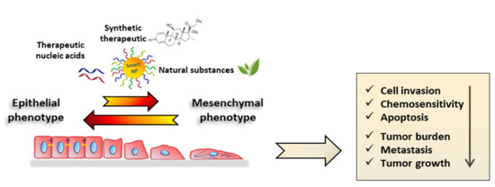
Remarkable Synergy When Combining EZH2 Inhibitors with YM155 Is H3K27me3-Independent
by Jun Yang and Andrew M. Davidoff
Cancers 2023, 15(1), 208; https://doi.org/10.3390/cancers15010208 - 29 Dec 2022
Cited by 2 | Viewed by 2173
Abstract
Targeting multiple molecules in the same biological network may maximize therapeutic efficacy. In this study, we identified a 27-gene module that is highly expressed in solid tumors, encoding actionable targets including EZH2 and BIRC5. The combination of EZH2 inhibitors and a BIRC5 inhibitor, [...] Read more.
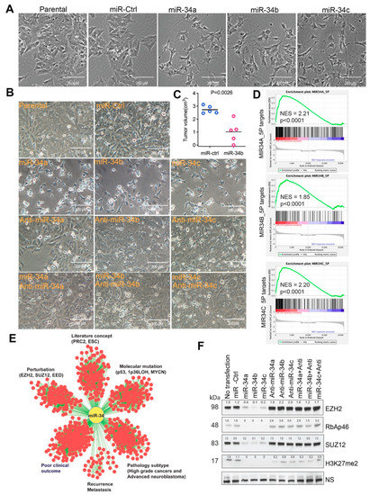
Targeting multiple molecules in the same biological network may maximize therapeutic efficacy. In this study, we identified a 27-gene module that is highly expressed in solid tumors, encoding actionable targets including EZH2 and BIRC5. The combination of EZH2 inhibitors and a BIRC5 inhibitor, YM155, results in a remarkable synergistic effect. The action of EZH2 inhibitors in this process is independent of the histone methyltransferase activity of polycomb repressive complex 2. Our study reveals a potential therapeutic approach for treating solid tumors by simultaneously targeting EZH2 and BIRC5. Full article
Show Figures

Figure 1

16 pages, 1923 KiB  
Article
Clinicopathological Features and Survival of Patients with Hepatocellular Carcinoma in Ethiopia: A Multicenter Study
by Getahun Befirdu Abza, Jemal Hussien Ahmed, Abdu Adem Yesufe, Edom Seife, Mengistu Erkie, Isabel Spriet, Legese Chelkeba and Pieter Annaert
Cancers 2023, 15(1), 193; https://doi.org/10.3390/cancers15010193 - 28 Dec 2022
Cited by 2 | Viewed by 2578
Abstract
(1) Background: Hepatocellular carcinoma (HCC) is one of the deadliest cancers globally, killing over 700,000 people each year. Despite the rising incidence and mortality rates of HCC in Ethiopia, only few single-centered studies have been conducted; therefore, we aimed to explore the clinicopathological [...] Read more.
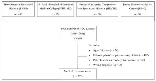
(1) Background: Hepatocellular carcinoma (HCC) is one of the deadliest cancers globally, killing over 700,000 people each year. Despite the rising incidence and mortality rates of HCC in Ethiopia, only few single-centered studies have been conducted; therefore, we aimed to explore the clinicopathological characteristics and survival of patients with HCC in multicenter settings. (2) Methods: We conducted a retrospective analysis of 369 patients with confirmed HCC diagnosed between 2016 and 2021. The survival of patients weas determined using the Kaplan–Meier method, and hazard ratios of the prognostic factors were estimated in Cox proportional hazard models. (3) Results: Majority patients were male (67%) and had a mean age of 52.0 ± 15.6 years. The majority of patients (87%) had a large tumor size (>5 cm) at diagnosis and presented with an advanced-stage condition. Cirrhosis (58%) and viral hepatitis (46.5%) were the main risk factors associated with HCC. The median overall survival was 141 days (95% CI: 117–165). Patients who took antivirals for HBV had a higher survival benefit compared to the untreated group (469 vs. 104 days; p < 0.001). The risk of death was 12 times higher in patients with Barcelona Clinic Liver Cancer-D (BCLC-D) terminal stage HCC compared to patients with an early stage (BCLC-A) HCC. The stage of HCC and treatment against HBV are the most significant survival predictors. (4) Conclusions: The overall survival of HCC patients in Ethiopia is poor. Cirrhosis and viral hepatitis are the primary risk factors linked with HCC. Patients who received antiviral therapy for HBV had a better survival outcome. Full article
Show Figures

Figure 1

20 pages, 1328 KiB  
Review
Anal Cancer in High-Risk Women: The Lost Tribe
by Micol Lupi, Danielle Brogden, Ann-Marie Howell, Paris Tekkis, Sarah Mills and Christos Kontovounisios
Cancers 2023, 15(1), 60; https://doi.org/10.3390/cancers15010060 - 22 Dec 2022
Cited by 5 | Viewed by 4426
Abstract
In developed countries the incidence of anal squamous cell carcinoma (SCC) has been rising; especially in women over the age of 60 years who present with more advanced disease stage than men. Historically, anal SCC screening has focused on people living with Human [...] Read more.
In developed countries the incidence of anal squamous cell carcinoma (SCC) has been rising; especially in women over the age of 60 years who present with more advanced disease stage than men. Historically, anal SCC screening has focused on people living with Human Immunodeficiency Virus (HIV) (PLWH) who are considered to be at the highest risk of anal SCC, and its precancerous lesion, anal squamous intraepithelial lesion (SIL). Despite this, women with vulval high-grade squamous epithelial lesions (HSIL) and SCCs have been shown to be as affected by anal HSIL and SCC as some PLWH. Nevertheless, there are no guidelines for the management of anal HSIL in this patient group. The ANCHOR trial demonstrated that treating anal HSIL significantly reduces the risk of anal SCC in PLWH, there is therefore an unmet requirement to clarify whether the screening and treatment of HSIL in women with a prior genital HSIL is also beneficial. This review presents the current evidence supporting the screening, treatment, and surveillance of anal HSIL in high-risk women with a previous history of genital HSIL and/or SCC. Full article
Show Figures

Figure 1

16 pages, 1409 KiB  
Review
A Review of Margetuximab-Based Therapies in Patients with HER2-Positive Metastatic Breast Cancer
by Moudi M. Alasmari
Cancers 2023, 15(1), 38; https://doi.org/10.3390/cancers15010038 - 21 Dec 2022
Cited by 15 | Viewed by 3067
Abstract
Breast cancer (BC) is the most commonly diagnosed cancer globally, with high mortality rates. Targeted drug therapies have revolutionized cancer treatment. For example, treatment with human epidermal receptor 2 (HER2) antagonists has markedly improved the prognosis of patients with HER2-positive BC (HER2 + [...] Read more.
Breast cancer (BC) is the most commonly diagnosed cancer globally, with high mortality rates. Targeted drug therapies have revolutionized cancer treatment. For example, treatment with human epidermal receptor 2 (HER2) antagonists has markedly improved the prognosis of patients with HER2-positive BC (HER2 + BC). However, HER2+ metastatic BC (MBC) remains prevalent owing to its resistance to conventional anti-HER2 drugs. Therefore, novel agents are needed to overcome the limitations of existing cancer treatments and to enhance the progression-free and overall survival rates. Progress has been made by optimizing the fragment crystallizable (Fc) domain of trastuzumab, an IgG1 monoclonal, chimeric anti-HER2 antibody, to develop margetuximab. The modified Fc domain of margetuximab enhances its binding affinity to CD16A and decreases its binding affinity to CD32B, thereby promoting its antitumor activity. This review summarizes studies on the efficacy of margetuximab, discusses its utility as an anti-HER2 monoclonal antibody drug for the treatment of HER2 + BC, and presents the latest advances in the treatment of BC. This review provides insights into the clinical implication of margetuximab in HER2 + MBC treatment. Full article
Show Figures

Figure 1

17 pages, 2113 KiB  
Review
Factors to Consider for the Correct Use of γH2AX in the Evaluation of DNA Double-Strand Breaks Damage Caused by Ionizing Radiation
by Davide Valente, Maria Pia Gentileschi, Antonino Guerrisi, Vicente Bruzzaniti, Aldo Morrone, Silvia Soddu and Alessandra Verdina
Cancers 2022, 14(24), 6204; https://doi.org/10.3390/cancers14246204 - 15 Dec 2022
Cited by 17 | Viewed by 7252
Abstract
People exposed to ionizing radiation (IR) both for diagnostic and therapeutic purposes is constantly increasing. Since the use of IR involves a risk of harmful effects, such as the DNA DSB induction, an accurate determination of this induced DNA damage and a correct [...] Read more.
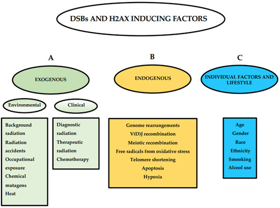
People exposed to ionizing radiation (IR) both for diagnostic and therapeutic purposes is constantly increasing. Since the use of IR involves a risk of harmful effects, such as the DNA DSB induction, an accurate determination of this induced DNA damage and a correct evaluation of the risk–benefit ratio in the clinical field are of key relevance. γH2AX (the phosphorylated form of the histone variant H2AX) is a very early marker of DSBs that can be induced both in physiological conditions, such as in the absence of specific external agents, and by external factors such as smoking, heat, background environmental radiation, and drugs. All these internal and external conditions result in a basal level of γH2AX which must be considered for the correct assessment of the DSBs after IR exposure. In this review we analyze the most common conditions that induce H2AX phosphorylation, including specific exogenous stimuli, cellular states, basic environmental factors, and lifestyles. Moreover, we discuss the most widely used methods for γH2AX determination and describe the principal applications of γH2AX scoring, paying particular attention to clinical studies. This knowledge will help us optimize the use of available methods in order to discern the specific γH2AX following IR-induced DSBs from the basal level of γH2AX in the cells. Full article
Show Figures

Figure 1

2020

Jump to: 2024, 2023, 2022, 2019

36 pages, 10946 KiB  
Review
The Landscape of CAR-T Cell Clinical Trials against Solid Tumors—A Comprehensive Overview
by Niels Schaft
Cancers 2020, 12(9), 2567; https://doi.org/10.3390/cancers12092567 - 9 Sep 2020
Cited by 65 | Viewed by 8744
Abstract
CAR-T cells showed great potential in the treatment of patients with hematologic tumors. However, the clinical efficacy of CAR-T cells against solid tumors lags behind. To obtain a comprehensive overview of the landscape of CAR-T cell clinical trials against this type of cancer, [...] Read more.
CAR-T cells showed great potential in the treatment of patients with hematologic tumors. However, the clinical efficacy of CAR-T cells against solid tumors lags behind. To obtain a comprehensive overview of the landscape of CAR-T cell clinical trials against this type of cancer, this review summarizes all the 196 studies registered at clinicaltrials.gov. Special focus is on: (1) geographical distribution; (2) targeted organs, tumor entities, and antigens; (3) CAR transfer methods, CAR formats, and extra features introduced into the T cells; and (4) patient pretreatments, injection sites, and safety measurements. Finally, the few data on clinical outcome are reported. The last assessment of clinicaltrials.gov for the data summarized in this paper was on 4 August 2020. Full article
Show Figures

Figure 1

15 pages, 1004 KiB  
Review
BRAF Mutated Colorectal Cancer: New Treatment Approaches
by Javier Molina-Cerrillo, María San Román, Javier Pozas, Teresa Alonso-Gordoa, Miguel Pozas, Elisa Conde, Marta Rosas, Enrique Grande, María Laura García-Bermejo and Alfredo Carrato
Cancers 2020, 12(6), 1571; https://doi.org/10.3390/cancers12061571 - 14 Jun 2020
Cited by 48 | Viewed by 7041
Abstract
Colon cancer is one of the most frequently diagnosed malignancies in adults, considering both its incidence and prevalence. Anatomically, the right colon is considered as being from the cecum to the splenic flexure, and the left colon is from the splenic flexure to [...] Read more.
Colon cancer is one of the most frequently diagnosed malignancies in adults, considering both its incidence and prevalence. Anatomically, the right colon is considered as being from the cecum to the splenic flexure, and the left colon is from the splenic flexure to the rectum. Sidedness is a surrogate of a wide spectrum of colorectal cancer (CRC) biology features (embryology, microbiome, methylation, microsatellite instability (MSI), BRAF, aging, KRAS, consensus molecular subtypes (CMS), etc.), which result in prognostic factors. Different molecular subtypes have been identified, according to genomic and transcriptomic criteria. A subgroup harboring a BRAF mutation has been described, and represents approximately 10% of the patients diagnosed with colon cancer. This subgroup has morphological, clinical, and therapeutic characteristics that differ substantially from patients who do not carry this genetic alteration. Unfortunately, there is no established standard of care for this particular cohort of patients. This manuscript aims to study the biology of this subgroup of colon cancer, to understand the current approach in clinical research. Full article
Show Figures

Graphical abstract

25 pages, 1037 KiB  
Review
Small Ones to Fight a Big Problem—Intervention of Cancer Metastasis by Small Molecules
by Dennis Kobelt, Mathias Dahlmann, Malti Dumbani, Nazli Güllü, Benedikt Kortüm, Miguel E. Alberto Vílchez, Ulrike Stein and Wolfgang Walther
Cancers 2020, 12(6), 1454; https://doi.org/10.3390/cancers12061454 - 3 Jun 2020
Cited by 5 | Viewed by 3621
Abstract
Metastasis represents the most lethal attribute of cancer and critically limits successful therapies in many tumor entities. The clinical need is defined by the fact that all cancer patients, who have or who will develop distant metastasis, will experience shorter survival. Thus, the [...] Read more.
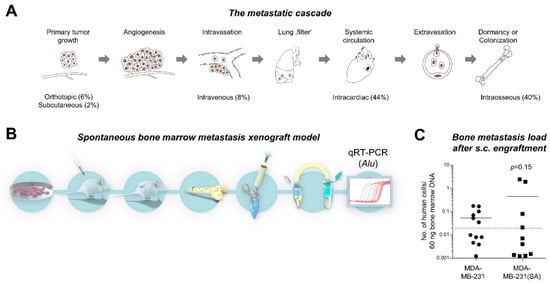
Metastasis represents the most lethal attribute of cancer and critically limits successful therapies in many tumor entities. The clinical need is defined by the fact that all cancer patients, who have or who will develop distant metastasis, will experience shorter survival. Thus, the ultimate goal in cancer therapy is the restriction of solid cancer metastasis by novel molecularly targeted small molecule based therapies. Biomarkers identifying cancer patients at high risk for metastasis and simultaneously acting as key drivers for metastasis are extremely desired. Clinical interventions targeting these key molecules will result in high efficiency in metastasis intervention. In result of this, personalized tailored interventions for restriction and prevention of cancer progression and metastasis will improve patient survival. This review defines crucial biological steps of the metastatic cascade, such as cell dissemination, migration and invasion as well as the action of metastasis suppressors. Targeting these biological steps with tailored therapeutic strategies of intervention or even prevention of metastasis using a wide range of small molecules will be discussed. Full article
Show Figures

Figure 1

21 pages, 2489 KiB  
Review
Fully Human Antibodies for Malignant Pleural Mesothelioma Targeting
by Fabio Nicolini, Martine Bocchini, Davide Angeli, Giuseppe Bronte, Angelo Delmonte, Lucio Crinò and Massimiliano Mazza
Cancers 2020, 12(4), 915; https://doi.org/10.3390/cancers12040915 - 8 Apr 2020
Cited by 4 | Viewed by 3739
Abstract
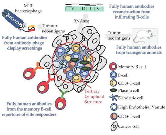
Immunotherapy is the most promising therapeutic approach against malignant pleural mesothelioma (MPM). Despite technological progress, the number of targetable antigens or specific antibodies is limited, thus hindering the full potential of recent therapeutic interventions. All possibilities of finding new targeting molecules must be [...] Read more.
Immunotherapy is the most promising therapeutic approach against malignant pleural mesothelioma (MPM). Despite technological progress, the number of targetable antigens or specific antibodies is limited, thus hindering the full potential of recent therapeutic interventions. All possibilities of finding new targeting molecules must be exploited. The specificity of targeting is guaranteed by the use of monoclonal antibodies, while fully human antibodies are preferred, as they are functional and generate no neutralizing antibodies. The aim of this review is to appraise the latest advances in screening methods dedicated to the identification and harnessing of fully human antibodies. The scope of identifying useful molecules proceeds along two avenues, i.e., through the antigen-first or binding-first approaches. The first relies on screening human antibody libraries or plasma from immunized transgenic mice or humans to isolate binders to specific antigens. The latter takes advantage of specific binding to tumor cells of antibodies present in phage display libraries or in responders’ plasma samples without prior knowledge of the antigens. Additionally, next-generation sequencing analysis of B-cell receptor repertoire pre- and post-therapy in memory B-cells from responders allows for the identification of clones expanded and matured upon treatment. Human antibodies identified can be subsequently reformatted to generate a plethora of therapeutics like antibody-drug conjugates, immunotoxins, and advanced cell-therapeutics such as chimeric antigen receptor-transduced T-cells. Full article
Show Figures

Graphical abstract

24 pages, 2060 KiB  
Review
MicroRNAs in Cancer Treatment-Induced Cardiotoxicity
by Laura Pellegrini, Sara Sileno, Marco D’Agostino, Eleonora Foglio, Maria Cristina Florio, Vincenzo Guzzanti, Matteo Antonio Russo, Federica Limana and Alessandra Magenta
Cancers 2020, 12(3), 704; https://doi.org/10.3390/cancers12030704 - 17 Mar 2020
Cited by 26 | Viewed by 4483
Abstract
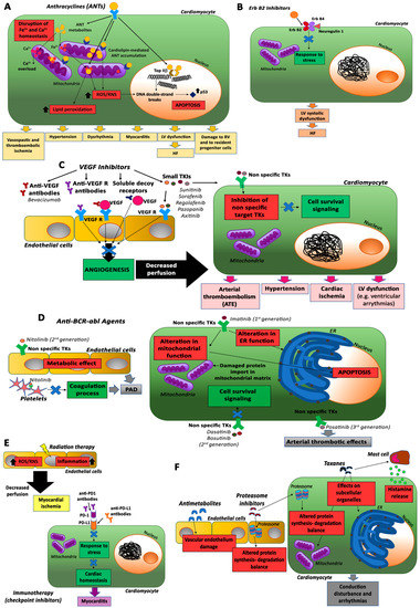
Cancer treatment has made significant progress in the cure of different types of tumors. Nevertheless, its clinical use is limited by unwanted cardiotoxicity. Aside from the conventional chemotherapy approaches, even the most newly developed, i.e., molecularly targeted therapy and immunotherapy, exhibit a similar [...] Read more.
Cancer treatment has made significant progress in the cure of different types of tumors. Nevertheless, its clinical use is limited by unwanted cardiotoxicity. Aside from the conventional chemotherapy approaches, even the most newly developed, i.e., molecularly targeted therapy and immunotherapy, exhibit a similar frequency and severity of toxicities that range from subclinical ventricular dysfunction to severe cardiomyopathy and, ultimately, congestive heart failure. Specific mechanisms leading to cardiotoxicity still remain to be elucidated. For instance, oxidative stress and DNA damage are considered key players in mediating cardiotoxicity in different treatments. microRNAs (miRNAs) act as key regulators in cell proliferation, cell death, apoptosis, and cell differentiation. Their dysregulation has been associated with adverse cardiac remodeling and toxicity. This review provides an overview of the cardiotoxicity induced by different oncologic treatments and potential miRNAs involved in this effect that could be used as possible therapeutic targets. Full article
Show Figures

Figure 1

20 pages, 753 KiB  
Review
Targeting Epigenetic Dependencies in Solid Tumors: Evolutionary Landscape Beyond Germ Layers Origin
by Francesca Citron and Linda Fabris
Cancers 2020, 12(3), 682; https://doi.org/10.3390/cancers12030682 - 13 Mar 2020
Cited by 7 | Viewed by 8300
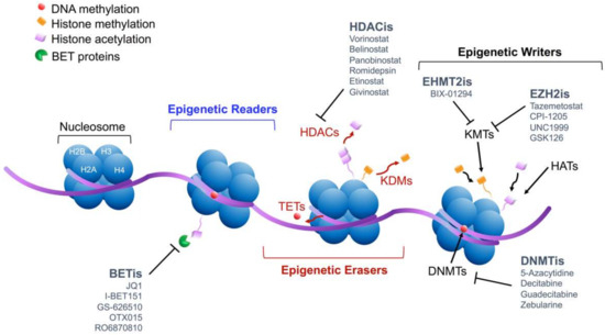
Abstract
Extensive efforts recently witnessed the complexity of cancer biology; however, molecular medicine still lacks the ability to elucidate hidden mechanisms for the maintenance of specific subclasses of rare tumors characterized by the silent onset and a poor prognosis (e.g., ovarian cancer, pancreatic cancer, [...] Read more.
Extensive efforts recently witnessed the complexity of cancer biology; however, molecular medicine still lacks the ability to elucidate hidden mechanisms for the maintenance of specific subclasses of rare tumors characterized by the silent onset and a poor prognosis (e.g., ovarian cancer, pancreatic cancer, and glioblastoma). Recent mutational fingerprints of human cancers highlighted genomic alteration occurring on epigenetic modulators. In this scenario, the epigenome dependency of cancer orchestrates a broad range of cellular processes critical for tumorigenesis and tumor progression, possibly mediating escaping mechanisms leading to drug resistance. Indeed, in this review, we discuss the pivotal role of chromatin remodeling in shaping the tumor architecture and modulating tumor fitness in a microenvironment-dependent context. We will also present recent advances in the epigenome targeting, posing a particular emphasis on how this knowledge could be translated into a feasible therapeutic approach to individualize clinical settings and improve patient outcomes. Full article
Show Figures

Figure 1

23 pages, 5572 KiB  
Article
Ependymoma Pediatric Brain Tumor Protein Fingerprinting by Integrated Mass Spectrometry Platforms: A Pilot Investigation
by Diana Valeria Rossetti, Luca Massimi, Claudia Martelli, Federica Vincenzoni, Susanna Di Silvestre, Gianluca Scorpio, Gianpiero Tamburrini, Massimo Caldarelli, Andrea Urbani and Claudia Desiderio
Cancers 2020, 12(3), 674; https://doi.org/10.3390/cancers12030674 - 13 Mar 2020
Cited by 6 | Viewed by 3436
Abstract
Ependymoma pediatric brain tumor occurs at approximate frequencies of 10–15% in supratentorial and 20–30% in posterior fossa regions. These tumors have an almost selective response to surgery and relative and confirmed resistance to radiotherapy and chemotherapic agents, respectively. Alongside histopathological grading, clinical and [...] Read more.
Ependymoma pediatric brain tumor occurs at approximate frequencies of 10–15% in supratentorial and 20–30% in posterior fossa regions. These tumors have an almost selective response to surgery and relative and confirmed resistance to radiotherapy and chemotherapic agents, respectively. Alongside histopathological grading, clinical and treatment evaluation of ependymomas currently consider the tumor localization and the genomic outlined associated molecular subgroups, with the supratentorial and the posterior fossa ependymomas nowadays considered diverse diseases. On these grounds and in trying to better understand the molecular features of these tumors, the present investigation aimed to originally investigate the proteomic profile of pediatric ependymoma tissues of different grade and localization by mass spectrometry platforms to disclose potential distinct protein phenotypes. To this purpose, acid-soluble and acid-insoluble fractions of ependymoma tumor tissues homogenates were analyzed by LC-MS following both the top-down and the shotgun proteomic approaches, respectively, to either investigate the intact proteome or its digested form. The two approaches were complementary in profiling the ependymoma tumor tissues and showed distinguished profiles for supratentorial and posterior fossa ependymomas and for WHO II and III tumor grades. Top-down proteomic analysis revealed statistically significant higher levels of thymosin beta 4, 10 kDa heat shock protein, non-histone chromosomal protein HMG-17, and mono-/uncitrullinated forms ratio of the glial fibrillary acidic protein (GFAP) fragment 388–432 in supratentorial ependymomas—the same GFAP fragment as well as the hemoglobin alpha- and the beta-chain marked grade II with respect to grade III posterior fossa ependymomas. Gene ontology classification of shotgun data of the identified cancer and the non-cancer related proteins disclosed protein elements exclusively marking tumor localization and pathways that were selectively overrepresented. These results, although preliminary, seem consistent with different protein profiles of ependymomas of diverse grade of aggressiveness and brain region development and contributed to enlarging the molecular knowledge of this still enigmatic tumor. Full article
Show Figures

Graphical abstract

24 pages, 2967 KiB  
Article
The Determination of Immunomodulation and Its Impact on Survival of Rectal Cancer Patients Depends on the Area Comprising a Tissue Microarray
by Elisabeth S. Gruber, Georg Oberhuber, Dietmar Pils, Theresa Stork, Katharina Sinn, Sylvia Gruber, Robert Nica, Dan Kolmer, Suzanne D. Turner, Michaela Schlederer, Joachim Widder, Wolfgang Doerr, Béla Teleky and Lukas Kenner
Cancers 2020, 12(3), 563; https://doi.org/10.3390/cancers12030563 - 29 Feb 2020
Cited by 7 | Viewed by 4081
Abstract
Background: T cell density in colorectal cancer (CRC) has proven to be of high prognostic importance. Here, we evaluated the influence of a hyperfractionated preoperative short-term radiation protocol (25 Gy) on immune cell density in tumor samples of rectal cancer (RC) patients and [...] Read more.
Background: T cell density in colorectal cancer (CRC) has proven to be of high prognostic importance. Here, we evaluated the influence of a hyperfractionated preoperative short-term radiation protocol (25 Gy) on immune cell density in tumor samples of rectal cancer (RC) patients and on patient survival. In addition, we assessed spatial tumor heterogeneity by comparison of analogue T cell quantification on full tissue sections with digital T cell quantification on a virtually established tissue microarray (TMA). Methods: A total of 75 RC patients (60 irradiated, 15 treatment-naïve) were defined for retrospective analysis. RC samples were processed for immunohistochemistry (CD3, CD8, PD-1, PD-L1). Analogue (score 0–3) as well as digital quantification (TMA: 2 cores vs. 6 cores, mean T cell count) of marker expression in 2 areas (central tumor, CT; invasive margin, IM) was performed. Survival was estimated on the basis of analogue as well as digital marker densities calculated from 2 cores (Immunoscore: CD3/CD8 ratio) and 6 cores per tumor area. Results: Irradiated RC samples showed a significant decrease in CD3 and CD8 positive T cells, independent of quantification mode. T cell densities of 6 virtual cores approximated to T cell densities of full tissue sections, independent of individual core density or location. Survival analysis based on full tissue section quantification demonstrated that CD3 and CD8 positive T cells as well as PD-1 positive tumor infiltrating leucocytes (TILs) in the CT and the IM had a significant impact on disease-free survival (DFS) as well as overall survival (OS). In addition, CD3 and CD8 positive T cells as well as PD-1 positive TILs in the IM proved as independent prognostic factors for DFS and OS; in the CT, PD-1 positive TILs predicted DFS and CD3 and CD8 positive T cells as well as PD-1 positive TILs predicted OS. Survival analysis based on virtual TMA showed no impact on DFS or OS. Conclusion: Spatial tumor heterogeneity might result in inadequate quantification of immune marker expression; however, if using a TMA, 6 cores per tumor area and patient sample represent comparable amounts of T cell densities to those quantified on full tissue sections. Consistently, the tissue area used for immune marker quantification represents a crucial factor for the evaluation of prognostic and predictive biomarker potential. Full article
Show Figures

Figure 1

20 pages, 3474 KiB  
Article
Novel Quinoline Compounds Active in Cancer Cells through Coupled DNA Methyltransferase Inhibition and Degradation
by Clemens Zwergel, Rossella Fioravanti, Giulia Stazi, Federica Sarno, Cecilia Battistelli, Annalisa Romanelli, Angela Nebbioso, Eduarda Mendes, Alexandra Paulo, Raffaele Strippoli, Marco Tripodi, Dany Pechalrieu, Paola B. Arimondo, Teresa De Luca, Donatella Del Bufalo, Daniela Trisciuoglio, Lucia Altucci, Sergio Valente and Antonello Mai
Cancers 2020, 12(2), 447; https://doi.org/10.3390/cancers12020447 - 14 Feb 2020
Cited by 7 | Viewed by 4114 | Correction
Abstract
DNA methyltransferases (DNMTs) play a relevant role in epigenetic control of cancer cell survival and proliferation. Since only two DNMT inhibitors (azacitidine and decitabine) have been approved to date for the treatment of hematological malignancies, the development of novel potent and specific inhibitors [...] Read more.
DNA methyltransferases (DNMTs) play a relevant role in epigenetic control of cancer cell survival and proliferation. Since only two DNMT inhibitors (azacitidine and decitabine) have been approved to date for the treatment of hematological malignancies, the development of novel potent and specific inhibitors is urgent. Here we describe the design, synthesis, and biological evaluation of a new series of compounds acting at the same time as DNMTs (mainly DNMT3A) inhibitors and degraders. Tested against leukemic and solid cancer cell lines, 2a–c and 4a–c (the last only for leukemias) displayed up to submicromolar antiproliferative activities. In HCT116 cells, such compounds induced EGFP gene expression in a promoter demethylation assay, confirming their demethylating activity in cells. In the same cell line, 2b and 4c chosen as representative samples induced DNMT1 and -3A protein degradation, suggesting for these compounds a double mechanism of DNMT3A inhibition and DNMT protein degradation. Full article
Show Figures

Graphical abstract

18 pages, 3803 KiB  
Article
Targeting Melanoma Hypoxia with the Food-Grade Lactic Acid Bacterium Lactococcus Lactis
by Rodolfo Garza-Morales, Beatriz E. Rendon, Mohammad Tariq Malik, Jeannete E. Garza-Cabrales, Anne Aucouturier, Luis G. Bermúdez-Humarán, Kelly M. McMasters, Lacey R. McNally and Jorge G. Gomez-Gutierrez
Cancers 2020, 12(2), 438; https://doi.org/10.3390/cancers12020438 - 13 Feb 2020
Cited by 15 | Viewed by 4315
Abstract
Melanoma is the most aggressive form of skin cancer. Hypoxia is a feature of the tumor microenvironment that reduces efficacy of immuno- and chemotherapies, resulting in poor clinical outcomes. Lactococcus lactis is a facultative anaerobic gram-positive lactic acid bacterium (LAB) that is Generally [...] Read more.
Melanoma is the most aggressive form of skin cancer. Hypoxia is a feature of the tumor microenvironment that reduces efficacy of immuno- and chemotherapies, resulting in poor clinical outcomes. Lactococcus lactis is a facultative anaerobic gram-positive lactic acid bacterium (LAB) that is Generally Recognized as Safe (GRAS). Recently, the use of LAB as a delivery vehicle has emerged as an alternative strategy to deliver therapeutic molecules; therefore, we investigated whether L. lactis can target and localize within melanoma hypoxic niches. To simulate hypoxic conditions in vitro, melanoma cells A2058, A375 and MeWo were cultured in a chamber with a gas mixture of 5% CO2, 94% N2 and 1% O2. Among the cell lines tested, MeWo cells displayed greater survival rates when compared to A2058 and A375 cells. Co-cultures of L. lactis expressing GFP or mCherry and MeWo cells revealed that L. lactis efficiently express the transgenes under hypoxic conditions. Moreover, multispectral optoacoustic tomography (MSOT), and near infrared (NIR) imaging of tumor-bearing BALB/c mice revealed that the intravenous injection of either L. lactis expressing β-galactosidase (β-gal) or infrared fluorescent protein (IRFP713) results in the establishment of the recombinant bacteria within tumor hypoxic niches. Overall, our data suggest that L. lactis represents an alternative strategy to target and deliver therapeutic molecules into the tumor hypoxic microenvironment. Full article
Show Figures

Figure 1

11 pages, 2550 KiB  
Case Report
Targeting Epidermal Growth Factor Receptor (EGFR) in Pediatric Colorectal Cancer
by Maria Debora De Pasquale, Alessandro Crocoli, Tamara Caldaro, Martina Rinelli, Gian Paolo Spinelli, Paola Francalanci, Raffaele Cozza, Alessandro Inserra and Evelina Miele
Cancers 2020, 12(2), 414; https://doi.org/10.3390/cancers12020414 - 11 Feb 2020
Cited by 3 | Viewed by 3297
Abstract
Background: Colorectal carcinoma (CRC) is very rare in the pediatric and adolescent age range and clinical management is performed according to adult protocols. We report, for the first time in the literature, a case of a child with metastatic CRC successfully treated [...] Read more.
Background: Colorectal carcinoma (CRC) is very rare in the pediatric and adolescent age range and clinical management is performed according to adult protocols. We report, for the first time in the literature, a case of a child with metastatic CRC successfully treated with panitumumab associated to chemotherapy. Methods: A twelve-year-old male was diagnosed with CRC with nodal metastasis and peritoneal neoplastic effusion. After performing a genetic evaluation, in light of the absence of mutations in RAS family genes, anti-Epidermal Growth Factor Receptor (EGFR) monoclonal antibody, panitumumab, was added to chemotherapy FOLFOXIRI. Results: The child successfully responded to therapy with normalization of the Carbohydrate Antigen (CA) 19.9 value after the third cycle of treatment. After the sixth cycle, he underwent surgery that consisted in sigmoid resection with complete D3 lymphadenectomy. At histological evaluation, no residual neoplastic cells were detectable in the surgical specimen. He completed 12 cycles of chemotherapy plus panitumomab and he is alive without disease 14 months from diagnosis. Conclusions: Our results suggest performing mutational screening for colorectal cancer also in the pediatric setting, in order to orient treatment that should include targeted therapies. Full article
Show Figures

Figure 1

15 pages, 4259 KiB  
Article
Modeling Spontaneous Bone Metastasis Formation of Solid Human Tumor Xenografts in Mice
by Vera Labitzky, Anke Baranowsky, Hanna Maar, Sandra Hanika, Sarah Starzonek, Ann-Kristin Ahlers, Katrin Stübke, Eva J. Koziolek, Markus Heine, Paula Schäfer, Sabine Windhorst, Manfred Jücker, Kristoffer Riecken, Michael Amling, Thorsten Schinke, Udo Schumacher, Ursula Valentiner and Tobias Lange
Cancers 2020, 12(2), 385; https://doi.org/10.3390/cancers12020385 - 7 Feb 2020
Cited by 14 | Viewed by 4319
Abstract
The majority of cancer-related deaths are due to hematogenous metastases, and the bone marrow (BM) represents one of the most frequent metastatic sites. To study BM metastasis formation in vivo, the most efficient approach is based on intracardiac injection of human tumor cells [...] Read more.
The majority of cancer-related deaths are due to hematogenous metastases, and the bone marrow (BM) represents one of the most frequent metastatic sites. To study BM metastasis formation in vivo, the most efficient approach is based on intracardiac injection of human tumor cells into immunodeficient mice. However, such a procedure circumvents the early steps of the metastatic cascade. Here we describe the development of xenograft mouse models (balb/c rag2-/- and severe combined immunodeficient (SCID)), in which BM metastases are spontaneously derived from subcutaneous (s.c.) primary tumors (PTs). As verified by histology, the described methodology including ex vivo bioluminescence imaging (BLI) even enabled the detection of micrometastases in the BM. Furthermore, we established sublines from xenograft primary tumors (PTs) and corresponding BM (BM) metastases using LAN-1 neuroblastoma xenografts as a first example. In vitro “metastasis” assays (viability, proliferation, transmigration, invasion, colony formation) partially indicated pro-metastatic features of the LAN-1-BM compared to the LAN-1-PT subline. Unexpectedly, after s.c. re-injection into mice, LAN-1-BM xenografts developed spontaneous BM metastases less frequently than LAN-1-PT xenografts. This study provides a novel methodologic approach for modelling the spontaneous metastatic cascade of human BM metastasis formation in mice. Moreover, our data indicate that putative bone-metastatic features get rapidly lost upon routine cell culture. Full article
Show Figures

Figure 1

14 pages, 1281 KiB  
Article
Circulating Levels of PD-L1 in Mesothelioma Patients from the NIBIT-MESO-1 Study: Correlation with Survival
by Carla Chiarucci, Sara Cannito, Maria Grazia Daffinà, Giovanni Amato, Gianluca Giacobini, Ornella Cutaia, Maria Fortunata Lofiego, Carolina Fazio, Diana Giannarelli, Riccardo Danielli, Anna Maria Di Giacomo, Sandra Coral, Luana Calabrò, Michele Maio and Alessia Covre
Cancers 2020, 12(2), 361; https://doi.org/10.3390/cancers12020361 - 5 Feb 2020
Cited by 21 | Viewed by 4032
Abstract
Targeting of the programmed cell death protein (PD)-1/programmed death-ligand 1 (PD-L1) axis has shown a significant clinical impact in several tumor types. Accordingly, our phase II NIBIT-MESO-1 study demonstrated an improved clinical efficacy in mesothelioma patients treated with the anti-PD-L1 durvalumab combined with [...] Read more.
Targeting of the programmed cell death protein (PD)-1/programmed death-ligand 1 (PD-L1) axis has shown a significant clinical impact in several tumor types. Accordingly, our phase II NIBIT-MESO-1 study demonstrated an improved clinical efficacy in mesothelioma patients treated with the anti-PD-L1 durvalumab combined with the anti-cytotoxic T-lymphocyte antigen (CTLA)-4 tremelimumab, as compared to tremelimumab alone. Due to the promising therapeutic activity of immune check-point inhibitors (ICIs) in mesothelioma patients, the identification of biomarkers predictive of response to treatment is of crucial relevance. The prognostic role of soluble PD-L1 (sPD-L1) proposed in cancer patients prompted us to investigate this protein in sera from mesothelioma patients (n = 40) enrolled in the NIBIT-MESO-1 study. A significant (p < 0.001) increase in sPD-L1 levels was detected in patients after the first cycle and during therapy vs. baseline. A longer overall survival (OS) was observed in patients with sPD-L1 concentrations below (at baseline, d1C2, d1C5 (p < 0.01)) or FC values above (p < 0.05 at d1C2, d1C3, d1C5) their statistically calculated optimal cut-offs. On the basis of these initial results, the specific role of CTLA-4-, PD-L1-, or PD-1-targeting on sPD-L1 release was then investigated in sera from 81 additional ICI-treated solid cancer patients. Results showed a significant (p < 0.001) increase of sPD-L1 levels during therapy compared to baseline only in anti-PD-L1-treated patients, supporting the specific involvement of PD-L1 targeting in the release of its soluble form. Our findings suggest that sPD-L1 represents a predictive biomarker of clinical response to anti-PD-L1 cancer immunotherapy. Full article
Show Figures

Figure 1

17 pages, 3435 KiB  
Article
TGF-Beta-Activated Cancer-Associated Fibroblasts Limit Cetuximab Efficacy in Preclinical Models of Head and Neck Cancer
by Ksenia M. Yegodayev, Ofra Novoplansky, Artemiy Golden, Manu Prasad, Liron Levin, Sankar Jagadeeshan, Jonathan Zorea, Orr Dimitstein, Ben-Zion Joshua, Limor Cohen, Ekaterina Khrameeva and Moshe Elkabets
Cancers 2020, 12(2), 339; https://doi.org/10.3390/cancers12020339 - 3 Feb 2020
Cited by 42 | Viewed by 5669
Abstract
Most head and neck cancer (HNC) patients are resistant to cetuximab, an antibody against the epidermal growth factor receptor. Such therapy resistance is known to be mediated, in part, by stromal cells surrounding the tumor cells; however, the mechanisms underlying such a resistance [...] Read more.
Most head and neck cancer (HNC) patients are resistant to cetuximab, an antibody against the epidermal growth factor receptor. Such therapy resistance is known to be mediated, in part, by stromal cells surrounding the tumor cells; however, the mechanisms underlying such a resistance phenotype remain unclear. To identify the mechanisms of cetuximab resistance in an unbiased manner, RNA-sequencing (RNA-seq) of HNC patient-derived xenografts (PDXs) was performed. Comparing the gene expression of HNC-PDXs before and after treatment with cetuximab indicated that the transforming growth factor-beta (TGF-beta) signaling pathway was upregulated in the stromal cells of PDXs that progressed on cetuximab treatment (CetuximabProg-PDX). However, in PDXs that were extremely sensitive to cetuximab (CetuximabSen-PDX), the TGF-beta pathway was downregulated in the stromal compartment. Histopathological analysis of PDXs showed that TGF-beta-activation was detected in cancer-associated fibroblasts (CAFs) of CetuximabProg-PDX. These TGF-beta-activated CAFs were sufficient to limit cetuximab efficacy in vitro and in vivo. Moreover, blocking the TGF-beta pathway using the SMAD3 inhibitor, SIS3, enhanced cetuximab efficacy and prevented the progression of CetuximabProg-PDX. Altogether, our findings indicate that TGF-beta-activated CAFs play a role in limiting cetuximab efficacy in HNC. Full article
Show Figures

Graphical abstract

20 pages, 993 KiB  
Review
Vimentin Intermediate Filaments as Potential Target for Cancer Treatment
by Katerina Strouhalova, Magdalena Přechová, Aneta Gandalovičová, Jan Brábek, Martin Gregor and Daniel Rosel
Cancers 2020, 12(1), 184; https://doi.org/10.3390/cancers12010184 - 11 Jan 2020
Cited by 156 | Viewed by 14249
Abstract
Intermediate filaments constitute the third component of the cellular skeleton. Unlike actin and microtubule cytoskeletons, the intermediate filaments are composed of a wide variety of structurally related proteins showing distinct expression patterns in tissues and cell types. Changes in the expression patterns of [...] Read more.
Intermediate filaments constitute the third component of the cellular skeleton. Unlike actin and microtubule cytoskeletons, the intermediate filaments are composed of a wide variety of structurally related proteins showing distinct expression patterns in tissues and cell types. Changes in the expression patterns of intermediate filaments are often associated with cancer progression; in particular with phenotypes leading to increased cellular migration and invasion. In this review we will describe the role of vimentin intermediate filaments in cancer cell migration, cell adhesion structures, and metastasis formation. The potential for targeting vimentin in cancer treatment and the development of drugs targeting vimentin will be reviewed. Full article
Show Figures

Figure 1

2019

Jump to: 2024, 2023, 2022, 2020

14 pages, 3262 KiB  
Article
Nanoparticles Loaded with the BET Inhibitor JQ1 Block the Growth of Triple Negative Breast Cancer Cells In Vitro and In Vivo
by Valentina Maggisano, Marilena Celano, Rocco Malivindi, Ines Barone, Donato Cosco, Catia Mio, Chiara Mignogna, Salvatore Panza, Giuseppe Damante, Massimo Fresta, Sebastiano Andò, Diego Russo, Stefania Catalano and Stefania Bulotta
Cancers 2020, 12(1), 91; https://doi.org/10.3390/cancers12010091 - 30 Dec 2019
Cited by 21 | Viewed by 4239
Abstract
Inhibition of bromo-and extra-terminal domain (BET) proteins, epigenetic regulators of genes involved in cell viability, has been efficiently tested in preclinical models of triple negative breast cancer (TNBC). However, the use of the selective BET-inhibitor JQ1 on humans is limited by its very [...] Read more.
Inhibition of bromo-and extra-terminal domain (BET) proteins, epigenetic regulators of genes involved in cell viability, has been efficiently tested in preclinical models of triple negative breast cancer (TNBC). However, the use of the selective BET-inhibitor JQ1 on humans is limited by its very short half-life. Herein, we developed, characterized and tested a novel formulation of nanoparticles containing JQ1 (N-JQ1) against TNBC in vitro and in vivo. N-JQ1, prepared using the nanoprecipitation method of preformedpoly-lactid-co-glycolic acid in an aqueous solution containing JQ1 and poloxamer-188 as a stabilizer, presented a high physico-chemical stability. Treatment of MDA-MB 157 and MDA-MB 231 TNBC cells with N-JQ1 determined a significant decrease in cell viability, adhesion and migration. Intra-peritoneal administration (5 days/week for two weeks) of N-JQ1 in nude mice hosting a xenograft TNBC after flank injection of MDA-MB-231 cells determined a great reduction in the growth and vascularity of the neoplasm. Moreover, the treatment resulted in a minimal infiltration of nearby tissues. Finally, the encapsulation of JQ1 in nanoparticles improved the anticancer efficacy of this epigenetic compound against TNBC in vitro and in vivo, opening the way to test it in the treatment of TNBC. Full article
Show Figures

Graphical abstract

14 pages, 24391 KiB  
Article
Salmonella Breaks Tumor Immune Tolerance by Downregulating Tumor Programmed Death-Ligand 1 Expression
by Man-Chin Chen, Christian Ronquillo Pangilinan and Che-Hsin Lee
Cancers 2020, 12(1), 57; https://doi.org/10.3390/cancers12010057 - 24 Dec 2019
Cited by 27 | Viewed by 4360
Abstract
Immunotherapy is becoming a popular treatment modality in combat against cancer, one of the world’s leading health problems. While tumor cells influence host immunity via expressing immune inhibitory signaling proteins, some bacteria possess immunomodulatory activities that counter the symptoms of tumors. The accumulation [...] Read more.
Immunotherapy is becoming a popular treatment modality in combat against cancer, one of the world’s leading health problems. While tumor cells influence host immunity via expressing immune inhibitory signaling proteins, some bacteria possess immunomodulatory activities that counter the symptoms of tumors. The accumulation of Salmonella in tumor sites influences tumor protein expression, resulting in T cell infiltration. However, the molecular mechanism by which Salmonella activates T cells remains elusive. Many tumors have been reported to have high expressions of programmed death-ligand 1 (PD-L1), which is an important immune checkpoint molecule involved in tumor immune escape. In this study, Salmonella reduced the expression of PD-L1 in tumor cells. The expression levels of phospho-protein kinase B (P-AKT), phospho-mammalian targets of rapamycin (P-mTOR), and the phospho-p70 ribosomal s6 kinase (P-p70s6K) pathway were revealed to be involved in the Salmonella-mediated downregulation of PD-L1. In a tumor-T cell coculture system, Salmonella increased T cell number and reduced T cell apoptosis. Systemic administration of Salmonella reduced the expressions of PD-L-1 in tumor-bearing mice. In addition, tumor growth was significantly inhibited along with an enhanced T cell infiltration following Salmonella treatment. These findings suggest that Salmonella acts upon the immune checkpoint, primarily PD-L1, to incapacitate protumor effects and thereby inhibit tumor growth. Full article
Show Figures

Figure 1

40 pages, 2632 KiB  
Review
Nanomaterials as Inhibitors of Epithelial Mesenchymal Transition in Cancer Treatment
by Marco Cordani, Raffaele Strippoli and Álvaro Somoza
Cancers 2020, 12(1), 25; https://doi.org/10.3390/cancers12010025 - 19 Dec 2019
Cited by 26 | Viewed by 5315
Abstract
Epithelial-mesenchymal transition (EMT) has emerged as a key regulator of cell invasion and metastasis in cancers. Besides the acquisition of migratory/invasive abilities, the EMT process is tightly connected with the generation of cancer stem cells (CSCs), thus contributing to chemoresistance. However, although EMT [...] Read more.
Epithelial-mesenchymal transition (EMT) has emerged as a key regulator of cell invasion and metastasis in cancers. Besides the acquisition of migratory/invasive abilities, the EMT process is tightly connected with the generation of cancer stem cells (CSCs), thus contributing to chemoresistance. However, although EMT represents a relevant therapeutic target for cancer treatment, its application in the clinic is still limited due to various reasons, including tumor-stage heterogeneity, molecular-cellular target specificity, and appropriate drug delivery. Concerning this last point, different nanomaterials may be used to counteract EMT induction, providing novel therapeutic tools against many different cancers. In this review, (1) we discuss the application of various nanomaterials for EMT-based therapies in cancer, (2) we summarize the therapeutic relevance of some of the proposed EMT targets, and (3) we review the potential benefits and weaknesses of each approach. Full article
Show Figures

Graphical abstract

15 pages, 2328 KiB  
Article
Metabolic Alterations in Pancreatic Cancer Progression
by Enza Vernucci, Jaime Abrego, Venugopal Gunda, Surendra K. Shukla, Aneesha Dasgupta, Vikrant Rai, Nina Chaika, Kyla Buettner, Alysha Illies, Fang Yu, Audrey J. Lazenby, Benjamin J. Swanson and Pankaj K. Singh
Cancers 2020, 12(1), 2; https://doi.org/10.3390/cancers12010002 - 18 Dec 2019
Cited by 33 | Viewed by 5555
Abstract
Pancreatic cancer is the third leading cause of cancer-related deaths in the USA. Pancreatic tumors are characterized by enhanced glycolytic metabolism promoted by a hypoxic tumor microenvironment and a resultant acidic milieu. The metabolic reprogramming allows cancer cells to survive hostile microenvironments. Through [...] Read more.
Pancreatic cancer is the third leading cause of cancer-related deaths in the USA. Pancreatic tumors are characterized by enhanced glycolytic metabolism promoted by a hypoxic tumor microenvironment and a resultant acidic milieu. The metabolic reprogramming allows cancer cells to survive hostile microenvironments. Through the analysis of the principal metabolic pathways, we identified the specific metabolites that are altered during pancreatic cancer progression in the spontaneous progression (KPC) mouse model. Genetically engineered mice exhibited metabolic alterations during PanINs formation, even before the tumor development. To account for other cells in the tumor microenvironment and to focus on metabolic adaptations concerning tumorigenic cells only, we compared the metabolic profile of KPC and orthotopic tumors with those obtained from KPC-tumor derived cell lines. We observed significant upregulation of glycolysis and the pentose phosphate pathway metabolites even at the early stages of pathogenesis. Other biosynthetic pathways also demonstrated a few common perturbations. While some of the metabolic changes in tumor cells are not detectable in orthotopic and spontaneous tumors, a significant number of tumor cell-intrinsic metabolic alterations are readily detectable in the animal models. Overall, we identified that metabolic alterations in precancerous lesions are maintained during cancer development and are largely mirrored by cancer cells in culture conditions. Full article
Show Figures

Figure 1

16 pages, 710 KiB  
Project Report
Standardization of Somatic Variant Classifications in Solid and Haematological Tumours by a Two-Level Approach of Biological and Clinical Classes: An Initiative of the Belgian ComPerMed Expert Panel
by Guy Froyen, Marie Le Mercier, Els Lierman, Karl Vandepoele, Friedel Nollet, Elke Boone, Joni Van der Meulen, Koen Jacobs, Suzan Lambin, Sara Vander Borght, Els Van Valckenborgh, Aline Antoniou and Aline Hébrant
Cancers 2019, 11(12), 2030; https://doi.org/10.3390/cancers11122030 - 16 Dec 2019
Cited by 32 | Viewed by 7002
Abstract
In most diagnostic laboratories, targeted next-generation sequencing (NGS) is currently the default assay for the detection of somatic variants in solid as well as haematological tumours. Independent of the method, the final outcome is a list of variants that differ from the human [...] Read more.
In most diagnostic laboratories, targeted next-generation sequencing (NGS) is currently the default assay for the detection of somatic variants in solid as well as haematological tumours. Independent of the method, the final outcome is a list of variants that differ from the human genome reference sequence of which some may relate to the establishment of the tumour in the patient. A critical point towards a uniform patient management is the assignment of the biological contribution of each variant to the malignancy and its subsequent clinical impact in a specific malignancy. These so-called biological and clinical classifications of somatic variants are currently not standardized and are vastly dependent on the subjective analysis of each laboratory. This subjectivity can thus result in a different classification and subsequent clinical interpretation of the same variant. Therefore, the ComPerMed panel of Belgian experts in cancer diagnostics set up a working group with the goal to harmonize the biological classification and clinical interpretation of somatic variants detected by NGS. This effort resulted in the establishment of a uniform, two-level classification workflow system that should enable high consistency in diagnosis, prognosis, treatment and follow-up of cancer patients. Variants are first classified into a tumour-independent biological five class system and subsequently in a four tier ACMG clinical classification. Here, we describe the ComPerMed workflow in detail including examples for each step of the pipeline. Moreover, this workflow can be implemented in variant classification software tools enabling automatic reporting of NGS data, independent of panel, method or analysis software. Full article
Show Figures

Figure 1

19 pages, 6009 KiB  
Article
Inhibition of Histone Demethylases LSD1 and UTX Regulates ERα Signaling in Breast Cancer
by Rosaria Benedetti, Carmela Dell’Aversana, Tommaso De Marchi, Dante Rotili, Ning Qing Liu, Boris Novakovic, Serena Boccella, Salvatore Di Maro, Sandro Cosconati, Alfonso Baldi, Emma Niméus, Johan Schultz, Urban Höglund, Sabatino Maione, Chiara Papulino, Ugo Chianese, Francesco Iovino, Antonio Federico, Antonello Mai, Hendrik G. Stunnenberg, Angela Nebbioso and Lucia Altucciadd Show full author list remove Hide full author list
Cancers 2019, 11(12), 2027; https://doi.org/10.3390/cancers11122027 - 16 Dec 2019
Cited by 36 | Viewed by 6564
Abstract
In breast cancer, Lysine-specific demethylase-1 (LSD1) and other lysine demethylases (KDMs), such as Lysine-specific demethylase 6A also known as Ubiquitously transcribed tetratricopeptide repeat, X chromosome (UTX), are co-expressed and co-localize with estrogen receptors (ERs), suggesting the potential use of hybrid (epi)molecules to target [...] Read more.
In breast cancer, Lysine-specific demethylase-1 (LSD1) and other lysine demethylases (KDMs), such as Lysine-specific demethylase 6A also known as Ubiquitously transcribed tetratricopeptide repeat, X chromosome (UTX), are co-expressed and co-localize with estrogen receptors (ERs), suggesting the potential use of hybrid (epi)molecules to target histone methylation and therefore regulate/redirect hormone receptor signaling. Here, we report on the biological activity of a dual-KDM inhibitor (MC3324), obtained by coupling the chemical properties of tranylcypromine, a known LSD1 inhibitor, with the 2OG competitive moiety developed for JmjC inhibition. MC3324 displays unique features not exhibited by the single moieties and well-characterized mono-pharmacological inhibitors. Inhibiting LSD1 and UTX, MC3324 induces significant growth arrest and apoptosis in hormone-responsive breast cancer model accompanied by a robust increase in H3K4me2 and H3K27me3. MC3324 down-regulates ERα in breast cancer at both transcriptional and non-transcriptional levels, mimicking the action of a selective endocrine receptor disruptor. MC3324 alters the histone methylation of ERα-regulated promoters, thereby affecting the transcription of genes involved in cell surveillance, hormone response, and death. MC3324 reduces cell proliferation in ex vivo breast cancers, as well as in breast models with acquired resistance to endocrine therapies. Similarly, MC3324 displays tumor-selective potential in vivo, in both xenograft mice and chicken embryo models, with no toxicity and good oral efficacy. This epigenetic multi-target approach is effective and may overcome potential mechanism(s) of resistance in breast cancer. Full article
Show Figures

Graphical abstract

14 pages, 1330 KiB  
Review
Potential and Challenges of Aptamers as Specific Carriers of Therapeutic Oligonucleotides for Precision Medicine in Cancer
by Silvia Nuzzo, Giuseppina Roscigno, Alessandra Affinito, Francesco Ingenito, Cristina Quintavalle and Gerolama Condorelli
Cancers 2019, 11(10), 1521; https://doi.org/10.3390/cancers11101521 - 10 Oct 2019
Cited by 31 | Viewed by 5109
Abstract
Due to the progress made in the area of precision and personalized medicine in the field of cancer therapy, strategies to selectively and specifically identify target molecules causative of the diseases are urgently needed. Efforts are being made by a number of different [...] Read more.
Due to the progress made in the area of precision and personalized medicine in the field of cancer therapy, strategies to selectively and specifically identify target molecules causative of the diseases are urgently needed. Efforts are being made by a number of different laboratories, companies, and researchers to develop therapeutic molecules that selectively recognize the tissues and the cells of interest, exhibit few or no off-target and side effects, are non-immunogenic, and have a strong action. Aptamers, artificially selected single-stranded DNA or RNA oligonucleotides, are promising molecules satisfying many of the requirements needed for diagnosis and precision medicine. Aptamers can also couple to their native mechanism of action the delivery of additional molecules (oligonucleotides, siRNAs, miRNAs) to target cells. In this review, we summarize recent progress in the aptamer-mediated strategy for the specific delivery of therapeutic oligonucleotides. Full article
Show Figures

Figure 1

17 pages, 3248 KiB  
Review
Kinase Inhibitors and Ovarian Cancer
by Periklis Katopodis, Dimple Chudasama, Gurleen Wander, Louise Sales, Juhi Kumar, Manreen Pandhal, Vladimir Anikin, Jayanta Chatterjee, Marcia Hall and Emmanouil Karteris
Cancers 2019, 11(9), 1357; https://doi.org/10.3390/cancers11091357 - 12 Sep 2019
Cited by 21 | Viewed by 12717
Abstract
Ovarian cancer is fifth in the rankings of cancer deaths among women, and accounts for more deaths than any other gynecological malignancy. Despite some improvement in overall-(OS) and progression-free survival (PFS) following surgery and first-line chemotherapy, there is a need for development of [...] Read more.
Ovarian cancer is fifth in the rankings of cancer deaths among women, and accounts for more deaths than any other gynecological malignancy. Despite some improvement in overall-(OS) and progression-free survival (PFS) following surgery and first-line chemotherapy, there is a need for development of novel and more effective therapeutic strategies. In this mini review, we provide a summary of the current landscape of the clinical use of tyrosine kinase inhibitors (TKIs) and mechanistic target of rapamycin (mTOR) inhibitors in ovarian cancer. Emerging data from phase I and II trials reveals that a combinatorial treatment that includes TKIs and chemotherapy agents seems promising in terms of PFS despite some adverse effects recorded; whereas the use of mTOR inhibitors seems less effective. There is a need for further research into the inhibition of multiple signaling pathways in ovarian cancer and progression to phase III trials for drugs that seem most promising. Full article
Show Figures

Figure 1

16 pages, 6127 KiB  
Article
Glycolipids Recognized by A2B5 Antibody Promote Proliferation, Migration, and Clonogenicity in Glioblastoma Cells
by Nathalie Baeza-Kallee, Raphaël Bergès, Aurélie Soubéran, Carole Colin, Emilie Denicolaï, Romain Appay, Aurélie Tchoghandjian and Dominique Figarella-Branger
Cancers 2019, 11(9), 1267; https://doi.org/10.3390/cancers11091267 - 28 Aug 2019
Cited by 21 | Viewed by 4386
Abstract
A2B5+ cells isolated from human glioblastomas exhibit cancer stem cell properties. The A2B5 epitope belongs to the sialoganglioside family and is synthetized by the ST8 alpha-N-acetyl-neuraminidase α-2,8-sialyltransferase 3 (ST8SIA3) enzyme. Glycolipids represent attractive targets for solid tumors; therefore, the aim of this study [...] Read more.
A2B5+ cells isolated from human glioblastomas exhibit cancer stem cell properties. The A2B5 epitope belongs to the sialoganglioside family and is synthetized by the ST8 alpha-N-acetyl-neuraminidase α-2,8-sialyltransferase 3 (ST8SIA3) enzyme. Glycolipids represent attractive targets for solid tumors; therefore, the aim of this study was to decipher A2B5 function in glioblastomas. To this end, we developed cell lines expressing various levels of A2B5 either by genetically manipulating ST8SIA3 or by using neuraminidase. The overexpression of ST8SIA3 in low-A2B5-expressing cells resulted in a dramatic increase of A2B5 immunoreactivity. ST8SIA3 overexpression increased cell proliferation, migration, and clonogenicity in vitro and tumor growth when cells were intracranially grafted. Conversely, lentiviral ST8SIA3 inactivation in low-A2B5-expressing cells resulted in reduced proliferation, migration, and clonogenicity in vitro and extended mouse survival. Furthermore, in the shST8SIA3 cells, we found an active apoptotic phenotype. In high-A2B5-expressing cancer stem cells, lentiviral delivery of shST8SIA3 stopped cell growth. Neuraminidase treatment, which modifies the A2B5 epitope, impaired cell survival, proliferation, self-renewal, and migration. Our findings prove the crucial role of the A2B5 epitope in the promotion of proliferation, migration, clonogenicity, and tumorigenesis, pointing at A2B5 as an attractive therapeutic target for glioblastomas. Full article
Show Figures

Figure 1

18 pages, 1581 KiB  
Review
Cancer Cell Lines Are Useful Model Systems for Medical Research
by Peppino Mirabelli, Luigi Coppola and Marco Salvatore
Cancers 2019, 11(8), 1098; https://doi.org/10.3390/cancers11081098 - 1 Aug 2019
Cited by 207 | Viewed by 15524
Abstract
Cell lines are in vitro model systems that are widely used in different fields of medical research, especially basic cancer research and drug discovery. Their usefulness is primarily linked to their ability to provide an indefinite source of biological material for experimental purposes. [...] Read more.
Cell lines are in vitro model systems that are widely used in different fields of medical research, especially basic cancer research and drug discovery. Their usefulness is primarily linked to their ability to provide an indefinite source of biological material for experimental purposes. Under the right conditions and with appropriate controls, authenticated cancer cell lines retain most of the genetic properties of the cancer of origin. During the last few years, comparing genomic data of most cancer cell lines has corroborated this statement and those that were observed studying the tumoral tissue equivalents included in the The Cancer Genome Atlas (TCGA) database. We are at the disposal of comprehensive open access cell line datasets describing their molecular and cellular alterations at an unprecedented level of accuracy. This aspect, in association with the possibility of setting up accurate culture conditions that mimic the in vivo microenvironment (e.g., three-dimensional (3D) coculture), has strengthened the importance of cancer cell lines for continuing to sustain medical research fields. However, it is important to consider that the appropriate use of cell lines needs to follow established guidelines for guaranteed data reproducibility and quality, and to prevent the occurrence of detrimental events (i.e., those that are linked to cross-contamination and mycoplasma contamination). Full article
Show Figures

Figure 1

10 pages, 1687 KiB  
Article
Circulating Tumor Cells in Right- and Left-Sided Colorectal Cancer
by Chiara Nicolazzo, Cristina Raimondi, Angela Gradilone, Alessandra Emiliani, Ann Zeuner, Federica Francescangeli, Francesca Belardinilli, Patrizia Seminara, Flavia Loreni, Valentina Magri, Silverio Tomao and Paola Gazzaniga
Cancers 2019, 11(8), 1042; https://doi.org/10.3390/cancers11081042 - 24 Jul 2019
Cited by 26 | Viewed by 5098
Abstract
Molecular alterations are not randomly distributed in colorectal cancer (CRC), but rather clustered on the basis of primary tumor location underlying the importance of colorectal cancer sidedness. We aimed to investigate whether circulating tumor cells (CTC) characterization might help clarify how different the [...] Read more.
Molecular alterations are not randomly distributed in colorectal cancer (CRC), but rather clustered on the basis of primary tumor location underlying the importance of colorectal cancer sidedness. We aimed to investigate whether circulating tumor cells (CTC) characterization might help clarify how different the patterns of dissemination might be relative to the behavior of left- (LCC) compared to right-sided (RCC) cancers. We retrospectively analyzed patients with metastatic CRC who had undergone standard baseline CTC evaluation before starting any first-line systemic treatment. Enumeration of CTC in left- and right-sided tumors were compared. The highest prognostic impact was exerted by CTC in left-sided primary cancer patients, even though the lowest median number of cells was detected in this subgroup of patients. CTC exhibit phenotypic heterogeneity, with a predominant mesenchymal phenotype found in CTC from distal compared to proximal primary tumors. Most CTC in RCC patients exhibited an apoptotic pattern. CTC in left-sided colon cancer patients exhibit a predominant mesenchymal phenotype. This might imply a substantial difference in the biology of proximal and distal cancers, associated with different patterns of tumor cells dissemination. The poor prognosis of right-sided CRC is not determined by the hematogenous dissemination of tumor cells, which appears to be predominantly a passive shedding of non-viable cells. Conversely, the subgroup of poor-prognosis left-sided CRC is reliably identified by the presence of mesenchymal CTC. Full article
Show Figures

Figure 1

21 pages, 4687 KiB  
Article
On the Impact of Chemo-Mechanically Induced Phenotypic Transitions in Gliomas
by Pietro Mascheroni, Juan Carlos López Alfonso, Maria Kalli, Triantafyllos Stylianopoulos, Michael Meyer-Hermann and Haralampos Hatzikirou
Cancers 2019, 11(5), 716; https://doi.org/10.3390/cancers11050716 - 24 May 2019
Cited by 9 | Viewed by 4358
Abstract
Tumor microenvironment is a critical player in glioma progression, and novel therapies for its targeting have been recently proposed. In particular, stress-alleviation strategies act on the tumor by reducing its stiffness, decreasing solid stresses and improving blood perfusion. However, these microenvironmental changes trigger [...] Read more.
Tumor microenvironment is a critical player in glioma progression, and novel therapies for its targeting have been recently proposed. In particular, stress-alleviation strategies act on the tumor by reducing its stiffness, decreasing solid stresses and improving blood perfusion. However, these microenvironmental changes trigger chemo–mechanically induced cellular phenotypic transitions whose impact on therapy outcomes is not completely understood. In this work we analyze the effects of mechanical compression on migration and proliferation of glioma cells. We derive a mathematical model of glioma progression focusing on cellular phenotypic plasticity. Our results reveal a trade-off between tumor infiltration and cellular content as a consequence of stress-alleviation approaches. We discuss how these novel findings increase the current understanding of glioma/microenvironment interactions and can contribute to new strategies for improved therapeutic outcomes. Full article
Show Figures

Figure 1

27 pages, 4751 KiB  
Article
NECTIN4 (PVRL4) as Putative Therapeutic Target for a Specific Subtype of High Grade Serous Ovarian Cancer—An Integrative Multi-Omics Approach
by Christine Bekos, Besnik Muqaku, Sabine Dekan, Reinhard Horvat, Stephan Polterauer, Christopher Gerner, Stefanie Aust and Dietmar Pils
Cancers 2019, 11(5), 698; https://doi.org/10.3390/cancers11050698 - 20 May 2019
Cited by 26 | Viewed by 5555
Abstract
In high grade serous ovarian cancer patients with peritoneal involvement and unfavorable outcome would benefit from targeted therapies. The aim of this study was to find a druggable target against peritoneal metastasis. We constructed a planar—scale free small world—co-association gene expression network and [...] Read more.
In high grade serous ovarian cancer patients with peritoneal involvement and unfavorable outcome would benefit from targeted therapies. The aim of this study was to find a druggable target against peritoneal metastasis. We constructed a planar—scale free small world—co-association gene expression network and searched for clusters with hub-genes associated to peritoneal spread. Protein expression and impact was validated via immunohistochemistry and correlations of deregulated pathways with comprehensive omics data were used for biological interpretation. A cluster up-regulated in miliary tumors with NECTIN4 as hub-gene was identified and impact on survival validated. High Nectin 4 protein expression was associated with unfavorable survival and (i) reduced expression of HLA genes (mainly MHC I); (ii) with reduced expression of genes from chromosome 22q11/12; (iii) higher BCAM in ascites and in a high-scoring expression cluster; (iv) higher Kallikrein gene and protein expressions; and (v) substantial immunologic differences; locally and systemically; e.g., reduced CD14 positive cells and reduction of different natural killer cell populations. Each three cell lines with high (miliary) or low NECTIN4 expression (non-miliary) were identified. An anti-Nectin 4 antibody with a linked antineoplastic drug–already under clinical investigation–could be a candidate for a targeted therapy in patients with extensive peritoneal involvement. Full article
Show Figures

Graphical abstract

13 pages, 2175 KiB  
Article
Interleukin-17F Has Anti-Tumor Effects in Oral Tongue Cancer
by Rabeia Almahmoudi, Abdelhakim Salem, Sakhr Murshid, Mauricio Rocha Dourado, Ehsanul Hoque Apu, Tuula Salo and Ahmed Al-Samadi
Cancers 2019, 11(5), 650; https://doi.org/10.3390/cancers11050650 - 11 May 2019
Cited by 13 | Viewed by 5038
Abstract
We recently showed that extracellular interleukin-17F (IL-17F) correlates with better disease-specific survival in oral tongue squamous cell carcinoma (OTSCC) patients. However, the underlying mechanisms of such effect remain obscure. Here, we used qRT-PCR to assess the expression of IL-17F and its receptors (IL-17RA [...] Read more.
We recently showed that extracellular interleukin-17F (IL-17F) correlates with better disease-specific survival in oral tongue squamous cell carcinoma (OTSCC) patients. However, the underlying mechanisms of such effect remain obscure. Here, we used qRT-PCR to assess the expression of IL-17F and its receptors (IL-17RA and IL-17RC) in two OTSCC cell lines (HSC-3 and SCC-25) and in normal human oral keratinocytes (HOKs). IL-17F effects on cancer cell proliferation, migration, and invasion were studied using a live-imaging IncuCyte system, and a Caspase-3/7 reagent was used for testing apoptosis. 3D tumor spheroids were utilized to assess the impact of IL-17F on invasion with or without cancer-associated fibroblasts (CAFs). Tube-formation assays were used to examine the effects of IL-17F on angiogenesis using human umbilical vein endothelial cells (HUVEC). OTSCC cells express low levels of IL-17F, IL-17RA, and IL-17RC mRNA compared with HOKs. IL-17F inhibited cell proliferation and random migration of highly invasive HSC-3 cells. CAFs promoted OTSCC invasion in tumor spheroids, whereas IL-17F eliminated such effect. IL-17F suppressed HUVEC tube formation in a dose-dependent manner. Collectively, we suggest that IL-17F counteracts the pro-tumorigenic activity in OTSCC. Due to its downregulation in tumor cells and inhibitory activity in in vitro cancer models, targeting IL-17F or its regulatory pathways could lead to promising immunotherapeutic strategies against OTSCC. Full article
Show Figures

Figure 1

17 pages, 862 KiB  
Review
Mutant p53 and Cellular Stress Pathways: A Criminal Alliance That Promotes Cancer Progression
by Gabriella D’Orazi and Mara Cirone
Cancers 2019, 11(5), 614; https://doi.org/10.3390/cancers11050614 - 2 May 2019
Cited by 52 | Viewed by 7867
Abstract
The capability of cancer cells to manage stress induced by hypoxia, nutrient shortage, acidosis, redox imbalance, loss of calcium homeostasis and exposure to drugs is a key factor to ensure cancer survival and chemoresistance. Among the protective mechanisms utilized by cancer cells to [...] Read more.
The capability of cancer cells to manage stress induced by hypoxia, nutrient shortage, acidosis, redox imbalance, loss of calcium homeostasis and exposure to drugs is a key factor to ensure cancer survival and chemoresistance. Among the protective mechanisms utilized by cancer cells to cope with stress a pivotal role is played by the activation of heat shock proteins (HSP) response, anti-oxidant response induced by nuclear factor erythroid 2-related factor 2 (NRF2), the hypoxia-inducible factor-1 (HIF-1), the unfolded protein response (UPR) and autophagy, cellular processes strictly interconnected. However, depending on the type, intensity or duration of cellular stress, the balance between pro-survival and pro-death pathways may change, and cell survival may be shifted into cell death. Mutations of p53 (mutp53), occurring in more than 50% of human cancers, may confer oncogenic gain-of-function (GOF) to the protein, mainly due to its stabilization and interaction with the above reported cellular pathways that help cancer cells to adapt to stress. This review will focus on the interplay of mutp53 with HSPs, NRF2, UPR, and autophagy and discuss how the manipulation of these interconnected processes may tip the balance towards cell death or survival, particularly in response to therapies. Full article
Show Figures

Figure 1

17 pages, 2257 KiB  
Article
Overactive IGF1/Insulin Receptors and NRASQ61R Mutation Drive Mechanisms of Resistance to Pazopanib and Define Rational Combination Strategies to Treat Synovial Sarcoma
by Cinzia Lanzi, Laura Dal Bo, Enrica Favini, Monica Tortoreto, Giovanni Luca Beretta, Noemi Arrighetti, Nadia Zaffaroni and Giuliana Cassinelli
Cancers 2019, 11(3), 408; https://doi.org/10.3390/cancers11030408 - 22 Mar 2019
Cited by 11 | Viewed by 4226
Abstract
Pazopanib is approved for treatment of advanced soft tissue sarcomas, but primary and secondary drug resistance limits its clinical utility. We investigated the molecular mechanisms mediating pazopanib resistance in human synovial sarcoma (SS) models. We found reduced cell sensitivity to pazopanib associated with [...] Read more.
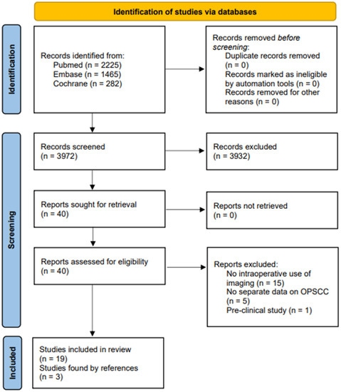
Pazopanib is approved for treatment of advanced soft tissue sarcomas, but primary and secondary drug resistance limits its clinical utility. We investigated the molecular mechanisms mediating pazopanib resistance in human synovial sarcoma (SS) models. We found reduced cell sensitivity to pazopanib associated with inefficient inhibition of the two critical signaling nodes, AKT and ERKs, despite strong inhibition of the main drug target, PDGFRα. In the CME-1 cell line, overactivation of IGF1 and Insulin receptors (IGF1R/InsR) sustained AKT activation and pazopanib resistance, which was overcome by a combination treatment with the double IGF1R/InsR inhibitor BMS754807. In the highly pazopanib resistant MoJo cell line, NRASQ61R mutation sustained constitutive ERK activation. Transfection of the NRAS mutant in the pazopanib sensitive SYO-1 cell line increased the drug IC50. MoJo cells treatment with pazopanib in combination with the MEK inhibitor trametinib restored ERK inhibition, synergistically inhibited cell growth, and induced apoptosis. The combination significantly enhanced the antitumor efficacy against MoJo orthotopic xenograft abrogating growth in 38% of mice. These findings identified two different mechanisms of intrinsic pazopanib resistance in SS cells, supporting molecular/immunohistochemical profiling of tumor specimens as a valuable approach to selecting patients who may benefit from rational drug combinations. Full article
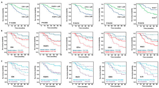
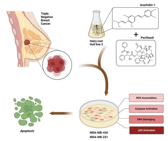
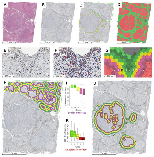
Show Figures

Figure 1

24 pages, 2556 KiB  
Review
Tumor Energy Metabolism and Potential of 3-Bromopyruvate as an Inhibitor of Aerobic Glycolysis: Implications in Tumor Treatment
by Tengjiao Fan, Guohui Sun, Xiaodong Sun, Lijiao Zhao, Rugang Zhong and Yongzhen Peng
Cancers 2019, 11(3), 317; https://doi.org/10.3390/cancers11030317 - 6 Mar 2019
Cited by 124 | Viewed by 15255
Abstract
Tumor formation and growth depend on various biological metabolism processes that are distinctly different with normal tissues. Abnormal energy metabolism is one of the typical characteristics of tumors. It has been proven that most tumor cells highly rely on aerobic glycolysis to obtain [...] Read more.
Tumor formation and growth depend on various biological metabolism processes that are distinctly different with normal tissues. Abnormal energy metabolism is one of the typical characteristics of tumors. It has been proven that most tumor cells highly rely on aerobic glycolysis to obtain energy rather than mitochondrial oxidative phosphorylation (OXPHOS) even in the presence of oxygen, a phenomenon called “Warburg effect”. Thus, inhibition of aerobic glycolysis becomes an attractive strategy to specifically kill tumor cells, while normal cells remain unaffected. In recent years, a small molecule alkylating agent, 3-bromopyruvate (3-BrPA), being an effective glycolytic inhibitor, has shown great potential as a promising antitumor drug. Not only it targets glycolysis process, but also inhibits mitochondrial OXPHOS in tumor cells. Excellent antitumor effects of 3-BrPA were observed in cultured cells and tumor-bearing animal models. In this review, we described the energy metabolic pathways of tumor cells, mechanism of action and cellular targets of 3-BrPA, antitumor effects, and the underlying mechanism of 3-BrPA alone or in combination with other antitumor drugs (e.g., cisplatin, doxorubicin, daunorubicin, 5-fluorouracil, etc.) in vitro and in vivo. In addition, few human case studies of 3-BrPA were also involved. Finally, the novel chemotherapeutic strategies of 3-BrPA, including wafer, liposomal nanoparticle, aerosol, and conjugate formulations, were also discussed for future clinical application. Full article
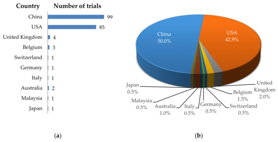
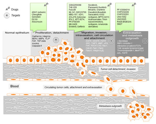
Show Figures

Figure 1

Back to TopTop