ijms-logo

Journal Browser

Journal Browser

Computational, Structural and Spectroscopic Studies of Enzyme Mechanisms, Inhibition and Dynamics

Editor


E-Mail Website
Collection Editor

Topical Collection Information

Dear Colleagues,

Enzymes are in the center of biochemical processes. They catalyze largest part of all chemical reactions in the living organisms (from viruses to human) and are characterized by unique capabilities to accelerate the reaction rates and to catalyze specific or very selective number of chemical transformations. Not surprisingly the enzymes received massive application in biomedicine, pharmacy, biotechnological and chemical industry.

The current progress in understanding enzymes underlines the new perspective of their applications and utilization in important areas for us. There is vastly growing amount of novel structures, spectroscopic data about intermediates, novel inhibitors synthesized and even enzymes with novel functions engineered.

The current thematic issue of International Journal of Molecular Sciences titled “Computational, Structural and Spectroscopic Studies of Enzyme Mechanisms, Inhibition and Dynamics” is focused on high quality studies by broad range of experimental and computational methods. Contributions focused on integrated modelling/experimental or combination between different experimental methods and the multilevel applications of computational methods are very welcome as well. Highly valued will be combined fundamental and innovative contributions focused on the applications of the enzyme mechanisms and in the all areas with impact for the society: industry, health, food etc. Finally we would like to support strengthen, develop, demonstrate and facilitate the independence of thinking, creativity, initiative of researchers at all levels.

Prof. Dr. Christo Z. Christov
Collection Editor

Manuscript Submission Information

Manuscripts for the topical collection can be submitted online at www.mdpi.com by registering and logging in to this website. Once you are registered, click here to go to the submission form. All papers will be peer-reviewed. Accepted papers will be published continuously in the journal (as soon as accepted) and will be listed together on this website. The topical collection considers regular research articles, short communications and review articles. A guide for authors and other relevant information for submission of manuscripts is available on the Instructions for Authors page.

Please visit the Instructions for Authors page before submitting a manuscript. The article processing charge (APC) for publication in this open access journal is 2900 CHF (Swiss Francs).

Keywords

  • enzyme mechanisms
  • computational modeling
  • qm / mm
  • molecular dynamics
  • enzyme spectroscopy
  • quantum mechanics
  • enzyme inhibition
  • enzyme crystallography
Published Papers (36 papers)

2017

Jump to: 2016, 2015, 2014, 2013

1122 KiB  
Review
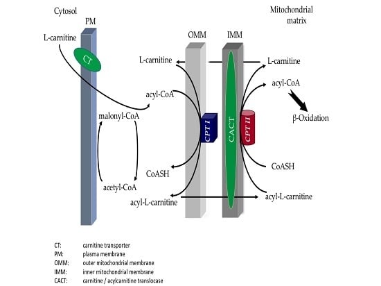
Muscle Carnitine Palmitoyltransferase II Deficiency: A Review of Enzymatic Controversy and Clinical Features
by Diana Lehmann, Leila Motlagh, Dina Robaa and Stephan Zierz
Int. J. Mol. Sci. 2017, 18(1), 82; https://doi.org/10.3390/ijms18010082 - 3 Jan 2017
Cited by 29 | Viewed by 8223
Abstract
CPT (carnitine palmitoyltransferase) II muscle deficiency is the most common form of muscle fatty acid metabolism disorders. In contrast to carnitine deficiency, it is clinically characterized by attacks of myalgia and rhabdomyolysis without persistent muscle weakness and lipid accumulation in muscle fibers. The [...] Read more.
CPT (carnitine palmitoyltransferase) II muscle deficiency is the most common form of muscle fatty acid metabolism disorders. In contrast to carnitine deficiency, it is clinically characterized by attacks of myalgia and rhabdomyolysis without persistent muscle weakness and lipid accumulation in muscle fibers. The biochemical consequences of the disease-causing mutations are still discussed controversially. CPT activity in muscles of patients with CPT II deficiency ranged from not detectable to reduced to normal. Based on the observation that in patients, total CPT is completely inhibited by malony-CoA, a deficiency of malonyl-CoA-insensitive CPT II has been suggested. In contrast, it has also been shown that in muscle CPT II deficiency, CPT II protein is present in normal concentrations with normal enzymatic activity. However, CPT II in patients is abnormally sensitive to inhibition by malonyl-CoA, Triton X-100 and fatty acid metabolites. A recent study on human recombinant CPT II enzymes (His6-N-hCPT2 and His6-N-hCPT2/S113L) revealed that the wild-type and the S113L variants showed the same enzymatic activity. However, the mutated enzyme showed an abnormal thermal destabilization at 40 and 45 °C and an abnormal sensitivity to inhibition by malony-CoA. The thermolability of the mutant enzyme might explain why symptoms in muscle CPT II deficiency mainly occur during prolonged exercise, infections and exposure to cold. In addition, the abnormally regulated enzyme might be mostly inhibited when the fatty acid metabolism is stressed. Full article
Show Figures

Graphical abstract

2016

Jump to: 2017, 2015, 2014, 2013

5238 KiB  
Review
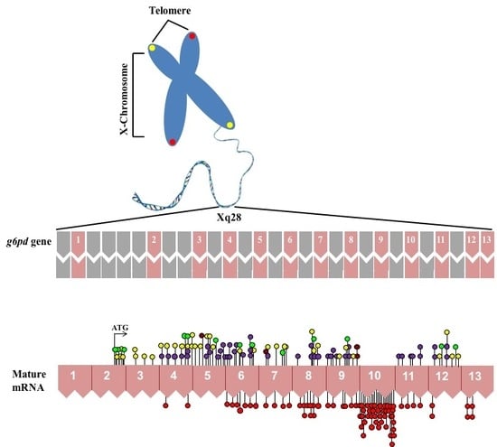
Glucose-6-Phosphate Dehydrogenase: Update and Analysis of New Mutations around the World
by Saúl Gómez-Manzo, Jaime Marcial-Quino, America Vanoye-Carlo, Hugo Serrano-Posada, Daniel Ortega-Cuellar, Abigail González-Valdez, Rosa Angélica Castillo-Rodríguez, Beatriz Hernández-Ochoa, Edgar Sierra-Palacios, Eduardo Rodríguez-Bustamante and Roberto Arreguin-Espinosa
Int. J. Mol. Sci. 2016, 17(12), 2069; https://doi.org/10.3390/ijms17122069 - 9 Dec 2016
Cited by 158 | Viewed by 24661
Abstract
Glucose-6-phosphate dehydrogenase (G6PD) is a key regulatory enzyme in the pentose phosphate pathway which produces nicotinamide adenine dinucleotide phosphate (NADPH) to maintain an adequate reducing environment in the cells and is especially important in red blood cells (RBC). Given its central role in [...] Read more.
Glucose-6-phosphate dehydrogenase (G6PD) is a key regulatory enzyme in the pentose phosphate pathway which produces nicotinamide adenine dinucleotide phosphate (NADPH) to maintain an adequate reducing environment in the cells and is especially important in red blood cells (RBC). Given its central role in the regulation of redox state, it is understandable that mutations in the gene encoding G6PD can cause deficiency of the protein activity leading to clinical manifestations such as neonatal jaundice and acute hemolytic anemia. Recently, an extensive review has been published about variants in the g6pd gene; recognizing 186 mutations. In this work, we review the state of the art in G6PD deficiency, describing 217 mutations in the g6pd gene; we also compile information about 31 new mutations, 16 that were not recognized and 15 more that have recently been reported. In order to get a better picture of the effects of new described mutations in g6pd gene, we locate the point mutations in the solved three-dimensional structure of the human G6PD protein. We found that class I mutations have the most deleterious effects on the structure and stability of the protein. Full article
Show Figures

Graphical abstract

6654 KiB  
Article
Exploring the Molecular Basis for Binding of Inhibitors by Threonyl-tRNA Synthetase from Brucella abortus: A Virtual Screening Study
by Ming Li, Fang Wen, Shengguo Zhao, Pengpeng Wang, Songli Li, Yangdong Zhang, Nan Zheng and Jiaqi Wang
Int. J. Mol. Sci. 2016, 17(7), 1078; https://doi.org/10.3390/ijms17071078 - 19 Jul 2016
Cited by 3 | Viewed by 5699
Abstract
Targeting threonyl-tRNA synthetase (ThrRS) of Brucella abortus is a promising approach to developing small-molecule drugs against bovine brucellosis. Using the BLASTp algorithm, we identified ThrRS from Escherichia coli (EThrRS, PDB ID 1QF6), which is 51% identical to ThrRS from Brucella abortus (BaThrRS) at [...] Read more.
Targeting threonyl-tRNA synthetase (ThrRS) of Brucella abortus is a promising approach to developing small-molecule drugs against bovine brucellosis. Using the BLASTp algorithm, we identified ThrRS from Escherichia coli (EThrRS, PDB ID 1QF6), which is 51% identical to ThrRS from Brucella abortus (BaThrRS) at the amino acid sequence level. EThrRS was used as the template to construct a BaThrRS homology model which was optimized using molecular dynamics simulations. To determine the residues important for substrate ATP binding, we identified the ATP-binding regions of BaThrRS, docked ATP to the protein, and identified the residues whose side chains surrounded bound ATP. We then used the binding site of ATP to virtually screen for BaThrRS inhibitors and got seven leads. We further characterized the BaThrRS-binding site of the compound with the highest predicted inhibitory activity. Our results should facilitate future experimental effects to find novel drugs for use against bovine brucellosis. Full article
Show Figures

Graphical abstract

1871 KiB  
Article
Ligand and Structure-Based Approaches for the Identification of Peptide Deformylase Inhibitors as Antibacterial Drugs
by Jian Gao, Li Liang, Yasheng Zhu, Shengzhi Qiu, Tao Wang and Ling Zhang
Int. J. Mol. Sci. 2016, 17(7), 1141; https://doi.org/10.3390/ijms17071141 - 15 Jul 2016
Cited by 32 | Viewed by 6671
Abstract
Peptide deformylase (PDF) is a metalloprotease catalyzing the removal of a formyl group from newly synthesized proteins, which makes it an important antibacterial drug target. Given the importance of PDF inhibitors like actinonin in antibacterial drug discovery, several reported potent PDF inhibitors were [...] Read more.
Peptide deformylase (PDF) is a metalloprotease catalyzing the removal of a formyl group from newly synthesized proteins, which makes it an important antibacterial drug target. Given the importance of PDF inhibitors like actinonin in antibacterial drug discovery, several reported potent PDF inhibitors were used to develop pharmacophore models using the Galahad module of Sybyl 7.1 software. Generated pharmacophore models were composed of two donor atom centers, four acceptor atom centers and two hydrophobic groups. Model-1 was screened against the Zinc database and several compounds were retrieved as hits. Compounds with Qfit values of more than 60 were employed to perform a molecular docking study with the receptor Escherichia coli PDF, then compounds with docking score values of more than 6 were used to predict the in silico pharmacokinetic and toxicity risk via OSIRIS property explorer. Two known PDF inhibitors were also used to perform a molecular docking study with E. coli PDF as reference molecules. The results of the molecular docking study were validated by reproducing the crystal structure of actinonin. Molecular docking and in silico pharmacokinetic and toxicity prediction studies suggested that ZINC08740166 has a relatively high docking score of 7.44 and a drug score of 0.78. Full article
Show Figures

Graphical abstract

12457 KiB  
Article
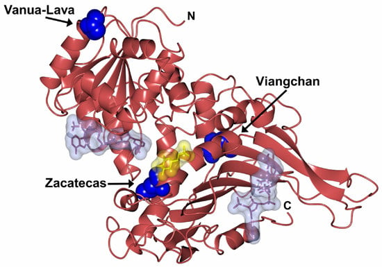
Functional and Biochemical Characterization of Three Recombinant Human Glucose-6-Phosphate Dehydrogenase Mutants: Zacatecas, Vanua-Lava and Viangchan
by Saúl Gómez-Manzo, Jaime Marcial-Quino, America Vanoye-Carlo, Hugo Serrano-Posada, Abigail González-Valdez, Víctor Martínez-Rosas, Beatriz Hernández-Ochoa, Edgar Sierra-Palacios, Rosa Angélica Castillo-Rodríguez, Miguel Cuevas-Cruz, Eduardo Rodríguez-Bustamante and Roberto Arreguin-Espinosa
Int. J. Mol. Sci. 2016, 17(5), 787; https://doi.org/10.3390/ijms17050787 - 21 May 2016
Cited by 23 | Viewed by 7841
Abstract
Glucose-6-phosphate dehydrogenase (G6PD) deficiency in humans causes severe disease, varying from mostly asymptomatic individuals to patients showing neonatal jaundice, acute hemolysis episodes or chronic nonspherocytic hemolytic anemia. In order to understand the effect of the mutations in G6PD gene function and its relation [...] Read more.
Glucose-6-phosphate dehydrogenase (G6PD) deficiency in humans causes severe disease, varying from mostly asymptomatic individuals to patients showing neonatal jaundice, acute hemolysis episodes or chronic nonspherocytic hemolytic anemia. In order to understand the effect of the mutations in G6PD gene function and its relation with G6PD deficiency severity, we report the construction, cloning and expression as well as the detailed kinetic and stability characterization of three purified clinical variants of G6PD that present in the Mexican population: G6PD Zacatecas (Class I), Vanua-Lava (Class II) and Viangchan (Class II). For all the G6PD mutants, we obtained low purification yield and altered kinetic parameters compared with Wild Type (WT). Our results show that the mutations, regardless of the distance from the active site where they are located, affect the catalytic properties and structural parameters and that these changes could be associated with the clinical presentation of the deficiency. Specifically, the structural characterization of the G6PD Zacatecas mutant suggests that the R257L mutation have a strong effect on the global stability of G6PD favoring an unstable active site. Using computational analysis, we offer a molecular explanation of the effects of these mutations on the active site. Full article
Show Figures

Graphical abstract

3896 KiB  
Article
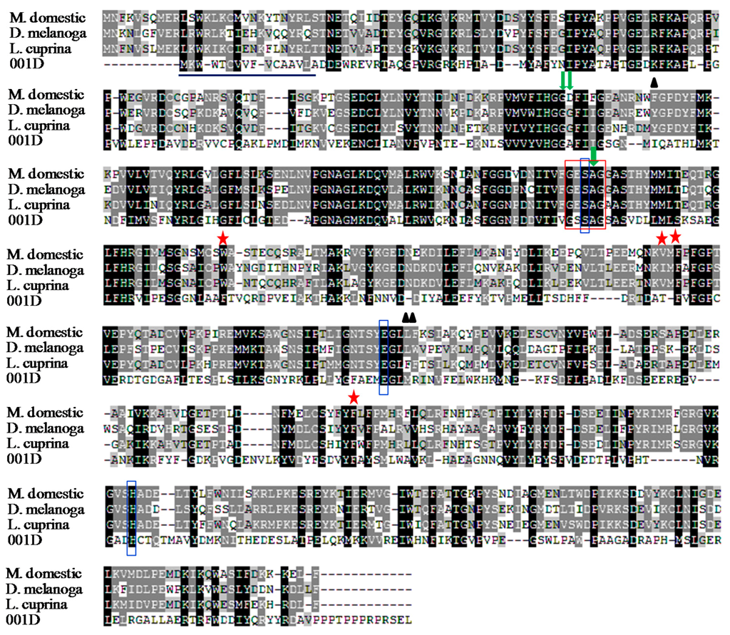
Bacterial Expression and Kinetic Analysis of Carboxylesterase 001D from Helicoverpa armigera
by Yongqiang Li, Jianwei Liu, Mei Lu, Zhiqing Ma, Chongling Cai, Yonghong Wang and Xing Zhang
Int. J. Mol. Sci. 2016, 17(4), 493; https://doi.org/10.3390/ijms17040493 - 2 Apr 2016
Cited by 18 | Viewed by 6589
Abstract
Carboxylesterasesare an important class of detoxification enzymes involved in insecticide resistance in insects. A subgroup of Helicoverpa armigera esterases, known as Clade 001, was implicated in organophosphate and pyrethroid insecticide resistance due to their overabundance in resistant strains. In this work, a novel [...] Read more.
Carboxylesterasesare an important class of detoxification enzymes involved in insecticide resistance in insects. A subgroup of Helicoverpa armigera esterases, known as Clade 001, was implicated in organophosphate and pyrethroid insecticide resistance due to their overabundance in resistant strains. In this work, a novel carboxylesterasegene 001D of H. armigera from China was cloned, which has an open reading frame of 1665 nucleotides encoding 554 amino acid residues. We used a series of fusion proteins to successfully express carboxylesterase 001D in Escherichia coli. Three different fusion proteins were generated and tested. The enzyme kinetic assay towards 1-naphthyl acetate showed all three purified fusion proteins are active with a Kcat between 0.35 and 2.29 s−1, and a Km between 7.61 and 19.72 μM. The HPLC assay showed all three purified fusion proteins had low but measurable hydrolase activity towards β-cypermethrin and fenvalerate insecticides (specific activities ranging from 0.13 to 0.67 μM·min−1·(μM−1·protein)). The enzyme was stable up to 40 °C and at pH 6.0–11.0. The results imply that carboxylesterase 001D is involved in detoxification, and this moderate insecticide hydrolysis may suggest that overexpression of the gene to enhance insecticide sequestration is necessary to allow carboxylesterases to confer resistance to these insecticides in H. armigera. Full article
Show Figures

Figure 1

2176 KiB  
Article
Computational Study on New Natural Compound Inhibitors of Pyruvate Dehydrogenase Kinases
by Xiaoli Zhou, Shanshan Yu, Jing Su and Liankun Sun
Int. J. Mol. Sci. 2016, 17(3), 340; https://doi.org/10.3390/ijms17030340 - 4 Mar 2016
Cited by 44 | Viewed by 10545
Abstract
Pyruvate dehydrogenase kinases (PDKs) are key enzymes in glucose metabolism, negatively regulating pyruvate dehyrogenase complex (PDC) activity through phosphorylation. Inhibiting PDKs could upregulate PDC activity and drive cells into more aerobic metabolism. Therefore, PDKs are potential targets for metabolism related diseases, such as [...] Read more.
Pyruvate dehydrogenase kinases (PDKs) are key enzymes in glucose metabolism, negatively regulating pyruvate dehyrogenase complex (PDC) activity through phosphorylation. Inhibiting PDKs could upregulate PDC activity and drive cells into more aerobic metabolism. Therefore, PDKs are potential targets for metabolism related diseases, such as cancers and diabetes. In this study, a series of computer-aided virtual screening techniques were utilized to discover potential inhibitors of PDKs. Structure-based screening using Libdock was carried out following by ADME (adsorption, distribution, metabolism, excretion) and toxicity prediction. Molecular docking was used to analyze the binding mechanism between these compounds and PDKs. Molecular dynamic simulation was utilized to confirm the stability of potential compound binding. From the computational results, two novel natural coumarins compounds (ZINC12296427 and ZINC12389251) from the ZINC database were found binding to PDKs with favorable interaction energy and predicted to be non-toxic. Our study provide valuable information of PDK-coumarins binding mechanisms in PDK inhibitor-based drug discovery. Full article
Show Figures

Graphical abstract

3348 KiB  
Article
Enzymatic Kinetic Properties of the Lactate Dehydrogenase Isoenzyme C4 of the Plateau Pika (Ochotona curzoniae)
by Yang Wang, Lian Wei, Dengbang Wei, Xiao Li, Lina Xu and Linna Wei
Int. J. Mol. Sci. 2016, 17(1), 39; https://doi.org/10.3390/ijms17010039 - 7 Jan 2016
Cited by 9 | Viewed by 11514
Abstract
Testis-specific lactate dehydrogenase (LDH-C4) is one of the lactate dehydrogenase (LDH) isozymes that catalyze the terminal reaction of pyruvate to lactate in the glycolytic pathway. LDH-C4 in mammals was previously thought to be expressed only in spermatozoa and testis and [...] Read more.
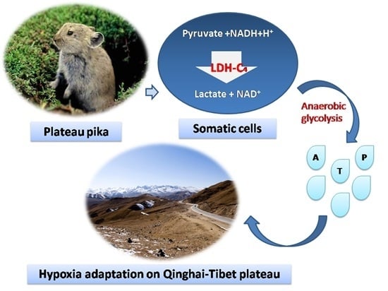
Testis-specific lactate dehydrogenase (LDH-C4) is one of the lactate dehydrogenase (LDH) isozymes that catalyze the terminal reaction of pyruvate to lactate in the glycolytic pathway. LDH-C4 in mammals was previously thought to be expressed only in spermatozoa and testis and not in other tissues. Plateau pika (Ochotona curzoniae) belongs to the genus Ochotona of the Ochotonidea family. It is a hypoxia-tolerant species living in remote mountain areas at altitudes of 3000–5000 m above sea level on the Qinghai-Tibet Plateau. Surprisingly, Ldh-c is expressed not only in its testis and sperm, but also in somatic tissues of plateau pika. To shed light on the function of LDH-C4 in somatic cells, Ldh-a, Ldh-b, and Ldh-c of plateau pika were subcloned into bacterial expression vectors. The pure enzymes of Lactate Dehydrogenase A4 (LDH-A4), Lactate Dehydrogenase B4 (LDH-B4), and LDH-C4 were prepared by a series of expression and purification processes, and the three enzymes were identified by the method of sodium dodecyl sulfate polyacrylamide gel electrophoresis (SDS-PAGE) and native polyacrylamide gel electrophoresis (PAGE). The enzymatic kinetics properties of these enzymes were studied by Lineweaver-Burk double-reciprocal plots. The results showed the Michaelis constant (Km) of LDH-C4 for pyruvate and lactate was 0.052 and 4.934 mmol/L, respectively, with an approximate 90 times higher affinity of LDH-C4 for pyruvate than for lactate. At relatively high concentrations of lactate, the inhibition constant (Ki) of the LDH isoenzymes varied: LDH-A4 (Ki = 26.900 mmol/L), LDH-B4 (Ki = 23.800 mmol/L), and LDH-C4 (Ki = 65.500 mmol/L). These data suggest that inhibition of lactate by LDH-A4 and LDH-B4 were stronger than LDH-C4. In light of the enzymatic kinetics properties, we suggest that the plateau pika can reduce reliance on oxygen supply and enhance its adaptation to the hypoxic environments due to increased anaerobic glycolysis by LDH-C4. Full article
Show Figures

Graphical abstract

2015

Jump to: 2017, 2016, 2014, 2013

4007 KiB  
Article
Domain Motions and Functionally-Key Residues of l-Alanine Dehydrogenase Revealed by an Elastic Network Model
by Xing-Yuan Li, Jing-Chao Zhang, Yan-Ying Zhu and Ji-Guo Su
Int. J. Mol. Sci. 2015, 16(12), 29383-29397; https://doi.org/10.3390/ijms161226170 - 9 Dec 2015
Cited by 10 | Viewed by 5965
Abstract
Mycobacterium tuberculosis l-alanine dehydrogenase (l-MtAlaDH) plays an important role in catalyzing l-alanine to ammonia and pyruvate, which has been considered to be a potential target for tuberculosis treatment. In the present work, the functional domain motions encoded in the structure of [...] Read more.
Mycobacterium tuberculosis l-alanine dehydrogenase (l-MtAlaDH) plays an important role in catalyzing l-alanine to ammonia and pyruvate, which has been considered to be a potential target for tuberculosis treatment. In the present work, the functional domain motions encoded in the structure of l-MtAlaDH were investigated by using the Gaussian network model (GNM) and the anisotropy network model (ANM). The slowest modes for the open-apo and closed-holo structures of the enzyme show that the domain motions have a common hinge axis centered in residues Met133 and Met301. Accompanying the conformational transition, both the 1,4-dihydronicotinamide adenine dinucleotide (NAD)-binding domain (NBD) and the substrate-binding domain (SBD) move in a highly coupled way. The first three slowest modes of ANM exhibit the open-closed, rotation and twist motions of l-MtAlaDH, respectively. The calculation of the fast modes reveals the residues responsible for the stability of the protein, and some of them are involved in the interaction with the ligand. Then, the functionally-important residues relevant to the binding of the ligand were identified by using a thermodynamic method. Our computational results are consistent with the experimental data, which will help us to understand the physical mechanism for the function of l-MtAlaDH. Full article
Show Figures

Figure 1

4175 KiB  
Article
Mutations of Glucose-6-Phosphate Dehydrogenase Durham, Santa-Maria and A+ Variants Are Associated with Loss Functional and Structural Stability of the Protein
by Saúl Gómez-Manzo, Jaime Marcial-Quino, America Vanoye-Carlo, Sergio Enríquez-Flores, Ignacio De la Mora-De la Mora, Abigail González-Valdez, Itzhel García-Torres, Víctor Martínez-Rosas, Edgar Sierra-Palacios, Fernando Lazcano-Pérez, Eduardo Rodríguez-Bustamante and Roberto Arreguin-Espinosa
Int. J. Mol. Sci. 2015, 16(12), 28657-28668; https://doi.org/10.3390/ijms161226124 - 2 Dec 2015
Cited by 28 | Viewed by 9091
Abstract
Glucose-6-phosphate dehydrogenase (G6PD) deficiency is the most common enzymopathy in the world. More than 160 mutations causing the disease have been identified, but only 10% of these variants have been studied at biochemical and biophysical levels. In this study we report on the [...] Read more.
Glucose-6-phosphate dehydrogenase (G6PD) deficiency is the most common enzymopathy in the world. More than 160 mutations causing the disease have been identified, but only 10% of these variants have been studied at biochemical and biophysical levels. In this study we report on the functional and structural characterization of three naturally occurring variants corresponding to different classes of disease severity: Class I G6PD Durham, Class II G6PD Santa Maria, and Class III G6PD A+. The results showed that the G6PD Durham (severe deficiency), and the G6PD Santa Maria and A+ (less severe deficiency) (Class I, II and III, respectively) affect the catalytic efficiency of these enzymes, are more sensitive to temperature denaturing, and affect the stability of the overall protein when compared to the wild type WT-G6PD. In the variants, the exposure of more and buried hydrophobic pockets was induced and monitored with 8-Anilinonaphthalene-1-sulfonic acid (ANS) fluorescence, directly affecting the compaction of structure at different levels and probably reducing the stability of the protein. The degree of functional and structural perturbation by each variant correlates with the clinical severity reported in different patients. Full article
Show Figures

Graphical abstract

4749 KiB  
Article
Repositioning of Thiourea-Containing Drugs as Tyrosinase Inhibitors
by Joonhyeok Choi and Jun-Goo Jee
Int. J. Mol. Sci. 2015, 16(12), 28534-28548; https://doi.org/10.3390/ijms161226114 - 2 Dec 2015
Cited by 33 | Viewed by 9761
Abstract
Tyrosinase catalyzes two distinct sequential reactions in melanin biosynthesis: The hydroxylation of tyrosine to dihydroxyphenylalanine (DOPA) and the oxidation of DOPA to dopaquinone. Developing functional modulators of tyrosinase is important for therapeutic and cosmetic purposes. Given the abundance of thiourea moiety in known [...] Read more.
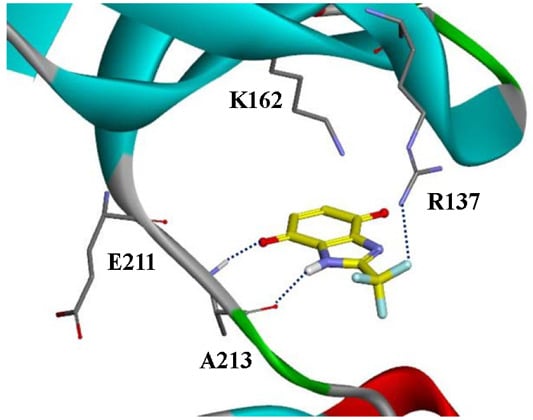
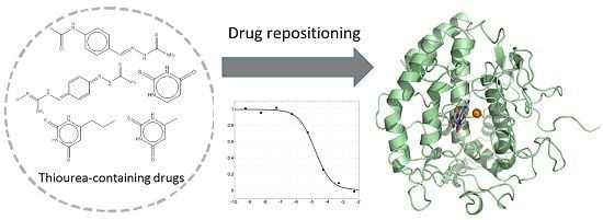
Tyrosinase catalyzes two distinct sequential reactions in melanin biosynthesis: The hydroxylation of tyrosine to dihydroxyphenylalanine (DOPA) and the oxidation of DOPA to dopaquinone. Developing functional modulators of tyrosinase is important for therapeutic and cosmetic purposes. Given the abundance of thiourea moiety in known tyrosinase inhibitors, we studied other thiourea-containing drugs as potential tyrosinase inhibitors. The thiourea-containing drugs in clinical use were retrieved and tested for their ability to inhibit tyrosinase. We observed that methimazole, thiouracil, methylthiouracil, propylthiouracil, ambazone, and thioacetazone inhibited mushroom tyrosinase. Except for methimazole, there was limited information regarding the activity of other drugs against tyrosinase. Both thioacetazone and ambazone significantly inhibited tyrosinase, with IC50 of 14 and 15 μM, respectively. Ambazone decreased melanin content without causing cellular toxicity at 20 μM in B16F10 cells. The activity of ambazone was stronger than that of kojic acid both in enzyme and melanin content assays. Kinetics of enzyme inhibition assigned the thiourea-containg drugs as non-competitive inhibitors. The complex models by docking simulation suggested that the intermolecular hydrogen bond via the nitrogen of thiourea and the contacts via thione were equally important for interacting with tyrosinase. These data were consistent with the results of enzyme assays with the analogues of thiourea. Full article
Show Figures

Graphical abstract

2428 KiB  
Article
An Investigation of Molecular Docking and Molecular Dynamic Simulation on Imidazopyridines as B-Raf Kinase Inhibitors
by Huiding Xie, Yupeng Li, Fang Yu, Xiaoguang Xie, Kaixiong Qiu and Jijun Fu
Int. J. Mol. Sci. 2015, 16(11), 27350-27361; https://doi.org/10.3390/ijms161126026 - 16 Nov 2015
Cited by 23 | Viewed by 6608
Abstract
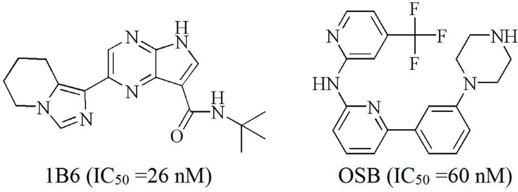
In the recent cancer treatment, B-Raf kinase is one of key targets. Nowadays, a group of imidazopyridines as B-Raf kinase inhibitors have been reported. In order to investigate the interaction between this group of inhibitors and B-Raf kinase, molecular docking, molecular dynamic (MD) [...] Read more.
In the recent cancer treatment, B-Raf kinase is one of key targets. Nowadays, a group of imidazopyridines as B-Raf kinase inhibitors have been reported. In order to investigate the interaction between this group of inhibitors and B-Raf kinase, molecular docking, molecular dynamic (MD) simulation and binding free energy (ΔGbind) calculation were performed in this work. Molecular docking was carried out to identify the key residues in the binding site, and MD simulations were performed to determine the detail binding mode. The results obtained from MD simulation reveal that the binding site is stable during the MD simulations, and some hydrogen bonds (H-bonds) in MD simulations are different from H-bonds in the docking mode. Based on the obtained MD trajectories, ΔGbind was computed by using Molecular Mechanics Generalized Born Surface Area (MM-GBSA), and the obtained energies are consistent with the activities. An energetic analysis reveals that both electrostatic and van der Waals contributions are important to ΔGbind, and the unfavorable polar solvation contribution results in the instability of the inhibitor with the lowest activity. These results are expected to understand the binding between B-Raf and imidazopyridines and provide some useful information to design potential B-Raf inhibitors. Full article
Show Figures

Graphical abstract

3630 KiB  
Review
From Genome to Structure and Back Again: A Family Portrait of the Transcarbamylases
by Dashuang Shi, Norma M. Allewell and Mendel Tuchman
Int. J. Mol. Sci. 2015, 16(8), 18836-18864; https://doi.org/10.3390/ijms160818836 - 12 Aug 2015
Cited by 19 | Viewed by 7954
Abstract
Enzymes in the transcarbamylase family catalyze the transfer of a carbamyl group from carbamyl phosphate (CP) to an amino group of a second substrate. The two best-characterized members, aspartate transcarbamylase (ATCase) and ornithine transcarbamylase (OTCase), are present in most organisms from bacteria to [...] Read more.
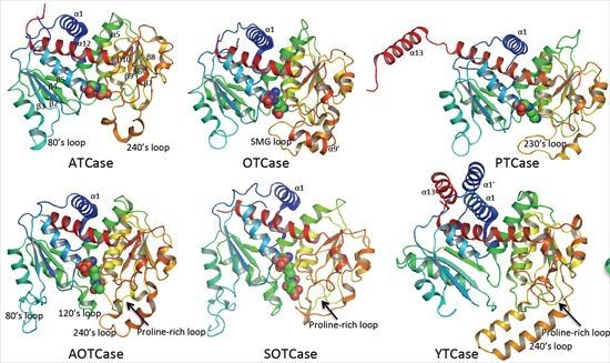
Enzymes in the transcarbamylase family catalyze the transfer of a carbamyl group from carbamyl phosphate (CP) to an amino group of a second substrate. The two best-characterized members, aspartate transcarbamylase (ATCase) and ornithine transcarbamylase (OTCase), are present in most organisms from bacteria to humans. Recently, structures of four new transcarbamylase members, N-acetyl-l-ornithine transcarbamylase (AOTCase), N-succinyl-l-ornithine transcarbamylase (SOTCase), ygeW encoded transcarbamylase (YTCase) and putrescine transcarbamylase (PTCase) have also been determined. Crystal structures of these enzymes have shown that they have a common overall fold with a trimer as their basic biological unit. The monomer structures share a common CP binding site in their N-terminal domain, but have different second substrate binding sites in their C-terminal domain. The discovery of three new transcarbamylases, l-2,3-diaminopropionate transcarbamylase (DPTCase), l-2,4-diaminobutyrate transcarbamylase (DBTCase) and ureidoglycine transcarbamylase (UGTCase), demonstrates that our knowledge and understanding of the spectrum of the transcarbamylase family is still incomplete. In this review, we summarize studies on the structures and function of transcarbamylases demonstrating how structural information helps to define biological function and how small structural differences govern enzyme specificity. Such information is important for correctly annotating transcarbamylase sequences in the genome databases and for identifying new members of the transcarbamylase family. Full article
Show Figures

Graphical abstract

2388 KiB  
Article
Biochemical Properties and Structure Analysis of a DAG-Like Lipase from Malassezia globosa
by Huan Xu, Dongming Lan, Bo Yang and Yonghua Wang
Int. J. Mol. Sci. 2015, 16(3), 4865-4879; https://doi.org/10.3390/ijms16034865 - 4 Mar 2015
Cited by 17 | Viewed by 7698
Abstract
Diacylglycerol (DAG)-like lipases are found to play an important role in the life sciences and industrial fields. A putative DAG-like lipase (MgMDL2) from Malassezia globosa was cloned and expressed in recombinant Pichia pastoris. The recombinant MgMDL2 was expressed as [...] Read more.
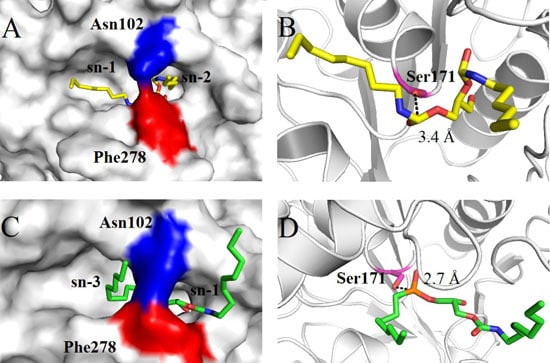
Diacylglycerol (DAG)-like lipases are found to play an important role in the life sciences and industrial fields. A putative DAG-like lipase (MgMDL2) from Malassezia globosa was cloned and expressed in recombinant Pichia pastoris. The recombinant MgMDL2 was expressed as a glycosylated protein and purified into homogeneity by anion exchange chromatography. The activity of recombinant MgMDL2 was optimal at 15 °C and pH 6.0, and it keeps over 50% of relative activity at 5 °C, suggesting that MgMDL2 was a cold active lipase. MgMDL2 retained over 80% of initial activity after incubation at 30 and 40 °C for 2.5 h, but it was not stable at 50 °C. Incubation of methanol and ethanol at a concentration of 30% for 2 h did not affect the recombinant enzyme activity, while metal ions, including Ca2+, Mn2+ and Ni2+, sharply inhibited the MgMDL2 activity at 5 mM by 42%, 35% and 36%, respectively. MgMDL2 exhibited a preference for medium chain-length esters with highest activity toward p-nitrophenyl caprylate, while it was active on mono- and diacylglycerol but not on triacylglycerol, indicating that it was a typical DAG-like lipase. By homology modeling, Phe278 was predicted to be involved in the preference of MgMDL2 for monoacyl- and diacyl-glyceride substrates, but not triglycerides. Full article
Show Figures

Graphical abstract

2014

Jump to: 2017, 2016, 2015, 2013

473 KiB  
Article
The Discovery of Aurora Kinase Inhibitor by Multi-Docking-Based Virtual Screening
by Jun-Tae Kim, Seo Hee Jung, Sun Young Kang, Chung-Kyu Ryu and Nam Sook Kang
Int. J. Mol. Sci. 2014, 15(11), 20403-20412; https://doi.org/10.3390/ijms151120403 - 6 Nov 2014
Cited by 4 | Viewed by 6812
Abstract
We report the discovery of aurora kinase inhibitor using the fragment-based virtual screening by multi-docking strategy. Among a number of fragments collected from eMololecules, we found four fragment molecules showing potent activity (>50% at 100 μM) against aurora kinase. Based on the explored [...] Read more.
We report the discovery of aurora kinase inhibitor using the fragment-based virtual screening by multi-docking strategy. Among a number of fragments collected from eMololecules, we found four fragment molecules showing potent activity (>50% at 100 μM) against aurora kinase. Based on the explored fragment scaffold, we selected two compounds in our synthesized library and validated the biological activity against Aurora kinase. Full article
Show Figures

Graphical abstract

5373 KiB  
Article
Molecular Modeling and MM-PBSA Free Energy Analysis of Endo-1,4-β-Xylanase from Ruminococcus albus 8
by Dongling Zhan, Lei Yu, Hanyong Jin, Shanshan Guan and Weiwei Han
Int. J. Mol. Sci. 2014, 15(10), 17284-17303; https://doi.org/10.3390/ijms151017284 - 26 Sep 2014
Cited by 18 | Viewed by 7976
Abstract
Endo-1,4-β-xylanase (EC 3.2.1.8) is the enzyme from Ruminococcus albus 8 (R. albus 8) (Xyn10A), and catalyzes the degradation of arabinoxylan, which is a major cell wall non-starch polysaccharide of cereals. The crystallographic structure of Xyn10A is still unknown. For this reason, we [...] Read more.
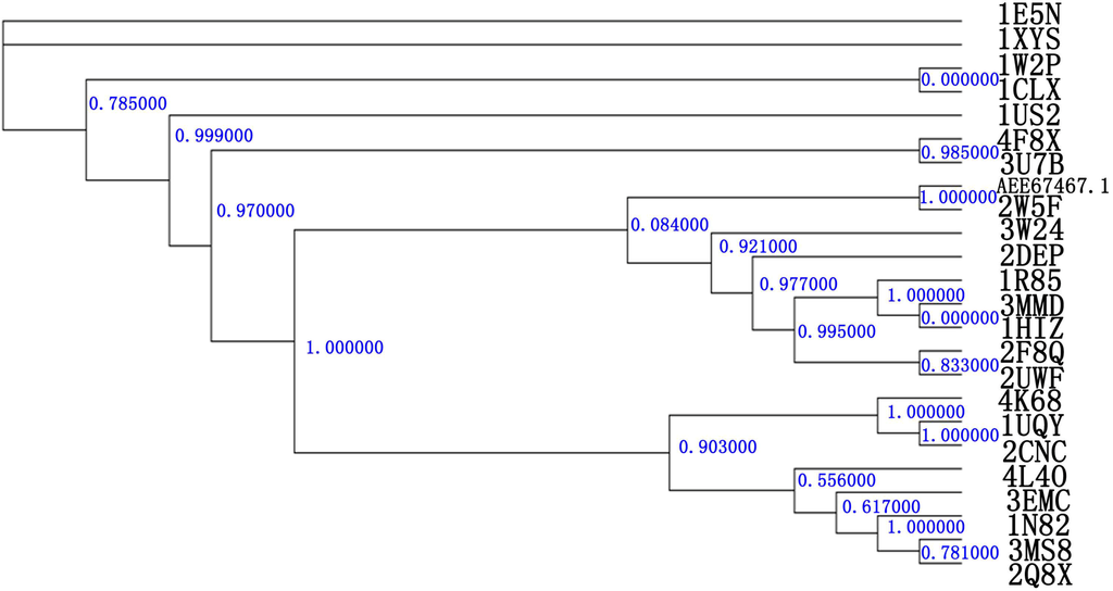
Endo-1,4-β-xylanase (EC 3.2.1.8) is the enzyme from Ruminococcus albus 8 (R. albus 8) (Xyn10A), and catalyzes the degradation of arabinoxylan, which is a major cell wall non-starch polysaccharide of cereals. The crystallographic structure of Xyn10A is still unknown. For this reason, we report a computer-assisted homology study conducted to build its three-dimensional structure based on the known sequence of amino acids of this enzyme. In this study, the best similarity was found with the Clostridium thermocellum (C. thermocellum) N-terminal endo-1,4-β-d-xylanase 10 b. Following the 100 ns molecular dynamics (MD) simulation, a reliable model was obtained for further studies. Molecular Mechanics/Poisson-Boltzmann Surface Area (MM-PBSA) methods were used for the substrate xylotetraose having the reactive sugar, which was bound in the −1 subsite of Xyn10A in the 4C1 (chair) and 2SO (skew boat) ground state conformations. According to the simulations and free energy analysis, Xyn10A binds the substrate with the −1 sugar in the 2SO conformation 39.27 kcal·mol−1 tighter than the substrate with the sugar in the 4C1 conformation. According to the Xyn10A-2SO Xylotetraose (X4(sb) interaction energies, the most important subsite for the substrate binding is subsite −1. The results of this study indicate that the substrate is bound in a skew boat conformation with Xyn10A and the −1 sugar subsite proceeds from the 4C1 conformation through 2SO to the transition state. MM-PBSA free energy analysis indicates that Asn187 and Trp344 in subsite −1 may an important residue for substrate binding. Our findings provide fundamental knowledge that may contribute to further enhancement of enzyme performance through molecular engineering. Full article
Show Figures

Figure 1

932 KiB  
Article
A Combined Experimental and Computational Study of Vam3, a Derivative of Resveratrol, and Syk Interaction
by Ming Jiang, Renping Liu, Ying Chen, Qisheng Zheng, Saijun Fan and Peixun Liu
Int. J. Mol. Sci. 2014, 15(9), 17188-17203; https://doi.org/10.3390/ijms150917188 - 25 Sep 2014
Cited by 10 | Viewed by 7333
Abstract
Spleen tyrosine kinase (Syk) plays an indispensable role through preliminary extracellular antigen-induced crosslinking of Fc receptor (FcR) in the pathogenesis of autoimmune disorders, such as rheumatoid arthritis. In this study, we identify Vam3, a dimeric derivative of resveratrol isolated from grapes, as an [...] Read more.
Spleen tyrosine kinase (Syk) plays an indispensable role through preliminary extracellular antigen-induced crosslinking of Fc receptor (FcR) in the pathogenesis of autoimmune disorders, such as rheumatoid arthritis. In this study, we identify Vam3, a dimeric derivative of resveratrol isolated from grapes, as an ATP-competitive inhibitor of Syk with an IC50 of 62.95 nM in an in vitro kinase assay. Moreover, docking and molecular dynamics simulation approaches were performed to get more detailed information about the binding mode of Vam3 and Syk. The results show that 11b-OH on ring-C and 4b-OH on ring-D could form two hydrogen bonds with Glu449 and Phe382 of Syk, respectively. In addition, arene-cation interaction between ring-D of Vam3 and Lys402 of Syk was also observed. These results indicate that ring-C and D play an essential role in Vam3–Syk interaction. Our studies may be helpful in the structural optimization of Vam3, and also aid the design of novel Syk inhibitors in the future. Full article
Show Figures

Figure 1

1032 KiB  
Article
Kinetics and Quantitative Structure—Activity Relationship Study on the Degradation Reaction from Perfluorooctanoic Acid to Trifluoroacetic Acid
by Chen Gong, Xiaomin Sun, Chenxi Zhang, Xue Zhang and Junfeng Niu
Int. J. Mol. Sci. 2014, 15(8), 14153-14165; https://doi.org/10.3390/ijms150814153 - 14 Aug 2014
Cited by 8 | Viewed by 7740
Abstract
Investigation of the degradation kinetics of perfluorooctanoic acid (PFOA) has been carried out to calculate rate constants of the main elementary reactions using the multichannel Rice-Ramsperger-Kassel-Marcus theory and canonical variational transition state theory with small-curvature tunneling correction over a temperature range of 200~500 [...] Read more.
Investigation of the degradation kinetics of perfluorooctanoic acid (PFOA) has been carried out to calculate rate constants of the main elementary reactions using the multichannel Rice-Ramsperger-Kassel-Marcus theory and canonical variational transition state theory with small-curvature tunneling correction over a temperature range of 200~500 K. The Arrhenius equations of rate constants of elementary reactions are fitted. The decarboxylation is role step in the degradation mechanism of PFOA. For the perfluorinated carboxylic acids from perfluorooctanoic acid to trifluoroacetic acid, the quantitative structure–activity relationship of the decarboxylation was analyzed with the genetic function approximation method and the structure–activity model was constructed. The main parameters governing rate constants of the decarboxylation reaction from the eight-carbon chain to the two-carbon chain were obtained. As the structure–activity model shows, the bond length and energy of C1–C2 (RC1–C2 and EC1–C2) are positively correlated to rate constants, while the volume (V), the energy difference between EHOMO and ELUMOE), and the net atomic charges on atom C2 (QC2) are negatively correlated. Full article
Show Figures

Graphical abstract

2809 KiB  
Article
Computational Study on Substrate Specificity of a Novel Cysteine Protease 1 Precursor from Zea mays
by Huimin Liu, Liangcheng Chen, Quan Li, Mingzhu Zheng and Jingsheng Liu
Int. J. Mol. Sci. 2014, 15(6), 10459-10478; https://doi.org/10.3390/ijms150610459 - 11 Jun 2014
Cited by 7 | Viewed by 8407
Abstract
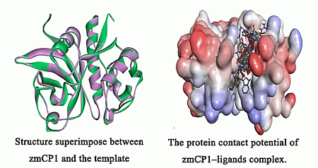
Cysteine protease 1 precursor from Zea mays (zmCP1) is classified as a member of the C1A family of peptidases (papain-like cysteine protease) in MEROPS (the Peptidase Database). The 3D structure and substrate specificity of the zmCP1 is still unknown. This study is the [...] Read more.
Cysteine protease 1 precursor from Zea mays (zmCP1) is classified as a member of the C1A family of peptidases (papain-like cysteine protease) in MEROPS (the Peptidase Database). The 3D structure and substrate specificity of the zmCP1 is still unknown. This study is the first one to build the 3D structure of zmCP1 by computer-assisted homology modeling. In order to determine the substrate specificity of zmCP1, docking study is used for rapid and convenient analysis of large populations of ligand–enzyme complexes. Docking results show that zmCP1 has preference for P1 position and P2 position for Arg and a large hydrophobic residue (such as Phe). Gly147, Gly191, Cys189, and Asp190 are predicted to function as active residues at the S1 subsite, and the S2 subsite contains Leu283, Leu193, Ala259, Met194, and Ala286. SIFt results indicate that Gly144, Arg268, Trp308, and Ser311 play important roles in substrate binding. Then Molecular Mechanics-Poisson-Boltzmann Surface Area (MM-PBSA) method was used to explain the substrate specificity for P1 position of zmCp1. This study provides insights into the molecular basis of zmCP1 activity and substrate specificity. Full article
Show Figures

Graphical abstract

745 KiB  
Article
Prediction of Protein S-Nitrosylation Sites Based on Adapted Normal Distribution Bi-Profile Bayes and Chou’s Pseudo Amino Acid Composition
by Cangzhi Jia, Xin Lin and Zhiping Wang
Int. J. Mol. Sci. 2014, 15(6), 10410-10423; https://doi.org/10.3390/ijms150610410 - 10 Jun 2014
Cited by 88 | Viewed by 8290
Abstract
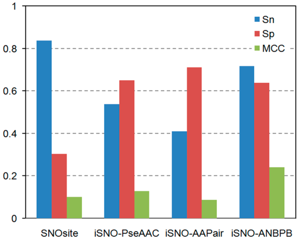
Protein S-nitrosylation is a reversible post-translational modification by covalent modification on the thiol group of cysteine residues by nitric oxide. Growing evidence shows that protein S-nitrosylation plays an important role in normal cellular function as well as in various pathophysiologic conditions. [...] Read more.
Protein S-nitrosylation is a reversible post-translational modification by covalent modification on the thiol group of cysteine residues by nitric oxide. Growing evidence shows that protein S-nitrosylation plays an important role in normal cellular function as well as in various pathophysiologic conditions. Because of the inherent chemical instability of the S-NO bond and the low abundance of endogenous S-nitrosylated proteins, the unambiguous identification of S-nitrosylation sites by commonly used proteomic approaches remains challenging. Therefore, computational prediction of S-nitrosylation sites has been considered as a powerful auxiliary tool. In this work, we mainly adopted an adapted normal distribution bi-profile Bayes (ANBPB) feature extraction model to characterize the distinction of position-specific amino acids in 784 S-nitrosylated and 1568 non-S-nitrosylated peptide sequences. We developed a support vector machine prediction model, iSNO-ANBPB, by incorporating ANBPB with the Chou’s pseudo amino acid composition. In jackknife cross-validation experiments, iSNO-ANBPB yielded an accuracy of 65.39% and a Matthew’s correlation coefficient (MCC) of 0.3014. When tested on an independent dataset, iSNO-ANBPB achieved an accuracy of 63.41% and a MCC of 0.2984, which are much higher than the values achieved by the existing predictors SNOSite, iSNO-PseAAC, the Li et al. algorithm, and iSNO-AAPair. On another training dataset, iSNO-ANBPB also outperformed GPS-SNO and iSNO-PseAAC in the 10-fold crossvalidation test. Full article
Show Figures

Figure 1

2569 KiB  
Review
Mechanisms of Enzyme-Catalyzed Reduction of Two Carcinogenic Nitro-Aromatics, 3-Nitrobenzanthrone and Aristolochic Acid I: Experimental and Theoretical Approaches
by Marie Stiborová, Eva Frei, Heinz H. Schmeiser, Volker M. Arlt and Václav Martínek
Int. J. Mol. Sci. 2014, 15(6), 10271-10295; https://doi.org/10.3390/ijms150610271 - 10 Jun 2014
Cited by 56 | Viewed by 9933
Abstract
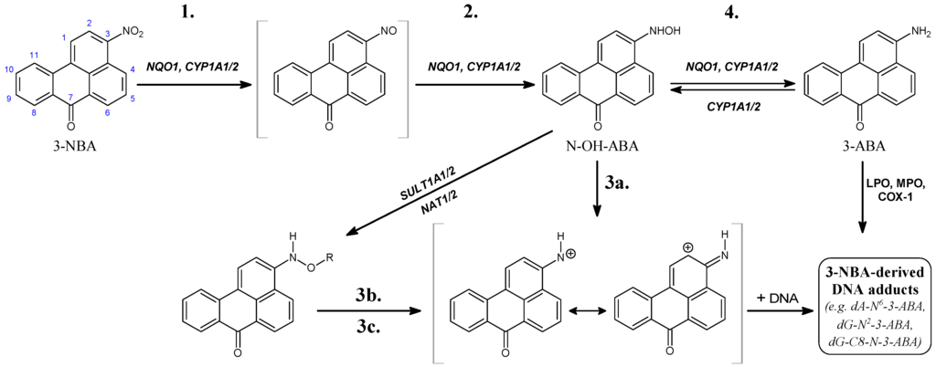
This review summarizes the results found in studies investigating the enzymatic activation of two genotoxic nitro-aromatics, an environmental pollutant and carcinogen 3-nitrobenzanthrone (3-NBA) and a natural plant nephrotoxin and carcinogen aristolochic acid I (AAI), to reactive species forming covalent DNA adducts. Experimental and [...] Read more.
This review summarizes the results found in studies investigating the enzymatic activation of two genotoxic nitro-aromatics, an environmental pollutant and carcinogen 3-nitrobenzanthrone (3-NBA) and a natural plant nephrotoxin and carcinogen aristolochic acid I (AAI), to reactive species forming covalent DNA adducts. Experimental and theoretical approaches determined the reasons why human NAD(P)H:quinone oxidoreductase (NQO1) and cytochromes P450 (CYP) 1A1 and 1A2 have the potential to reductively activate both nitro-aromatics. The results also contributed to the elucidation of the molecular mechanisms of these reactions. The contribution of conjugation enzymes such as N,O-acetyltransferases (NATs) and sulfotransferases (SULTs) to the activation of 3-NBA and AAI was also examined. The results indicated differences in the abilities of 3-NBA and AAI metabolites to be further activated by these conjugation enzymes. The formation of DNA adducts generated by both carcinogens during their reductive activation by the NOQ1 and CYP1A1/2 enzymes was investigated with pure enzymes, enzymes present in subcellular cytosolic and microsomal fractions, selective inhibitors, and animal models (including knock-out and humanized animals). For the theoretical approaches, flexible in silico docking methods as well as ab initio calculations were employed. The results summarized in this review demonstrate that a combination of experimental and theoretical approaches is a useful tool to study the enzyme-mediated reaction mechanisms of 3-NBA and AAI reduction. Full article
Show Figures

Figure 1

1150 KiB  
Article
The Application of an Emerging Technique for Protein–Protein Interaction Interface Mapping: The Combination of Photo-Initiated Cross-Linking Protein Nanoprobes with Mass Spectrometry
by Renata Ptáčková, Tomáš Ječmen, Petr Novák, Jiří Hudeček, Marie Stiborová and Miroslav Šulc
Int. J. Mol. Sci. 2014, 15(6), 9224-9241; https://doi.org/10.3390/ijms15069224 - 26 May 2014
Cited by 9 | Viewed by 8181
Abstract
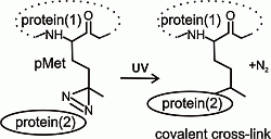
Protein–protein interaction was investigated using a protein nanoprobe capable of photo-initiated cross-linking in combination with high-resolution and tandem mass spectrometry. This emerging experimental approach introduces photo-analogs of amino acids within a protein sequence during its recombinant expression, preserves native protein structure and is [...] Read more.
Protein–protein interaction was investigated using a protein nanoprobe capable of photo-initiated cross-linking in combination with high-resolution and tandem mass spectrometry. This emerging experimental approach introduces photo-analogs of amino acids within a protein sequence during its recombinant expression, preserves native protein structure and is suitable for mapping the contact between two proteins. The contact surface regions involved in the well-characterized interaction between two molecules of human 14-3-3ζ regulatory protein were used as a model. The employed photo-initiated cross-linking techniques extend the number of residues shown to be within interaction distance in the contact surface of the 14-3-3ζ dimer (Gln8–Met78). The results of this study are in agreement with our previously published data from molecular dynamic calculations based on high-resolution chemical cross-linking data and Hydrogen/Deuterium exchange mass spectrometry. The observed contact is also in accord with the 14-3-3ζ X-ray crystal structure (PDB 3dhr). The results of the present work are relevant to the structural biology of transient interaction in the 14-3-3ζ protein, and demonstrate the ability of the chosen methodology (the combination of photo-initiated cross-linking protein nanoprobes and mass spectrometry analysis) to map the protein-protein interface or regions with a flexible structure. Full article
Show Figures

Graphical abstract

373 KiB  
Article
Application of Computational Methods for the Design of BACE-1 Inhibitors: Validation of in Silico Modelling
by Marek Bajda, Jakub Jończyk, Barbara Malawska and Sławomir Filipek
Int. J. Mol. Sci. 2014, 15(3), 5128-5139; https://doi.org/10.3390/ijms15035128 - 24 Mar 2014
Cited by 18 | Viewed by 7852
Abstract
β-Secretase (BACE-1) constitutes an important target for search of anti-Alzheimer’s drugs. The first inhibitors of this enzyme were peptidic compounds with high molecular weight and low bioavailability. Therefore, the search for new efficient non-peptidic inhibitors has been undertaken by many scientific groups. We [...] Read more.
β-Secretase (BACE-1) constitutes an important target for search of anti-Alzheimer’s drugs. The first inhibitors of this enzyme were peptidic compounds with high molecular weight and low bioavailability. Therefore, the search for new efficient non-peptidic inhibitors has been undertaken by many scientific groups. We started our work from the development of in silico methodology for the design of novel BACE-1 ligands. It was validated on the basis of crystal structures of complexes with inhibitors, redocking, cross-docking and training/test sets of reference ligands. The presented procedure of assessment of the novel compounds as β-secretase inhibitors could be widely used in the design process. Full article
Show Figures

Graphical abstract

3708 KiB  
Article
Comparative Molecular Dynamics Simulations of Mitogen-Activated Protein Kinase-Activated Protein Kinase 5
by Inger Lindin, Yimingjiang Wuxiuer, Aina Westrheim Ravna, Ugo Moens and Ingebrigt Sylte
Int. J. Mol. Sci. 2014, 15(3), 4878-4902; https://doi.org/10.3390/ijms15034878 - 19 Mar 2014
Cited by 7 | Viewed by 11529
Abstract
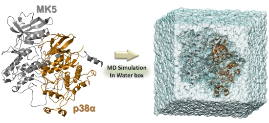
The mitogen-activated protein kinase-activated protein kinase MK5 is a substrate of the mitogen-activated protein kinases p38, ERK3 and ERK4. Cell culture and animal studies have demonstrated that MK5 is involved in tumour suppression and promotion, embryogenesis, anxiety, cell motility and cell cycle regulation [...] Read more.
The mitogen-activated protein kinase-activated protein kinase MK5 is a substrate of the mitogen-activated protein kinases p38, ERK3 and ERK4. Cell culture and animal studies have demonstrated that MK5 is involved in tumour suppression and promotion, embryogenesis, anxiety, cell motility and cell cycle regulation. In the present study, homology models of MK5 were used for molecular dynamics (MD) simulations of: (1) MK5 alone; (2) MK5 in complex with an inhibitor; and (3) MK5 in complex with the interaction partner p38α. The calculations showed that the inhibitor occupied the active site and disrupted the intramolecular network of amino acids. However, intramolecular interactions consistent with an inactive protein kinase fold were not formed. MD with p38α showed that not only the p38 docking region, but also amino acids in the activation segment, αH helix, P-loop, regulatory phosphorylation region and the C-terminal of MK5 may be involved in forming a very stable MK5-p38α complex, and that p38α binding decreases the residual fluctuation of the MK5 model. Electrostatic Potential Surface (EPS) calculations of MK5 and p38α showed that electrostatic interactions are important for recognition and binding. Full article
Show Figures

Graphical abstract

723 KiB  
Communication
Rationalization of Activity Cliffs of a Sulfonamide Inhibitor of DNA Methyltransferases with Induced-Fit Docking
by José L. Medina-Franco, Oscar Méndez-Lucio and Jakyung Yoo
Int. J. Mol. Sci. 2014, 15(2), 3253-3261; https://doi.org/10.3390/ijms15023253 - 21 Feb 2014
Cited by 25 | Viewed by 10028
Abstract
Inhibitors of human DNA methyltransferases (DNMT) are of increasing interest to develop novel epi-drugs for the treatment of cancer and other diseases. As the number of compounds with reported DNMT inhibition is increasing, molecular docking is shedding light to elucidate their mechanism of [...] Read more.
Inhibitors of human DNA methyltransferases (DNMT) are of increasing interest to develop novel epi-drugs for the treatment of cancer and other diseases. As the number of compounds with reported DNMT inhibition is increasing, molecular docking is shedding light to elucidate their mechanism of action and further interpret structure–activity relationships. Herein, we present a structure-based rationalization of the activity of SW155246, a distinct sulfonamide compound recently reported as an inhibitor of human DNMT1 obtained from high-throughput screening. We used flexible and induce-fit docking to develop a binding model of SW155246 with a crystallographic structure of human DNMT1. Results were in excellent agreement with experimental information providing a three-dimensional structural interpretation of ‘activity cliffs’, e.g., analogues of SW155246 with a high structural similarity to the sulfonamide compound, but with no activity in the enzymatic assay. Full article
Show Figures

Graphical abstract

1659 KiB  
Review
Large-Scale Domain Motions and Pyridoxal-5'-Phosphate Assisted Radical Catalysis in Coenzyme B12-Dependent Aminomutases
by Amarendra Nath Maity, Yung-Han Chen and Shyue-Chu Ke
Int. J. Mol. Sci. 2014, 15(2), 3064-3087; https://doi.org/10.3390/ijms15023064 - 20 Feb 2014
Cited by 14 | Viewed by 9209
Abstract
Lysine 5,6-aminomutase (5,6-LAM) and ornithine 4,5-aminomutase (4,5-OAM) are two of the rare enzymes that use assistance of two vitamins as cofactors. These enzymes employ radical generating capability of coenzyme B12 (5'-deoxyadenosylcobalamin, dAdoCbl) and ability of pyridoxal-5'-phosphate (PLP, vitamin B6) to [...] Read more.
Lysine 5,6-aminomutase (5,6-LAM) and ornithine 4,5-aminomutase (4,5-OAM) are two of the rare enzymes that use assistance of two vitamins as cofactors. These enzymes employ radical generating capability of coenzyme B12 (5'-deoxyadenosylcobalamin, dAdoCbl) and ability of pyridoxal-5'-phosphate (PLP, vitamin B6) to stabilize high-energy intermediates for performing challenging 1,2-amino rearrangements between adjacent carbons. A large-scale domain movement is required for interconversion between the catalytically inactive open form and the catalytically active closed form. In spite of all the similarities, these enzymes differ in substrate specificities. 4,5-OAM is highly specific for D-ornithine as a substrate while 5,6-LAM can accept D-lysine and L-β-lysine. This review focuses on recent computational, spectroscopic and structural studies of these enzymes and their implications on the related enzymes. Additionally, we also discuss the potential biosynthetic application of 5,6-LAM. Full article
Show Figures

4513 KiB  
Article
Insight into Conformational Change for 14-3-3σ Protein by Molecular Dynamics Simulation
by Guodong Hu, Haiyan Li, Jing-Yuan Liu and Jihua Wang
Int. J. Mol. Sci. 2014, 15(2), 2794-2810; https://doi.org/10.3390/ijms15022794 - 18 Feb 2014
Cited by 19 | Viewed by 8266
Abstract
14-3-3σ is a member of a highly conserved family of 14-3-3 proteins that has a double-edged sword role in human cancers. Former reports have indicated that the 14-3-3 protein may be in an open or closed state. In this work, we found that [...] Read more.
14-3-3σ is a member of a highly conserved family of 14-3-3 proteins that has a double-edged sword role in human cancers. Former reports have indicated that the 14-3-3 protein may be in an open or closed state. In this work, we found that the apo-14-3-3σ is in an open state compared with the phosphopeptide bound 14-3-3σ complex which is in a more closed state based on our 80 ns molecular dynamics (MD) simulations. The interaction between the two monomers of 14-3-3σ in the open state is the same as that in the closed state. In both open and closed states, helices A to D, which are involved in dimerization, are stable. However, large differences are found in helices E and F. The hydrophobic contacts and hydrogen bonds between helices E and G in apo-14-3-3σ are different from those in the bound 14-3-3σ complex. The restrained and the mutated (Arg56 or Arg129 to alanine) MD simulations indicate that the conformation of four residues (Lys49, Arg56, Arg129 and Tyr130) may play an important role to keep the 14-3-3σ protein in an open or closed state. These results would be useful to evaluate the 14-3-3σ protein structure-function relationship. Full article
Show Figures

461 KiB  
Article
Structural Data on the Periplasmic Aldehyde Oxidoreductase PaoABC from Escherichia coli: SAXS and Preliminary X-ray Crystallography Analysis
by Ana Rita Otrelo-Cardoso, Márcia Alexandra Da Silva Correia, Viola Schwuchow, Dmitri I. Svergun, Maria João Romão, Silke Leimkühler and Teresa Santos-Silva
Int. J. Mol. Sci. 2014, 15(2), 2223-2236; https://doi.org/10.3390/ijms15022223 - 31 Jan 2014
Cited by 11 | Viewed by 9633
Abstract
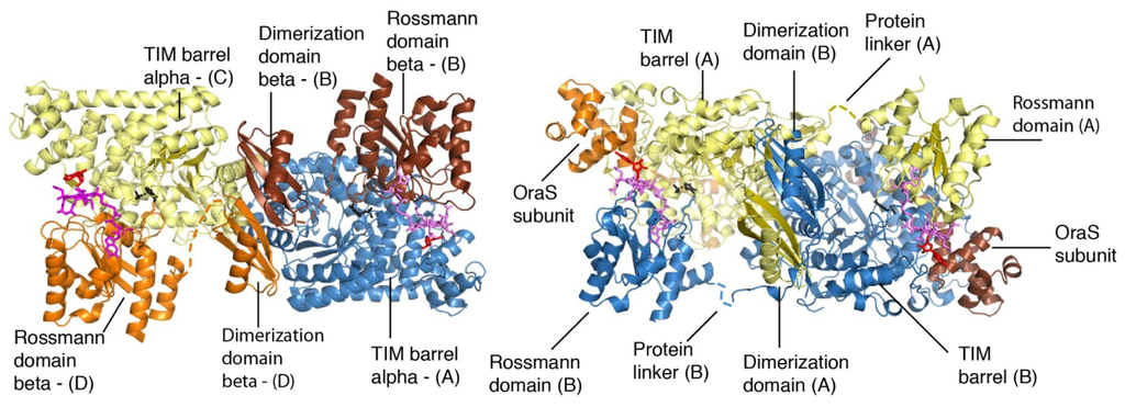
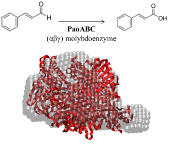
The periplasmic aldehyde oxidoreductase PaoABC from Escherichia coli is a molybdenum enzyme involved in detoxification of aldehydes in the cell. It is an example of an αβγ heterotrimeric enzyme of the xanthine oxidase family of enzymes which does not dimerize via its molybdenum [...] Read more.
The periplasmic aldehyde oxidoreductase PaoABC from Escherichia coli is a molybdenum enzyme involved in detoxification of aldehydes in the cell. It is an example of an αβγ heterotrimeric enzyme of the xanthine oxidase family of enzymes which does not dimerize via its molybdenum cofactor binding domain. In order to structurally characterize PaoABC, X-ray crystallography and small angle X-ray scattering (SAXS) have been carried out. The protein crystallizes in the presence of 20% (w/v) polyethylene glycol 3350 using the hanging-drop vapour diffusion method. Although crystals were initially twinned, several experiments were done to overcome twinning and lowering the crystallization temperature (293 K to 277 K) was the solution to the problem. The non-twinned crystals used to solve the structure diffract X-rays to beyond 1.80 Å and belong to the C2 space group, with cell parameters a = 109.42 Å, b = 78.08 Å, c = 151.77 Å, β = 99.77°, and one molecule in the asymmetric unit. A molecular replacement solution was found for each subunit separately, using several proteins as search models. SAXS data of PaoABC were also collected showing that, in solution, the protein is also an αβγ heterotrimer. Full article
Show Figures

Graphical abstract

1300 KiB  
Article
Exploring the Molecular Basis for Selective Binding of Homoserine Dehydrogenase from Mycobacterium leprae TN toward Inhibitors: A Virtual Screening Study
by Dongling Zhan, Dongmei Wang, Weihong Min and Weiwei Han
Int. J. Mol. Sci. 2014, 15(2), 1826-1841; https://doi.org/10.3390/ijms15021826 - 24 Jan 2014
Cited by 5 | Viewed by 8392
Abstract
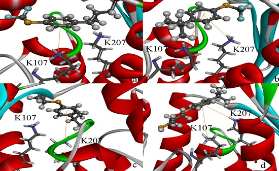
Homoserine dehydrogenase (HSD) from Mycobacterium leprae TN is an antifungal target for antifungal properties including efficacy against the human pathogen. The 3D structure of HSD has been firmly established by homology modeling methods. Using the template, homoserine dehydrogenase from Thiobacillus denitrificans (PDB Id [...] Read more.
Homoserine dehydrogenase (HSD) from Mycobacterium leprae TN is an antifungal target for antifungal properties including efficacy against the human pathogen. The 3D structure of HSD has been firmly established by homology modeling methods. Using the template, homoserine dehydrogenase from Thiobacillus denitrificans (PDB Id 3MTJ), a sequence identity of 40% was found and molecular dynamics simulation was used to optimize a reliable structure. The substrate and co-factor-binding regions in HSD were identified. In order to determine the important residues of the substrate (l-aspartate semialdehyde (l-ASA)) binding, the ASA was docked to the protein; Thr163, Asp198, and Glu192 may be important because they form a hydrogen bond with HSD through AutoDock 4.2 software. neuraminidaseAfter use of a virtual screening technique of HSD, the four top-scoring docking hits all seemed to cation–π ion pair with the key recognition residue Lys107, and Lys207. These ligands therefore seemed to be new chemotypes for HSD. Our results may be helpful for further experimental investigations. Full article
Show Figures

Graphical abstract

520 KiB  
Review
In Silico Discovery of Aminoacyl-tRNA Synthetase Inhibitors
by Yaxue Zhao, Qingqing Meng, Linquan Bai and Huchen Zhou
Int. J. Mol. Sci. 2014, 15(1), 1358-1373; https://doi.org/10.3390/ijms15011358 - 20 Jan 2014
Cited by 14 | Viewed by 8779
Abstract
Aminoacyl-tRNA synthetases (aaRSs) are enzymes that catalyze the transfer of amino acids to their cognate tRNA. They play a pivotal role in protein synthesis and are essential for cell growth and survival. The aaRSs are one of the leading targets for development of [...] Read more.
Aminoacyl-tRNA synthetases (aaRSs) are enzymes that catalyze the transfer of amino acids to their cognate tRNA. They play a pivotal role in protein synthesis and are essential for cell growth and survival. The aaRSs are one of the leading targets for development of antibiotic agents. In this review, we mainly focused on aaRS inhibitor discovery and development using in silico methods including virtual screening and structure-based drug design. These computational methods are relatively fast and cheap, and are proving to be of great benefit for the rational development of more potent aaRS inhibitors and other pharmaceutical agents that may usher in a much needed generation of new antibiotics. Full article
Show Figures

2013

Jump to: 2017, 2016, 2015, 2014

858 KiB  
Review
Multi-Scale Computational Enzymology: Enhancing Our Understanding of Enzymatic Catalysis
by Rami Gherib, Hisham M. Dokainish and James W. Gauld
Int. J. Mol. Sci. 2014, 15(1), 401-422; https://doi.org/10.3390/ijms15010401 - 31 Dec 2013
Cited by 8 | Viewed by 9981
Abstract
Elucidating the origin of enzymatic catalysis stands as one the great challenges of contemporary biochemistry and biophysics. The recent emergence of computational enzymology has enhanced our atomistic-level description of biocatalysis as well the kinetic and thermodynamic properties of their mechanisms. There exists a [...] Read more.
Elucidating the origin of enzymatic catalysis stands as one the great challenges of contemporary biochemistry and biophysics. The recent emergence of computational enzymology has enhanced our atomistic-level description of biocatalysis as well the kinetic and thermodynamic properties of their mechanisms. There exists a diversity of computational methods allowing the investigation of specific enzymatic properties. Small or large density functional theory models allow the comparison of a plethora of mechanistic reactive species and divergent catalytic pathways. Molecular docking can model different substrate conformations embedded within enzyme active sites and determine those with optimal binding affinities. Molecular dynamics simulations provide insights into the dynamics and roles of active site components as well as the interactions between substrate and enzymes. Hybrid quantum mechanical/molecular mechanical (QM/MM) can model reactions in active sites while considering steric and electrostatic contributions provided by the surrounding environment. Using previous studies done within our group, on OvoA, EgtB, ThrRS, LuxS and MsrA enzymatic systems, we will review how these methods can be used either independently or cooperatively to get insights into enzymatic catalysis. Full article
Show Figures

1088 KiB  
Article
Importance of H-Abstraction in the Final Step of Nitrosoalkane Formation in the Mechanism-Based Inactivation of Cytochrome P450 by Amine-Containing Drugs
by Hajime Hirao, Nandun M. Thellamurege, Pratanphorn Chuanprasit and Kai Xu
Int. J. Mol. Sci. 2013, 14(12), 24692-24705; https://doi.org/10.3390/ijms141224692 - 18 Dec 2013
Cited by 23 | Viewed by 8151
Abstract
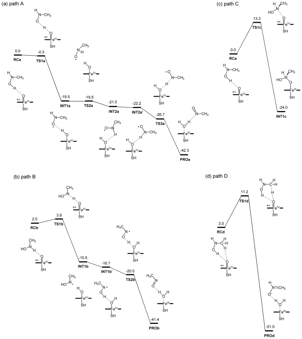
The metabolism of amine-containing drugs by cytochrome P450 enzymes (P450s) is prone to form a nitrosoalkane metabolic intermediate (MI), which subsequently coordinates to the heme iron of a P450, to produce a metabolic-intermediate complex (MIC). This type of P450 inhibition, referred to as [...] Read more.
The metabolism of amine-containing drugs by cytochrome P450 enzymes (P450s) is prone to form a nitrosoalkane metabolic intermediate (MI), which subsequently coordinates to the heme iron of a P450, to produce a metabolic-intermediate complex (MIC). This type of P450 inhibition, referred to as mechanism-based inactivation (MBI), presents a serious concern in drug discovery processes. We applied density functional theory (DFT) to the reaction between N-methylhydroxylamine (NMH) and the compound I reactive species of P450, in an effort to elucidate the mechanism of the putative final step of the MI formation in the alkylamine metabolism. Our DFT calculations show that H-abstraction from the hydroxyl group of NMH is the most favorable pathway via which the nitrosoalkane intermediate is produced spontaneously. H-abstraction from the N–H bond was slightly less favorable. In contrast, N-oxidation and H-abstraction from the C–H bond of the methyl group had much higher energy barriers. Hence, if the conversion of NMH to nitrosoalkane is catalyzed by a P450, the reaction should proceed preferentially via H-abstraction, either from the O–H bond or from the N–H bond. Our theoretical analysis of the interaction between the MI and pentacoordinate heme moieties provided further insights into the coordination bond in the MIC. Full article
Show Figures

785 KiB  
Article
Small Molecule Binding, Docking, and Characterization of the Interaction between Pth1 and Peptidyl-tRNA
by Mary C. Hames, Hana McFeeters, W. Blake Holloway, Christopher B. Stanley, Volker S. Urban and Robert L. McFeeters
Int. J. Mol. Sci. 2013, 14(11), 22741-22752; https://doi.org/10.3390/ijms141122741 - 19 Nov 2013
Cited by 10 | Viewed by 9579
Abstract
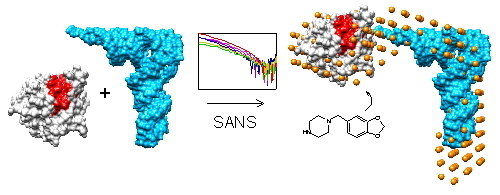
Bacterial Pth1 is essential for viability. Pth1 cleaves the ester bond between the peptide and nucleotide of peptidyl-tRNA generated from aborted translation, expression of mini-genes, and short ORFs. We have determined the shape of the Pth1:peptidyl-tRNA complex using small angle neutron scattering. Binding [...] Read more.
Bacterial Pth1 is essential for viability. Pth1 cleaves the ester bond between the peptide and nucleotide of peptidyl-tRNA generated from aborted translation, expression of mini-genes, and short ORFs. We have determined the shape of the Pth1:peptidyl-tRNA complex using small angle neutron scattering. Binding of piperonylpiperazine, a small molecule constituent of a combinatorial synthetic library common to most compounds with inhibitory activity, was mapped to Pth1 via NMR spectroscopy. We also report computational docking results, modeling piperonylpiperazine binding based on chemical shift perturbation mapping. Overall these studies promote Pth1 as a novel antibiotic target, contribute to understanding how Pth1 interacts with its substrate, advance the current model for cleavage, and demonstrate feasibility of small molecule inhibition. Full article
Show Figures

Graphical abstract

1448 KiB  
Article
Oximes: Inhibitors of Human Recombinant Acetylcholinesterase. A Structure-Activity Relationship (SAR) Study
by Vendula Sepsova, Jana Zdarova Karasova, Jan Korabecny, Rafael Dolezal, Filip Zemek, Brian J. Bennion and Kamil Kuca
Int. J. Mol. Sci. 2013, 14(8), 16882-16900; https://doi.org/10.3390/ijms140816882 - 16 Aug 2013
Cited by 42 | Viewed by 9620
Abstract
Acetylcholinesterase (AChE) reactivators were developed for the treatment of organophosphate intoxication. Standard care involves the use of anticonvulsants (e.g., diazepam), parasympatolytics (e.g., atropine) and oximes that restore AChE activity. However, oximes also bind to the active site of AChE, simultaneously acting as reversible [...] Read more.
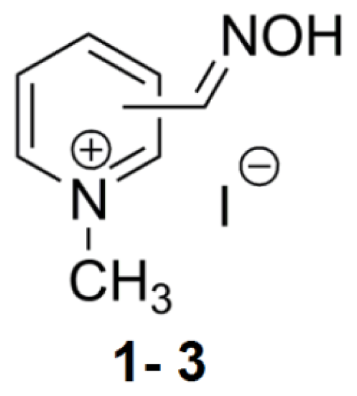
Acetylcholinesterase (AChE) reactivators were developed for the treatment of organophosphate intoxication. Standard care involves the use of anticonvulsants (e.g., diazepam), parasympatolytics (e.g., atropine) and oximes that restore AChE activity. However, oximes also bind to the active site of AChE, simultaneously acting as reversible inhibitors. The goal of the present study is to determine how oxime structure influences the inhibition of human recombinant AChE (hrAChE). Therefore, 24 structurally different oximes were tested and the results compared to the previous eel AChE (EeAChE) experiments. Structural factors that were tested included the number of pyridinium rings, the length and structural features of the linker, and the number and position of the oxime group on the pyridinium ring. Full article
Show Figures

410 KiB  
Article
Design, Synthesis, Biological Activity and Molecular Dynamics Studies of Specific Protein Tyrosine Phosphatase 1B Inhibitors over SHP-2
by Su-Xia Sun, Xiao-Bo Li, Wen-Bo Liu, Ying Ma, Run-Ling Wang, Xian-Chao Cheng, Shu-Qing Wang and Wei Liu
Int. J. Mol. Sci. 2013, 14(6), 12661-12674; https://doi.org/10.3390/ijms140612661 - 17 Jun 2013
Cited by 8 | Viewed by 10268
Abstract
Over expressing in PTPN1 (encoding Protein tyrosine phosphatase 1B, PTP1B), a protein tyrosine phosphatase (PTP) that plays an overall positive role in insulin signaling, is linked to the pathogenesis of diabetes and obesity. The relationship between PTP1B and human diseases exhibits PTP1B as [...] Read more.
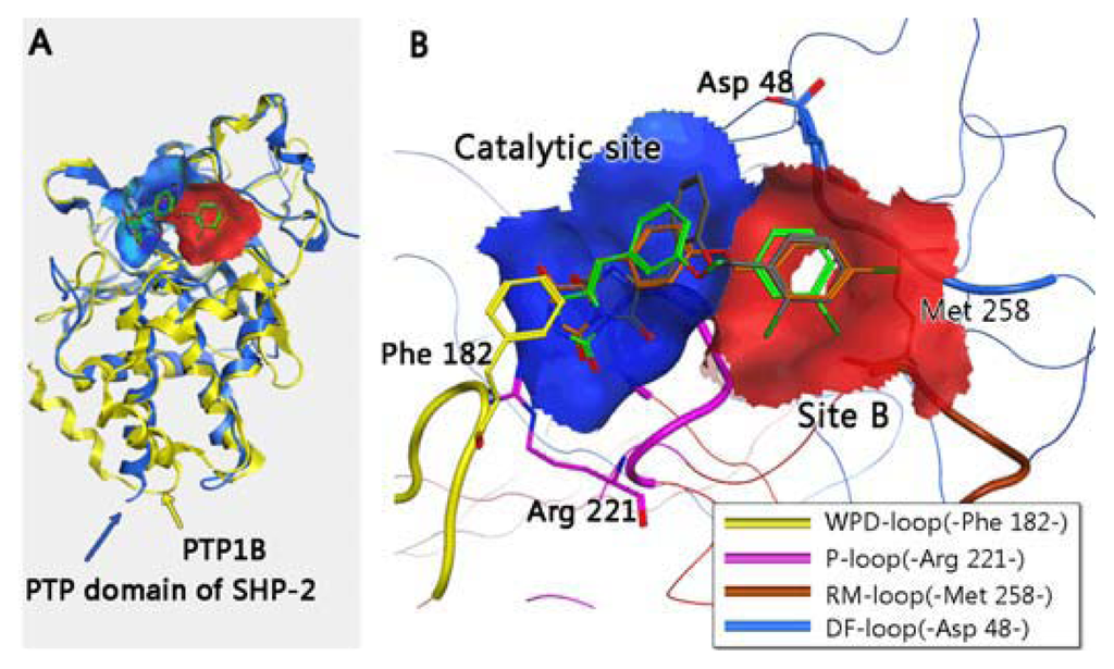
Over expressing in PTPN1 (encoding Protein tyrosine phosphatase 1B, PTP1B), a protein tyrosine phosphatase (PTP) that plays an overall positive role in insulin signaling, is linked to the pathogenesis of diabetes and obesity. The relationship between PTP1B and human diseases exhibits PTP1B as the target to treat these diseases. In this article, small weight molecules of the imidazolidine series were screened from databases and optimized on silicon as the inhibitors of PTP1B based on the steric conformation and electronic configuration of thiazolidinedione (TZD) compounds. The top three candidates were tested using an in vitro biological assay after synthesis. Finally, we report a novel inhibitor, Compound 13, that specifically inhibits PTP1B over the closely related phosphatase Src homology 2 (SH2) domain-containing phosphatase 2 (SHP-2) at 80 μΜ. Its IC50 values are reported in this paper as well. This compound was further verified by computer analysis for its ability to combine the catalytic domains of PTP1B and SHP-2 by molecular dynamics (MD) simulations. Full article
Show Figures

837 KiB  
Article
Molecular Dynamics Simulation of Tryptophan Hydroxylase-1: Binding Modes and Free Energy Analysis to Phenylalanine Derivative Inhibitors
by Hao Zhong, Wei Huang, Gu He, Cheng Peng, Fengbo Wu and Liang Ouyang
Int. J. Mol. Sci. 2013, 14(5), 9947-9962; https://doi.org/10.3390/ijms14059947 - 10 May 2013
Cited by 35 | Viewed by 9348
Abstract
Serotonin is a neurotransmitter that modulates many central and peripheral functions. Tryptophan hydroxylase-1 (TPH1) is a key enzyme of serotonin synthesis. In the current study, the interaction mechanism of phenylalanine derivative TPH1 inhibitors was investigated using molecular dynamics (MD) simulations, free energy calculations, [...] Read more.
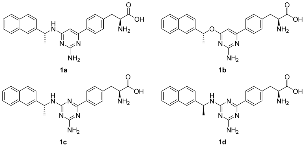
Serotonin is a neurotransmitter that modulates many central and peripheral functions. Tryptophan hydroxylase-1 (TPH1) is a key enzyme of serotonin synthesis. In the current study, the interaction mechanism of phenylalanine derivative TPH1 inhibitors was investigated using molecular dynamics (MD) simulations, free energy calculations, free energy decomposition analysis and computational alanine scanning. The predicted binding free energies of these complexes are consistent with the experimental data. The analysis of the individual energy terms indicates that although the van der Waals and electrostatics interaction contributions are important in distinguishing the binding affinities of these inhibitors, the electrostatic contribution plays a more crucial role in that. Moreover, it is observed that different configurations of the naphthalene substituent could form different binding patterns with protein, yet lead to similar inhibitory potency. The combination of different molecular modeling techniques is an efficient way to interpret the interaction mechanism of inhibitors and our work could provide valuable information for the TPH1 inhibitor design in the future. Full article
Show Figures

Back to TopTop