Next Issue
Volume 10, June
Previous Issue
Volume 10, April


Toxins, Volume 10, Issue 5 (May 2018) – 43 articles

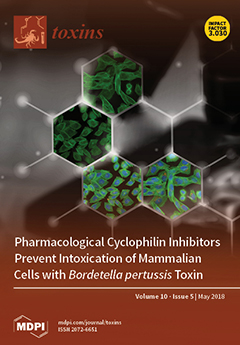
Cover Story (view full-size image): The cyclophilin inhibitor cyclosporine A protects cells from intoxication with pertussis toxin by reducing the amount of the enzymatic subunit PTS1 of this toxin in the cytosol of target cells demonstrated by fluorescence microscopy. Thereby, the ADP-ribosylation of inhibitory alpha subunits of trimeric G proteins by PTS1 is reduced and the subsequent disturbed signaling causing the severe symptoms of whooping cough averted. Thus, cyclophilin inhibitors might represent promising starting points for development of novel therapeutic strategies against whooping cough. Blue = nucleus, Green = F-actin, Red = PTS1. View the paper here.
  • Issues are regarded as officially published after their release is announced to the table of contents alert mailing list.
  • You may sign up for e-mail alerts to receive table of contents of newly released issues.
  • PDF is the official format for papers published in both, html and pdf forms. To view the papers in pdf format, click on the "PDF Full-text" link, and use the free Adobe Reader to open them.
Order results
Result details
Section
Select all
Export citation of selected articles as:
21 pages, 2262 KiB  
Review
Mechanisms of Action and Cell Death Associated with Clostridium perfringens Toxins
by Mauricio A. Navarro, Bruce A. McClane and Francisco A. Uzal
Toxins 2018, 10(5), 212; https://doi.org/10.3390/toxins10050212 - 22 May 2018
Cited by 152 | Viewed by 20363
Abstract
Clostridium perfringens uses its large arsenal of protein toxins to produce histotoxic, neurologic and intestinal infections in humans and animals. The major toxins involved in diseases are alpha (CPA), beta (CPB), epsilon (ETX), iota (ITX), enterotoxin (CPE), and necrotic B-like (NetB) toxins. CPA [...] Read more.
Clostridium perfringens uses its large arsenal of protein toxins to produce histotoxic, neurologic and intestinal infections in humans and animals. The major toxins involved in diseases are alpha (CPA), beta (CPB), epsilon (ETX), iota (ITX), enterotoxin (CPE), and necrotic B-like (NetB) toxins. CPA is the main virulence factor involved in gas gangrene in humans, whereas its role in animal diseases is limited and controversial. CPB is responsible for necrotizing enteritis and enterotoxemia, mostly in neonatal individuals of many animal species, including humans. ETX is the main toxin involved in enterotoxemia of sheep and goats. ITX has been implicated in cases of enteritis in rabbits and other animal species; however, its specific role in causing disease has not been proved. CPE is responsible for human food-poisoning and non-foodborne C. perfringens-mediated diarrhea. NetB is the cause of necrotic enteritis in chickens. In most cases, host–toxin interaction starts on the plasma membrane of target cells via specific receptors, resulting in the activation of intracellular pathways with a variety of effects, commonly including cell death. In general, the molecular mechanisms of cell death associated with C. perfringens toxins involve features of apoptosis, necrosis and/or necroptosis. Full article
Show Figures

Figure 1

9 pages, 272 KiB  
Article
Development of a Highly Sensitive FcMito qPCR Assay for the Quantification of the Toxigenic Fungal Plant Pathogen Fusarium culmorum
by Katarzyna Bilska, Tomasz Kulik, Anna Ostrowska-Kołodziejczak, Maciej Buśko, Matias Pasquali, Marco Beyer, Anna Baturo-Cieśniewska, Marcin Juda, Dariusz Załuski, Kinga Treder, Joerg Denekas and Juliusz Perkowski
Toxins 2018, 10(5), 211; https://doi.org/10.3390/toxins10050211 - 21 May 2018
Cited by 18 | Viewed by 4671
Abstract
Fusarium culmorum is a ubiquitous, soil-borne fungus (ascomycete) causing foot and root rot and Fusarium head blight on cereals. It is responsible for yield and quality losses as well as grain contamination with mycotoxins, which are a potential health hazard. An extremely sensitive [...] Read more.
Fusarium culmorum is a ubiquitous, soil-borne fungus (ascomycete) causing foot and root rot and Fusarium head blight on cereals. It is responsible for yield and quality losses as well as grain contamination with mycotoxins, which are a potential health hazard. An extremely sensitive mitochondrial-based qPCR assay (FcMito qPCR) for quantification of F. culmorum was developed in this study. To provide specificity, the FcMito assay was successfully validated against 85 F. culmorum strains and 53 isolates of 30 other fungal species. The assay efficiency and sensitivity were evaluated against different F. culmorum strains with various amounts of pure fungal DNA and in the presence of background wheat DNA. The results demonstrated the high efficiency of the assay (97.2–106.0%, R2-values > 0.99). It was also shown that, in the presence of background DNA, 0.01 pg of fungal template could be reliably quantified. The FcMito assay was used to quantify F. culmorum DNA using 108 grain samples with different trichothecene levels. A significant positive correlation was found between fungal DNA quantity and the total trichothecene content. The obtained results showed that the sensitivity of the FcMito assay was much higher than the nuclear-based qPCR assay for F. culmorum. Full article
(This article belongs to the Special Issue Recent Advances in Fusarium Research)
12 pages, 1557 KiB  
Article
Domain II of Pseudomonas Exotoxin Is Critical for Efficacy of Bolus Doses in a Xenograft Model of Acute Lymphoblastic Leukemia
by Fabian Müller, Tyler Cunningham, Richard Beers, Tapan K. Bera, Alan S. Wayne and Ira Pastan
Toxins 2018, 10(5), 210; https://doi.org/10.3390/toxins10050210 - 21 May 2018
Cited by 8 | Viewed by 4209
Abstract
Moxetumomab pasudotox is a fusion protein of a CD22-targeting antibody and Pseudomonas exotoxin. Minutes of exposure to Moxetumomab achieves similar cell killing than hours of exposure to a novel deimmunized variant against some acute lymphoblastic leukemia (ALL). Because blood levels fall quickly, Moxetumomab [...] Read more.
Moxetumomab pasudotox is a fusion protein of a CD22-targeting antibody and Pseudomonas exotoxin. Minutes of exposure to Moxetumomab achieves similar cell killing than hours of exposure to a novel deimmunized variant against some acute lymphoblastic leukemia (ALL). Because blood levels fall quickly, Moxetumomab is more than 1000-fold more active than the deimmunized variant in vivo. We aimed to identify which part of Moxetumomab increases in vivo efficacy and generated five immunotoxins, tested time-dependent activity, and determined the efficacy in a KOPN-8 xenograft model. Full domain II shortened the time cells had to be exposed to die to only a few minutes for some ALL; deimmunized domain III consistently extended the time. Against KOPN-8, full domain II accelerated time to arrest protein synthesis by three-fold and tripled PARP-cleavage. In vivo efficacy was increased by more than 10-fold by domain II and increasing size, and therefore half-life enhanced efficacy two- to four-fold. In summary, in vivo efficacy is determined by the time cells have to be exposed to immunotoxin to die and serum half-life. Thus, domain II is most critical for activity against some ALL treated with bolus doses; however, immunotoxins lacking all but the furin-cleavage site of domain II may be advantageous when treating continuously. Full article
(This article belongs to the Special Issue Venom and Toxin as Targeted Therapy)
Show Figures

Figure 1

11 pages, 1676 KiB  
Article
Acid Sphingomyelinase Promotes Cellular Internalization of Clostridium perfringens Iota-Toxin
by Masahiro Nagahama, Masaya Takehara, Kazuaki Miyamoto, Kazumi Ishidoh and Keiko Kobayashi
Toxins 2018, 10(5), 209; https://doi.org/10.3390/toxins10050209 - 20 May 2018
Cited by 7 | Viewed by 4220
Abstract
Clostridium perfringens iota-toxin is a binary actin-ADP-ribosylating toxin composed of the enzymatic component Ia and receptor binding component Ib. Ib binds to a cell surface receptor, forms Ib oligomer in lipid rafts, and associates with Ia. The Ia-Ib complex then internalizes by endocytosis. [...] Read more.
Clostridium perfringens iota-toxin is a binary actin-ADP-ribosylating toxin composed of the enzymatic component Ia and receptor binding component Ib. Ib binds to a cell surface receptor, forms Ib oligomer in lipid rafts, and associates with Ia. The Ia-Ib complex then internalizes by endocytosis. Here, we showed that acid sphingomyelinase (ASMase) facilitates the cellular uptake of iota-toxin. Inhibitions of ASMase and lysosomal exocytosis by respective blockers depressed cell rounding induced by iota-toxin. The cytotoxicity of the toxin increased in the presence of Ca2+ in extracellular fluids. Ib entered target cells in the presence but not the absence of Ca2+. Ib induced the extracellular release of ASMase in the presence of Ca2+. ASMase siRNA prevented the cell rounding induced by iota-toxin. Furthermore, treatment of the cells with Ib resulted in the production of ceramide in cytoplasmic vesicles. These observations showed that ASMase promotes the internalization of iota-toxin into target cells. Full article
(This article belongs to the Special Issue Bacterial Pore-Forming Toxins)
Show Figures

Figure 1

27 pages, 954 KiB  
Review
The Expanding Therapeutic Utility of Botulinum Neurotoxins
by Elena Fonfria, Jacquie Maignel, Stephane Lezmi, Vincent Martin, Andrew Splevins, Saif Shubber, Mikhail Kalinichev, Keith Foster, Philippe Picaut and Johannes Krupp
Toxins 2018, 10(5), 208; https://doi.org/10.3390/toxins10050208 - 18 May 2018
Cited by 85 | Viewed by 9207
Abstract
Botulinum neurotoxin (BoNT) is a major therapeutic agent that is licensed in neurological indications, such as dystonia and spasticity. The BoNT family, which is produced in nature by clostridial bacteria, comprises several pharmacologically distinct proteins with distinct properties. In this review, we present [...] Read more.
Botulinum neurotoxin (BoNT) is a major therapeutic agent that is licensed in neurological indications, such as dystonia and spasticity. The BoNT family, which is produced in nature by clostridial bacteria, comprises several pharmacologically distinct proteins with distinct properties. In this review, we present an overview of the current therapeutic landscape and explore the diversity of BoNT proteins as future therapeutics. In recent years, novel indications have emerged in the fields of pain, migraine, overactive bladder, osteoarthritis, and wound healing. The study of biological effects distal to the injection site could provide future opportunities for disease-tailored BoNT therapies. However, there are some challenges in the pharmaceutical development of BoNTs, such as liquid and slow-release BoNT formulations; and, transdermal, transurothelial, and transepithelial delivery. Innovative approaches in the areas of formulation and delivery, together with highly sensitive analytical tools, will be key for the success of next generation BoNT clinical products. Full article
Show Figures

Graphical abstract

12 pages, 2627 KiB  
Article
Transcriptomic Analysis of Pseudoscorpion Venom Reveals a Unique Cocktail Dominated by Enzymes and Protease Inhibitors
by Carlos E. Santibáñez-López, Andrew Z. Ontano, Mark S. Harvey and Prashant P. Sharma
Toxins 2018, 10(5), 207; https://doi.org/10.3390/toxins10050207 - 18 May 2018
Cited by 22 | Viewed by 8711
Abstract
Transcriptomic and genomic analyses have illuminated the diversity of venoms in three of the four venomous arachnid orders (scorpions, spiders, and ticks). To date, no venom gland transcriptome analysis has been available for pseudoscorpions, the fourth venomous arachnid lineage. To redress this gap, [...] Read more.
Transcriptomic and genomic analyses have illuminated the diversity of venoms in three of the four venomous arachnid orders (scorpions, spiders, and ticks). To date, no venom gland transcriptome analysis has been available for pseudoscorpions, the fourth venomous arachnid lineage. To redress this gap, we sequenced an mRNA library generated from the venom glands of the species Synsphyronus apimelus (Garypidae). High-throughput sequencing by the Illumina protocol, followed by de novo assembly, resulted in a total of 238,331 transcripts. From those, we annotated 131 transcripts, which code for putative peptides/proteins with similar sequences to previously reported venom components available from different arachnid species in protein databases. Transcripts putatively coding for enzymes showed the richest diversity, followed by other venom components such as peptidase inhibitors, cysteine-rich peptides, and thyroglobulin 1-like peptides. Only 11 transcripts were found that code for putatively low molecular mass spider toxins. This study constitutes the first report of the diversity of components within pseudoscorpion venom. Full article
(This article belongs to the Section Animal Venoms)
Show Figures

Figure 1

12 pages, 254 KiB  
Article
Effects of Dietary Arginine, Ornithine, and Zeolite Supplementation on Uremic Toxins in Cats
by Nadine Paßlack and Jürgen Zentek
Toxins 2018, 10(5), 206; https://doi.org/10.3390/toxins10050206 - 18 May 2018
Cited by 3 | Viewed by 4262
Abstract
To test if arginine and ornithine, both components of the Krebs-Henseleit cycle, or zeolite, a potential ammonium absorber, can modulate the excretion of harmful bacterial metabolites, intestinal microbial protein fermentation was stimulated by feeding a high-protein (60.3%) diet as a single daily meal [...] Read more.
To test if arginine and ornithine, both components of the Krebs-Henseleit cycle, or zeolite, a potential ammonium absorber, can modulate the excretion of harmful bacterial metabolites, intestinal microbial protein fermentation was stimulated by feeding a high-protein (60.3%) diet as a single daily meal to 10 adult cats. The diet was supplemented without or with arginine (+50, 75, 100% compared to arginine in the basal diet), ornithine (+100, 150, 200% compared to arginine in the basal diet), or zeolite (0.125, 0.25, 0.375 g/kg body weight/day). The cats received each diet for 11 days. Urine, feces, and blood were collected during the last 4 days. Arginine and ornithine enhanced the postprandial increase of blood urea, but renal urea excretion was not increased. Zeolite decreased renal ammonium excretion and fecal biogenic amines. The data indicate an increased detoxification rate of ammonia by arginine and ornithine supplementation. However, as urea was not increasingly excreted, detrimental effects on renal function cannot be excluded. Zeolite had beneficial effects on the intestinal nitrogen metabolism, which should be further evaluated in diseased cats. Clinical studies should investigate whether dietary arginine and ornithine might improve hepatic ammonia detoxification or could be detrimental for renal function. Full article
(This article belongs to the Special Issue Disposition of Uremic Toxins: The Challenges in Uremia)
16 pages, 1517 KiB  
Article
Is 3-Carboxy-4-methyl-5-propyl-2-furanpropionate (CMPF) a Clinically Relevant Uremic Toxin in Haemodialysis Patients?
by Mathilde Luce, Anais Bouchara, Myriam Pastural, Samuel Granjon, Jean Christophe Szelag, Maurice Laville, Walid Arkouche, Denis Fouque, Christophe O. Soulage and Laetitia Koppe
Toxins 2018, 10(5), 205; https://doi.org/10.3390/toxins10050205 - 18 May 2018
Cited by 14 | Viewed by 4844
Abstract
3-Carboxy-4-methyl-5-propyl-2-furanpropionate (CMPF) is a metabolite of furan fatty acid and a marker of fish oil intake. CMPF is described as a protein-bound uremic toxin and interacts with free oxygen radicals, which can induce cell damages. However, the clinical consequences of CMPF accumulation in [...] Read more.
3-Carboxy-4-methyl-5-propyl-2-furanpropionate (CMPF) is a metabolite of furan fatty acid and a marker of fish oil intake. CMPF is described as a protein-bound uremic toxin and interacts with free oxygen radicals, which can induce cell damages. However, the clinical consequences of CMPF accumulation in haemodialysis patients remain poorly documented. The aims of this study are to investigate potential association between CMPF levels and (i) biochemical and nutritional parameters; (ii) cardiovascular events and (iii) mortality. Two hundred and fifty-two patients undergoing maintenance haemodialysis were included. Routine clinical biochemistry tests and assay for CMPF by HPLC technique were performed at the inclusion. Body composition parameters were measured using a bioimpedance spectroscopy method. The enrolled patients were prospectively monitored for cardiovascular events and mortality. CMPF level was positively correlated with nutritional parameters and lean mass and is significantly higher in patients without protein-energy wasting. However, the multivariate linear regression analysis indicated that CMPF level was not independently associated with albumin, prealbumin, creatinemia and body mass index. Elevated serum CMPF was not associated with mortality and cardiovascular morbidity. Our results indicate that CMPF is not a relevant uremic toxin in haemodialysis and in contrast could be a marker of healthy diet and omega 3 intakes. Full article
(This article belongs to the Section Uremic Toxins)
Show Figures

Figure 1

15 pages, 3160 KiB  
Article
Distal Colon Motor Dysfunction in Mice with Chronic Kidney Disease: Putative Role of Uremic Toxins
by Elsa Hoibian, Nans Florens, Laetitia Koppe, Hubert Vidal and Christophe O. Soulage
Toxins 2018, 10(5), 204; https://doi.org/10.3390/toxins10050204 - 16 May 2018
Cited by 26 | Viewed by 6161
Abstract
Although gastrointestinal complications are a common feature of patients with chronic kidney disease (CKD), the impact of uremia on bowel motility remains poorly understood. The present study was, therefore, designed to investigate the impact of uremia on gut motility. Kidney failure was induced [...] Read more.
Although gastrointestinal complications are a common feature of patients with chronic kidney disease (CKD), the impact of uremia on bowel motility remains poorly understood. The present study was, therefore, designed to investigate the impact of uremia on gut motility. Kidney failure was induced in mice by chemical nephrectomy using an adenine diet (0.25% w/w). Gastrointestinal transit time and colon motility were explored in vivo and ex vivo. Colons from control mice were incubated with uremic plasma or uremic toxins (urea, indoxyl-sulfate or p-cresyl-sulfate) at concentrations encountered in patients with end-stage renal disease. Mice fed an adenine diet for 3 weeks exhibited a 3-fold increase in plasma urea (p < 0.001) evidencing kidney failure. The median gastrointestinal transit time was doubled (1.8-fold, p < 0.001) while a reduction in colonic propulsive motility was observed in CKD mice (3-fold, p < 0.001). Colon from CKD mice exhibited an abnormal pattern of contraction associated with a blunted maximal force of contraction. Control colons incubated with plasma from hemodialysis patients exhibited a blunted level of maximal contraction (p < 0.01). Incubation with urea did not elicit any difference but incubation with indoxyl-sulfate or p-cresyl-sulfate decreased the maximal force of contraction (−66% and −55%, respectively. p < 0.01). Taken together, these data suggest that uremia impairs colon motility probably through the retention of uremic toxins. Colon dysmotility might contribute to the gastrointestinal symptoms often reported in patients with CKD. Full article
(This article belongs to the Special Issue The Intestine and Uremia)
Show Figures

Figure 1

12 pages, 1173 KiB  
Article
Botulinum Toxin Type A Injection for Cervical Dystonia in Adults with Dyskinetic Cerebral Palsy
by You Gyoung Yi, Keewon Kim, Youbin Yi, Young-Ah Choi, Ja-Ho Leigh and Moon Suk Bang
Toxins 2018, 10(5), 203; https://doi.org/10.3390/toxins10050203 - 16 May 2018
Cited by 10 | Viewed by 5657
Abstract
We aimed to evaluate the efficacy and safety of injecting botulinum toxin A (BoNT-A) into the neck muscles to treat cervical dystonia (CD) in patients with dyskinetic cerebral palsy (CP). This was a randomized, double-blinded, placebo-controlled trial with cross-over design. We prospectively enrolled [...] Read more.
We aimed to evaluate the efficacy and safety of injecting botulinum toxin A (BoNT-A) into the neck muscles to treat cervical dystonia (CD) in patients with dyskinetic cerebral palsy (CP). This was a randomized, double-blinded, placebo-controlled trial with cross-over design. We prospectively enrolled adults with dyskinetic CP who were over 20 years old and had been clinically diagnosed with CD for more than one year. The primary outcome measure was the change in Toronto Western Spasmodic Torticollis Rating Scale (TWSTRS) at four weeks from the baseline TWSTRS. Seventeen patients were initially enrolled, but one patient was excluded after the final evaluation because of a violation of the study protocol. At four weeks, the BoNT-A injections showed significant improvement in TWSTRS total scores compared to the saline injections (p = 0.0286 for ANCOVA). At 12 weeks, the BoNT-A injections resulted in greater improvements in TWSTRS total scores than the saline injections without statistical significance (p = 0.0783 for ANCOVA). Dysphagia occurred in three out of 16 patients: two after BoNT-A and one after saline. The dysphagia was transient and improved naturally within two weeks without any special treatment. BoNT-A injection for CD in adults with dyskinetic CP is relatively safe and improves pain and disability. Full article
(This article belongs to the Special Issue Botulinum Toxin Treatment of Movement Disorders)
Show Figures

Figure 1

18 pages, 944 KiB  
Review
Role of Uremic Toxins for Kidney, Cardiovascular, and Bone Dysfunction
by Hideki Fujii, Shunsuke Goto and Masafumi Fukagawa
Toxins 2018, 10(5), 202; https://doi.org/10.3390/toxins10050202 - 16 May 2018
Cited by 97 | Viewed by 7036
Abstract
With decreasing kidney function, cardiovascular disease (CVD) and mineral bone disorders frequently emerge in patients with chronic kidney disease (CKD). For these patients, in addition to the traditional risk factors, non-traditional CKD-specific risk factors are also associated with such diseases and conditions. One [...] Read more.
With decreasing kidney function, cardiovascular disease (CVD) and mineral bone disorders frequently emerge in patients with chronic kidney disease (CKD). For these patients, in addition to the traditional risk factors, non-traditional CKD-specific risk factors are also associated with such diseases and conditions. One of these non-traditional risk factors is the accumulation of uremic toxins (UTs). In addition, the accumulation of UTs further deteriorates kidney function. Recently, a huge number of UTs have been identified. Although many experimental and clinical studies have reported associations between UTs and the progression of CKD, CVD, and bone disease, these relationships are very complex and have not been fully elucidated. Among the UTs, indoxyl sulfate, asymmetric dimethylarginine, and p-cresylsulfate have been of particular focus, up until now. In this review, we summarize the pathophysiological influences of these UTs on the kidney, cardiovascular system, and bone, and discuss the clinical data regarding the harmful effects of these UTs on diseases and conditions. Full article
(This article belongs to the Special Issue Uremia and Cardiovascular Disease)
Show Figures

Figure 1

17 pages, 2833 KiB  
Article
Molecular Mechanisms of Apoptosis in HepaRG Cell Line Induced by Polyphyllin VI via the Fas Death Pathway and Mitochondrial-Dependent Pathway
by Yi Liu, Xiaoxv Dong, Wenping Wang, Longtai You, Xingbin Yin, Chunjing Yang, Na Sai, Xin Leng and Jian Ni
Toxins 2018, 10(5), 201; https://doi.org/10.3390/toxins10050201 - 15 May 2018
Cited by 29 | Viewed by 4917
Abstract
Polyphyllin VI, which is an active saponin, is mainly isolated from traditional medicinal plant Paris polyphylla, which causes liver damage in rats. In the present study, we aimed to explore the potential cytotoxicity of polyphyllin VI on the growth of HepaRG cells [...] Read more.
Polyphyllin VI, which is an active saponin, is mainly isolated from traditional medicinal plant Paris polyphylla, which causes liver damage in rats. In the present study, we aimed to explore the potential cytotoxicity of polyphyllin VI on the growth of HepaRG cells and to determine the molecular mechanism. The results revealed that polyphyllin VI changed cell morphology and induced apoptosis in HepaRG cells. Flow cytometric assay displayed that polyphyllin VI promoted the generation of reactive oxygen species (ROS), depolarized the mitochondrial membrane potential (MMP), and induced S phase cell cycle arrest by decreasing the expression of cyclin A2 and CDK2, while significantly increasing the expression of p21 protein. Polyphyllin VI induced the release of cytochrome c from the mitochondria to the cytosol and activated Fas, caspase-3, -8, -9, and PARP proteins. Pretreatment with NAC and Z-VAD-FMK (ROS scavenger and caspase inhibitor, respectively) on HepaRG cells increased the percentage of viable cells, which indicated that polyphyllin VI induced cell apoptosis through mitochondrial pathway by the generation of ROS and Fas death-dependent pathway. All of the effects are in dose- and time-dependent manners. Taken together, these findings emphasize the necessity of risk assessment to polyphyllin VI and offer an insight into polyphyllin VI-induced apoptosis of HepaRG cells. Full article
(This article belongs to the Special Issue Toxicity of Plant Toxins in Medical Herbs)
Show Figures

Figure 1

10 pages, 1165 KiB  
Article
High Production of LukMF’ in Staphylococcus aureus Field Strains Is Associated with Clinical Bovine Mastitis
by Jurriaan Hoekstra, Victor Rutten, Laura Sommeling, Tine Van Werven, Mirlin Spaninks, Birgitta Duim, Lindert Benedictus and Gerrit Koop
Toxins 2018, 10(5), 200; https://doi.org/10.3390/toxins10050200 - 15 May 2018
Cited by 28 | Viewed by 5607
Abstract
Staphylococcus aureus, a major cause of bovine mastitis, produces a wide range of immune-evasion molecules. The bi-component leukocidin LukMF’ is a potent killer of bovine neutrophils in vitro. Since the role of LukMF’ in development of bovine mastitis has not been studied [...] Read more.
Staphylococcus aureus, a major cause of bovine mastitis, produces a wide range of immune-evasion molecules. The bi-component leukocidin LukMF’ is a potent killer of bovine neutrophils in vitro. Since the role of LukMF’ in development of bovine mastitis has not been studied in natural infections, we aimed to clarify whether presence of the lukM-lukF’ genes and production levels of LukMF’ are associated with clinical severity of the disease. Staphylococcus aureus was isolated from mastitis milk samples (38 clinical and 17 subclinical cases) from 33 different farms. The lukM-lukF’ genes were present in 96% of the isolates. Remarkably, 22% of the lukM-lukF’-positive S. aureus isolates displayed a 10-fold higher in vitro LukMF’ production than the average of the lower-producing ones. These high producing isolates were cultured significantly more frequently from clinical than subclinical mastitis cases. Also, the detection of LukM protein in milk samples was significantly associated with clinical mastitis and high production in vitro. The high producing LukMF’ strains all belonged to the same genetic lineage, spa-type t543. Analysis of their global toxin gene regulators revealed a point mutation in the Repressor of toxins (rot) gene which results in a non-functional start codon, preventing translation of rot. This mutation was only identified in high LukMF’ producing isolates and not in low LukMF’ producing isolates. Since rot suppresses the expression of various toxins including leukocidins, this mutation is a possible explanation for increased LukMF’ production. Identification of high LukMF’ producing strains is of clinical relevance and can potentially be used as a prognostic marker for severity of mastitis. Full article
(This article belongs to the Special Issue Leukotoxins)
Show Figures

Figure 1

19 pages, 2045 KiB  
Article
Saccharomyces cerevisiae Boulardii Reduces the Deoxynivalenol-Induced Alteration of the Intestinal Transcriptome
by Imourana Alassane-Kpembi, Philippe Pinton, Jean-François Hupé, Manon Neves, Yannick Lippi, Sylvie Combes, Mathieu Castex and Isabelle P. Oswald
Toxins 2018, 10(5), 199; https://doi.org/10.3390/toxins10050199 - 15 May 2018
Cited by 25 | Viewed by 5470
Abstract
Type B trichothecene mycotoxin deoxynivalenol (DON) is one of the most frequently occurring food contaminants. By inducing trans-activation of a number of pro-inflammatory cytokines and increasing the stability of their mRNA, trichothecene can impair intestinal health. Several yeast products, especially Saccharomyces cerevisiae, [...] Read more.
Type B trichothecene mycotoxin deoxynivalenol (DON) is one of the most frequently occurring food contaminants. By inducing trans-activation of a number of pro-inflammatory cytokines and increasing the stability of their mRNA, trichothecene can impair intestinal health. Several yeast products, especially Saccharomyces cerevisiae, have the potential for improving the enteric health of piglets, but little is known about the mechanisms by which the administration of yeast counteracts the DON-induced intestinal alterations. Using a pig jejunum explant model, a whole-transcriptome analysis was performed to decipher the early response of the small intestine to the deleterious effects of DON after administration of S. cerevisiae boulardii strain CNCM I-1079. Compared to the control condition, no differentially expressed gene (DE) was observed after treatment by yeast only. By contrast, 3619 probes—corresponding to 2771 genes—were differentially expressed following exposure to DON, and 32 signaling pathways were identified from the IPA software functional analysis of the set of DE genes. When the intestinal explants were treated with S. cerevisiae boulardii prior to DON exposure, the number of DE genes decreased by half (1718 probes corresponding to 1384 genes). Prototypical inflammation signaling pathways triggered by DON, including NF-κB and p38 MAPK, were reversed, although the yeast demonstrated limited efficacy toward some other pathways. S. cerevisiae boulardii also restored the lipid metabolism signaling pathway, and reversed the down-regulation of the antioxidant action of vitamin C signaling pathway. The latter effect could reduce the burden of DON-induced oxidative stress. Altogether, the results show that S. cerevisiae boulardii reduces the DON-induced alteration of intestinal transcriptome, and point to new mechanisms for the healing of tissue injury by yeast. Full article
(This article belongs to the Special Issue Effects of Mycotoxins on the Intestine)
Show Figures

Figure 1

9 pages, 2426 KiB  
Article
Label-Free G-Quadruplex Aptamer Fluorescence Assay for Ochratoxin A Using a Thioflavin T Probe
by Kefeng Wu, Changbei Ma, Han Zhao, Hailun He and Hanchun Chen
Toxins 2018, 10(5), 198; https://doi.org/10.3390/toxins10050198 - 12 May 2018
Cited by 53 | Viewed by 6077
Abstract
Ochratoxin A (OTA) is one of the most common mycotoxins contaminating feed and foodstuffs. Therefore, a great deal of concern is associated with AFB1 toxicity. In this work, a fast and sensitive fluorescence aptamer biosensor has been proposed for the OTA assay. In [...] Read more.
Ochratoxin A (OTA) is one of the most common mycotoxins contaminating feed and foodstuffs. Therefore, a great deal of concern is associated with AFB1 toxicity. In this work, a fast and sensitive fluorescence aptamer biosensor has been proposed for the OTA assay. In the absence of OTA, the OTA aptamer can form a G-quadruplex structure with thioflavin T (ThT) dye, which results in increased fluorescence. After joining OTA, OTA aptamer combines with OTA and the G-quadruplex can be formed. Only faint fluorescence was finally observed when ThT weakly reacts with the quadruplex. Through this test method, the entire reaction and analysis process of OTA can be completed in 10 min. Under optimal experimental conditions (600 nM OTA-APT, 7 μM ThT, and 3 min incubation time), this proposed assay has a good limit of detection (LOD) of 0.4 ng/mL and shows a good linear relationship within the range of 1.2–200 ng/mL under the best experimental conditions. This method has a high specificity for OTA relative to Ochratoxin B (23%) and Aflatoxin B1 (13%). In addition, the quantitative determination of this method in real samples has been validated using a sample of red wine supplemented with a range of OTA concentrations (1.2 ng/mL, 12 ng/mL, and 40 ng/mL) with recoveries of 96.5% to 107%. Full article
(This article belongs to the Special Issue Advanced Sensors for Toxins)
Show Figures

Figure 1

19 pages, 1478 KiB  
Review
Designed Strategies for Fluorescence-Based Biosensors for the Detection of Mycotoxins
by Atul Sharma, Reem Khan, Gaelle Catanante, Tauqir A. Sherazi, Sunil Bhand, Akhtar Hayat and Jean Louis Marty
Toxins 2018, 10(5), 197; https://doi.org/10.3390/toxins10050197 - 11 May 2018
Cited by 75 | Viewed by 8538
Abstract
Small molecule toxins such as mycotoxins with low molecular weight are the most widely studied biological toxins. These biological toxins are responsible for food poisoning and have the potential to be used as biological warfare agents at the toxic dose. Due to the [...] Read more.
Small molecule toxins such as mycotoxins with low molecular weight are the most widely studied biological toxins. These biological toxins are responsible for food poisoning and have the potential to be used as biological warfare agents at the toxic dose. Due to the poisonous nature of mycotoxins, effective analysis techniques for quantifying their toxicity are indispensable. In this context, biosensors have been emerged as a powerful tool to monitors toxins at extremely low level. Recently, biosensors based on fluorescence detection have attained special interest with the incorporation of nanomaterials. This review paper will focus on the development of fluorescence-based biosensors for mycotoxin detection, with particular emphasis on their design as well as properties such as sensitivity and specificity. A number of these fluorescent biosensors have shown promising results in food samples for the detection of mycotoxins, suggesting their future potential for food applications. Full article
(This article belongs to the Special Issue Advanced Sensors for Toxins)
Show Figures

Figure 1

13 pages, 1929 KiB  
Article
Electrochemical Immunosensor for Detection of Aflatoxin B1 Based on Indirect Competitive ELISA
by Farah Asilah Azri, Rashidah Sukor, Jinap Selamat, Fatimah Abu Bakar, Nor Azah Yusof and Reza Hajian
Toxins 2018, 10(5), 196; https://doi.org/10.3390/toxins10050196 - 11 May 2018
Cited by 51 | Viewed by 7436
Abstract
Mycotoxins are the secondary toxic metabolites produced naturally by fungi. Analysis of mycotoxins is essential to minimize the consumption of contaminated food and feed. In this present work, an ultrasensitive electrochemical immunosensor for the detection of aflatoxin B1 (AFB1) was [...] Read more.
Mycotoxins are the secondary toxic metabolites produced naturally by fungi. Analysis of mycotoxins is essential to minimize the consumption of contaminated food and feed. In this present work, an ultrasensitive electrochemical immunosensor for the detection of aflatoxin B1 (AFB1) was successfully developed based on an indirect competitive enzyme-linked immunosorbent assay (ELISA). Various parameters of ELISA, including antigen–antibody concentration, blocking agents, incubation time, temperature and pH of reagents, were first optimized in a 96-well microtiter plate to study the antigen–antibody interaction and optimize the optimum parameters of the assay. The optimized assay was transferred onto the multi-walled carbon nanotubes/chitosan/screen-printed carbon electrode (MWCNTs/CS/SPCE) by covalent attachment with the aid of 1-Ethyl-3-(3-dimetylaminopropyl)-carbodiimide (EDC) and N-hydroxysuccinimide (NHS). Competition occurred between aflatoxin B1-bovine serum albumin (AFB1–BSA) and free AFB1 (in peanut sample and standard) for the binding site of a fixed amount of anti-AFB1 antibody. Differential pulse voltammetry (DPV) analysis was used for the detection based on the reduction peak of TMB(ox). The developed immunosensor showed a linear range of 0.0001 to 10 ng/mL with detection limit of 0.3 pg/mL. AFB1 analysis in spiked peanut samples resulted in recoveries between 80% and 127%. The precision of the developed immunosensor was evaluated by RSD values (n = 5) as 4.78% and 2.71% for reproducibility and repeatability, respectively. Full article
(This article belongs to the Special Issue Advanced Sensors for Toxins)
Show Figures

Figure 1

18 pages, 1912 KiB  
Article
Purification and Characterization of Recombinant Botulinum Neurotoxin Serotype FA, Also Known as Serotype H
by Gavin Hackett, Kevin Moore, David Burgin, Fraser Hornby, Bryony Gray, Mark Elliott, Imran Mir and Matthew Beard
Toxins 2018, 10(5), 195; https://doi.org/10.3390/toxins10050195 - 11 May 2018
Cited by 20 | Viewed by 8241
Abstract
We have purified and characterized recombinant botulinum neurotoxin serotype FA (BoNT/FA). This protein has also been named as a new serotype (serotype H), but the classification has been controversial. A lack of well-characterized, highly pure material has been a roadblock to study. Here [...] Read more.
We have purified and characterized recombinant botulinum neurotoxin serotype FA (BoNT/FA). This protein has also been named as a new serotype (serotype H), but the classification has been controversial. A lack of well-characterized, highly pure material has been a roadblock to study. Here we report purification and characterization of enzymatically active, and of inactive nontoxic, recombinant forms of BoNT/FA as tractable alternatives to purifying this neurotoxin from native Clostridium botulinum. BoNT/FA cleaves the same intracellular target proteins as BoNT/F1 and other F serotype BoNTs; the intracellular targets are vesicle associated membrane proteins (VAMP) 1, 2 and 3. BoNT/FA cleaves the same site in VAMP-2 as BoNT/F5, which is different from the cleavage site of other F serotype BoNTs. BoNT/FA has slower enzyme kinetics than BoNT/F1 in a cell-free protease assay and is less potent at inhibiting ex vivo nerve-stimulated skeletal muscle contraction. In contrast, BoNT/FA is more potent at inhibiting neurotransmitter release from cultured neurons. Full article
(This article belongs to the Special Issue Novel BoNTs and Toxin Engineering)
Show Figures

Figure 1

22 pages, 4408 KiB  
Article
Proteomic Investigation to Identify Anticancer Targets of Nemopilema nomurai Jellyfish Venom in Human Hepatocarcinoma HepG2 Cells
by Indu Choudhary, Hyunkyoung Lee, Min Jung Pyo, Yunwi Heo, Jinho Chae, Seung Shic Yum, Changkeun Kang and Euikyung Kim
Toxins 2018, 10(5), 194; https://doi.org/10.3390/toxins10050194 - 10 May 2018
Cited by 15 | Viewed by 6234
Abstract
Nemopilema nomurai is a giant jellyfish that blooms in East Asian seas. Recently, N. nomurai venom (NnV) was characterized from a toxicological and pharmacological point of view. A mild dose of NnV inhibits the growth of various kinds of cancer cells, mainly hepatic [...] Read more.
Nemopilema nomurai is a giant jellyfish that blooms in East Asian seas. Recently, N. nomurai venom (NnV) was characterized from a toxicological and pharmacological point of view. A mild dose of NnV inhibits the growth of various kinds of cancer cells, mainly hepatic cancer cells. The present study aims to identify the potential therapeutic targets and mechanism of NnV in the growth inhibition of cancer cells. Human hepatocellular carcinoma (HepG2) cells were treated with NnV, and its proteome was analyzed using two-dimensional gel electrophoresis, followed by matrix-assisted laser desorption/ionization time-of-flight mass spectrometry (MALDI/TOF/MS). The quantity of twenty four proteins in NnV-treated HepG2 cells varied compared to non-treated control cells. Among them, the amounts of fourteen proteins decreased and ten proteins showed elevated levels. We also found that the amounts of several cancer biomarkers and oncoproteins, which usually increase in various types of cancer cells, decreased after NnV treatment. The representative proteins included proliferating cell nuclear antigen (PCNA), glucose-regulated protein 78 (GRP78), glucose-6-phosphate dehydrogenase (G6PD), elongation factor 1γ (EF1γ), nucleolar and spindle-associated protein (NuSAP), and activator of 90 kDa heat shock protein ATPase homolog 1 (AHSA1). Western blotting also confirmed altered levels of PCNA, GRP78, and G6PD in NnV-treated HepG2 cells. In summary, the proteomic approach explains the mode of action of NnV as an anticancer agent. Further characterization of NnV may help to unveil novel therapeutic agents in cancer treatment. Full article
(This article belongs to the Special Issue Evolution and Molecular Biology of Marine Biotoxins)
Show Figures

Graphical abstract

18 pages, 1428 KiB  
Article
A Genomic and Proteomic Approach to Identify and Quantify the Expressed Bacillus thuringiensis Proteins in the Supernatant and Parasporal Crystal
by Joaquín Gomis-Cebolla, Ana Paula Scaramal Ricietto and Juan Ferré
Toxins 2018, 10(5), 193; https://doi.org/10.3390/toxins10050193 - 10 May 2018
Cited by 8 | Viewed by 4756
Abstract
The combined analysis of genomic and proteomic data allowed us to determine which cry and vip genes are present in a Bacillus thuringiensis (Bt) isolate and which ones are being expressed. Nine Bt isolates were selected from Spanish collections of Bt [...] Read more.
The combined analysis of genomic and proteomic data allowed us to determine which cry and vip genes are present in a Bacillus thuringiensis (Bt) isolate and which ones are being expressed. Nine Bt isolates were selected from Spanish collections of Bt based on their vip1 and vip2 gene content. As a first step, nine isolates were analyzed by PCR to select those Bt isolates that contained genes with the lowest similarity to already described vip1 and vip2 genes (isolates E-SE10.2 and O-V84.2). Two selected isolates were subjected to a combined genomic and proteomic analysis. The results showed that the Bt isolate E-SE10.2 codifies for two new vegetative proteins, Vip2Ac-like_1 and Sip1Aa-like_1, that do not show expression differences at 24 h vs. 48 h and are expressed in a low amount. The Bt isolate O-V84.2 codifies for three new vegetative proteins, Vip4Aa-like_1, Vip4Aa-like_2, and Vip2Ac-like_2, that are marginally expressed. The Vip4Aa-like_1 protein was two-fold more abundant at 24 h vs. 48 h, while the Vip4Aa-like_2 was detected only at 24 h. For Vip2Ac-like_2, no differences in expression were found at 24 h vs. 48 h. Moreover, the parasporal crystal of the E-SE10.2 isolate contains a single type of crystal protein, Cry23Aa-like, while the parasporal crystal from O-V84.2 contains three kinds of crystal proteins: 7.0–9.8% weight of Cry45Aa-like proteins, 35–37% weight of Cry32-like proteins and 2.8–4.3% weight of Cry73-like protein. Full article
(This article belongs to the Special Issue Insecticidal Toxins from Bacillus thuringiensis)
Show Figures

Graphical abstract

13 pages, 1417 KiB  
Article
Transcriptomic Analysis of Ciguatoxin-Induced Changes in Gene Expression in Primary Cultures of Mice Cortical Neurons
by Juan Andrés Rubiolo, Carmen Vale, Andrea Boente-Juncal, Masahiro Hirama, Shuji Yamashita, Mercedes Camiña, Mercedes R. Vieytes and Luis M. Botana
Toxins 2018, 10(5), 192; https://doi.org/10.3390/toxins10050192 - 10 May 2018
Cited by 11 | Viewed by 4226
Abstract
Ciguatoxins are polyether marine toxins that act as sodium channel activators. These toxins cause ciguatera, one of the most widespread nonbacterial forms of food poisoning, which presents several symptoms in humans including long-term neurological alterations. Earlier work has shown that both acute and [...] Read more.
Ciguatoxins are polyether marine toxins that act as sodium channel activators. These toxins cause ciguatera, one of the most widespread nonbacterial forms of food poisoning, which presents several symptoms in humans including long-term neurological alterations. Earlier work has shown that both acute and chronic exposure of primary cortical neurons to synthetic ciguatoxin CTX3C have profound impacts on neuronal function. Thus, the present work aimed to identify relevant neuronal genes and metabolic pathways that could be altered by ciguatoxin exposure. To study the effect of ciguatoxins in primary neurons in culture, we performed a transcriptomic analysis using whole mouse genome microarrays, for primary cortical neurons exposed during 6, 24, or 72 h in culture to CTX3C. Here, we have shown that the effects of the toxin on gene expression differ with the exposure time. The results presented here have identified several relevant genes and pathways related to the effect of ciguatoxins on neurons and may assist in future research or even treatment of ciguatera. Moreover, we demonstrated that the effects of the toxin on gene expression were exclusively consequential of its action as a voltage-gated sodium channel activator, since all the effects of CTX3C were avoided by preincubation of the neurons with the sodium channel blocker tetrodotoxin. Full article
(This article belongs to the Section Marine and Freshwater Toxins)
Show Figures

Figure 1

1 pages, 210 KiB  
Correction
Correction: Occurrence of β-N-methylamino-l-alanine (BMAA) and Isomers in Aquatic Environments and Aquatic Food Sources for Humans. Toxins 2018, 10, 83
by Emilie Lance, Nathalie Arnich, Thomas Maignien and Ronel Biré
Toxins 2018, 10(5), 191; https://doi.org/10.3390/toxins10050191 - 10 May 2018
Cited by 8 | Viewed by 3154
Abstract
The authors wish to correct the citation of some references in this paper[...] Full article
(This article belongs to the Section Marine and Freshwater Toxins)
18 pages, 829 KiB  
Review
Novel Botulinum Neurotoxins: Exploring Underneath the Iceberg Tip
by Domenico Azarnia Tehran and Marco Pirazzini
Toxins 2018, 10(5), 190; https://doi.org/10.3390/toxins10050190 - 10 May 2018
Cited by 55 | Viewed by 8365
Abstract
Botulinum neurotoxins (BoNTs), the etiological agents of botulism, are the deadliest toxins known to humans. Yet, thanks to their biological and toxicological features, BoNTs have become sophisticated tools to study neuronal physiology and valuable therapeutics for an increasing number of human disorders. BoNTs [...] Read more.
Botulinum neurotoxins (BoNTs), the etiological agents of botulism, are the deadliest toxins known to humans. Yet, thanks to their biological and toxicological features, BoNTs have become sophisticated tools to study neuronal physiology and valuable therapeutics for an increasing number of human disorders. BoNTs are produced by multiple bacteria of the genus Clostridium and, on the basis of their different immunological properties, were classified as seven distinct types of toxin. BoNT classification remained stagnant for the last 50 years until, via bioinformatics and high-throughput sequencing techniques, dozens of BoNT variants, novel serotypes as well as BoNT-like toxins within non-clostridial species have been discovered. Here, we discuss how the now “booming field” of botulinum neurotoxin may shed light on their evolutionary origin and open exciting avenues for future therapeutic applications. Full article
(This article belongs to the Special Issue Novel BoNTs and Toxin Engineering)
Show Figures

Graphical abstract

13 pages, 2570 KiB  
Article
Tissue Distribution and Elimination of Ciguatoxins in Tridacna maxima (Tridacnidae, Bivalvia) Fed Gambierdiscus polynesiensis
by Mélanie Roué, Hélène Taiana Darius, André Ung, Jérôme Viallon, Manoella Sibat, Philipp Hess, Zouher Amzil and Mireille Chinain
Toxins 2018, 10(5), 189; https://doi.org/10.3390/toxins10050189 - 10 May 2018
Cited by 20 | Viewed by 4725
Abstract
Ciguatera is a foodborne disease caused by the consumption of seafood contaminated with ciguatoxins (CTXs). Ciguatera-like poisoning events involving giant clams (Tridacna maxima) are reported occasionally from Pacific islands communities. The present study aimed at providing insights into CTXs tissue distribution [...] Read more.
Ciguatera is a foodborne disease caused by the consumption of seafood contaminated with ciguatoxins (CTXs). Ciguatera-like poisoning events involving giant clams (Tridacna maxima) are reported occasionally from Pacific islands communities. The present study aimed at providing insights into CTXs tissue distribution and detoxification rate in giant clams exposed to toxic cells of Gambierdiscus polynesiensis, in the framework of seafood safety assessment. In a first experiment, three groups of tissue (viscera, flesh and mantle) were dissected from exposed individuals, and analyzed for their toxicity using the neuroblastoma cell-based assay (CBA-N2a) and liquid chromatography-tandem mass spectrometry (LC-MS/MS) analyses. The viscera, flesh, and mantle were shown to retain 65%, 25%, and 10% of the total toxin burden, respectively. All tissues reached levels above the safety limit recommended for human consumption, suggesting that evisceration alone, a practice widely used among local populations, is not enough to ensure seafood safety. In a second experiment, the toxin content in contaminated giant clams was followed at different time points (0, 2, 4, and 6 days post-exposure). Observations suggest that no toxin elimination is visible in T. maxima throughout 6 days of detoxification. Full article
(This article belongs to the Special Issue Food Safety and Natural Toxins)
Show Figures

Figure 1

2 pages, 1194 KiB  
Article
Chronic Effects of Fusarium Mycotoxins in Rations with or without Increased Concentrate Proportion on the Insulin Sensitivity in Lactating Dairy Cows
by Asako Kinoshita, Christina Keese, Ulrich Meyer, Alexander Starke, Christine Wrenzycki, Sven Dänicke and Jürgen Rehage
Toxins 2018, 10(5), 188; https://doi.org/10.3390/toxins10050188 - 8 May 2018
Cited by 10 | Viewed by 5139
Abstract
The objective of this study was to investigate the effect of long-term exposure to a Fusarium toxin deoxynivalenol (DON, 5 mg/kg DM) on the energy metabolism in lactating cows fed diets with different amounts of concentrate. In Period 1 27 German Holstein cows [...] Read more.
The objective of this study was to investigate the effect of long-term exposure to a Fusarium toxin deoxynivalenol (DON, 5 mg/kg DM) on the energy metabolism in lactating cows fed diets with different amounts of concentrate. In Period 1 27 German Holstein cows were assigned to two groups and fed a control or mycotoxin-contaminated diet with 50% concentrate for 11 weeks. In Period 2 each group was further divided and fed either a diet containing 30% or 60% concentrate for 16 weeks. Blood samples were collected in week 0, 4, 8, 15, 21, and 27 for calculation of the Revised Quantitative Insulin Sensitivity Check Index and biopsy samples of skeletal muscle and the liver in w 0, 15, and 27 for analysis by real-time RT-qPCR. The DON-fed groups presented lower insulin sensitivities than controls at week 27. Concomitantly, muscular mRNA expression of insulin receptors and hepatic mRNA expression of glucose transporter 2 and key enzymes for gluconeogenesis and fatty acid metabolism were lower in DON-fed cows compared to the control. The study revealed no consistent evidence that DON effects were modified by dietary concentrate levels. In conclusion, long-term dietary DON intake appears to have mild effects on energy metabolism in lactating dairy cows. Full article
(This article belongs to the Collection Fusarium Toxins – Relevance for Human and Animal Health)
Show Figures

Graphical abstract

22 pages, 3101 KiB  
Article
Metabolites Identified during Varied Doses of Aspergillus Species in Zea mays Grains, and Their Correlation with Aflatoxin Levels
by Titilayo D. O. Falade, Panagiotis K. Chrysanthopoulos, Mark P. Hodson, Yasmina Sultanbawa, Mary Fletcher, Ross Darnell, Sam Korie and Glen Fox
Toxins 2018, 10(5), 187; https://doi.org/10.3390/toxins10050187 - 7 May 2018
Cited by 10 | Viewed by 5952
Abstract
Aflatoxin contamination is associated with the development of aflatoxigenic fungi such as Aspergillus flavus and A. parasiticus on food grains. This study was aimed at investigating metabolites produced during fungal development on maize and their correlation with aflatoxin levels. Maize cobs were harvested [...] Read more.
Aflatoxin contamination is associated with the development of aflatoxigenic fungi such as Aspergillus flavus and A. parasiticus on food grains. This study was aimed at investigating metabolites produced during fungal development on maize and their correlation with aflatoxin levels. Maize cobs were harvested at R3 (milk), R4 (dough), and R5 (dent) stages of maturity. Individual kernels were inoculated in petri dishes with four doses of fungal spores. Fungal colonisation, metabolite profile, and aflatoxin levels were examined. Grain colonisation decreased with kernel maturity: milk-, dough-, and dent-stage kernels by approximately 100%, 60%, and 30% respectively. Aflatoxin levels increased with dose at dough and dent stages. Polar metabolites including alanine, proline, serine, valine, inositol, iso-leucine, sucrose, fructose, trehalose, turanose, mannitol, glycerol, arabitol, inositol, myo-inositol, and some intermediates of the tricarboxylic acid cycle (TCA—also known as citric acid or Krebs cycle) were important for dose classification. Important non-polar metabolites included arachidic, palmitic, stearic, 3,4-xylylic, and margaric acids. Aflatoxin levels correlated with levels of several polar metabolites. The strongest positive and negative correlations were with arabitol (R = 0.48) and turanose and (R = −0.53), respectively. Several metabolites were interconnected with the TCA; interconnections of the metabolites with the TCA cycle varied depending upon the grain maturity. Full article
(This article belongs to the Special Issue Metabolomics in Mycotoxin Research)
Show Figures

Figure 1

19 pages, 6906 KiB  
Article
Microbial Diversity and Toxin Risk in Tropical Freshwater Reservoirs of Cape Verde
by Ana P. Semedo-Aguiar, Jose B. Pereira-Leal and Ricardo B. Leite
Toxins 2018, 10(5), 186; https://doi.org/10.3390/toxins10050186 - 5 May 2018
Cited by 9 | Viewed by 8628
Abstract
The Cape Verde islands are part of the African Sahelian arid belt that possesses an erratic rain pattern prompting the need for water reservoirs, which are now critical for the country’s sustainability. Worldwide, freshwater cyanobacterial blooms are increasing in frequency due to global [...] Read more.
The Cape Verde islands are part of the African Sahelian arid belt that possesses an erratic rain pattern prompting the need for water reservoirs, which are now critical for the country’s sustainability. Worldwide, freshwater cyanobacterial blooms are increasing in frequency due to global climate change and the eutrophication of water bodies, particularly in reservoirs. To date, there have been no risk assessments of cyanobacterial toxin production in these man-made structures. We evaluated this potential risk using 16S rRNA gene amplicon sequencing and full metagenome sequencing in freshwater reservoirs of Cape Verde. Our analysis revealed the presence of several potentially toxic cyanobacterial genera in all sampled reservoirs. Faveta potentially toxic and bloom-forming Microcystis sp., dominated our samples, while a Cryptomonas green algae and Gammaproteobacteria dominated Saquinho and Poilão reservoirs. We reconstructed and assembled the Microcystis genome, extracted from the metagenome of bulk DNA from Faveta water. Phylogenetic analysis of Microcystis cf. aeruginosa CV01’s genome revealed its close relationship with other Microcystis genomes, as well as clustering with other continental African strains, suggesting geographical coherency. In addition, it revealed several clusters of known toxin-producing genes. This survey reinforces the need to better understand the country’s microbial ecology as a whole of water reservoirs on the rise. Full article
(This article belongs to the Special Issue Public Health Outreach to Prevention of Aquatic Toxin Exposure)
Show Figures

Figure 1

15 pages, 16670 KiB  
Article
The Role of Pseudomonas aeruginosa ExoY in an Acute Mouse Lung Infection Model
by Christina Kloth, Bastian Schirmer, Antje Munder, Tane Stelzer, Justin Rothschuh and Roland Seifert
Toxins 2018, 10(5), 185; https://doi.org/10.3390/toxins10050185 - 4 May 2018
Cited by 23 | Viewed by 7319
Abstract
The effector protein Exotoxin Y (ExoY) produced by Pseudomonas aeruginosa is injected via the type III secretion system (T3SS) into host cells. ExoY acts as nucleotidyl cyclase promoting the intracellular accumulation of cyclic nucleotides. To what extent nucleotidyl cyclase activity contributes to the [...] Read more.
The effector protein Exotoxin Y (ExoY) produced by Pseudomonas aeruginosa is injected via the type III secretion system (T3SS) into host cells. ExoY acts as nucleotidyl cyclase promoting the intracellular accumulation of cyclic nucleotides. To what extent nucleotidyl cyclase activity contributes to the pathogenicity of ExoY and which mechanisms participate in the manifestation of lung infection is still unclear. Here, we used an acute airway infection model in mice to address the role of ExoY in lung infection. In infected lungs, a dose-dependent phenotype of infection with bacteria-expressing ExoY was mirrored by haemorrhage, formation of interstitial oedema in alveolar septa, and infiltration of the perivascular space with erythrocytes and neutrophilic granulocytes. Analyses of the infection process on the cellular and organismal level comparing infections with Pseudomonas aeruginosa mutants expressing either nucleotidyl cyclase-active or -inactive ExoY revealed differential cytokine secretion, increased prevalence of apoptosis, and a break of lung barrier integrity in mice infected with cyclase-active ExoY. Notably, of all measured cyclic nucleotides, only the increase of cyclic UMP in infected mouse lungs coincides temporally with the observed early pathologic changes. In summary, our results suggest that the nucleotidyl cyclase activity of ExoY can contribute to P. aeruginosa acute pathogenicity. Full article
(This article belongs to the Section Bacterial Toxins)
Show Figures

Graphical abstract

12 pages, 335 KiB  
Review
Current Status of Mycotoxin Contamination of Food Commodities in Zimbabwe
by Nancy Nleya, Modupeade Christianah Adetunji and Mulunda Mwanza
Toxins 2018, 10(5), 89; https://doi.org/10.3390/toxins10050089 - 3 May 2018
Cited by 41 | Viewed by 9660
Abstract
Agricultural products, especially cereal grains, serve as staple foods in sub-Saharan Africa. However, climatic conditions in this region can lead to contamination of these commodities by moulds, with subsequent production of mycotoxins posing health risks to both humans and animals. There is limited [...] Read more.
Agricultural products, especially cereal grains, serve as staple foods in sub-Saharan Africa. However, climatic conditions in this region can lead to contamination of these commodities by moulds, with subsequent production of mycotoxins posing health risks to both humans and animals. There is limited documentation on the occurrence of mycotoxins in sub-Saharan African countries, leading to the exposure of their populations to a wide variety of mycotoxins through consumption of contaminated foods. This review aims at highlighting the current status of mycotoxin contamination of food products in Zimbabwe and recommended strategies of reducing this problem. Zimbabwe is one of the African countries with very little information with regards to mycotoxin contamination of its food commodities, both on the market and at household levels. Even though evidence of multitoxin occurrence in some food commodities such as maize and other staple foods exist, available published research focuses only on Aspergillus and Fusarium mycotoxins, namely aflatoxins, deoxynivalenol (DON), trichothecenes, fumonisins, and zearalenone (ZEA). Occurrence of mycotoxins in the food chain has been mainly associated with poor agricultural practices. Analysis of mycotoxins has been done mainly using chromatographic and immunological methods. Zimbabwe has adopted European standards, but the legislation is quite flexible, with testing for mycotoxin contamination in food commodities being done voluntarily or upon request. Therefore, the country needs to tighten its legislation as well as adopt stricter standards that will improve the food safety and security of the masses. Full article
(This article belongs to the Collection Understanding Mycotoxin Occurrence in Food and Feed Chains)
Show Figures

Graphical abstract

17 pages, 1345 KiB  
Review
Zearalenone Promotes Cell Proliferation or Causes Cell Death?
by Wanglong Zheng, Bingjie Wang, Xi Li, Tao Wang, Hui Zou, Jianhong Gu, Yan Yuan, Xuezhong Liu, Jianfa Bai, Jianchun Bian and Zongping Liu
Toxins 2018, 10(5), 184; https://doi.org/10.3390/toxins10050184 - 2 May 2018
Cited by 78 | Viewed by 8645
Abstract
Zearalenone (ZEA), one of the mycotoxins, exerts different mechanisms of toxicity in different cell types at different doses. It can not only stimulate cell proliferation but also inhibit cell viability, induce cell apoptosis, and cause cell death. Thus, the objective of this review [...] Read more.
Zearalenone (ZEA), one of the mycotoxins, exerts different mechanisms of toxicity in different cell types at different doses. It can not only stimulate cell proliferation but also inhibit cell viability, induce cell apoptosis, and cause cell death. Thus, the objective of this review is to summarize the available mechanisms and current evidence of what is known about the cell proliferation or cell death induced by ZEA. An increasing number of studies have suggested that ZEA promoted cell proliferation attributing to its estrogen-like effects and carcinogenic properties. What’s more, many studies have indicated that ZEA caused cell death via affecting the distribution of the cell cycle, stimulating oxidative stress and inducing apoptosis. In addition, several studies have revealed that autophagy and some antioxidants can reverse the damage or cell death induced by ZEA. This review thoroughly summarized the metabolic process of ZEA and the molecular mechanisms of ZEA stimulating cell proliferation and cell death. It concluded that a low dose of ZEA can exert estrogen-like effects and carcinogenic properties, which can stimulate the proliferation of cells. While, in addition, a high dose of ZEA can cause cell death through inducing cell cycle arrest, oxidative stress, DNA damage, mitochondrial damage, and apoptosis. Full article
(This article belongs to the Special Issue Recent Advances in Fusarium Research)
Show Figures

Figure 1

Previous Issue
Next Issue
Back to TopTop