ijms-logo

Journal Browser

Journal Browser

Computational Studies of Biomolecules (Closed)

A topical collection in International Journal of Molecular Sciences (ISSN 1422-0067). This collection belongs to the section "Molecular Biophysics".

Viewed by 153522

Editors


E-Mail Website
Collection Editor
Department of Chemistry, Michigan Technological University, Houghton, MI 49931, USA
Interests: computational chemical biology; enzyme mechanisms; catalytic activity and inhibition; computer-aided drug design; conformational dynamics of proteins and nucleic acids; biomolecular spectroscopy; bioinorganic enzymology
Special Issues, Collections and Topics in MDPI journals

Topical Collection Information

Dear Colleagues,

Computational chemistry methods are nowadays widely applied for studying biomolecular structure, mechanisms, dynamics, and function. Molecular dynamic (MD) simulation methods, quantum mechanic (QM) methods, combined quantum mechanics/molecular mechanics (QM/MM), molecular docking, and other computational techniques have proven to be very useful for fundamental understanding of structure–function relationships in biomolecules, but also very useful for applications in drug design, chemical biology, and biotechnology. Importantly, the increased computational power and the development of high-performance computing have made it possible to achieve growth in synergistic computational–experimental studies in the most topical areas of biomolecular sciences in a timely manner.  

The current Topical Collection aims to attract high-quality contributions of modeling biomolecular structures, dynamics, functions, and interactions with the potential of interpretation of experimental data and applications in drug design and protein design.

Topics of interest:

  • Development and validation of new computational modeling methods;
  • Computational studies of proteins’ structure–function relationships;
  • Computational investigations of nucleic acids’ structure–function relationships;
  • Modeling of protein and nucleic acid dynamics;
  • Protein docking;
  • Protein–ligand interactions;
  • Nucleic acid–ligand interactions;
  • Protein design;
  • Computational enzymology–enzymatic reaction mechanisms;
  • Protein homology modeling.

Prof. Dr. Christo Z. Christov
Dr. Tatyana Karabencheva-Christova
Collection Editors

Manuscript Submission Information

Manuscripts should be submitted online at www.mdpi.com by registering and logging in to this website. Once you are registered, click here to go to the submission form. Manuscripts can be submitted until the deadline. All submissions that pass pre-check are peer-reviewed. Accepted papers will be published continuously in the journal (as soon as accepted) and will be listed together on the collection website. Research articles, review articles as well as short communications are invited. For planned papers, a title and short abstract (about 100 words) can be sent to the Editorial Office for announcement on this website.

Submitted manuscripts should not have been published previously, nor be under consideration for publication elsewhere (except conference proceedings papers). All manuscripts are thoroughly refereed through a single-blind peer-review process. A guide for authors and other relevant information for submission of manuscripts is available on the Instructions for Authors page. International Journal of Molecular Sciences is an international peer-reviewed open access semimonthly journal published by MDPI.

Please visit the Instructions for Authors page before submitting a manuscript. There is an Article Processing Charge (APC) for publication in this open access journal. For details about the APC please see here. Submitted papers should be well formatted and use good English. Authors may use MDPI's English editing service prior to publication or during author revisions.

Published Papers (43 papers)

2023

Jump to: 2022, 2021, 2020, 2019

21 pages, 3481 KiB  
Article
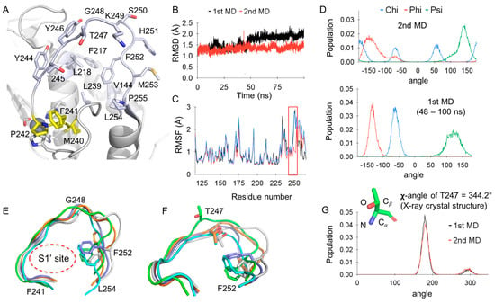
Molecular Dynamics Simulations of Matrix Metalloproteinase 13 and the Analysis of the Specificity Loop and the S1′−Site
by Jun Yong Choi and Eugene Chung
Int. J. Mol. Sci. 2023, 24(13), 10577; https://doi.org/10.3390/ijms241310577 - 24 Jun 2023
Cited by 2 | Viewed by 1767
Abstract
The specificity loop of Matrix Metalloproteinases (MMPs) is known to regulate recognition of their substrates, and the S1′−site surrounded by the loop is a unique place to address the selectivity of ligands toward each MMP. Molecular dynamics (MD) simulations of apo−MMP−13 and its [...] Read more.
The specificity loop of Matrix Metalloproteinases (MMPs) is known to regulate recognition of their substrates, and the S1′−site surrounded by the loop is a unique place to address the selectivity of ligands toward each MMP. Molecular dynamics (MD) simulations of apo−MMP−13 and its complex forms with various ligands were conducted to identify the role of the specificity loop for the ligand binding to MMP−13. The MD simulations showed the dual role of T247 as a hydrogen bond donor to the ligand, as well as a contributor to the formation of the van der Waal surface area, with T245 and K249 on the S1′−site. The hydrophobic surface area mediated by T247 blocks the access of water molecules to the S1′−site of MMP−13 and stabilizes the ligand in the site. The F252 residue is flexible in order to search for the optimum location in the S1′−site of the apo−MMP−13, but once a ligand binds to the S1′−site, it can form offset π−π or edge−to−π stacking interactions with the ligand. Lastly, H222 and Y244 provide the offset π−π and π−CH(Cβ) interactions on each side of the phenyl ring of the ligand, and this sandwiched interaction could be critical for the ligand binding to MMP−13. Full article
Show Figures

Figure 1

18 pages, 4450 KiB  
Article
Triterpenoids from Kochiae Fructus: Glucose Uptake in 3T3-L1 Adipocytes and α-Glucosidase Inhibition, In Silico Molecular Docking
by Xue-Lin Chen, Kun Zhang, Xia Zhao, Han-Lei Wang, Mei Han, Ru Li, Zhen-Nan Zhang and Yu-Mei Zhang
Int. J. Mol. Sci. 2023, 24(3), 2454; https://doi.org/10.3390/ijms24032454 - 26 Jan 2023
Cited by 4 | Viewed by 2403
Abstract
In this study, three new triterpenes (13) and fourteen known triterpenoids (417) were isolated from the ethanol extract of Kochiae Fructus, and their structures were elucidated by analyzing UV, IR, HR-ESI-MS, 1D, and 2D NMR [...] Read more.
In this study, three new triterpenes (13) and fourteen known triterpenoids (417) were isolated from the ethanol extract of Kochiae Fructus, and their structures were elucidated by analyzing UV, IR, HR-ESI-MS, 1D, and 2D NMR spectroscopic data. Among them, compounds 6, 8, and 1117 were isolated for the first time from this plant. The screening results of the glucose uptake experiment indicated that compound 13 had a potent effect on glucose uptake in 3T3-L1 adipocytes at 20 μM. Meanwhile, compounds 3, 9 and 13 exhibited significant inhibitory activities against α-glucosidase, with IC50 values of 23.50 ± 3.37, 4.29 ± 0.52, and 16.99 ± 2.70 µM, respectively, and their α-glucosidase inhibitory activities were reported for the first time. According to the enzyme kinetics using Lineweaver–Burk and Dixon plots, we found that compounds 3, 9 and 13 were α-glucosidase mixed-type inhibitors with Ki values of 56.86 ± 1.23, 48.88 ± 0.07 and 13.63 ± 0.42 μM, respectively. In silico molecular docking analysis showed that compounds 3 and 13 possessed superior binding capacities with α-glucosidase (3A4A AutoDock score: −4.99 and −4.63 kcal/mol). Whereas compound 9 showed +2.74 kcal/mol, which indicated compound 9 exerted the effect of inhibiting α-glucosidase activity by preferentially binding to the enzyme−substrate complex. As a result, compounds 3, 9 and 13 could have therapeutic potentials for type 2 diabetes mellitus, due to their potent hypoglycemic activities. Full article
Show Figures

Graphical abstract

2022

Jump to: 2023, 2021, 2020, 2019

16 pages, 2476 KiB  
Article
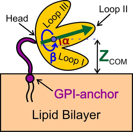
Orientational Preferences of GPI-Anchored Ly6/uPAR Proteins
by Maxim M. Zaigraev, Ekaterina N. Lyukmanova, Alexander S. Paramonov, Zakhar O. Shenkarev and Anton O. Chugunov
Int. J. Mol. Sci. 2023, 24(1), 11; https://doi.org/10.3390/ijms24010011 - 20 Dec 2022
Cited by 5 | Viewed by 2553
Abstract
Ly6/uPAR proteins regulate many essential functions in the nervous and immune systems and epithelium. Most of these proteins contain single β-structural LU domains with three protruding loops and are glycosylphosphatidylinositol (GPI)-anchored to a membrane. The GPI-anchor role is currently poorly studied. Here, we [...] Read more.
Ly6/uPAR proteins regulate many essential functions in the nervous and immune systems and epithelium. Most of these proteins contain single β-structural LU domains with three protruding loops and are glycosylphosphatidylinositol (GPI)-anchored to a membrane. The GPI-anchor role is currently poorly studied. Here, we investigated the positional and orientational preferences of six GPI-anchored proteins in the receptor-unbound state by molecular dynamics simulations. Regardless of the linker length between the LU domain and GPI-anchor, the proteins interacted with the membrane by polypeptide parts and N-/O-glycans. Lynx1, Lynx2, Lypd6B, and Ly6H contacted the membrane by the loop regions responsible for interactions with nicotinic acetylcholine receptors, while Lypd6 and CD59 demonstrated unique orientations with accessible receptor-binding sites. Thus, GPI-anchoring does not guarantee an optimal ‘pre-orientation’ of the LU domain for the receptor interaction. Full article
Show Figures

Graphical abstract

10 pages, 2627 KiB  
Article
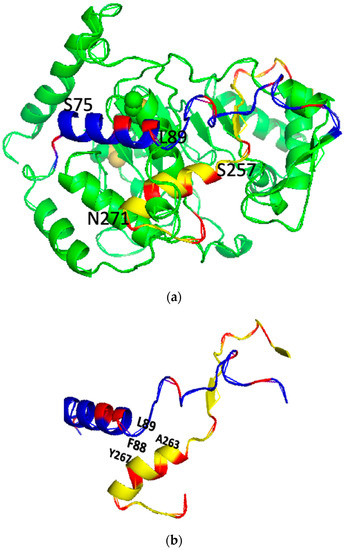
The Graphical Studies of the Major Molecular Interactions for Neural Cell Adhesion Molecule (NCAM) Polysialylation by Incorporating Wenxiang Diagram into NMR Spectroscopy
by Guo-Ping Zhou and Ri-Bo Huang
Int. J. Mol. Sci. 2022, 23(23), 15128; https://doi.org/10.3390/ijms232315128 - 1 Dec 2022
Cited by 3 | Viewed by 1342
Abstract
Polysialylation is a process of polysialic acid (polySia) addition to neural cell adhesion molecule (NCAM), which is associated with tumor cell migration and progression in many metastatic cancers and neurocognition. Polysialylation can be catalyzed by two highly homologous mammalian polysialyltransferases (polySTs), ST8Sia II [...] Read more.
Polysialylation is a process of polysialic acid (polySia) addition to neural cell adhesion molecule (NCAM), which is associated with tumor cell migration and progression in many metastatic cancers and neurocognition. Polysialylation can be catalyzed by two highly homologous mammalian polysialyltransferases (polySTs), ST8Sia II (STX) and ST8Sia IV (PST). It has been proposed that two polybasic domains, polybasic region (PBR) and polysialyltransferase domain (PSTD) in polySTs, are possible binding sites for the intermolecular interactions of polyST–NCAM and polyST–polySia, respectively, as well as the intramolecular interaction of PSTD–PBR. In this study, Chou’s wenxiang diagrams of the PSTD and PBR are used to determine the key amino acids of these intermolecular and intramolecular interactions, and thus it may be helpful for the identification of the crucial amino acids in the polyST and for the understanding of the molecular mechanism of NCAM polysialylation by incorporating the wenxiang diagram and molecular modeling into NMR spectroscopy. Full article
Show Figures

Figure 1

18 pages, 3665 KiB  
Article
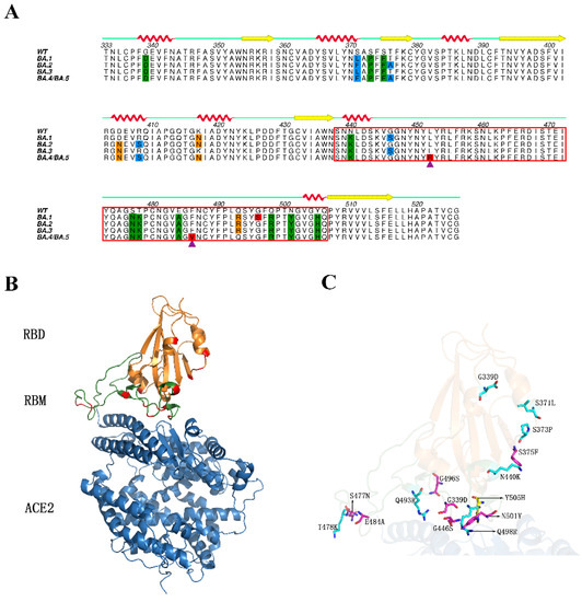
Electrostatic Interactions Are the Primary Determinant of the Binding Affinity of SARS-CoV-2 Spike RBD to ACE2: A Computational Case Study of Omicron Variants
by Peng Sang, Yong-Qin Chen, Meng-Ting Liu, Yu-Ting Wang, Ting Yue, Yi Li, Yi-Rui Yin and Li-Quan Yang
Int. J. Mol. Sci. 2022, 23(23), 14796; https://doi.org/10.3390/ijms232314796 - 26 Nov 2022
Cited by 17 | Viewed by 2522
Abstract
To explore the mechanistic origin that determines the binding affinity of SARS-CoV-2 spike receptor binding domain (RBD) to human angiotensin converting enzyme 2 (ACE2), we constructed the homology models of RBD-ACE2 complexes of four Omicron subvariants (BA.1, BA.2, BA.3 and BA.4/5), and compared [...] Read more.
To explore the mechanistic origin that determines the binding affinity of SARS-CoV-2 spike receptor binding domain (RBD) to human angiotensin converting enzyme 2 (ACE2), we constructed the homology models of RBD-ACE2 complexes of four Omicron subvariants (BA.1, BA.2, BA.3 and BA.4/5), and compared them with wild type complex (RBDWT-ACE2) in terms of various structural dynamic properties by molecular dynamics (MD) simulations and binding free energy (BFE) calculations. The results of MD simulations suggest that the RBDs of all the Omicron subvariants (RBDOMIs) feature increased global structural fluctuations when compared with RBDWT. Detailed comparison of BFE components reveals that the enhanced electrostatic attractive interactions are the main determinant of the higher ACE2-binding affinity of RBDOMIs than RBDWT, while the weakened electrostatic attractive interactions determine RBD of BA.4/5 subvariant (RBDBA.4/5) lowest ACE2-binding affinity among all Omicron subvariants. The per-residue BFE decompositions and the hydrogen bond (HB) networks analyses indicate that the enhanced electrostatic attractive interactions are mainly through gain/loss of the positively/negatively charged residues, and the formation or destruction of the interfacial HBs and salt bridges can also largely affect the ACE2-binding affinity of RBD. It is worth pointing out that since Q493R plays the most important positive contribution in enhancing binding affinity, the absence of this mutation in RBDBA.4/5 results in a significantly weaker binding affinity to ACE2 than other Omicron subvariants. Our results provide insight into the role of electrostatic interactions in determining of the binding affinity of SARS-CoV-2 RBD to human ACE2. Full article
Show Figures

Figure 1

26 pages, 11747 KiB  
Article
In Silico Identification of Multi-Target Ligands as Promising Hit Compounds for Neurodegenerative Diseases Drug Development
by Petko Alov, Hristo Stoimenov, Iglika Lessigiarska, Tania Pencheva, Nikolay T. Tzvetkov, Ilza Pajeva and Ivanka Tsakovska
Int. J. Mol. Sci. 2022, 23(21), 13650; https://doi.org/10.3390/ijms232113650 - 7 Nov 2022
Cited by 3 | Viewed by 2895
Abstract
The conventional treatment of neurodegenerative diseases (NDDs) is based on the “one molecule—one target” paradigm. To combat the multifactorial nature of NDDs, the focus is now shifted toward the development of small-molecule-based compounds that can modulate more than one protein target, known as [...] Read more.
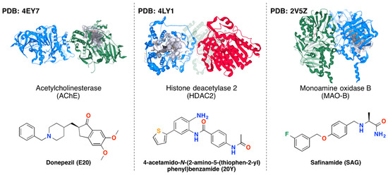
The conventional treatment of neurodegenerative diseases (NDDs) is based on the “one molecule—one target” paradigm. To combat the multifactorial nature of NDDs, the focus is now shifted toward the development of small-molecule-based compounds that can modulate more than one protein target, known as “multi-target-directed ligands” (MTDLs), while having low affinity for proteins that are irrelevant for the therapy. The in silico approaches have demonstrated a potential to be a suitable tool for the identification of MTDLs as promising drug candidates with reduction in cost and time for research and development. In this study more than 650,000 compounds were screened by a series of in silico approaches to identify drug-like compounds with predicted activity simultaneously towards three important proteins in the NDDs symptomatic treatment: acetylcholinesterase (AChE), histone deacetylase 2 (HDAC2), and monoamine oxidase B (MAO-B). The compounds with affinities below 5.0 µM for all studied targets were additionally filtered to remove known non-specifically binding or unstable compounds. The selected four hits underwent subsequent refinement through in silico blood-brain barrier penetration estimation, safety evaluation, and molecular dynamics simulations resulting in two hit compounds that constitute a rational basis for further development of multi-target active compounds against NDDs. Full article
Show Figures

Figure 1

35 pages, 3151 KiB  
Review
Application of Computational Biology and Artificial Intelligence in Drug Design
by Yue Zhang, Mengqi Luo, Peng Wu, Song Wu, Tzong-Yi Lee and Chen Bai
Int. J. Mol. Sci. 2022, 23(21), 13568; https://doi.org/10.3390/ijms232113568 - 5 Nov 2022
Cited by 38 | Viewed by 14637
Abstract
Traditional drug design requires a great amount of research time and developmental expense. Booming computational approaches, including computational biology, computer-aided drug design, and artificial intelligence, have the potential to expedite the efficiency of drug discovery by minimizing the time and financial cost. In [...] Read more.
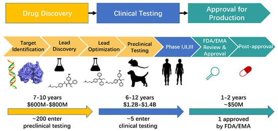
Traditional drug design requires a great amount of research time and developmental expense. Booming computational approaches, including computational biology, computer-aided drug design, and artificial intelligence, have the potential to expedite the efficiency of drug discovery by minimizing the time and financial cost. In recent years, computational approaches are being widely used to improve the efficacy and effectiveness of drug discovery and pipeline, leading to the approval of plenty of new drugs for marketing. The present review emphasizes on the applications of these indispensable computational approaches in aiding target identification, lead discovery, and lead optimization. Some challenges of using these approaches for drug design are also discussed. Moreover, we propose a methodology for integrating various computational techniques into new drug discovery and design. Full article
Show Figures

Figure 1

16 pages, 5508 KiB  
Article
Effect of Silica Microparticles on Interactions in Mono- and Multicomponent Membranes
by Beata Tim, Monika Rojewska and Krystyna Prochaska
Int. J. Mol. Sci. 2022, 23(21), 12822; https://doi.org/10.3390/ijms232112822 - 24 Oct 2022
Cited by 6 | Viewed by 1929
Abstract
Advancing our understanding of the mechanism of the interaction between inhaled pollutant microparticles and cell membrane components is useful to study the impact of fine particulate matter on human health. In this paper, we focus on the effect of cholesterol (Chol) molecules on [...] Read more.
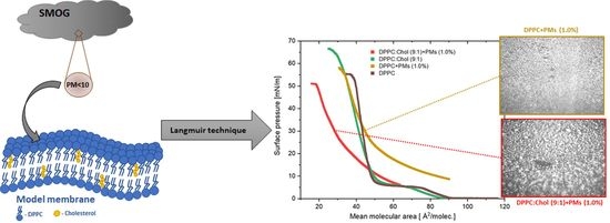
Advancing our understanding of the mechanism of the interaction between inhaled pollutant microparticles and cell membrane components is useful to study the impact of fine particulate matter on human health. In this paper, we focus on the effect of cholesterol (Chol) molecules on the surface properties of a model membrane in the presence of silica microparticles (MPs). Mixed monolayers containing phospholipid-dipalmitoylphosphatidylcholine (DPPC), Chol and silica particle dispersions (MPs; 0.033% w/w, 0.33% w/w and 0.83% w/w) were formed and studied using the Langmuir monolayer technique complemented by Brewster Angle Microscopy (BAM) images. It was shown that Chol caused a condensation of the DPPC monolayer, which influenced the penetration of MPs and their interactions with the model membrane. The relaxation experiments of the lipid–MP monolayer proved that the presence of Chol molecules in the monolayer led to the formation of lipid and MP complexes. Strong interactions between Chol and MPs contributed to the formation of more stable monolayers. The presented results can be useful to better comprehend the interaction between particulate materials and the lipid components of biomembranes. Full article
Show Figures

Graphical abstract

19 pages, 3881 KiB  
Article
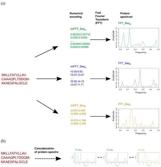
PD-BertEDL: An Ensemble Deep Learning Method Using BERT and Multivariate Representation to Predict Peptide Detectability
by Huiqing Wang, Juan Wang, Zhipeng Feng, Ying Li and Hong Zhao
Int. J. Mol. Sci. 2022, 23(20), 12385; https://doi.org/10.3390/ijms232012385 - 16 Oct 2022
Cited by 2 | Viewed by 1770
Abstract
Peptide detectability is defined as the probability of identifying a peptide from a mixture of standard samples, which is a key step in protein identification and analysis. Exploring effective methods for predicting peptide detectability is helpful for disease treatment and clinical research. However, [...] Read more.
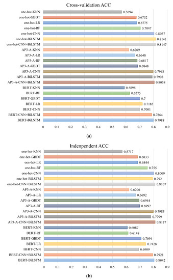
Peptide detectability is defined as the probability of identifying a peptide from a mixture of standard samples, which is a key step in protein identification and analysis. Exploring effective methods for predicting peptide detectability is helpful for disease treatment and clinical research. However, most existing computational methods for predicting peptide detectability rely on a single information. With the increasing complexity of feature representation, it is necessary to explore the influence of multivariate information on peptide detectability. Thus, we propose an ensemble deep learning method, PD-BertEDL. Bidirectional encoder representations from transformers (BERT) is introduced to capture the context information of peptides. Context information, sequence information, and physicochemical information of peptides were combined to construct the multivariate feature space of peptides. We use different deep learning methods to capture the high-quality features of different categories of peptides information and use the average fusion strategy to integrate three model prediction results to solve the heterogeneity problem and to enhance the robustness and adaptability of the model. The experimental results show that PD-BertEDL is superior to the existing prediction methods, which can effectively predict peptide detectability and provide strong support for protein identification and quantitative analysis, as well as disease treatment. Full article
Show Figures

Figure 1

15 pages, 6154 KiB  
Article
Interspecies Comparison of Interaction Energies between Photosynthetic Protein RuBisCO and 2CABP Ligand
by Masayasu Fujii and Shigenori Tanaka
Int. J. Mol. Sci. 2022, 23(19), 11347; https://doi.org/10.3390/ijms231911347 - 26 Sep 2022
Cited by 2 | Viewed by 2104
Abstract
Ribulose 1,5-bisphosphate carboxylase/oxygenase (RuBisCO) functions as the initial enzyme in the dark reactions of photosynthesis, catalyzing reactions that extract CO2 from the atmosphere and fix CO2 into organic compounds. RuBisCO is classified into four types (isoforms I–IV) according to sequence-based phylogenetic [...] Read more.
Ribulose 1,5-bisphosphate carboxylase/oxygenase (RuBisCO) functions as the initial enzyme in the dark reactions of photosynthesis, catalyzing reactions that extract CO2 from the atmosphere and fix CO2 into organic compounds. RuBisCO is classified into four types (isoforms I–IV) according to sequence-based phylogenetic trees. Given its size, the computational cost of accurate quantum-chemical calculations for functional analysis of RuBisCO is high; however, recent advances in hardware performance and the use of the fragment molecular orbital (FMO) method have enabled the ab initio analyses of RuBisCO. Here, we performed FMO calculations on multiple structural datasets for various complexes with the 2′-carboxylarabinitol 1,5-bisphosphate (2CABP) ligand as a substrate analog and investigated whether phylogenetic relationships based on sequence information are physicochemically relevant as well as whether novel information unobtainable from sequence information can be revealed. We extracted features similar to the phylogenetic relationships found in sequence analysis, and in terms of singular value decomposition, we identified residues that strongly interacted with the ligand and the characteristics of the isoforms for each principal component. These results identified a strong correlation between phylogenetic relationships obtained by sequence analysis and residue interaction energies with the ligand. Notably, some important residues were located far from the ligand, making comparisons among species using only residues proximal to the ligand insufficient. Full article
Show Figures

Figure 1

15 pages, 2082 KiB  
Article
Balanced Force Field ff03CMAP Improving the Dynamics Conformation Sampling of Phosphorylation Site
by Bozitao Zhong, Ge Song and Hai-Feng Chen
Int. J. Mol. Sci. 2022, 23(19), 11285; https://doi.org/10.3390/ijms231911285 - 25 Sep 2022
Cited by 3 | Viewed by 2334
Abstract
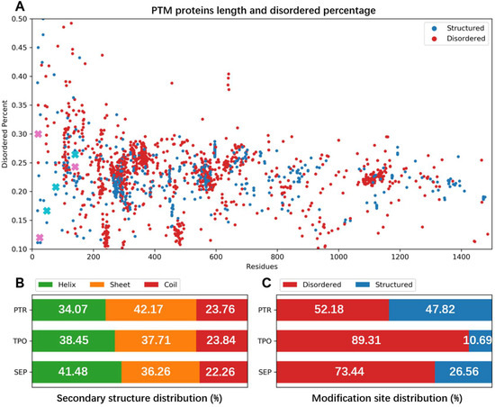
Phosphorylation plays a key role in plant biology, such as the accumulation of plant cells to form the observed proteome. Statistical analysis found that many phosphorylation sites are located in disordered regions. However, current force fields are mainly trained for structural proteins, which [...] Read more.
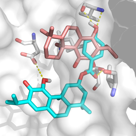
Phosphorylation plays a key role in plant biology, such as the accumulation of plant cells to form the observed proteome. Statistical analysis found that many phosphorylation sites are located in disordered regions. However, current force fields are mainly trained for structural proteins, which might not have the capacity to perfectly capture the dynamic conformation of the phosphorylated proteins. Therefore, we evaluated the performance of ff03CMAP, a balanced force field between structural and disordered proteins, for the sampling of the phosphorylated proteins. The test results of 11 different phosphorylated systems, including dipeptides, disordered proteins, folded proteins, and their complex, indicate that the ff03CMAP force field can better sample the conformations of phosphorylation sites for disordered proteins and disordered regions than ff03. For the solvent model, the results strongly suggest that the ff03CMAP force field with the TIP4PD water model is the best combination for the conformer sampling. Additional tests of CHARMM36m and FB18 force fields on two phosphorylated systems suggest that the overall performance of ff03CMAP is similar to that of FB18 and better than that of CHARMM36m. These results can help other researchers to choose suitable force field and solvent models to investigate the dynamic properties of phosphorylation proteins. Full article
Show Figures

Figure 1

21 pages, 3679 KiB  
Article
PSP-GNM: Predicting Protein Stability Changes upon Point Mutations with a Gaussian Network Model
by Sambit Kumar Mishra
Int. J. Mol. Sci. 2022, 23(18), 10711; https://doi.org/10.3390/ijms231810711 - 14 Sep 2022
Cited by 2 | Viewed by 3152
Abstract
Understanding the effects of missense mutations on protein stability is a widely acknowledged significant biological problem. Genomic missense mutations may alter one or more amino acids, leading to increased or decreased stability of the encoded proteins. In this study, we describe a novel [...] Read more.
Understanding the effects of missense mutations on protein stability is a widely acknowledged significant biological problem. Genomic missense mutations may alter one or more amino acids, leading to increased or decreased stability of the encoded proteins. In this study, we describe a novel approach—Protein Stability Prediction with a Gaussian Network Model (PSP-GNM)—to measure the unfolding Gibbs free energy change (ΔΔG) and evaluate the effects of single amino acid substitutions on protein stability. Specifically, PSP-GNM employs a coarse-grained Gaussian Network Model (GNM) that has interactions between amino acids weighted by the Miyazawa–Jernigan statistical potential. We used PSP-GNM to simulate partial unfolding of the wildtype and mutant protein structures, and then used the difference in the energies and entropies of the unfolded wildtype and mutant proteins to calculate ΔΔG. The extent of the agreement between the ΔΔG calculated by PSP-GNM and the experimental ΔΔG was evaluated on three benchmark datasets: 350 forward mutations (S350 dataset), 669 forward and reverse mutations (S669 dataset) and 611 forward and reverse mutations (S611 dataset). We observed a Pearson correlation coefficient as high as 0.61, which is comparable to many of the existing state-of-the-art methods. The agreement with experimental ΔΔG further increased when we considered only those measurements made close to 25 °C and neutral pH, suggesting dependence on experimental conditions. We also assessed for the antisymmetry (ΔΔGreverse = −ΔΔGforward) between the forward and reverse mutations on the Ssym+ dataset, which has 352 forward and reverse mutations. While most available methods do not display significant antisymmetry, PSP-GNM demonstrated near-perfect antisymmetry, with a Pearson correlation of −0.97. PSP-GNM is written in Python and can be downloaded as a stand-alone code. Full article
Show Figures

Figure 1

12 pages, 4611 KiB  
Article
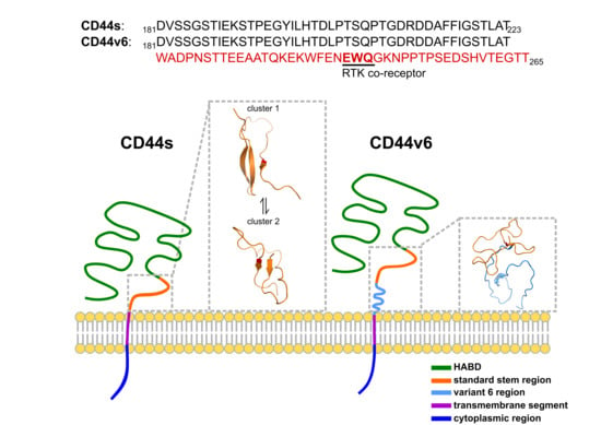
Kozak Similarity Score Algorithm Identifies Alternative Translation Initiation Codons Implicated in Cancers
by Alec C. Gleason, Ghanashyam Ghadge, Yoshifumi Sonobe and Raymond P. Roos
Int. J. Mol. Sci. 2022, 23(18), 10564; https://doi.org/10.3390/ijms231810564 - 12 Sep 2022
Cited by 6 | Viewed by 3481
Abstract
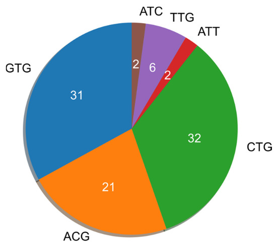
Ribosome profiling and mass spectroscopy have identified canonical and noncanonical translation initiation codons (TICs) that are upstream of the main translation initiation site and used to translate oncogenic proteins. There have previously been conflicting reports about the patterns of nucleotides that surround noncanonical [...] Read more.
Ribosome profiling and mass spectroscopy have identified canonical and noncanonical translation initiation codons (TICs) that are upstream of the main translation initiation site and used to translate oncogenic proteins. There have previously been conflicting reports about the patterns of nucleotides that surround noncanonical TICs. Here, we use a Kozak Similarity Score algorithm to find that nearly all of these TICs have flanking nucleotides closely matching the Kozak sequence. Remarkably, the nucleotides flanking alternative noncanonical TICs are frequently closer to the Kozak sequence than the nucleotides flanking TICs used to translate the gene’s main protein. Of note, the 5′ untranslated region (5‘UTR) of cancer-associated genes with an upstream TIC tend to be significantly longer than the same region in genes not associated with cancer. The presence of a longer-than-typical 5′UTR increases the likelihood of ribosome binding to upstream noncanonical TICs, and may be a distinguishing feature of a number of genes overexpressed in cancer. Noncanonical TICs that are located in the 5′UTR, although thought by some to be disadvantageous and suppressed by evolution, may translate oncogenic proteins because of their flanking nucleotides. Full article
Show Figures

Figure 1

15 pages, 2676 KiB  
Article
Electronic Structure and Solvation Effects from Core and Valence Photoelectron Spectroscopy of Serum Albumin
by Jean-Philippe Renault, Lucie Huart, Aleksandar R. Milosavljević, John D. Bozek, Jerôme Palaudoux, Jean-Michel Guigner, Laurent Marichal, Jocelyne Leroy, Frank Wien, Marie-Anne Hervé Du Penhoat and Christophe Nicolas
Int. J. Mol. Sci. 2022, 23(15), 8227; https://doi.org/10.3390/ijms23158227 - 26 Jul 2022
Cited by 2 | Viewed by 2707
Abstract
X-ray photoelectron spectroscopy of bovine serum albumin (BSA) in a liquid jet is used to investigate the electronic structure of a solvated protein, yielding insight into charge transfer mechanisms in biological systems in their natural environment. No structural damage was observed in BSA [...] Read more.
X-ray photoelectron spectroscopy of bovine serum albumin (BSA) in a liquid jet is used to investigate the electronic structure of a solvated protein, yielding insight into charge transfer mechanisms in biological systems in their natural environment. No structural damage was observed in BSA following X-ray photoelectron spectroscopy in a liquid jet sample environment. Carbon and nitrogen atoms in different chemical environments were resolved in the X-ray photoelectron spectra of both solid and solvated BSA. The calculations of charge distributions demonstrate the difficulty of assigning chemical contributions in complex systems in an aqueous environment. The high-resolution X-ray core electron spectra recorded are unchanged upon solvation. A comparison of the valence bands of BSA in both phases is also presented. These bands display a higher sensitivity to solvation effects. The ionization energy of the solvated BSA is determined at 5.7 ± 0.3 eV. Experimental results are compared with theoretical calculations to distinguish the contributions of various molecular components to the electronic structure. This comparison points towards the role of water in hole delocalization in proteins. Full article
Show Figures

Figure 1

20 pages, 5260 KiB  
Article
The 2-hydroxy-3-(4-aryl-1-piperazinyl)propyl Phthalimide Derivatives as Prodrugs—Spectroscopic and Theoretical Binding Studies with Plasma Proteins
by Aleksandra Marciniak, Aleksandra Kotynia, Dominika Szkatuła and Edward Krzyżak
Int. J. Mol. Sci. 2022, 23(13), 7003; https://doi.org/10.3390/ijms23137003 - 23 Jun 2022
Cited by 4 | Viewed by 1871
Abstract
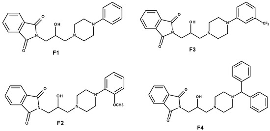
Many publications in databases deal with the interactions of new drugs with albumin. However, it is not only albumin that is responsible for binding pharmaceutical molecules to proteins in the human body. There are many more proteins in plasma that are important for [...] Read more.
Many publications in databases deal with the interactions of new drugs with albumin. However, it is not only albumin that is responsible for binding pharmaceutical molecules to proteins in the human body. There are many more proteins in plasma that are important for the study of the ADME pathway. Therefore, in this study, we have shown the results of the interactions between the plasma proteins albumin, orosomucoid, and gamma globulins and non-toxic anti-inflammatory phthalimide analogs, which due to the promising obtained results, may be potential candidates in the group of analgesic and anti-inflammatory drugs. Using spectroscopic methods and molecular modeling, we showed that all four tested compounds form complexes with the analyzed proteins. The formation of a complex with proteins raises the pharmacological efficacy of the drug. Therefore, the obtained results could be a step in the study of the pharmacokinetics and pharmacodynamics of new potential pharmaceuticals. Full article
Show Figures

Figure 1

15 pages, 3282 KiB  
Article
Molecular Dynamics Studies on the Structural Stability Prediction of SARS-CoV-2 Variants Including Multiple Mutants
by Kwang-Eun Choi, Jeong-Min Kim, Jee Eun Rhee, Ae Kyung Park, Eun-Jin Kim, Cheon Kwon Yoo and Nam Sook Kang
Int. J. Mol. Sci. 2022, 23(9), 4956; https://doi.org/10.3390/ijms23094956 - 29 Apr 2022
Cited by 6 | Viewed by 2533
Abstract
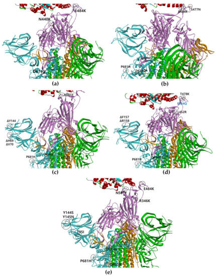
Severe Acute Respiratory Syndrome Coronavirus 2 (SARS-CoV-2) has caused the Coronavirus Disease (COVID-19) pandemic worldwide. The spike protein in SARS-CoV-2 fuses with and invades cells in the host respiratory system by binding to angiotensin-converting enzyme 2 (ACE2). The spike protein, however, undergoes continuous [...] Read more.
Severe Acute Respiratory Syndrome Coronavirus 2 (SARS-CoV-2) has caused the Coronavirus Disease (COVID-19) pandemic worldwide. The spike protein in SARS-CoV-2 fuses with and invades cells in the host respiratory system by binding to angiotensin-converting enzyme 2 (ACE2). The spike protein, however, undergoes continuous mutation from a D614G single mutant to an omicron variant, including multiple mutants. In this study, variants, including multiple mutants (double, triple mutants, B.1.620, delta, alpha, delta_E484Q, mu, and omicron) were investigated in patients. The 3D structure of the full-length spike protein was used in conformational analysis depending on the SARS-CoV-2 variants. The structural stability of the variant types was analyzed based on the distance between the receptor-binding domain (RBD) of each chain in the spike protein and the binding free energy between the spike protein and bound ACE2 in the one-, two-, and three-open-complex forms using molecular dynamics (MD) simulation. Omicron variants, the most prevalent in the recent history of the global pandemic, which consist of 32 mutations, showed higher stability in all open-complex forms compared with that of the wild type and other variants. We suggest that the conformational stability of the spike protein is the one of the important determinants for the differences in viral infectivity among variants, including multiple mutants. Full article
Show Figures

Figure 1

13 pages, 3489 KiB  
Article
Design and Nonadiabatic Photoisomerization Dynamics Study of a Three-Stroke Light-Driven Molecular Rotary Motor
by Jianzheng Ma, Sujie Yang, Di Zhao, Chenwei Jiang, Zhenggang Lan and Fuli Li
Int. J. Mol. Sci. 2022, 23(7), 3908; https://doi.org/10.3390/ijms23073908 - 31 Mar 2022
Cited by 6 | Viewed by 2263
Abstract
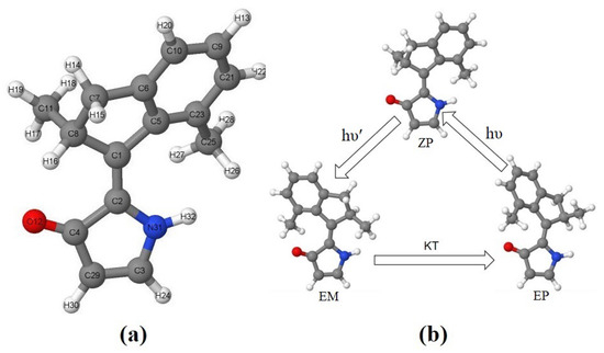
Working cycle of conventional light-driven molecular rotary motors (LDMRMs), especially Feringa-type motors, usually have four steps, two photoisomerization steps, and two thermal helix inversion (THI) steps. THI steps hinder the ability of the motor to operate at lower temperatures and limit the rotation [...] Read more.
Working cycle of conventional light-driven molecular rotary motors (LDMRMs), especially Feringa-type motors, usually have four steps, two photoisomerization steps, and two thermal helix inversion (THI) steps. THI steps hinder the ability of the motor to operate at lower temperatures and limit the rotation speed of LDMRMs. A three-stroke LDMRM, 2-(2,7-dimethyl-2,3-dihydro-1H-inden-1-ylidene)-1,2-dihydro-3H-pyrrol-3-one (DDIY), is proposed, which is capable of completing an unidirectional rotation by two photoisomerization steps and one thermal helix inversion step at room temperature. On the basis of trajectory surface-hopping simulation at the semi-empirical OM2/MRCI level, the EP→ZP and ZP→EM nonadiabatic photoisomerization dynamics of DDIY were systematically analyzed. Quantum yields of EP→ZP and ZP→EM photoisomerization of DDIY are ca. 34% and 18%, respectively. Both EP→ZP and ZP→EM photoisomerization processes occur on an ultrafast time scale (ca. 100–300 fs). This three-stroke LDMRM may stimulate further research for the development of new families of more efficient LDMRMs. Full article
Show Figures

Figure 1

18 pages, 8864 KiB  
Article
Identification of Novel GSK-3β Hits Using Competitive Biophysical Assays
by Beatrice Balboni, Shailesh Kumar Tripathi, Marina Veronesi, Debora Russo, Ilaria Penna, Barbara Giabbai, Tiziano Bandiera, Paola Storici, Stefania Girotto and Andrea Cavalli
Int. J. Mol. Sci. 2022, 23(7), 3856; https://doi.org/10.3390/ijms23073856 - 31 Mar 2022
Cited by 4 | Viewed by 2860
Abstract
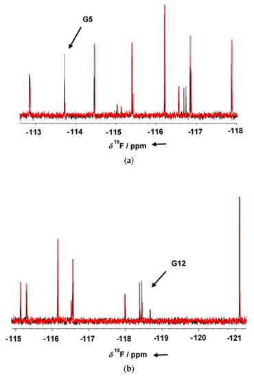
Glycogen synthase kinase 3 beta (GSK-3β) is an evolutionarily conserved serine-threonine kinase dysregulated in numerous pathologies, such as Alzheimer’s disease and cancer. Even though GSK-3β is a validated pharmacological target most of its inhibitors have two main limitations: the lack of selectivity due [...] Read more.
Glycogen synthase kinase 3 beta (GSK-3β) is an evolutionarily conserved serine-threonine kinase dysregulated in numerous pathologies, such as Alzheimer’s disease and cancer. Even though GSK-3β is a validated pharmacological target most of its inhibitors have two main limitations: the lack of selectivity due to the high homology that characterizes the ATP binding site of most kinases, and the toxicity that emerges from GSK-3β complete inhibition which translates into the impairment of the plethora of pathways GSK-3β is involved in. Starting from a 1D 19F NMR fragment screening, we set up several biophysical assays for the identification of GSK-3β inhibitors capable of binding protein hotspots other than the ATP binding pocket or to the ATP binding pocket, but with an affinity able of competing with a reference binder. A phosphorylation activity assay on a panel of several kinases provided selectivity data that were further rationalized and corroborated by structural information on GSK-3β in complex with the hit compounds. In this study, we identified promising fragments, inhibitors of GSK-3β, while proposing an alternative screening workflow that allows facing the flaws that characterize the most common GSK-3β inhibitors through the identification of selective inhibitors and/or inhibitors able to modulate GSK-3β activity without leading to its complete inhibition. Full article
Show Figures

Figure 1

12 pages, 6131 KiB  
Article
Discovery of SARS-CoV-2 3CLPro Peptidomimetic Inhibitors through the Catalytic Dyad Histidine-Specific Protein–Ligand Interactions
by Yaxin Wang, Binghong Xu, Sen Ma, Hao Wang, Luqing Shang, Cheng Zhu and Sheng Ye
Int. J. Mol. Sci. 2022, 23(4), 2392; https://doi.org/10.3390/ijms23042392 - 21 Feb 2022
Cited by 7 | Viewed by 3252
Abstract
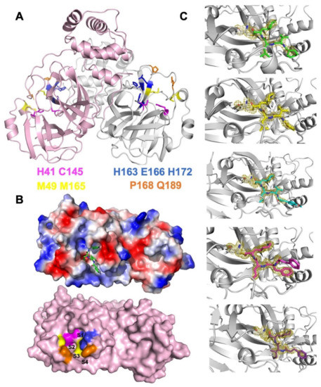
As the etiological agent for the coronavirus disease 2019, severe acute respiratory syndrome coronavirus 2 (SARS-CoV-2) challenges the ongoing efforts of vaccine development and drug design. Due to the accumulating cases of breakthrough infections, there are urgent needs for broad-spectrum antiviral medicines. Here, [...] Read more.
As the etiological agent for the coronavirus disease 2019, severe acute respiratory syndrome coronavirus 2 (SARS-CoV-2) challenges the ongoing efforts of vaccine development and drug design. Due to the accumulating cases of breakthrough infections, there are urgent needs for broad-spectrum antiviral medicines. Here, we designed and examined five new tetrapeptidomimetic anti-SARS-CoV-2 inhibitors targeting the 3C-Like protease (3CLPro), which is highly conserved among coronaviruses and essential for viral replications. We significantly improved the efficacy of a ketoamide lead compound based on high-resolution co-crystal structures, all-atom simulations, and binding energy calculations. The inhibitors successfully engaged the catalytic dyad histidine residue (H41) of 3CLPro as designed, and they exhibited nanomolar inhibitory capacity as well as mitigated the viral loads of SARS-CoV-2 in cellular assays. As a widely applicable design principle, our results revealed that the potencies of 3CLPro-specific drug candidates were determined by the interplay between 3CLPro H41 residue and the peptidomimetic inhibitors. Full article
Show Figures

Figure 1

2021

Jump to: 2023, 2022, 2020, 2019

21 pages, 5270 KiB  
Article
A Lumenal Loop Associated with Catalytic Asymmetry in Plant Vacuolar H+-Translocating Pyrophosphatase
by Viktor A. Anashkin and Alexander A. Baykov
Int. J. Mol. Sci. 2021, 22(23), 12902; https://doi.org/10.3390/ijms222312902 - 29 Nov 2021
Cited by 3 | Viewed by 1757
Abstract
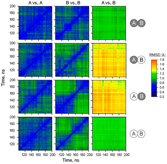
Membrane-integral inorganic pyrophosphatases (mPPases) couple pyrophosphate hydrolysis with H+ and Na+ pumping in plants and microbes. mPPases are homodimeric transporters with two catalytic sites facing the cytoplasm and demonstrating highly different substrate-binding affinities and activities. The structural aspects of the functional [...] Read more.
Membrane-integral inorganic pyrophosphatases (mPPases) couple pyrophosphate hydrolysis with H+ and Na+ pumping in plants and microbes. mPPases are homodimeric transporters with two catalytic sites facing the cytoplasm and demonstrating highly different substrate-binding affinities and activities. The structural aspects of the functional asymmetry are still poorly understood because the structure of the physiologically relevant dimer form with only one active site occupied by the substrate is unknown. We addressed this issue by molecular dynamics (MD) simulations of the H+-transporting mPPase of Vigna radiata, starting from its crystal structure containing a close substrate analog (imidodiphosphate, IDP) in both active sites. The MD simulations revealed pre-existing subunit asymmetry, which increased upon IDP binding to one subunit and persisted in the fully occupied dimer. The most significant asymmetrical change caused by IDP binding is a ‘rigid body’-like displacement of the lumenal loop connecting α-helices 2 and 3 in the partner subunit and opening its exit channel for water. This highly conserved 14–19-residue loop is found only in plant vacuolar mPPases and may have a regulatory function, such as pH sensing in the vacuole. Our data define the structural link between the loop and active sites and are consistent with the published structural and functional data. Full article
Show Figures

Figure 1

25 pages, 3563 KiB  
Article
DOPA Homeostasis by Dopamine: A Control-Theoretic View
by Rune Kleppe, Qaiser Waheed and Peter Ruoff
Int. J. Mol. Sci. 2021, 22(23), 12862; https://doi.org/10.3390/ijms222312862 - 28 Nov 2021
Cited by 7 | Viewed by 3399
Abstract
Dopamine (DA) is an important signal mediator in the brain as well as in the periphery. The term “dopamine homeostasis” occasionally found in the literature refers to the fact that abnormal DA levels can be associated with a variety of neuropsychiatric disorders. An [...] Read more.
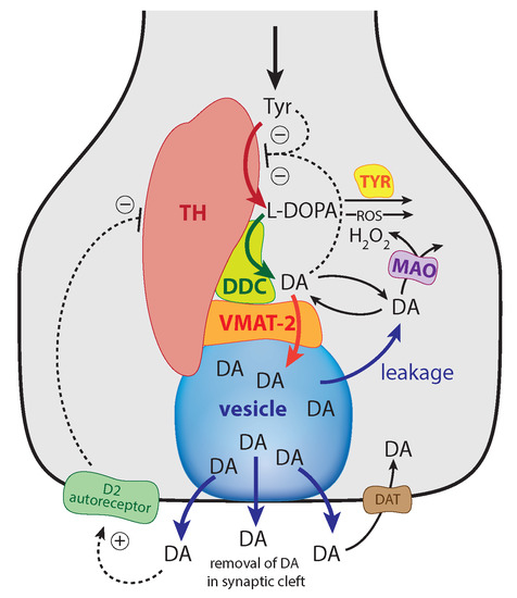
Dopamine (DA) is an important signal mediator in the brain as well as in the periphery. The term “dopamine homeostasis” occasionally found in the literature refers to the fact that abnormal DA levels can be associated with a variety of neuropsychiatric disorders. An analysis of the negative feedback inhibition of tyrosine hydroxylase (TH) by DA indicates, with support from the experimental data, that the TH-DA negative feedback loop has developed to exhibit 3,4-dihydroxyphenylalanine (DOPA) homeostasis by using DA as a derepression regulator. DA levels generally decline when DOPA is removed, for example, by increased oxidative stress. Robust DOPA regulation by DA further implies that maximum vesicular DA levels are established, which appear necessary for a reliable translation of neural activity into a corresponding chemical transmitter signal. An uncontrolled continuous rise (windup) in DA occurs when Levodopa treatment exceeds a critical dose. Increased oxidative stress leads to the successive breakdown of DOPA homeostasis and to a corresponding reduction in DA levels. To keep DOPA regulation robust, the vesicular DA loading requires close to zero-order kinetics combined with a sufficiently high compensatory flux provided by TH. The protection of DOPA and DA due to a channeling complex is discussed. Full article
Show Figures

Figure 1

24 pages, 455 KiB  
Article
Ensemble of Template-Free and Template-Based Classifiers for Protein Secondary Structure Prediction
by Gabriel Bianchin de Oliveira, Helio Pedrini and Zanoni Dias
Int. J. Mol. Sci. 2021, 22(21), 11449; https://doi.org/10.3390/ijms222111449 - 23 Oct 2021
Cited by 1 | Viewed by 2085
Abstract
Protein secondary structures are important in many biological processes and applications. Due to advances in sequencing methods, there are many proteins sequenced, but fewer proteins with secondary structures defined by laboratory methods. With the development of computer technology, computational methods have (started to) [...] Read more.
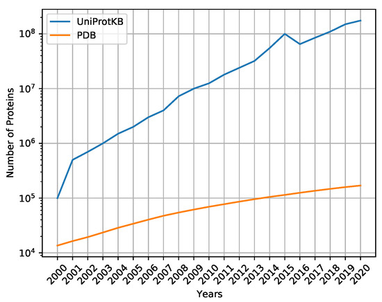
Protein secondary structures are important in many biological processes and applications. Due to advances in sequencing methods, there are many proteins sequenced, but fewer proteins with secondary structures defined by laboratory methods. With the development of computer technology, computational methods have (started to) become the most important methodologies for predicting secondary structures. We evaluated two different approaches to this problem—driven by the recent results obtained by computational methods in this task—(i) template-free classifiers, based on machine learning techniques; and (ii) template-based classifiers, based on searching tools. Both approaches are formed by different sub-classifiers—six for template-free and two for template-based, each with a specific view of the protein. Our results show that these ensembles improve the results of each approach individually. Full article
Show Figures

Figure 1

28 pages, 16064 KiB  
Article
A Comparative Study to Decipher the Structural and Dynamics Determinants Underlying the Activity and Thermal Stability of GH-11 Xylanases
by Jelena Vucinic, Gleb Novikov, Cédric Y. Montanier, Claire Dumon, Thomas Schiex and Sophie Barbe
Int. J. Mol. Sci. 2021, 22(11), 5961; https://doi.org/10.3390/ijms22115961 - 31 May 2021
Cited by 13 | Viewed by 3156
Abstract
With the growing need for renewable sources of energy, the interest for enzymes capable of biomass degradation has been increasing. In this paper, we consider two different xylanases from the GH-11 family: the particularly active GH-11 xylanase from Neocallimastix patriciarum, NpXyn11A, [...] Read more.
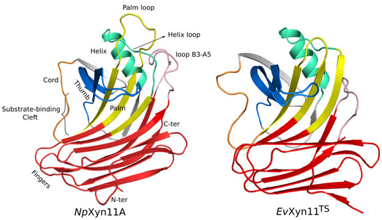
With the growing need for renewable sources of energy, the interest for enzymes capable of biomass degradation has been increasing. In this paper, we consider two different xylanases from the GH-11 family: the particularly active GH-11 xylanase from Neocallimastix patriciarum, NpXyn11A, and the hyper-thermostable mutant of the environmentally isolated GH-11 xylanase, EvXyn11TS. Our aim is to identify the molecular determinants underlying the enhanced capacities of these two enzymes to ultimately graft the abilities of one on the other. Molecular dynamics simulations of the respective free-enzymes and enzyme–xylohexaose complexes were carried out at temperatures of 300, 340, and 500 K. An in-depth analysis of these MD simulations showed how differences in dynamics influence the activity and stability of these two enzymes and allowed us to study and understand in greater depth the molecular and structural basis of these two systems. In light of the results presented in this paper, the thumb region and the larger substrate binding cleft of NpXyn11A seem to play a major role on the activity of this enzyme. Its lower thermal stability may instead be caused by the higher flexibility of certain regions located further from the active site. Regions such as the N-ter, the loops located in the fingers region, the palm loop, and the helix loop seem to be less stable than in the hyper-thermostable EvXyn11TS. By identifying molecular regions that are critical for the stability of these enzymes, this study allowed us to identify promising targets for engineering GH-11 xylanases. Eventually, we identify NpXyn11A as the ideal host for grafting the thermostabilizing traits of EvXyn11TS. Full article
Show Figures

Figure 1

20 pages, 6437 KiB  
Article
Molecular Mechanism of Small-Molecule Inhibitors in Blocking the PD-1/PD-L1 Pathway through PD-L1 Dimerization
by Yan Guo, Yulong Jin, Bingfeng Wang and Boping Liu
Int. J. Mol. Sci. 2021, 22(9), 4766; https://doi.org/10.3390/ijms22094766 - 30 Apr 2021
Cited by 27 | Viewed by 3488
Abstract
Programmed cell death-1 (PD-1), which is a molecule involved in the inhibitory signal in the immune system and is important due to blocking of the interactions between PD-1 and programmed cell death ligand-1 (PD-L1), has emerged as a promising immunotherapy for treating cancer. [...] Read more.
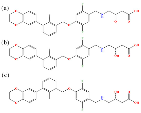
Programmed cell death-1 (PD-1), which is a molecule involved in the inhibitory signal in the immune system and is important due to blocking of the interactions between PD-1 and programmed cell death ligand-1 (PD-L1), has emerged as a promising immunotherapy for treating cancer. In this work, molecular dynamics simulations were performed on complex systems consisting of the PD-L1 dimer with (S)-BMS-200, (R)-BMS-200 and (MOD)-BMS-200 (i.e., S, R and MOD systems) to systematically evaluate the inhibitory mechanism of BMS-200-related small-molecule inhibitors in detail. Among them, (MOD)-BMS-200 was modified from the original (S)-BMS-200 by replacing the hydroxyl group with a carbonyl to remove its chirality. Binding free energy analysis indicates that BMS-200-related inhibitors can promote the dimerization of PD-L1. Meanwhile, no significant differences were observed between the S and MOD systems, though the R system exhibited a slightly higher energy. Residue energy decomposition, nonbonded interaction, and contact number analyses show that the inhibitors mainly bind with the C, F and G regions of the PD-L1 dimer, while nonpolar interactions of key residues Ile54, Tyr56, Met115, Ala121 and Tyr123 on both PD-L1 monomers are the dominant binding-related stability factors. Furthermore, compared with (S)-BMS-200, (R)-BMS-200 is more likely to form hydrogen bonds with charged residues. Finally, free energy landscape and protein–protein interaction analyses show that the key residues of the PD-L1 dimer undergo remarkable conformational changes induced by (S)-BMS-200, which boosts its intimate interactions. This systematic investigation provides a comprehensive molecular insight into the ligand recognition process, which will benefit the design of new small-molecule inhibitors targeting PD-L1 for use in anticancer therapy. Full article
Show Figures

Figure 1

25 pages, 4432 KiB  
Article
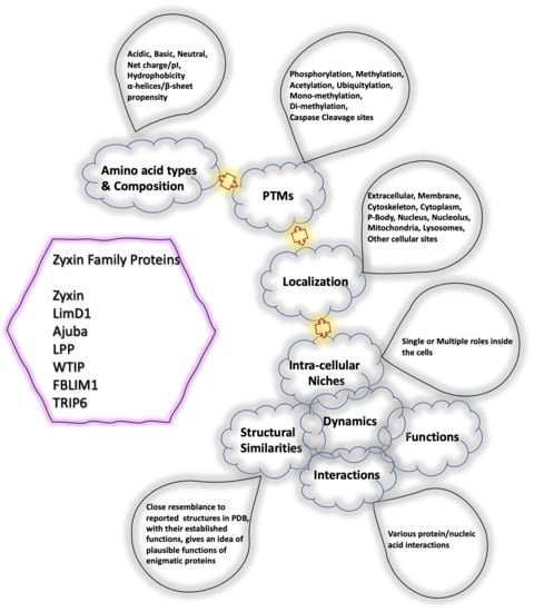
Bioinformatic Analysis of Structure and Function of LIM Domains of Human Zyxin Family Proteins
by M. Quadir Siddiqui, Maulik D. Badmalia and Trushar R. Patel
Int. J. Mol. Sci. 2021, 22(5), 2647; https://doi.org/10.3390/ijms22052647 - 5 Mar 2021
Cited by 16 | Viewed by 3552
Abstract
Members of the human Zyxin family are LIM domain-containing proteins that perform critical cellular functions and are indispensable for cellular integrity. Despite their importance, not much is known about their structure, functions, interactions and dynamics. To provide insights into these, we used a [...] Read more.
Members of the human Zyxin family are LIM domain-containing proteins that perform critical cellular functions and are indispensable for cellular integrity. Despite their importance, not much is known about their structure, functions, interactions and dynamics. To provide insights into these, we used a set of in-silico tools and databases and analyzed their amino acid sequence, phylogeny, post-translational modifications, structure-dynamics, molecular interactions, and functions. Our analysis revealed that zyxin members are ohnologs. Presence of a conserved nuclear export signal composed of LxxLxL/LxxxLxL consensus sequence, as well as a possible nuclear localization signal, suggesting that Zyxin family members may have nuclear and cytoplasmic roles. The molecular modeling and structural analysis indicated that Zyxin family LIM domains share similarities with transcriptional regulators and have positively charged electrostatic patches, which may indicate that they have previously unanticipated nucleic acid binding properties. Intrinsic dynamics analysis of Lim domains suggest that only Lim1 has similar internal dynamics properties, unlike Lim2/3. Furthermore, we analyzed protein expression and mutational frequency in various malignancies, as well as mapped protein-protein interaction networks they are involved in. Overall, our comprehensive bioinformatic analysis suggests that these proteins may play important roles in mediating protein-protein and protein-nucleic acid interactions. Full article
Show Figures

Graphical abstract

2020

Jump to: 2023, 2022, 2021, 2019

32 pages, 11150 KiB  
Article
Alpha-Carbonic Anhydrases from Hydrothermal Vent Sources as Potential Carbon Dioxide Sequestration Agents: In Silico Sequence, Structure and Dynamics Analyses
by Colleen Varaidzo Manyumwa, Reza Zolfaghari Emameh and Özlem Tastan Bishop
Int. J. Mol. Sci. 2020, 21(21), 8066; https://doi.org/10.3390/ijms21218066 - 29 Oct 2020
Cited by 8 | Viewed by 4487
Abstract
With the increase in CO2 emissions worldwide and its dire effects, there is a need to reduce CO2 concentrations in the atmosphere. Alpha-carbonic anhydrases (α-CAs) have been identified as suitable sequestration agents. This study reports the sequence and structural analysis of [...] Read more.
With the increase in CO2 emissions worldwide and its dire effects, there is a need to reduce CO2 concentrations in the atmosphere. Alpha-carbonic anhydrases (α-CAs) have been identified as suitable sequestration agents. This study reports the sequence and structural analysis of 15 α-CAs from bacteria, originating from hydrothermal vent systems. Structural analysis of the multimers enabled the identification of hotspot and interface residues. Molecular dynamics simulations of the homo-multimers were performed at 300 K, 363 K, 393 K and 423 K to unearth potentially thermostable α-CAs. Average betweenness centrality (BC) calculations confirmed the relevance of some hotspot and interface residues. The key residues responsible for dimer thermostability were identified by comparing fluctuating interfaces with stable ones, and were part of conserved motifs. Crucial long-lived hydrogen bond networks were observed around residues with high BC values. Dynamic cross correlation fortified the relevance of oligomerization of these proteins, thus the importance of simulating them in their multimeric forms. A consensus of the simulation analyses used in this study suggested high thermostability for the α-CA from Nitratiruptor tergarcus. Overall, our novel findings enhance the potential of biotechnology applications through the discovery of alternative thermostable CO2 sequestration agents and their potential protein design. Full article
Show Figures

Graphical abstract

15 pages, 5292 KiB  
Article
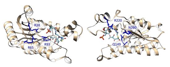
Molecular Basis for Polyketide Ketoreductase–Substrate Interactions
by Shiji Zhao, Fanglue Ni, Tianyin Qiu, Jacob T. Wolff, Shiou-Chuan Tsai and Ray Luo
Int. J. Mol. Sci. 2020, 21(20), 7562; https://doi.org/10.3390/ijms21207562 - 13 Oct 2020
Cited by 15 | Viewed by 3171
Abstract
Polyketides are a large class of structurally and functionally diverse natural products with important bioactivities. Many polyketides are synthesized by reducing type II polyketide synthases (PKSs), containing transiently interacting standalone enzymes. During synthesis, ketoreductase (KR) catalyzes regiospecific carbonyl to hydroxyl reduction, determining the [...] Read more.
Polyketides are a large class of structurally and functionally diverse natural products with important bioactivities. Many polyketides are synthesized by reducing type II polyketide synthases (PKSs), containing transiently interacting standalone enzymes. During synthesis, ketoreductase (KR) catalyzes regiospecific carbonyl to hydroxyl reduction, determining the product outcome, yet little is known about what drives specific KR–substrate interactions. In this study, computational approaches were used to explore KR–substrate interactions based on previously solved apo and mimic cocrystal structures. We found five key factors guiding KR–substrate binding. First, two major substrate binding motifs were identified. Second, substrate length is the key determinant of substrate binding position. Third, two key residues in chain length specificity were confirmed. Fourth, phosphorylation of substrates is critical for binding. Finally, packing/hydrophobic effects primarily determine the binding stability. The molecular bases revealed here will help further engineering of type II PKSs and directed biosynthesis of new polyketides. Full article
Show Figures

Graphical abstract

14 pages, 3634 KiB  
Article
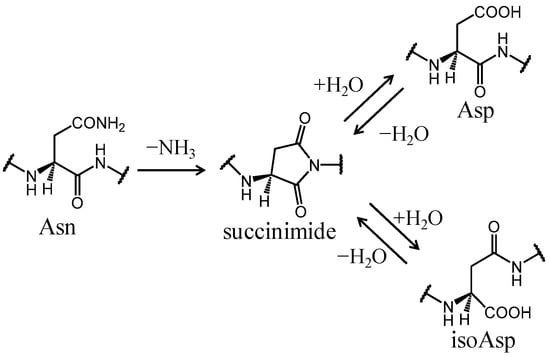
Mechanisms of Deamidation of Asparagine Residues and Effects of Main-Chain Conformation on Activation Energy
by Koichi Kato, Tomoki Nakayoshi, Eiji Kurimoto and Akifumi Oda
Int. J. Mol. Sci. 2020, 21(19), 7035; https://doi.org/10.3390/ijms21197035 - 24 Sep 2020
Cited by 25 | Viewed by 6707
Abstract
Deamidation of asparagine (Asn) residues is a nonenzymatic post-translational modification of proteins. Asn deamidation is associated with pathogenesis of age-related diseases and hypofunction of monoclonal antibodies. Deamidation rate is known to be affected by the residue following Asn on the carboxyl side and [...] Read more.
Deamidation of asparagine (Asn) residues is a nonenzymatic post-translational modification of proteins. Asn deamidation is associated with pathogenesis of age-related diseases and hypofunction of monoclonal antibodies. Deamidation rate is known to be affected by the residue following Asn on the carboxyl side and by secondary structure. Information about main-chain conformation of Asn residues is necessary to accurately predict deamidation rate. In this study, the effect of main-chain conformation of Asn residues on deamidation rate was computationally investigated using molecular dynamics (MD) simulations and quantum chemical calculations. The results of MD simulations for γS-crystallin suggested that frequently deamidated Asn residues have common main-chain conformations on the N-terminal side. Based on the simulated structure, initial structures for the quantum chemical calculations were constructed and optimized geometries were obtained using the B3LYP density functional method. Structures that were frequently deamidated had a lower activation energy barrier than that of the little deamidated structure. We also showed that dihydrogen phosphate and bicarbonate ions are important catalysts for deamidation of Asn residues. Full article
Show Figures

Figure 1

13 pages, 1989 KiB  
Article
Understanding the Interaction Modes and Reactivity of Trimedoxime toward MmAChE Inhibited by Nerve Agents: Theoretical and Experimental Aspects
by Alexandre A. de Castro, Daniel A. Polisel, Bruna T. L. Pereira, Elaine F. F. da Cunha, Kamil Kuca, Eugenie Nepovimova and Teodorico C. Ramalho
Int. J. Mol. Sci. 2020, 21(18), 6510; https://doi.org/10.3390/ijms21186510 - 5 Sep 2020
Cited by 2 | Viewed by 2297
Abstract
Organophosphorus (OP) compounds are used as both chemical weapons and pesticides. However, these agents are very dangerous and toxic to humans, animals, and the environment. Thus, investigations with reactivators have been deeply developed in order to design new antidotes with better efficiency, as [...] Read more.
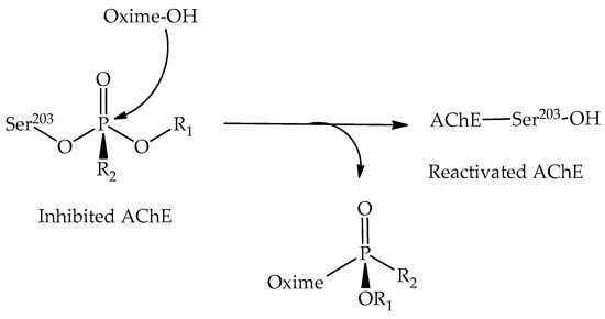
Organophosphorus (OP) compounds are used as both chemical weapons and pesticides. However, these agents are very dangerous and toxic to humans, animals, and the environment. Thus, investigations with reactivators have been deeply developed in order to design new antidotes with better efficiency, as well as a greater spectrum of action in the acetylcholinesterase (AChE) reactivation process. With that in mind, in this work, we investigated the behavior of trimedoxime toward the Mus musculus acetylcholinesterase (MmAChE) inhibited by a range of nerve agents, such as chemical weapons. From experimental assays, reactivation percentages were obtained for the reactivation of different AChE–OP complexes. On the other hand, theoretical calculations were performed to assess the differences in interaction modes and the reactivity of trimedoxime within the AChE active site. Comparing theoretical and experimental data, it is possible to notice that the oxime, in most cases, showed better reactivation percentages at higher concentrations, with the best result for the reactivation of the AChE–VX adduct. From this work, it was revealed that the mechanistic process contributes most to the oxime efficiency than the interaction in the site. In this way, this study is important to better understand the reactivation process through trimedoxime, contributing to the proposal of novel antidotes. Full article
Show Figures

Figure 1

11 pages, 3683 KiB  
Article
Plasticity of the 340-Loop in Influenza Neuraminidase Offers New Insight for Antiviral Drug Development
by Nanyu Han, Justin Tze Yang Ng, Yanpeng Li, Yuguang Mu and Zunxi Huang
Int. J. Mol. Sci. 2020, 21(16), 5655; https://doi.org/10.3390/ijms21165655 - 6 Aug 2020
Cited by 2 | Viewed by 2336
Abstract
The recently discovered 340-cavity in influenza neuraminidase (NA) N6 and N7 subtypes has introduced new possibilities for rational structure-based drug design. However, the plasticity of the 340-loop (residues 342–347) and the role of the 340-loop in NA activity and substrate binding have not [...] Read more.
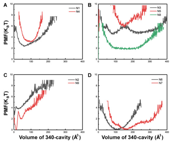
The recently discovered 340-cavity in influenza neuraminidase (NA) N6 and N7 subtypes has introduced new possibilities for rational structure-based drug design. However, the plasticity of the 340-loop (residues 342–347) and the role of the 340-loop in NA activity and substrate binding have not been deeply exploited. Here, we investigate the mechanism of 340-cavity formation and demonstrate for the first time that seven of nine NA subtypes are able to adopt an open 340-cavity over 1.8 μs total molecular dynamics simulation time. The finding that the 340-loop plays a role in the sialic acid binding pathway suggests that the 340-cavity can function as a druggable pocket. Comparing the open and closed conformations of the 340-loop, the side chain orientation of residue 344 was found to govern the formation of the 340-cavity. Additionally, the conserved calcium ion was found to substantially influence the stability of the 340-loop. Our study provides dynamical evidence supporting the 340-cavity as a druggable hotspot at the atomic level and offers new structural insight in designing antiviral drugs. Full article
Show Figures

Figure 1

13 pages, 4427 KiB  
Article
A Structural Model for Bax∆2-Mediated Activation of Caspase 8-Dependent Apoptosis
by Bing Xie, Qi Yao, Jialing Xiang and David D.L. Minh
Int. J. Mol. Sci. 2020, 21(15), 5476; https://doi.org/10.3390/ijms21155476 - 31 Jul 2020
Cited by 2 | Viewed by 2866
Abstract
Bax∆2 is a pro-apoptotic anti-tumor protein in the Bax family. While most of the Bax family causes cell death by targeting mitochondria, Bax∆2 forms cytosolic aggregates and activates caspase 8-dependent cell death. We previously showed that the Bax∆2 helix α9 is critical for [...] Read more.
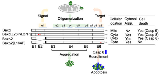
Bax∆2 is a pro-apoptotic anti-tumor protein in the Bax family. While most of the Bax family causes cell death by targeting mitochondria, Bax∆2 forms cytosolic aggregates and activates caspase 8-dependent cell death. We previously showed that the Bax∆2 helix α9 is critical for caspase 8 recruitment. However, the interaction between these two proteins at the structural level is unknown. In this in silico study, we performed molecular dynamics (MD) simulations and protein–protein docking on Bax∆2 variants. The results suggest that the Bax∆2 variants have different stable states. Mutating the Baxα mitochondria-targeting signal [L26P/L27P] appears to introduce a kink into helix α1. Protein–protein docking suggests that helices α9 of both wild-type Bax∆2 and Bax∆2 caspase 8 binding-deficient mutant [L164P] can fit in the same caspase 8 binding site, but the mutant is unable to fit as well as wild-type Bax∆2. Together, these data point to a structural basis for explaining Bax∆2 function in caspase 8-dependent cell death. Full article
Show Figures

Figure 1

24 pages, 6257 KiB  
Article
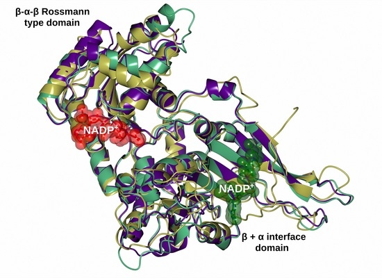
Characterizing the Fused TvG6PD::6PGL Protein from the Protozoan Trichomonas vaginalis, and Effects of the NADP+ Molecule on Enzyme Stability
by Laura Morales-Luna, Beatriz Hernández-Ochoa, Edson Jiovany Ramírez-Nava, Víctor Martínez-Rosas, Paulina Ortiz-Ramírez, Fabiola Fernández-Rosario, Abigail González-Valdez, Noemí Cárdenas-Rodríguez, Hugo Serrano-Posada, Sara Centeno-Leija, Roberto Arreguin-Espinosa, Miguel Cuevas-Cruz, Daniel Ortega-Cuellar, Verónica Pérez de la Cruz, Luz María Rocha-Ramírez, Edgar Sierra-Palacios, Rosa Angélica Castillo-Rodríguez, Vanesa Vega-García, Yadira Rufino-González, Jaime Marcial-Quino and Saúl Gómez-Manzoadd Show full author list remove Hide full author list
Int. J. Mol. Sci. 2020, 21(14), 4831; https://doi.org/10.3390/ijms21144831 - 8 Jul 2020
Cited by 6 | Viewed by 3135
Abstract
This report describes a functional and structural analysis of fused glucose-6-phosphate dehydrogenase dehydrogenase-phosphogluconolactonase protein from the protozoan Trichomonas vaginalis (T. vaginalis). The glucose-6-phosphate dehydrogenase (g6pd) gene from T. vaginalis was isolated by PCR and the sequence of the [...] Read more.
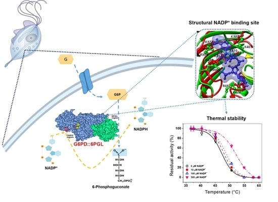
This report describes a functional and structural analysis of fused glucose-6-phosphate dehydrogenase dehydrogenase-phosphogluconolactonase protein from the protozoan Trichomonas vaginalis (T. vaginalis). The glucose-6-phosphate dehydrogenase (g6pd) gene from T. vaginalis was isolated by PCR and the sequence of the product showed that is fused with 6pgl gene. The fused Tvg6pd::6pgl gene was cloned and overexpressed in a heterologous system. The recombinant protein was purified by affinity chromatography, and the oligomeric state of the TvG6PD::6PGL protein was found as tetramer, with an optimal pH of 8.0. The kinetic parameters for the G6PD domain were determined using glucose-6-phosphate (G6P) and nicotinamide adenine dinucleotide phosphate (NADP+) as substrates. Biochemical assays as the effects of temperature, susceptibility to trypsin digestion, and analysis of hydrochloride of guanidine on protein stability in the presence or absence of NADP+ were performed. These results revealed that the protein becomes more stable in the presence of the NADP+. In addition, we determined the dissociation constant for the binding (Kd) of NADP+ in the protein and suggests the possible structural site in the fused TvG6PD::6PGL protein. Finally, computational modeling studies were performed to obtain an approximation of the structure of TvG6PD::6PGL. The generated model showed differences with the GlG6PD::6PGL protein (even more so with human G6PD) despite both being fused. Full article
Show Figures

Graphical abstract

17 pages, 2353 KiB  
Article
Activity to Breast Cancer Cell Lines of Different Malignancy and Predicted Interaction with Protein Kinase C Isoforms of Royleanones
by Vera M. S. Isca, Milan Sencanski, Nenad Filipovic, Daniel J. V. A. Dos Santos, Ana Čipak Gašparović, Lucília Saraíva, Carlos A. M. Afonso, Patrícia Rijo and Alfonso T. García-Sosa
Int. J. Mol. Sci. 2020, 21(10), 3671; https://doi.org/10.3390/ijms21103671 - 23 May 2020
Cited by 9 | Viewed by 4464
Abstract
Plants have been used for centuries to treat several illnesses. The Plectranthus genus has a vast variety of species that has allowed the isolation of cytotoxic compounds with notable activities. The abietane diterpenes 6,7-dehydroroyleanone (DeRoy, 1), 7α-acetoxy-6β-hydroxyroyleanone (Roy, 2), and Parvifloron [...] Read more.
Plants have been used for centuries to treat several illnesses. The Plectranthus genus has a vast variety of species that has allowed the isolation of cytotoxic compounds with notable activities. The abietane diterpenes 6,7-dehydroroyleanone (DeRoy, 1), 7α-acetoxy-6β-hydroxyroyleanone (Roy, 2), and Parvifloron D (ParvD, 3) were obtained from Plectranthus spp. and showed promising biological activities, such as cytotoxicity. The inhibitory effects of the different natural abietanes (1-3) were compared in MFC7, SkBr3, and SUM159 cell lines, as well as SUM159 grown in cancer stem cell-inducing conditions. Based on the royleanones’ bioactivity, the derivatives RoyBz (4), RoyBzCl (5), RoyPr2 (6), and DihydroxyRoy (7), previously obtained from 2, were selected for further studies. Protein kinases C (PKCs) are involved in several carcinogenic processes. Thus, PKCs are potential targets for cancer therapy. To date, the portfolio of available PKC modulators remains very limited due to the difficulty of designing isozyme-selective PKC modulators. As such, molecular docking was used to evaluate royleanones 1-6 as predicted isozyme-selective PKC binders. Subtle changes in the binding site of each PKC isoform change the predicted interaction profiles of the ligands. Subtle changes in royleanone substitution patterns, such as a double substitution only with non-substituted phenyls, or hydroxybenzoate at position four that flips the binding mode of ParvD (3), can increase the predicted interactions in certain PKC subtypes. Full article
Show Figures

Graphical abstract

14 pages, 4282 KiB  
Article
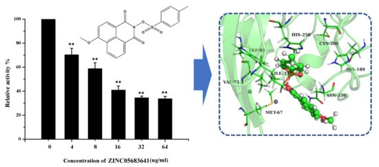
Discovery of the Novel Inhibitor Against New Delhi Metallo-β-Lactamase Based on Virtual Screening and Molecular Modelling
by Xiyan Wang, Yanan Yang, Yawen Gao and Xiaodi Niu
Int. J. Mol. Sci. 2020, 21(10), 3567; https://doi.org/10.3390/ijms21103567 - 18 May 2020
Cited by 18 | Viewed by 3683
Abstract
New Delhi metallo-β-lactamase (NDM-1), one of the metallo-β-lactamases (MBLs), leads to antibiotic resistance in clinical treatments due to the strong ability of hydrolysis to almost all kinds of β-lactam antibiotics. Therefore, there is the urgent need for the research and development of the [...] Read more.
New Delhi metallo-β-lactamase (NDM-1), one of the metallo-β-lactamases (MBLs), leads to antibiotic resistance in clinical treatments due to the strong ability of hydrolysis to almost all kinds of β-lactam antibiotics. Therefore, there is the urgent need for the research and development of the novel drug-resistant inhibitors targeting NDM-1. In this study, ZINC05683641 was screened as potential NDM-1 inhibitor by virtual screening and the inhibitor mechanism of this compound was explored based on molecular dynamics simulation. The nitrocefin assay showed that the IC50 value of ZINC05683641 was 13.59 ± 0.52 μM, indicating that the hydrolytic activity of NDM-1 can be obviously suppressed by ZINC05683641. Further, the binding mode of ZINC05683641 with NDM-1 was obtained by molecular modeling, binding free energy calculation, mutagenesis assays and fluorescence-quenching assays. As results, ILE-35, MET-67, VAL-73, TRP-93, CYS-208, ASN-220 and HIS-250 played the key roles in the binding of NDM-1 with ZINC05683641. Interestingly, these key residues were exactly located in the catalytic activity region of NDM-1, implying that the inhibitor mechanism of ZINC05683641 against NDM-1 was the competitive inhibition. These findings will provide an available approach to research and develop new drug against NDM-1 and treatment for bacterial resistance. Full article
Show Figures

Graphical abstract

23 pages, 9059 KiB  
Article
Effects of Single and Double Mutants in Human Glucose-6-Phosphate Dehydrogenase Variants Present in the Mexican Population: Biochemical and Structural Analysis
by Víctor Martínez-Rosas, Merit Valeria Juárez-Cruz, Edson Jiovany Ramírez-Nava, Beatriz Hernández-Ochoa, Laura Morales-Luna, Abigail González-Valdez, Hugo Serrano-Posada, Noemí Cárdenas-Rodríguez, Paulina Ortiz-Ramírez, Sara Centeno-Leija, Roberto Arreguin-Espinosa, Miguel Cuevas-Cruz, Daniel Ortega-Cuellar, Verónica Pérez de la Cruz, Luz María Rocha-Ramírez, Edgar Sierra-Palacios, Rosa Angélica Castillo-Rodríguez, Isabel Baeza-Ramírez, Jaime Marcial-Quino and Saúl Gómez-Manzo
Int. J. Mol. Sci. 2020, 21(8), 2732; https://doi.org/10.3390/ijms21082732 - 15 Apr 2020
Cited by 13 | Viewed by 4623
Abstract
Glucose-6-phosphate dehydrogenase (G6PD) deficiency is the most frequent human enzymopathy, affecting over 400 million people globally. Worldwide, 217 mutations have been reported at the genetic level, and only 19 have been found in Mexico. The objective of this work was to contribute to [...] Read more.
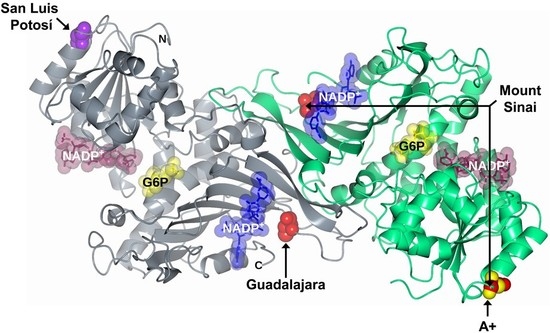
Glucose-6-phosphate dehydrogenase (G6PD) deficiency is the most frequent human enzymopathy, affecting over 400 million people globally. Worldwide, 217 mutations have been reported at the genetic level, and only 19 have been found in Mexico. The objective of this work was to contribute to the knowledge of the function and structure of three single natural variants (G6PD A+, G6PD San Luis Potosi, and G6PD Guadalajara) and a double mutant (G6PD Mount Sinai), each localized in a different region of the three-dimensional (3D) structure. In the functional characterization of the mutants, we observed a decrease in specific activity, protein expression and purification, catalytic efficiency, and substrate affinity in comparison with wild-type (WT) G6PD. Moreover, the analysis of the effect of all mutations on the structural stability showed that its presence increases denaturation and lability with temperature and it is more sensible to trypsin digestion protease and guanidine hydrochloride compared with WT G6PD. This could be explained by accelerated degradation of the variant enzymes due to reduced stability of the protein, as is shown in patients with G6PD deficiency. Full article
Show Figures

Graphical abstract

23 pages, 2320 KiB  
Review
Dynamics, a Powerful Component of Current and Future in Silico Approaches for Protein Design and Engineering
by Bartłomiej Surpeta, Carlos Eduardo Sequeiros-Borja and Jan Brezovsky
Int. J. Mol. Sci. 2020, 21(8), 2713; https://doi.org/10.3390/ijms21082713 - 14 Apr 2020
Cited by 14 | Viewed by 7037
Abstract
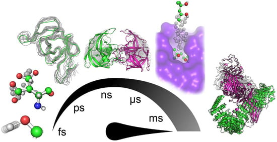
Computational prediction has become an indispensable aid in the processes of engineering and designing proteins for various biotechnological applications. With the tremendous progress in more powerful computer hardware and more efficient algorithms, some of in silico tools and methods have started to apply [...] Read more.
Computational prediction has become an indispensable aid in the processes of engineering and designing proteins for various biotechnological applications. With the tremendous progress in more powerful computer hardware and more efficient algorithms, some of in silico tools and methods have started to apply the more realistic description of proteins as their conformational ensembles, making protein dynamics an integral part of their prediction workflows. To help protein engineers to harness benefits of considering dynamics in their designs, we surveyed new tools developed for analyses of conformational ensembles in order to select engineering hotspots and design mutations. Next, we discussed the collective evolution towards more flexible protein design methods, including ensemble-based approaches, knowledge-assisted methods, and provable algorithms. Finally, we highlighted apparent challenges that current approaches are facing and provided our perspectives on their further development. Full article
Show Figures

Figure 1

16 pages, 3136 KiB  
Article
Docking and Molecular Dynamics Predictions of Pesticide Binding to the Calyx of Bovine β-Lactoglobulin
by Paulina Cortes-Hernandez, Roberto Vázquez Nuñez and Lenin Domínguez-Ramírez
Int. J. Mol. Sci. 2020, 21(6), 1988; https://doi.org/10.3390/ijms21061988 - 14 Mar 2020
Cited by 8 | Viewed by 4004
Abstract
Pesticides are used extensively in agriculture, and their residues in food must be monitored to prevent toxicity. The most abundant protein in cow’s milk, β-lactoglobulin (BLG), shows high affinity for diverse hydrophobic ligands in its central binding pocket, called the calyx. Several of [...] Read more.
Pesticides are used extensively in agriculture, and their residues in food must be monitored to prevent toxicity. The most abundant protein in cow’s milk, β-lactoglobulin (BLG), shows high affinity for diverse hydrophobic ligands in its central binding pocket, called the calyx. Several of the most frequently used pesticides are hydrophobic. To predict if BLG may be an unintended carrier for pesticides, we tested its ability to bind 555 pesticides and their isomers, for a total of 889 compounds, in a rigid docking screen. We focused on the analysis of 60 unique molecules belonging to the five pesticide classes defined by the World Health Organization, that docked into BLG’s calyx with ΔGs ranging from −8.2 to −12 kcal mol−1, chosen by statistical criteria. These “potential ligands” were further analyzed using molecular dynamic simulations, and the binding energies were explored with Molecular Mechanics/Generalized Born/Surface Area (MMGBSA). Hydrophobic pyrethroid insecticides, like cypermethrin, were found to bind as deeply and tightly into the calyx as BLG’s natural ligand, palmitate; while polar compounds, like paraquat, were expelled. Our results suggest that BLG could be a carrier for pesticides, in particular for pyrethroid insecticides, allowing for their accumulation in cow’s milk beyond their solubility restrictions. This analysis opens possibilities for pesticide biosensor design based on BLG. Full article
Show Figures

Graphical abstract

16 pages, 3031 KiB  
Article
Ligand Binding Mechanism and Its Relationship with Conformational Changes in Adenine Riboswitch
by Guodong Hu, Haiyan Li, Shicai Xu and Jihua Wang
Int. J. Mol. Sci. 2020, 21(6), 1926; https://doi.org/10.3390/ijms21061926 - 11 Mar 2020
Cited by 18 | Viewed by 3327
Abstract
Riboswitches are naturally occurring RNA aptamers that control the expression of essential bacterial genes by binding to specific small molecules. The binding with both high affinity and specificity induces conformational changes. Thus, riboswitches were proposed as a possible molecular target for developing antibiotics [...] Read more.
Riboswitches are naturally occurring RNA aptamers that control the expression of essential bacterial genes by binding to specific small molecules. The binding with both high affinity and specificity induces conformational changes. Thus, riboswitches were proposed as a possible molecular target for developing antibiotics and chemical tools. The adenine riboswitch can bind not only to purine analogues but also to pyrimidine analogues. Here, long molecular dynamics (MD) simulations and molecular mechanics Poisson–Boltzmann surface area (MM-PBSA) computational methodologies were carried out to show the differences in the binding model and the conformational changes upon five ligands binding. The binding free energies of the guanine riboswitch aptamer with C74U mutation complexes were compared to the binding free energies of the adenine riboswitch (AR) aptamer complexes. The calculated results are in agreement with the experimental data. The differences for the same ligand binding to two different aptamers are related to the electrostatic contribution. Binding dynamical analysis suggests a flexible binding pocket for the pyrimidine ligand in comparison with the purine ligand. The 18 μs of MD simulations in total indicate that both ligand-unbound and ligand-bound aptamers transfer their conformation between open and closed states. The ligand binding obviously affects the conformational change. The conformational states of the aptamer are associated with the distance between the mass center of two key nucleotides (U51 and A52) and the mass center of the other two key nucleotides (C74 and C75). The results suggest that the dynamical character of the binding pocket would affect its biofunction. To design new ligands of the adenine riboswitch, it is recommended to consider the binding affinities of the ligand and the conformational change of the ligand binding pocket. Full article
Show Figures

Graphical abstract

21 pages, 4398 KiB  
Article
Elucidating the Structural Basis of the Intracellular pH Sensing Mechanism of TASK-2 K2P Channels
by Daniel Bustos, Mauricio Bedoya, David Ramírez, Guierdy Concha, Leandro Zúñiga, Niels Decher, Erix W. Hernández-Rodríguez, Francisco V. Sepúlveda, Leandro Martínez and Wendy González
Int. J. Mol. Sci. 2020, 21(2), 532; https://doi.org/10.3390/ijms21020532 - 14 Jan 2020
Cited by 6 | Viewed by 4044
Abstract
Two-pore domain potassium (K2P) channels maintain the cell’s background conductance by stabilizing the resting membrane potential. They assemble as dimers possessing four transmembrane helices in each subunit. K2P channels were crystallized in “up” and “down” states. The movements of [...] Read more.
Two-pore domain potassium (K2P) channels maintain the cell’s background conductance by stabilizing the resting membrane potential. They assemble as dimers possessing four transmembrane helices in each subunit. K2P channels were crystallized in “up” and “down” states. The movements of the pore-lining transmembrane TM4 helix produce the aperture or closure of side fenestrations that connect the lipid membrane with the central cavity. When the TM4 helix is in the up-state, the fenestrations are closed, while they are open in the down-state. It is thought that the fenestration states are related to the activity of K2P channels and the opening of the channels preferentially occurs from the up-state. TASK-2, a member of the TALK subfamily of K2P channels, is opened by intracellular alkalization leading the deprotonation of the K245 residue at the end of the TM4 helix. This charge neutralization of K245 could be sensitive or coupled to the fenestration state. Here, we describe the relationship between the states of the intramembrane fenestrations and K245 residue in TASK-2 channel. By using molecular modeling and simulations, we show that the protonated state of K245 (K245+) favors the open fenestration state and, symmetrically, that the open fenestration state favors the protonated state of the lysine residue. We show that the channel can be completely blocked by Prozac, which is known to induce fenestration opening in TREK-2. K245 protonation and fenestration aperture have an additive effect on the conductance of the channel. The opening of the fenestrations with K245+ increases the entrance of lipids into the selectivity filter, blocking the channel. At the same time, the protonation of K245 introduces electrostatic potential energy barriers to ion entrance. We computed the free energy profiles of ion penetration into the channel in different fenestration and K245 protonation states, to show that the effects of the two transformations are summed up, leading to maximum channel blocking. Estimated rates of ion transport are in qualitative agreement with experimental results and support the hypothesis that the most important barrier for ion transport under K245+ and open fenestration conditions is the entrance of the ions into the channel. Full article
Show Figures

Figure 1

18 pages, 5610 KiB  
Article
Structural Characterization of the CD44 Stem Region for Standard and Cancer-Associated Isoforms
by Kun-Lin Chen, Deng Li, Ting-Xuan Lu and Shu-Wei Chang
Int. J. Mol. Sci. 2020, 21(1), 336; https://doi.org/10.3390/ijms21010336 - 3 Jan 2020
Cited by 13 | Viewed by 5289
Abstract
CD44 is widely expressed in most vertebrate cells, whereas the expression of CD44v6 is restricted to only a few tissues and has been considered to be associated with tumor progression and metastasis. Thus, CD44v6 has been recognized as a promising prognostic biomarker and [...] Read more.
CD44 is widely expressed in most vertebrate cells, whereas the expression of CD44v6 is restricted to only a few tissues and has been considered to be associated with tumor progression and metastasis. Thus, CD44v6 has been recognized as a promising prognostic biomarker and therapeutic target for various cancers for more than a decade. However, despite many experimental studies, the structural dynamics and differences between CD44s and CD44v6, particularly in their stem region, still remain elusive. Here, a computational study was conducted to address these problems. We found that the stem of CD44s adopted predominantly two conformations, one featuring antiparallel β-sheets and the other featuring parallel β-sheets, whereas the stem of CD44v6 adopted mainly one conformation with relatively highly suppressed β-sheet contents. Moreover, Phe215 was found to be essential in the β-sheets of both CD44s and CD44v6. We finally found intramolecular Phe215–Trp224 hydrogen-bonding interactions and hydrophobic interactions with Phe215 that cooperatively drove conformational differences upon the addition of the v6 region to CD44. Our study elucidated the structural differences between the stem regions of CD44s and CD44v6 and thus can offer useful structural information for drug design to specifically target CD44v6 in promising clinical applications. Full article
Show Figures

Graphical abstract

2019

Jump to: 2023, 2022, 2021, 2020

26 pages, 3950 KiB  
Article
Novel Descriptors and Digital Signal Processing- Based Method for Protein Sequence Activity Relationship Study
by Nicolas T. Fontaine, Xavier F. Cadet and Iyanar Vetrivel
Int. J. Mol. Sci. 2019, 20(22), 5640; https://doi.org/10.3390/ijms20225640 - 11 Nov 2019
Cited by 11 | Viewed by 4981
Abstract
The work aiming to unravel the correlation between protein sequence and function in the absence of structural information can be highly rewarding. We present a new way of considering descriptors from the amino acids index database for modeling and predicting the fitness value [...] Read more.
The work aiming to unravel the correlation between protein sequence and function in the absence of structural information can be highly rewarding. We present a new way of considering descriptors from the amino acids index database for modeling and predicting the fitness value of a polypeptide chain. This approach includes the following steps: (i) Calculating Q elementary numerical sequences (Ele_SEQ) depending on the encoding of the amino acid residues, (ii) determining an extended numerical sequence (Ext_SEQ) by concatenating the Q elementary numerical sequences, wherein at least one elementary numerical sequence is a protein spectrum obtained by applying fast Fourier transformation (FFT), and (iii) predicting a value of fitness for polypeptide variants (train and/or validation set). These new descriptors were tested on four sets of proteins of different lengths (GLP-2, TNF alpha, cytochrome P450, and epoxide hydrolase) and activities (cAMP activation, binding affinity, thermostability and enantioselectivity). We show that the use of multiple physicochemical descriptors coupled with the implementation of the FFT, taking into account the interactions between residues of amino acids within the protein sequence, could lead to very significant improvement in the quality of models and predictions. The choice of the descriptor or of the combination of descriptors and/or FFT is dependent on the couple protein/fitness. This approach can provide potential users with value added to existing mutant libraries where screening efforts have so far been unsuccessful in finding improved polypeptide mutants for useful applications. Full article
Show Figures

Figure 1

17 pages, 1766 KiB  
Article
Molecular Cloning and Exploration of the Biochemical and Functional Analysis of Recombinant Glucose-6-Phosphate Dehydrogenase from Gluconoacetobacter diazotrophicus PAL5
by Edson Jiovany Ramírez-Nava, Daniel Ortega-Cuellar, Abigail González-Valdez, Rosa Angélica Castillo-Rodríguez, Gabriel Yaxal Ponce-Soto, Beatriz Hernández-Ochoa, Noemí Cárdenas-Rodríguez, Víctor Martínez-Rosas, Laura Morales-Luna, Hugo Serrano-Posada, Edgar Sierra-Palacios, Roberto Arreguin-Espinosa, Miguel Cuevas-Cruz, Luz María Rocha-Ramírez, Verónica Pérez de la Cruz, Jaime Marcial-Quino and Saúl Gómez-Manzo
Int. J. Mol. Sci. 2019, 20(21), 5279; https://doi.org/10.3390/ijms20215279 - 24 Oct 2019
Cited by 5 | Viewed by 3900
Abstract
Gluconacetobacter diazotrophicus PAL5 (GDI) is an endophytic bacterium with potential biotechnological applications in industry and agronomy. The recent description of its complete genome and its principal metabolic enzymes suggests that glucose metabolism is accomplished through the pentose phosphate pathway (PPP); however, the enzymes [...] Read more.
Gluconacetobacter diazotrophicus PAL5 (GDI) is an endophytic bacterium with potential biotechnological applications in industry and agronomy. The recent description of its complete genome and its principal metabolic enzymes suggests that glucose metabolism is accomplished through the pentose phosphate pathway (PPP); however, the enzymes participating in this pathway have not yet been characterized in detail. The objective of the present work was to clone, purify, and biochemically and physicochemically characterize glucose-6-phosphate dehydrogenase (G6PD) from GDI. The gene was cloned and expressed as a tagged protein in E. coli to be purified by affinity chromatography. The native state of the G6PD protein in the solution was found to be a tetramer with optimal activity at pH 8.8 and a temperature between 37 and 50 °C. The apparent Km values for G6P and nicotinamide adenine dinucleotide phosphate (NADP+) were 63 and 7.2 μM, respectively. Finally, from the amino acid sequence a three-dimensional (3D) model was obtained, which allowed the arrangement of the amino acids involved in the catalytic activity, which are conserved (RIDHYLGKE, GxGGDLT, and EKPxG) with those of other species, to be identified. This characterization of the enzyme could help to identify new environmental conditions for the knowledge of the plant–microorganism interactions and a better use of GDI in new technological applications. Full article
Show Figures

Graphical abstract

23 pages, 4187 KiB  
Article
Influence of the First Chromophore-Forming Residue on Photobleaching and Oxidative Photoconversion of EGFP and EYFP
by Tirthendu Sen, Anastasia V. Mamontova, Anastasia V. Titelmayer, Aleksander M. Shakhov, Artyom A. Astafiev, Atanu Acharya, Konstantin A. Lukyanov, Anna I. Krylov and Alexey M. Bogdanov
Int. J. Mol. Sci. 2019, 20(20), 5229; https://doi.org/10.3390/ijms20205229 - 22 Oct 2019
Cited by 15 | Viewed by 5227
Abstract
Enhanced green fluorescent protein (EGFP)—one of the most widely applied genetically encoded fluorescent probes—carries the threonine-tyrosine-glycine (TYG) chromophore. EGFP efficiently undergoes green-to-red oxidative photoconversion (“redding”) with electron acceptors. Enhanced yellow fluorescent protein (EYFP), a close EGFP homologue (five amino acid substitutions), has a [...] Read more.
Enhanced green fluorescent protein (EGFP)—one of the most widely applied genetically encoded fluorescent probes—carries the threonine-tyrosine-glycine (TYG) chromophore. EGFP efficiently undergoes green-to-red oxidative photoconversion (“redding”) with electron acceptors. Enhanced yellow fluorescent protein (EYFP), a close EGFP homologue (five amino acid substitutions), has a glycine-tyrosine-glycine (GYG) chromophore and is much less susceptible to redding, requiring halide ions in addition to the oxidants. In this contribution we aim to clarify the role of the first chromophore-forming amino acid in photoinduced behavior of these fluorescent proteins. To that end, we compared photobleaching and redding kinetics of EGFP, EYFP, and their mutants with reciprocally substituted chromophore residues, EGFP-T65G and EYFP-G65T. Measurements showed that T65G mutation significantly increases EGFP photostability and inhibits its excited-state oxidation efficiency. Remarkably, while EYFP-G65T demonstrated highly increased spectral sensitivity to chloride, it is also able to undergo redding chloride-independently. Atomistic calculations reveal that the GYG chromophore has an increased flexibility, which facilitates radiationless relaxation leading to the reduced fluorescence quantum yield in the T65G mutant. The GYG chromophore also has larger oscillator strength as compared to TYG, which leads to a shorter radiative lifetime (i.e., a faster rate of fluorescence). The faster fluorescence rate partially compensates for the loss of quantum efficiency due to radiationless relaxation. The shorter excited-state lifetime of the GYG chromophore is responsible for its increased photostability and resistance to redding. In EYFP and EYFP-G65T, the chromophore is stabilized by π-stacking with Tyr203, which suppresses its twisting motions relative to EGFP. Full article
Show Figures

Figure 1

Back to TopTop